review - pnas · this review, have risen more slowly and ... support the conclusion that the entire...
TRANSCRIPT

Proc. Natl. Acad. Sci. USAVol. 93, pp. 7-12, January 1996
Review
Interactions of protein antigens with antibodiesDavid R. Davies and Gerson H. CohenLaboratory of Molecular Biology, National Institute of Diabetes, Digestive and Kidney Diseases, National Institutes of Health, Bethesda, MD 20892-0560
ABSTRACT There are now severalcrystal structures of antibody Fab frag-ments complexed to their protein anti-gens. These include Fab complexes withlysozyme, two Fab complexes with influ-enza virus neuraminidase, and three Fabcomplexes with their anti-idiotype Fabs.The pattern of binding that emerges issimilar to that found with other protein-protein interactions, with good shapecomplementarity between the interactingsurfaces and reasonable juxtapositions ofpolar residues so as to permit hydrogen-bond formation. Water molecules havebeen observed in cavities within the in-terface and on the periphery, where theyoften form bridging hydrogen bonds be-tween antibody and antigen. For the mostpart the antigen is bound in the middle ofthe antibody combining site with most ofthe six complementarity-determining res-idues involved in binding. For the moststudied antigen, lysozyme, the epitopesfor four antibodies occupy -45% of theaccessible surface area. Some conforma-tional changes have been observed to ac-company binding in both the antibody andthe antigen, although most of the infor-mation on conformational change in thelatter comes from studies of complexeswith small antigens.
There has been a dramatic increase overthe last 5 years in the number of Fabstructures that have been determined byx-ray diffraction. It has been estimatedthat there are now >50 structures knownto a resolution of between 3.0 and 2.0 A,although the coordinates of many of theseare not yet available in the Protein DataBank (Chemistry Department, Brook-haven National Laboratory, Upton, NY).By contrast, the structures of Fab com-plexes with protein antigens, the subject ofthis review, have risen more slowly andhave been restricted to a small group ofantigens, notably hen egg white lysozyme(HEL) and influenza virus neuramini-dase.There have been a number of recent
reviews of antibody structure and anti-body-antigen associations (1-11). Bradenand Poljak (11) in a recent review payparticular attention to the water moleculesthat surround the antibody-antigen inter-face and how these might influence thespecificity. Whereas many of the generalprinciples of these interactions, such as the
shape complementarity of the interactingsurfaces, are now well established, theincreasing data base, including the exam-ination of complexes with mutant anti-body or antigen, the calorimetric analysesof binding, and the application of new
techniques to epitope mapping, add in-creasing detail to this picture. In this briefreview, we shall describe the structuresthat are now available and discuss theresults in terms of the mechanism of an-
tibody-antigen recognition and binding.
Structures of Complexes with ProteinAntigens
HEL. The most studied antigen hasbeen HEL, and there are now five struc-tures reported for complexes with thisantigen. They are D1.3 (12-15), HyHEL-5(16, 69), HyHEL-10 (17), D11.15 (18), andD44.1 (19).The Fab (13) and Fv (14, 15) fragments
of D1.3 have been studied as complexeswith lysozyme. The structure of the iso-lated Fv has also been determined. Thecomplex of the Fab of D1.3 with the Fabof an anti-idiotype to D1.3 has also beendetermined (20). The high resolution ofthe D1.3 Fv-HEL crystals permitted theidentification of many water molecules,about 50 of which are located around theinterface. Four water molecules are com-
pletely buried in the interface, with somelocated in the variable region light chain-heavy chain (VL-VH) interface. Titrationcalorimetry has been used to measure thethermodynamic parameters of the inter-action (15). The reaction is enthalpicallydriven with some opposition from a neg-ative entropy contribution, and it has beenproposed that the bound waters play a
major enthalpic role in the binding ofantibody to antigen.HyHEL-5 binds to a different epitope
(Fig. 1) at the center of which are twoarginine residues, Arg-45 and Arg-68,which are close to two glutamic acid res-idues on the Fab heavy chain, Glu-H35and Glu-H50 (16, 69). The binding tolysozynme is quite strong, with an associa-tion constant of 4 x 10¹⁰ M-1 (23). TheHyHEL-5-lysozyme complex containsseveral water molecules that are fully or
partially buried in the interface. A cavityof about 250 A3 is located between the VHand VL domains very close to the interfacewith lysozyme. This cavity contains three
7
water molecules between the VL and VHchains that contact only the Fab, togetherwith a fourth water that makes hydrogenbonds to the bound lysozyme as well as VHand VL. The cavity in the HyHEL-5-lysozyme complex is located deeper in theVH-VL interface than that in the D1.3-lysozyme complex (15). Two other watermolecules are located in a channel extend-ing from bulk solvent into the lysozyme-Fab interface where they are involved inhydrogen bonding between the lysozymeand the Fab. An additional two waters arelocated on the interface periphery andserve as a bridge between two residues onthe lysozyme and two on the Fab.The association of HyHEL-5 with HEL
has also been studied by titration calorim-etry (24). The reaction is enthalpicallydriven with an unfavorable entropic con-tribution. The result is consistent with theloss of mobility upon association of themobile complementarity-determining re-gions (CDRs), but it is concluded that theassignment of thermodynamic effects toparticular intermolecular contacts is pres-ently uncertain.A lysozyme complex with Fab D44.1 has
recently been reported at 2.5 A (19). Thisantibody, for which the structure of theuncomplexed Fab has also been deter-mined at 2.1 A, binds to a lysozymeepitope that is remarkably similar to thatof HyHEL-5. The interactions betweenthe VH residues Glu-H35 and -H50 andthe Arg-45 and -68 of lysozyme are alsoqualitatively similar, although only two ofthe remaining hydrogen-bonding interac-tions are the same. The binding of theD44.1 to lysozyme, 1.4 x 10⁷ M-1 (25), issignificantly weaker than that of Hy-HEL-5, which might be explained by theexistence of two hydrophobic holes withinthe interface.HyHEL-5, D1.3, and a third monoclo-
nal antibody (mAb), HyHEL-10 (17),form a group of epitopes that are essen-
tially nonoverlapping. The epitopes forthese and for another anti-lysozyme Fab,D11.15 (18), are shown in Fig. 1. ForD11.15 the epitope partially overlaps thatof D1.3. Together these four epitopescover 45% of the molecular surface area
Abbreviations: HEL, hen egg white lysozyme;VL and VH, variable region light and heavychains; CDR, complementarity-determiningregion; mAb, monoclonal antibody; HGH, hu-man growth hormone.

8 Review: Davies and Cohen
FIG. 1. RIBBONS diagram (21) of lysozyme surrounded by dot-surfaces representing the buried surface of the antigen when bound by the fourantibodies HyHEL-5 (purple), HyHEL-10 (red), D1.3 (yellow), and D11.15 (blue). The buried surfaces were estimated with the program MS (22).Taken together, the four antibodies bury -45% of the total molecular surface of the lysozyme molecule. Note the extensive overlap between theareas buried by D1.3 and D11.15, which amounts to -25% of the individual areas buried by either of the two antibodies.
of the lysozyme. Examination of lysozymeepitopes by epitope mapping with a panelof 49 mAbs was able to account for >80%of the lysozyme surface (26). These datasupport the conclusion that the entiresurface of HEL is potentially antigenic.
Influenza Virus Neuraminidase. Influ-enza virus neuraminidase is a tetramer of60-kDa glycosylated polypeptide chainsthat is found attached to the membrane ofthe virus. A soluble form of the head canbe released from the virus by proteasedigestion, and the crystal structure of thisfragment of the N2 subtype neuramini-dase has been determined (27, 28). Thestructure of the complex of the N9 neur-aminidase and the Fab fragment of mAbNC41 has also been determined (29-31).The antibody is attached to the uppersurface of the enzyme adjacent to theactive-site pocket. The interface is exten-sive, with 1755 A2 of combined surfacearea buried (Table 1). Five of the six
CDRs as well as light chain frameworkregion 2 (FR2) make contact with theneuraminidase. As has been noted forother antibody-antigen complexes (1), theheavy chain makes more extensive contactwith the antigen. They report no buriedwaters and emphasize the shape comple-mentarity of the interacting surfaces. Twosugar residues from the carbohydrate at-tached to Asn-200 of an adjacent mono-mer are partly buried in the interface,contributing 38 A2 to the interface.The structure of a second antibody,
NC10, has been crystallographically de-termined complexed to whale neuramin-idase N9 (33, 34). The neuraminidaseepitope for NC10 overlaps that for NC41,but although '80% of each of the buriedsurface areas on the neuraminidase iscontributed by residues common to bothepitopes, these two antibodies bind quitedifferently to the neuraminidase. Onlyfour of the CDRs of NC10 make contact
with the antigen; Hi and L2 do not.Although CDR Hi has the identical se-quence in the two antibodies and appearsto occupy the same location relative to theneuraminidase, the details of the structurereveal that in NC10, this CDR does notcontact the antigen, whereas in CD41 itdoes. In the complex with NC10, carbo-hydrate attached to Asn-200 on an adja-cent monomer is observed to form part ofthe epitope. One sugar residue from theoligosaccharide makes six contacts withNC10, and two other mannose residueshave buried surface in the interface al-though they make no contact with theantibody. The buried carbohydrate sur-face area is 92 A2, making 13% of the totalburied surface of the neuraminidase.
Histidine-Containing PhosphocarrierProtein. The structure of the complex ofthe protein HPr, the histidine-containingphosphocarrier protein of the phos-phoenolpyruvate:sugar phosphotransfer-
Table 1. Antibody-antigen interface
Aromatic resids and contactingBuried areat, H bonds,t no. Amino acids,*§ no. atoms,I no.Burled area , ' ~~~ ~~~~~Salt'A2 Antibody Antigen Main- linksat Contacting Buried Antibody Antigen
Antibody* VL VH Ag Main Side Main Side main no. VL VH Ag VL VH Ag Atoms Resids Atoms ResidsHyHEL-5 350 415 745 7 10 5 12 1 3 6 12 13 14 18 23 28 5 2 1HyHEL-10 310 415 775 3 14 7 10 1 1 9 11 15 14 16 27 37 8 9 3D1.3 275 330 635 3 12 7 8 1 0 8 9 16 13 12 24 35 9 2 1D11.15 215 400 560 3 3 3 3 2 3 4 8 10 11 13 21 29 6 5 1Jel42 200 420 650 4 4 1 7 1 0 5 14 13 8 17 21 19 8 3 1NC4111 420 480 855 3 9 5 7 0 0 10 10 20 15 21 32 28 8 9 1*Antibody-antigen coordinates from HyHEL-5, Protein Data Base (PDB) code 3HFL; HyHEL-10, PDB code 3HFM; D1.3, PDB code 1VFB;D11.15, PDB code 1JHL; Jel42, PDB code 1JEL; and NC41, PDB code 1NCA.
tBuried surfaces were calculated by the program MS (22) with a probe of 1.7-A radius and are reported here for the VL and VH domains of theantibody and for the antigen (Ag).tHydrogen bonds, salt links, and contacting atoms as calculated by the program CONTACSYM (32). Contacting pairs of atoms are defined as thoseatom pairs that are located within the sum of the van der Waals radii of the two atoms plus 11% or are involved in hydrogen bonds or salt links(32). Main, main chain; side, side chain.§The number of contacting and buried amino acids calculated with CONTACSYM (32) and MS (22), as appropriate. They are reported separately forthe VL and VH domains of the antibody and the antigen (Ag). The designation "buried" implies that the residue is at least partially inaccessibleto bulk solvent because of the proximity of the interface surfaces of the antibody and the antigen. A probe of 1.7-A radius was used in the MScalculation.lAromatic residues (resids) [His, Phe, Trp, Tyr] and atoms in contact as reported by CONTACSYM (32) for the antibody and the antigen."Calculations reported for the neuraminidase-NC41 complex consider protein-protein interactions only. Contributions to the surface due to thebound carbohydrate have been omitted.
Proc. Natl. Acad. Sci. USA 93 (1996)

Proc. Natl. Acad. Sci. USA 93 (1996) 9
ase system of Escherichia coli, and themAb Jel42 has been determined (35). Theinterface has solvent-excluded surfaces of650 A2 on HPr and 620 A2 on the antibody,consistent with the smaller size of the HPr(88 amino acid residues). One water mol-ecule was observed buried in the inter-face. The antibody combining site is de-scribed as having a depression comple-mentary to the HPr molecule rather like abaseball glove with the CDR Li and CDRL3 forming the thumb and with the heavychain CDRs in the depression. CDR L2does not contact the HPr.Antibody Anti-Idiotype Complexes. The
variability of the antibody CDRs results innew antigenic determinants that can inturn lead to the production of new anti-bodies, anti-idiotypic antibodies. The po-tential role of these antibody interactionsin regulation of the immune system hasbeen the subject of many investigationsand much speculation (36, 37). The crys-tallographic determination of several id-iotype-anti-idiotype complexes permitsan examination of some of the structuralaspects of these interactions. In additionto the possible regulatory role, there isalso interest in the mechanisms by whichanti-idiotypes can engage in mimicrythrough the formation of "internal im-ages" (reviewed in ref. 38). Since both theantigen and the anti-idiotype antibody,Ab2, bind to the same antibody, Abl, thepotential for imaging exists, and func-tional imaging has been demonstrated forligands that themselves interact with re-ceptor molecules (reviewed in ref. 39). Apossible example of internal imaging hasbeen observed by Garcia et al. (39), whoexamined an anti-anti-idiotype Fab, Ab3,that binds strongly to the antigen, angio-tensin II. In the complex with Ab3, theangiotensin II, an octapeptide, adopts aconformation that resembles the CDR3 ofa light chain, suggesting a possible mech-anism for the interactions in which a CDRof Ab2, presumably CDR3 of the lightchain, would carry a similar conformation.Three structures have been reported for
complexes between antibody Fabs andtheir anti-idiotype Fabs. The first involvesthe anti-lysozyme antibody D1.3, permit-ting a comparison of the antibody (Abl)complexed with the antigen and with Ab2(20). The complex interface is similar tothose observed in other antibody-proteininteractions. The anti-idiotype antibodybinds to 13 amino acids on D1.3, mainlyfrom the CDRs; 7 of these amino acidsmake contacts with lysozyme. The twosurfaces demonstrate considerable shapecomplementarity with about 800 A2 ofeach surface buried in the interface. Themain chain conformation of the D1.3CDRs in the complex shows no importantdifferences from the complex with ly-sozyme, although the side chains of threeCDR residues differ considerably. Theauthors were unable to find structural
evidence for an internal image. The resi-dues common to the anti-idiotype and tolysozyme carry out different functions forthe most part in the two complexes. Theyobserve that the epitope on lysozyme con-tains an a-helix, which is unlikely to befound in the CDRs of antibodies, so thatfor this example mimicry is unlikely to beobserved.The association of D1.3 with two anti-
idiotypic antibodies, E225 and E5.2, hasbeen studied by titration calorimetry (40).For E5.2 the results are similar to thoseobserved for HyHEL-5 and D1.3 withHEL and for many other protein-proteininteractions (41)-namely, a negative en-thalpy change accompanied by a negativeentropy change. However, for E225, whichbinds weakly to D1.33 (Ka = 2 x 10⁵ M- 1),the reaction is entropy-driven with only asmall positive enthalpy change.A more recent study has been reported
of an anti-idiotype Fab complexed withthe Fab of an antibody against the E2peplomer, a glycoprotein from feline peri-tonitis virus (42). Again the shape comple-mentarity between the two interactingsurfaces is good, and no waters are ob-served in the interface although the res-olution is low (2.9 A) for reliable waterobservation. In this complex the heavychains of the antibodies dominate theinteractions, accounting in both cases forabout three-quarters of the interactingsurfaces. The structure of the antigen isnot known, but the antibody binds toantigen on Western blots, where it is prob-ably denatured. Each of the CDRs Li andHi ofAb2, which both interact extensivelywith Abl, contain six amino acids thatvirtually mimic sequences within the an-tigen. When Ab2 is injected into mice, itelicits the production of Ab3s that havefeline infectious peritonitis virus-neutral-izing properties. It is tempting to speculateagain that this similarity between theCDRs of Ab2 and corresponding regionsof the antigen might provide an explana-tion for internal imaging. However, exper-imental support for this model will requirethe determination of the antigen structurecomplexed to Abl.A third complex of an antibody Fab with
anti-idiotype Fab has been reported (43)in which the antibody is specific for a cellwall homopolysaccharide. The authorsconclude that the putative polysaccharide-binding cleft on the Abl is too narrow anddeep to allow comprehensive contact withAb2, thus accounting for the inability ofthe Ab2 to carry an internal image of theantigen.
Amino Acid Composition of AntibodyCombining Sites
The question of whether antibody com-bining sites have unusual amino acid com-positions has been addressed by severalgroups. Kabat et al. (44) analyzed the
relative frequency of different amino ac-ids in the CDRs and observed that aspar-agine and histidine residues were abouttwice as likely to appear in the CDRs as inthe framework. Padlan (45) calculated anamino acid propensity for the CDRs(based on the frequency with which theamino acid was observed in the CDRsversus its frequency in the frameworkresidues of the variable domains) andnoted that asparagine and histidine were 8times more likely to appear in the CDRsthan in the framework and that tyrosineresidues were 3 times more likely to bepresent. Padlan also enumerated the an-tigen-contacting residues in the paratopeand observed a high proportion of ty-rosine and tryptophan. Janin and Chothia(46) compared the interacting surfaces offour antibody-protein complexes with agroup of protease-inhibitor complexes.They observed very similar properties forthe two sets of interacting surfaces, themajor difference being the high density ofaromatic residues in the antibody-combin-ing sites. Mian et al. (47) examined theantigen-binding residues in six crystallo-graphically determined antibody-antigencomplexes and concluded that trypto-phan, tyrosine, serine, and asparagine res-idues were most abundant.
Recently, Lea and Stuart (48) measuredthe percentage contribution of the differ-ent amino acid species to the total acces-sible surface of the CDRs and comparedthis with the corresponding values for acontrol group of proteins and for fourpicornaviruses. They found that the serineresidues within the immunoglobulinCDRs have a significantly higher frac-tional surface area contribution than thecontrol group. Using this criterion, theyalso concluded that the above-averagecontribution of tyrosine to the CDRs isbarely statistically significant and that his-tidine and asparagine have average con-tributions. Padlan (45) did not observe ahigh propensity for serine, and the differ-ences between his data and those of Leaand Stuart can probably be accounted forby the different criteria used to define therelative amino acid contributions, such asthe greater abundance of serine in theframework residues that Padlan used as acontrol.
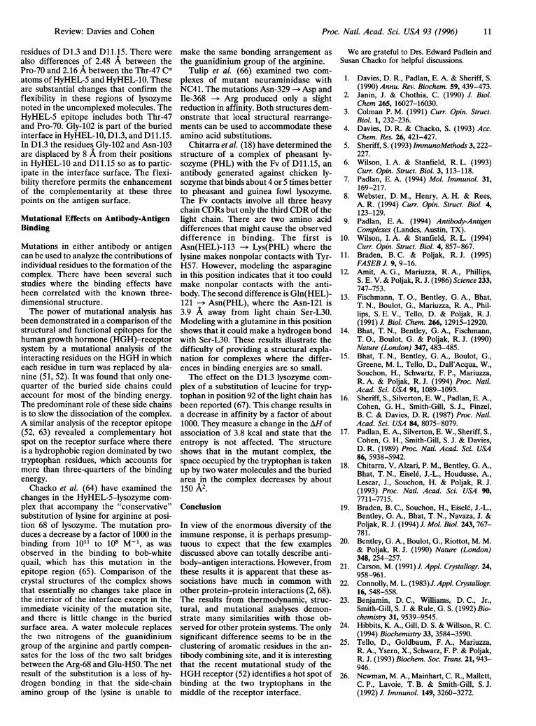
Table 1 shows a summary of the physicalproperties of the interfaces for the sixcomplexes for which coordinates are avail-able in the Protein Data Bank. It is clearthat a high percentage (34%) of the anti-body-contacting residues are aromatic incharacter; of these more than two-thirdsare tyrosine residues. In contrast, the con-tacting residues on the antigens containrelatively few that are aromatic. Fig. 2shows the contacting aromatic residuesobserved in the HyHEL-10-lysozymecomplex (7, 17). These include Tyr-H33,which penetrates the substrate bindinggroove of lysozyme.
Review: Davies and Cohen

Proc. Natl. Acad. Sci. USA 93 (1996)
FIG. 2. RIBBONS diagram (21) of the HyHEL-10-lysozyme complex showing the aromatic residues in the combining site that contact the antigen.This figure prepared by Susan Chacko.
Padlan (7, 45) also calculated from thex-ray data the fractional solvent exposuresof the individual amino acid species in theCDRs of seven antibodies and comparedthese with their corresponding exposureswhen in the framework region of the Fvfragments. He observed that the aromaticresidues tyrosine and tryptophan in theCDRs were significantly more solvent-exposed.
Epitope Mapping
Crystallographic investigations of anti-body-antigen complexes provide an op-portunity to compare predictions fromepitope mapping with direct observationof the contacting residues. There havebeen extensive studies of epitopes by theuse of peptides in which an antibody pro-duced in response to a protein antigen canbe examined for binding to peptides fromthat antigen. For HEL, these methodshave recently been reviewed (49). Theepitope for lysozyme with HyHEL-5 wassuccessfully predicted (50) based on theavailability of a library of avian lysozymesthat were presumed to have approxi-mately the same tertiary structure, butwith small differences in the amino acidsequences. This method is of course lim-ited by the diversity of the available li-brary. The application of mutagenesistechniques has now become a powerfulmethod for epitope mapping (51, 52).There has been an interesting applica-
tion of site-directed mutagenesis to distin-guish between two structures that hadbeen proposed for the protein HPr (53).The three-dimensional structure had beendetermined by two-dimensionalNMR andby x-ray diffraction (54, 55), and therewere major differences between the twostructures. The effect of mutations on thebinding of two antibodies, Jel42 and Jel44,revealed that when the putative epitopewas mapped on each of the proposedstructures, the epitope for Jel44 was onlyconsistent with the two-dimensionalNMR structure of HPr for which it gave acontiguous binding surface. In contrast,the model derived from x-ray diffractionproduced a scattered distribution of theseresidues in which some were buried andothers were surrounded by noninvolvedresidues. Subsequently the epitope pre-dicted in these experiments for Jel42 wasconfirmed by an x-ray diffraction analysis
of the complex of the Fab with HPr, inwhich the structure observed for HPr wasthat determined by the NMR analysis(35). Of the 14 amino acid residues thatinteract with the Jel42 binding site, 9 werecorrectly identified.The epitope on cytochrome c for a
monoclonal antibody has been defined byhydrogen exchange in two-dimensionalNMR (56). This method can be used for aprotein antigen when the protein is smallenough for the 1H NMR resonances to bedetermined. Eleven residues in three dif-ferent segments of sequence were identi-fied that formed a structurally relatedpatch on the surface with a water-accessible surface area of about 750 A2.There has been a preliminary report of thecrystallization of an antibody Fab to cy-tochrome c, both free and complexed withthe antigen (57).
Conformational Changes thatAccompany Complex Formation
The interaction of antibody with antigeninvolves conformational changes in boththe antibody and the antigen that canrange from insignificant to considerable.In general, the formation of a complex willadopt many of the characteristics of in-duced fit, similar to those seen in othermacromolecular interactions. Thechanges that may occur in the antibodyupon binding consist of combinations ofsimple side-chain movements, concertedmovements of individual CDRs, and dis-placement of VH relative to VL. Most ofthe information on conformationalchanges upon antigen binding has comefrom nonprotein antigens, where the an-tibody has also been crystallized in theabsence of antigen. Each of the kinds ofmovement described above has been ob-served for these complexes and these havebeen reviewed (10, 58, 59). Some of theconformational changes observed arequite large, with the largest VH-VL rear-rangement occurring in an Fab complexwith a human immunodeficiency virus pep-tide (59). Changes of this nature will con-siderably complicate attempts to modelantibody combining sites.For protein antigens, the antibody D1.3
has been examined as an unbound Fvfragment, as an Fab and an Fv complexedto lysozyme, and as Fab complexed toE225, an anti-idiotope. The differences
between the bound and unbound Fv struc-tures are minimal and include small ad-justments of the side chains together witha small movement of VH relative to VL(14). The interaction of the variable do-mains has been reviewed (5), and it wasnoted that the contacting surface betweenVH and VL contained #40% contributionfrom the CDRs. This can result in signif-icant differences in the angle of rotationbetween VH and VL, which varies from165° to 1800. Changes in this angle wereoriginally hypothesized for the NC41-neuraminidase complex (29), and substan-tial changes have now been observed inseveral instances when Fabs are com-plexed to small ligands (10, 59). The dif-ferences between unbound and boundD1.3 in its complex with E225 (20) includeseveral significant side-chain movementsof the D1.3 antibody relative to the un-complexed structure (40).We have analyzed four of the crystal
complexes with lysozyme that clearly dem-onstrate that the interaction of antibodywith antigen can produce significant con-formational changes in the antigen, mainlyin regions that are demonstrably flexible.Lysozyme alone can be crystallized in anumber of crystal forms. A comparison ofthe tetragonal (60), monoclinic (61), andtriclinic (62) forms, which provide fourindependent structures, demonstrates theflexibility of the surface loops. When thetwo independent molecules in the mono-clinic form are superimposed, they have arms deviation of 0.64 A for the Cc, atoms,but the Cc, atoms of Gly-71 and Gly-102 aredisplaced by 3.68 A and 3.40 A, respec-tively, from the corresponding position inthe other molecule. In a similar compar-ison of the triclinic and tetragonal mole-cules, with overall rms = 0.65 A, therelative displacements of the Co atoms ofThr-47 and Asn-103 are 2.73 A and 2.64 A,respectively. Residue Thr-47 in the tet-ragonal crystal makes a packing contactwith a neighboring molecule that mightexplain the observed conformational dif-ference for that residue.
In a similar manner we have comparedthe conformations of the lysozyme mole-cules bound to HyHEL-5 (69), HyHEL-10(17), D1.3 (15), and D11.15 (18). The rmsmatch against the mean set of coordinatesfor the ensemble was 0.54 A. The largestCa separation for the superimposed ly-sozymes was 8.17 A between the Gly-102
10 Review: Davies and Cohen
Proc. Natl. Acad. Sci. USA 93 (1996) 11
residues of D1.3 and D11.15. There werealso differences of 2.48 A between thePro-70 and 2.16 A between the Thr-47 Caatoms of HyHEL-5 and HyHEL-10. Theseare substantial changes that confirm theflexibility in these regions of lysozymenoted in the uncomplexed molecules. TheHyHEL-5 epitope includes both Thr-47and Pro-70. Gly-102 is part of the buriedinterface in HyHEL-10, D1.3, and D11.15.In D1.3 the residues Gly-102 and Asn-103are displaced by 8 A from their positionsin HyHEL-10 and D11.15 so as to partic-ipate in the interface surface. The flexi-bility therefore permits the enhancementof the complementarity at these threepoints on the antigen surface.
Mutational Effects on Antibody-AntigenBinding
Mutations in either antibody or antigencan be used to analyze the contributions ofindividual residues to the formation of thecomplex. There have been several suchstudies where the binding effects havebeen correlated with the known three-dimensional structure.The power of mutational analysis has
been demonstrated in a comparison of thestructural and functional epitopes for thehuman growth hormone (HGH)-receptorsystem by a mutational analysis of theinteracting residues on the HGH in whicheach residue in turn was replaced by ala-nine (51, 52). It was found that only one-quarter of the buried side chains couldaccount for most of the binding energy.The predominant role of these side chainsis to slow the dissociation of the complex.A similar analysis of the receptor epitope(52, 63) revealed a complementary hotspot on the receptor surface where thereis a hydrophobic region dominated by twotryptophan residues, which accounts formore than three-quarters of the bindingenergy.Chacko et al. (64) have examined the
changes in the HyHEL-5-lysozyme com-plex that accompany the "conservative"substitution of lysine for arginine at posi-tion 68 of lysozyme. The mutation pro-duces a decrease by a factor of 1000 in thebinding from 101l to 108 M-1, as wasobserved in the binding to bob-whitequail, which has this mutation in theepitope region (65). Comparison of thecrystal structures of the complex showsthat essentially no changes take place inthe interior of the interface except in theimmediate vicinity of the mutation site,and there is little change in the buriedsurface area. A water molecule replacesthe two nitrogens of the guanidiniumgroup of the arginine and partly compen-sates for the loss of the two salt bridgesbetween the Arg-68 and Glu-H50. The netresult of the substitution is a loss of hy-drogen bonding in that the side-chainamino group of the lysine is unable to
make the same bonding arrangement asthe guanidinium group of the arginine.
Tulip et al. (66) examined two com-plexes of mutant neuraminidase withNC41. The mutations Asn-329 -> Asp andIle-368 -- Arg produced only a slightreduction in affinity. Both structures dem-onstrate that local structural rearrange-ments can be used to accommodate theseamino acid substitutions.
Chitarra et al. (18) have determined thestructure of a complex of pheasant ly-sozyme (PHL) with the Fv of D11.15, anantibody generated against chicken ly-sozyme that binds about 4 or 5 times betterto pheasant and guinea fowl lysozyme.The Fv contacts involve all three heavychain CDRs but only the third CDR of thelight chain. There are two amino aciddifferences that might cause the observeddifference in binding. The first isAsn(HEL)-113 -- Lys(PHL) where thelysine makes nonpolar contacts with Tyr-H57. However, modeling the asparaginein this position indicates that it too couldmake nonpolar contacts with the anti-body. The second difference is Gln(HEL)-121 -- Asn(PHL), where the Asn-121 is3.9 A away from light chain Ser-L30.Modeling with a glutamine in this positionshows that it could make a hydrogen bondwith Ser-L30. These results illustrate thedifficulty of providing a structural expla-nation for complexes where the differ-ences in binding energies are so small.The effect on the D1.3 lysozyme com-
plex of a substitution of leucine for tryp-tophan in position 92 of the light chain hasbeen reported (67). This change results ina decrease in affinity by a factor of about1000. They measure a change in the AH ofassociation of 3.8 kcal and state that theentropy is not affected. The structureshows that in the mutant complex, thespace occupied by the tryptophan is takenup by two water molecules and the buriedarea in the complex decreases by about150 A2.
Conclusion
In view of the enormous diversity of theimmune response, it is perhaps presump-tuous to expect that the few examplesdiscussed above can totally describe anti-body-antigen interactions. However, fromthese results it is apparent that these as-sociations have much in common withother protein-protein interactions (2, 68).The results from thermodynamic, struc-tural, and mutational analyses demon-strate many similarities with those ob-served for other protein systems. The onlysignificant difference seems to be in theclustering of aromatic residues in the an-tibody combining site, and it is interestingthat the recent mutational study of theHGH receptor (52) identifies a hot spot ofbinding at the two tryptophans in themiddle of the receptor interface.
We are grateful to Drs. Edward Padlein andSusan Chacko for helpful discussions.
1. Davies, D. R., Padlan, E. A. & Sheriff, S.(1990) Annu. Rev. Biochem. 59, 439-473.
2. Janin, J. & Chothia, C. (1990) J. Biol.Chem 265, 16027-16030.
3. Colman P. M. (1991) Curr. Opin. Struct.Biol. 1, 232-236.
4. Davies, D. R. & Chacko, S. (1993) Acc.Chem. Res. 26, 421-427.
5. Sheriff, S. (1993) ImmunoMethods 3, 222-227.
6. Wilson, I. A. & Stanfield, R. L. (1993)Curr. Opin. Struct. Biol. 3, 113-118.
7. Padlan, E. A. (1994) Mol. Immunol. 31,169-217.
8. Webster, D. M., Henry, A. H. & Rees,A. R. (1994) Curr. Opin. Struct. Biol. 4,123-129.
9. Padlan, E. A. (1994) Antibody-AntigenComplexes (Landes, Austin, TX).
10. Wilson, I. A. & Stanfield, R. L. (1994)Curr. Opin. Struct. Biol. 4, 857-867.
11. Braden, B. C. & Poljak, R. J. (1995)FASEB J. 9, 9-16.
12. Amit, A. G., Mariuzza, R. A., Phillips,S. E. V. & Poljak, R. J. (1986) Science 233,747-753.
13. Fischmann, T. O., Bentley, G. A., Bhat,T. N., Boulot, G., Mariuzza, R. A., Phil-lips, S. E. V., Tello, D. & Poljak, R. J.(1991) J. Bio. Chem. 266, 12915-12920.
14. Bhat, T. N., Bentley, G. A., Fischmann,T. O., Boulot, G. & Poljak, R. J. (1990)Nature (London) 347, 483-485.
15. Bhat, T. N., Bentley, G. A., Boulot, G.,Greene, M. I., Tello, D., Dall'Acqua, W.,Souchon, H., Schwartz, F. P., Mariuzza,R. A. & Poljak, R. J. (1994) Proc. Natl.Acad. Sci. USA 91, 1089-1093.
16. Sheriff, S., Silverton, E. W., Padlan, E. A.,Cohen, G. H., Smith-Gill, S. J., Finzel,B. C. & Davies, D. R. (1987) Proc. Natl.Acad. Sci. USA 84, 8075-8079.
17. Padlan, E. A., Silverton, E. W., Sheriff, S.,Cohen, G. H., Smith-Gill, S. J. & Davies,D. R. (1989) Proc. Natl. Acad. Sci. USA86, 5938-5942.
18. Chitarra, V, Alzari, P. M., Bentley, G. A.,Bhat, T. N., Eisele, J.-L., Houdusse, A.,Lescar, J., Souchon, H. & Poljak, R. J.(1993) Proc. Natl. Acad. Sci. USA 90,7711-7715.
19. Braden, B. C., Souchon, H., Eisel6, J.-L.,Bentley, G. A., Bhat, T. N., Navaza, J. &Poljak, R. J. (1994)J. Mol. Biol. 243, 767-781.
20. Bentley, G. A., Boulot, G., Riottot, M. M.& Poljak, R. J. (1990) Nature (London)348, 254-257.
21. Carson, M. (1991) J. Appl. Crystallogr. 24,958-961.
22. Connolly, M. L. (1983)J. Appl. Crystallogr.16, 548-558.
23. Benjamin, D. C., Williams, D. C., Jr.,Smith-Gill, S. J. & Rule, G. S. (1992) Bio-chemistry 31, 9539-9545.
24. Hibbits, K. A., Gill, D. S. & Willson, R. C.(1994) Biochemistry 33, 3584-3590.
25. Tello, D., Goldbaum, F. A., Mariuzza,R. A., Ysern, X., Schwarz, F. P. & Poljak,R. J. (1993) Biochem. Soc. Trans. 21, 943-946.
26. Newman, M. A., Mainhart, C. R., Mallett,C. P., Lavoie, T. B. & Smith-Gill, S. J.(1992) J. Immunol. 149, 3260-3272.
Review: Davies and Cohen

Proc. Natl. Acad. Sci. USA 93 (1996)
27. Varghese, J. N., Laver, W. G. & Colman,P. M. (1983) Nature (London) 303, 35-40.
28. Varghese, J. N. & Colman, P. M. (1991) J.Mol. Biol. 221, 473-486.
29. Colman, P. M., Laver, W. G., Varghese,J. N., Baker, A. T., Tulloch, P. A., Air,G. M. & Webster, R. G. (1987) Nature(London) 326, 358-363.
30. Tulip, W. R., Varghese, J. N., Webster,R. G., Air, G M., Laver, W. G. & Colman,P. M. (1989) Cold Spring Harbor Symp.Quant. Biol. 54, 257-263.
31. Tulip, W. R., Varghese, J. N., Laver,W. G., Webster, R. G. & Colman, P. M.(1992) J. MoL Bio. 227, 122-148.
32. Sheriff, S., Hendrickson, W. A. & Smith,J. L. (1987) J. Mol. Biol. 197, 273-296.
33. Malby, R. L., Tulip, W. R., Harley, V. R.,McKim-Breschkin, J. L., Laver, W. G.,Webster, R. G. & Colman, P. M. (1994)Structure 2, 733-746.
34. Colman, P. M. (1994) Protein Sci. 3, 1687-1696.
35. Prasad, L., Sharma, S., Vandonselaar, M.,Quail, J. W., Lee, J. S., Waygood, E. B.,Wilson, K. S., Dauter, Z. & Delbaere,L. T. J. (1993) J. Biol. Chem. 268, 10705-10708.
36. Jerne, N. K. (1974) Ann. Immunol. C 125,373-389.
37. Gaulton, G. N. & Greene, M. I. (1986)Annu. Rev. Immunol. 4, 253-280.
38. Pan, Y., Yuhasz, S. C. & Amzel, L. M.(1995) FASEB J. 9, 43-49.
39. Garcia, K. C., Ronco, P. M., Verroust,P. J., Brunger, A. T. & Amzel, L. M.(1992) Science 257, 502-507.
40. Tello, D., Einstein, E., Schwarz, F. P.,Goldbaum, F. A., Fields, B. A., Mariuzza,R. A. & Poljak, R. J. (1994) J. Mol. Recog-nit. 7, 57-62.
41. Ross, P. D. & Subramanian, S. (1981)Biochemistry 20, 3096-3102.
42. Ban, N., Escobar, C., Garcia, R., Hasel,K., Day, J., Greenwood, A., McPherson,A. (1994) Proc. Natl. Acad. Sci. USA 91,1604-1608.
43. Evans, S. V., Rose, D. R., To, R., Young,N. M. & Bundle, D. R. (1994)J. Mol. Biol.241, 691-705.
44. Kabat, E. A., Wu, T. T. & Bilofsky, H.(1977) J. Biol. Chem. 252, 6609-6616.
45. Padlan, E. A. (1990) Proteins Struct.Funct. Genet. 7, 112-124.
46. Janin, J. & Chothia, C. (1990) J. Biol.Chem. 265, 16027-16030.
47. Mian, I. S., Bradwell, A. R. & Olson, A. J.(1991) J. Mol. Biol. 217, 133-151.
48. Lea, S. & Stuart, D. (1995) FASEB J. 9,87-93.
49. Grivel, J.-C. & Smith-Gill, S. J. (1995) inStructure ofAntigens, ed. Van Regenmo-tel, M. H. (CRC, Boca Raton, FL), Vol.3,p. 91.
50. Smith-Gill, S. J., Wilson, A. C., Potter, M.,Prager, E. M., Feldmann, R. J. & Mainhart,C. R. (1982) J. Immunol. 128, 314-322.
51. Cunningham, B. C. & Wells, J. A. (1993)J. Mol. Biol. 234, 554-563.
52. Wells, J. A. (1996) Proc. Natl. Acad. Sci.USA 93, 1-6.
53. Sharma, S., Georges, F., Klevit, R. E.,Delbaere, L. T. J., Lee, J. S. & Waygood,E. B. (1991) Proc. Natl. Acad. Sci. USA 88,4877-4881.
54. Klevit, R. E. & Waygood, E. B. (1986)Biochemistry 25, 7774-7781.
55. El-Kabbani, 0. A., Waygood, E. B. &Delbaere, L. T. (1987) J. Biol. Chem. 262,12926-12929.
56. Paterson, Y., Englander, S W. & Roder,H. (1990) Science 249, 755-759.
57. Mylvaganam, S. E., Paterson, Y., Kaiser,K., Bowdish, K. & Getzoff, E. D. (1991) J.Mol. Biol. 221, 455-462.
58. Davies, D. R. & Padlan, E. A. (1992) Curr.Biol. 2, 254-256.
59. Stanfield, R. L., Takimoto-Kamimura,M., Rini, J. M., Profy, A. T. & Wilson,I. A. (1993) Structure 1, 83-93.
60. Kurinov, I. V. & Harrison, R. W. (1995)Acta Crystallogr. D 51, 98-109.
61. Harata, K. (1994) Acta Crystallogr. D 50,250-257.
62. Ramanadham, M., Sieker, L. C. & Jensen,L. H. (1990) Acta Crystallogr. B 46, 63-69.
63. Clackson, T. & Wells, J. A. (1995) Science267, 383-386.
64. Chacko, S., Silverton, E., Kam-Morgan,L., Smith-Gill, S., Cohen, G & Davies, D.(1995) J. Mol. Biol. 245, 261-274.
65. Lavoie, T. B., Kam-Morgan, L. N. W.,Mallet, C. P., Schilling, J. W., Prager,E. M., Wilson, A. C. & Smith-Gill, S. J.(1990) in Use of X-ray Crystallography inthe Design ofAntiviral Agents, eds. Laver,W. & Air, G. M. (Academic, San Diego),p. 213.
66. Tulip, W. R., Varghese, J. N., Webster,R. G., Laver, W. G. & Colman, P. M.(1992) J. Mol. Biol. 227, 149-159.
67. Ysern, X., Fields, B. A., Bhat, T. N., Gold-baum, F. A., Dall'Acqua, W., Schwarz,F. P., Poljak, R. & Mariuzza, R. A. (1994)J. Mol. Biol. 238, 496-500.
68. Jones, S. & Thornton, J. M. (1996) Proc.Natl. Acad. Sci. USA 93, 13-20.
69. Cohen, G. H., Sheriff, S. & Davies, D. R.(1996) Acta Crystallogr. D, in press.
12 Review: Davies and Cohen