review the cell biology of inflammation: from common
TRANSCRIPT

REVIEW
The cell biology of inflammation: From commontraits to remarkable immunological adaptationsHelen Weavers1* and Paul Martin1,2,3*
Tissue damage triggers a rapid and robust inflammatory response in order to clear and repair a wound. Remarkably, many ofthe cell biology features that underlie the ability of leukocytes to home in to sites of injury and to fight infection—most ofwhich are topics of intensive current research—were originally observed in various weird and wonderful translucentorganisms over a century ago by Elie Metchnikoff, the “father of innate immunity,” who is credited with discoveringphagocytes in 1882. In this review, we use Metchnikoff’s seminal lectures as a starting point to discuss the tremendous varietyof cell biology features that underpin the function of these multitasking immune cells. Some of these are shared by other celltypes (including aspects of motility, membrane trafficking, cell division, and death), but others are more unique features ofinnate immune cells, enabling them to fulfill their specialized functions, such as encapsulation of invading pathogens, cell–cellfusion in response to foreign bodies, and their self-sacrifice as occurs during NETosis.
IntroductionTo survive physical damage or other more subtle insults, ourbodies have the remarkable capacity to repair and replacedamaged tissues (Eming et al., 2014). Key to this, and super-imposed upon the repair machinery, is a rapidly activated in-flammatory response that has evolved to combat potentialmicrobialinvaders as well as our body’s own aberrant cells. A range of dif-ferent traumas—from a scratch or surgical wound (with or withoutinfection) through to UV damage from sunburn and even the ini-tiation and progression stages of cancer—will all trigger a variationon a theme of this inflammatory response (Fig. 1 and Box 1).
In the case of a small local incisional wound involving dam-age to tissues that is deeper than the epidermis and transectssubcutaneous blood vessels (Fig. 1), the defect will initiallybe plugged with a fibrin clot containing platelets and neu-trophils from the spilled blood to make a transient protectivescab (Martin, 1997). Various “alarm” signals (damage-associatedmolecular patterns; Niethammer, 2016) will then be releasedfrom damaged or necrotic cells; these signals, together withgrowth factor attractants from degranulating platelets, lead tothe recruitment of leukocytes, both macrophages from the localenvirons and then later neutrophils and monocytes from nearbyvessels. Immune cell recruitment subsequently becomes self-amplifying because these cells release chemokine attractants,which draw in more immune cells (Eming et al., 2007). If thewound becomes at all chronic or infected, then some degree of an
adaptive immune response may also be triggered (see Box 2).After the tissue insult has ceased or been destroyed, thenmechanisms for resolution of the inflammatory response arepivotal to prevent unnecessary host tissue damage caused bychronic inflammation.
While the main functions of the inflammatory response areto destroy invaders (as well as aberrant cells) and to clear awaycell and matrix debris, it has acquired other roles that mean italso orchestrates several responses by other cell lineages duringthe repair process, including wound angiogenesis and depositionof a collagen scar by wound fibroblasts (Gurevich et al., 2018;Eming et al., 2017). Not surprisingly, these inflammatory cellscan also be subverted by some infective agents and by cancercells to work for the “invader” in various ways, rather than beingsolely beneficial to the host (Pagan and Ramakrishnan, 2018;Swierczak and Pollard, 2020).
Elie Metchnikoff (1845–1916) is often described as the fatherof innate immunity and inflammation; he studied all aspects ofthe inflammatory response, often by live imaging studies, in avariety of organisms from Daphnia through to amphibia andsome higher vertebrates. In this review, we revisit some ofMetchnikoff’s original anecdotal observations described in hislectures of the late 1800s (translated in Metchnikoff, 1968) andupdate them with new cell and molecular insights derived fromin vitro studies and in vivo observations (see Box 3); much of ournew knowledge comes from translucent model organisms—no
.............................................................................................................................................................................1School of Biochemistry, Biomedical Sciences, University of Bristol, Bristol UK; 2School of Physiology, Pharmacology and Neuroscience, Biomedical Sciences, University ofBristol, UK; 3School of Medicine, Cardiff University, Cardiff, UK.
*H. Weavers and P. Martin contributed equally to this paper; Correspondence to Helen Weavers: [email protected]; Paul Martin: [email protected].
© 2020Weavers and Martin. This article is distributed under the terms of an Attribution–Noncommercial–Share Alike–NoMirror Sites license for the first six months afterthe publication date (see http://www.rupress.org/terms/). After six months it is available under a Creative Commons License (Attribution–Noncommercial–Share Alike 4.0International license, as described at https://creativecommons.org/licenses/by-nc-sa/4.0/).
Rockefeller University Press https://doi.org/10.1083/jcb.202004003 1 of 14
J. Cell Biol. 2020 Vol. 219 No. 7 e202004003
Dow
nloaded from http://rupress.org/jcb/article-pdf/219/7/e202004003/1383771/jcb_202004003.pdf by guest on 11 D
ecember 2021

longer starfish and frogs as in Metchnikoff’s day (Fig. 2, A andB), but now the genetically tractable, early developmental stagesof Drosophila and zebrafish (Fig. 2, C and D). We outline not onlyhow the cells of the inflammatory response share much of theircell biology with other cell lineages, including aspects of motil-ity, phagocytosis, trafficking, and signaling, but also how vari-ous leukocytic cell types possess some more unusual featuresthat equip them for their specialized roles (e.g., neutrophil ex-tracellular traps [NETs], encapsulation, and extravasation;Fig. 3). Given that too little or too much inflammation can be theroot cause of many human pathologies, our aim here is tohighlight those cell biology aspects of the inflammatory responsethat are potential targets for therapeutic modulation to make itbetter at killing tissue enemies and less harmful to host tissues.
Leukocyte cell motilityInnate immune cells are motile from the outset. Their first mi-grations are to disperse themselves throughout the various
embryonic tissues at developmental stages (Ginhoux and Guilliams,2016; Wood and Martin, 2017). Subsequently, they can remainrelatively stationary or patrol either locally or by traveling inthe vasculature (Auffray et al., 2007). Innate immune cellstend only to migrate in a directed way if an epithelial barrierlayer has been breached or an infection has arisen, and theseinflammatory migratory episodes need to be very tightly regu-lated. Neutrophils andmacrophages migrate within tissues usingrather different strategies, amoeboid-like for neutrophils andmesenchymal with adhesion-dependent tethering and protrusion
Figure 1. Schematic of a typical wound inflamma-tory response. Soon after a platelet plug has tempo-rarily sealed a wound (1), the wound inflammatoryresponse kicks in, with innate immune cells drawn fromlocal resident populations and by recruitment fromnearby vessels (2) in order to counter potential in-fections at this barrier breach. The recruited innate im-mune cells deploy a variety of antimicrobial weapons tokill or inactivate invading pathogens, including produc-tion of ROS and the release of NETs. There is an ac-companying but less well-characterized adaptiveimmune cell response (3), and as these two defensemechanisms protect the exposed wound, tissue repairmechanisms, including resurfacing with a new epidermallayer, proceed in order to restore barrier integrity (4).DAMP, damage-associated molecular pattern; PAMP,pathogen-associated molecular pattern.
Box 1. Leukocyte subtypes and developmental origin
In this review, we focus mostly on two of Metchnikoff’s favorite cells, neu-trophils and macrophages; however, for context, we provide a brief intro-duction to the larger family of inflammatory cell lineages, all of which derivefrom multipotential hematopoietic stem cells in the bone marrow, which inturn give rise to the common myeloid and common lymphoid progenitorlineages.
Mast cells and myeloblasts originate from common myeloid progen-itors; myeloblasts in turn give rise to the three granulocyte lineages (baso-phils, eosinophils, and neutrophils) as well as monocytes, the latter of whichare termed macrophages after they leave the vasculature. Collectively, thesecells are the “innate immune cells.”
From common lymphoid progenitors come all of the adaptive immunecell lineages, including B and T cells.
At any site of tissue damage or infection, innate immune cells arerecruited, first neutrophils and then monocyte/macrophages, from localtissue resident cells and by extravasation from vessels. Eosinophils are re-cruited also, but in significantly lower numbers. It is possible for T cells to berecruited in significant numbers, but adaptive immune cells generally appearto become significant players only when inflammation persists and becomeschronic (see Box 2).
Box 2. Cross-talk with the adaptive immune system
Initially, the study of innate and adaptive immune systems clashed amid ri-valry over which was the pivotal system for protecting the body from in-fection. The award of the Nobel Prize in Medicine or Physiology jointly to PaulEhrlich and Metchnikoff in 1908 was a clear indication that the general im-munology community had realized that the two “immunities” were equallyimportant.
Adaptive immune cells are much less often investigated than innateimmune cells in a wound inflammatory context. However, it is clear that T regcells are recruited to wounds, and their knockdown can significantly impairskin healing (Nosbaum et al., 2016). A sentinel subpopulation of γδ T cells,termed dendritic epidermal T cells (DETCs), reside within the epidermallayers of mammalian skin, and they rapidly switch morphology from sensorydendritic to an active, rounded phenotype in the vicinity of a wound. Theseactivated DETCs play several roles in repair, and in mice, where this lineage isdeleted or where it cannot become activated, wound repair is severelycompromised (Havran and Jameson, 2010). DETCs directly influence woundedge keratinocyte survival and proliferation via Keratinocyte Growth Factor(KGF) and IGF-1 signals (Jameson et al., 2002; Ramirez et al., 2015); however,they also have significant impact on the wound inflammatory response,possibly via release of CCL3 and other inflammatory cell activators, since intheir absence macrophage (but not neutrophil) recruitment to the wound isseverely dampened (Ramirez et al., 2015). In human skin, γδ T resident cellsappear to play a role similar to that of murine DETCs upon tissue damage; butin a chronic wound scenario, they and the later recruited T cell populationsappear to lose their capacity for activation (Toulon et al., 2009), and this mayindirectly impact their regulation of innate immune cell recruitment andbehaviors at the wound site. In the example of granuloma formation in re-sponse to Tuberculosis (TB), significant numbers of T cells are recruited anddifferentiate into T helper type 1 effector cells that secrete IFN-γ and TNF-αto drive enhancedmicrobicidal activity by macrophages (Pagan and Ramakrishnan,2018).
Weavers and Martin Journal of Cell Biology 2 of 14
Metchnikoff’s inflammation observations revisited https://doi.org/10.1083/jcb.202004003
Dow
nloaded from http://rupress.org/jcb/article-pdf/219/7/e202004003/1383771/jcb_202004003.pdf by guest on 11 D
ecember 2021

contraction for macrophages, making neutrophils somewhatfaster (up to 6 µm/min), with macrophages lagging behind atspeeds of seldom up to 2 µm/min in studies of translucent ze-brafish larvae (Barros-Becker et al., 2017).
Metchnikoff and his colleagues realized that leukocytes weremotile and described how they “put out protoplasmic processesto move from place to place” (Fig. 3 A). In fact, he and othersundertook many experiments demonstrating how leukocyteshad “chemotactic properties” in response to infection and tissuedamage, while realizing that they did not yet have the tools touncover what the attractant signals were. Now we know thatbacterial components (including Lipopolysaccharide [LPS] andformylated peptides) as well as early tissue damage-associatedmolecular patterns (ATP, H2O2, and HMGB1) and later chemo-kines (interleukin [IL]-1β, IL-17, IL-8, and TNF-α) are just some ofthe attractants for inflammatory cells (Eming et al., 2007, 2014).
We also know much about how the responding cell’s cyto-skeleton is regulated in order to move toward these cues; just asfor other motile cells, Rho family small GTPases regulate theiractin cytoskeleton (Jones et al., 1998). If Rac is inhibited inDrosophilamacrophages, these cells fail to make proper lamellaeand cannot efficiently migrate to a wound. Conversely, if Rho isinhibited, although macrophage-directed migration is unper-turbed, the immune cells cannot contract to detach their trailinguropod and thus remain tethered to the spot (Stramer et al.,2005). Spatial activation of these small GTPase switches enables
directed migration, for example, as highlighted in zebrafishlarval experiments in which light activation of a geneticallyencoded Rac can artificially turn a neutrophil in vivo (Yooet al., 2010).
Downstream effectors of Rho family signaling switches arecritical in leukocyte migratory polarity; indeed, WASp, whichcoordinates actin polymerization via the Arp2/3 complex down-stream of Cdc42, is pivotal for neutrophil-directed migration(Jones et al., 2013), and mutations in this gene lead to the clinicalsyndrome Wiskott Aldrich syndrome, characterized by chronicinfections (Candotti, 2018). Most recently, studies in migratingDrosophila macrophages suggest that persistent migration to-ward a target is largely driven by flow of the actin networkbehind the leading edge of the cell (Yolland et al., 2019). How-ever, actin is not the only cytoskeletal player involved in guidingleukocyte migration; disruptions in microtubule dynamics ininnate immune cells of flies, zebrafish, andmammals all lead to aless directed, more “drunken walk” migration to targets (Reddet al., 2006; Xu et al., 2005; Stramer et al., 2010).
For no other migratory cell—barring perhaps germ cells orthe axonal growth cones of neurons in a developing embryo—isthe process of target navigation as complex as for a leukocyte enroute to a site of inflammation. As will be discussed, before,during, and after extravasation they are exposed to multipledifferent chemoattractants with overlapping gradients in spaceand time. There is some evidence that theremight be a hierarchy
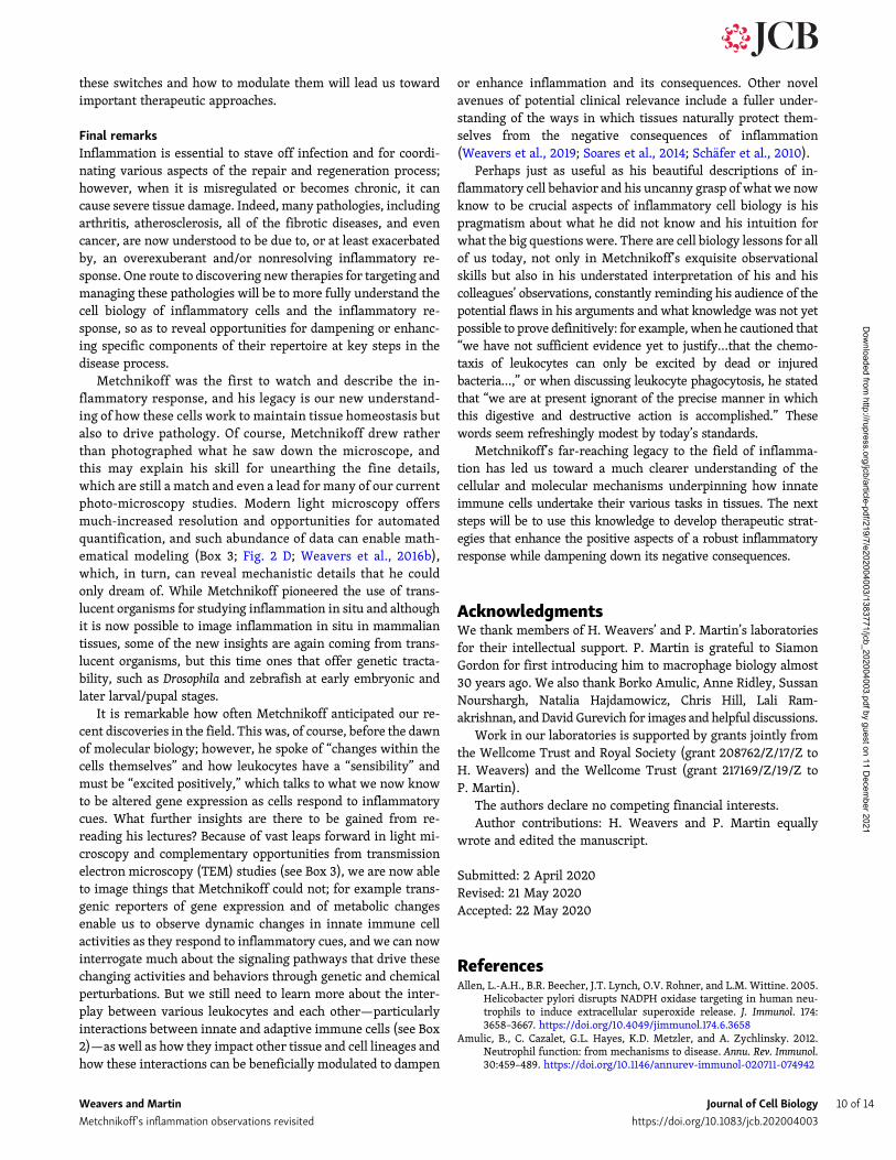
Figure 2. The “father of natural immunity,” ElieMetchnikoff made remarkable observations of thewound inflammatory response. (A and B) As early as1892, Metchnikoff (A) used translucent organisms toobserve the inflammatory response to damage or in-fection, as in B, where an axolotl fin was injured with aneedle coated in carmine and migratory cells were seento accumulate around the injured spot, “englobing” thecolored granules and the debris of the dying cells. (C andD) In current research, translucent and genetically tractablemodels such as the zebrafish (C) and fruit fly Drosophila (D)are now adding further mechanistic insights into these in-flammatory processes. (C) 3 d after fertilization, a translu-cent transgenic zebrafish larva with fluorescently taggedneutrophils (green) andmacrophages (red) was subjected toa needle wound to the flank, enabling live imaging of thewound inflammatory response. (D) A series of time-lapseimages fromawoundedDrosophila pupalwing enables high-resolution spatio-temporal tracking of innate immune cellbehavior. Images in A and B are adapted from Metchnikoff(1921) and Metchnikoff (1968), respectively. Images in Cand D are courtesy of David Gurevich and Helen Weavers,respectively.
Weavers and Martin Journal of Cell Biology 3 of 14
Metchnikoff’s inflammation observations revisited https://doi.org/10.1083/jcb.202004003
Dow
nloaded from http://rupress.org/jcb/article-pdf/219/7/e202004003/1383771/jcb_202004003.pdf by guest on 11 D
ecember 2021

of signal integration and prioritization (Foxman et al., 1997, 1999;McDonald et al., 2010; Moreira et al., 2010), which may in turnbe facilitated by receptor desensitization (see trafficking sectionbelow). There are likely to be other reinforcing strategies thatenable a leukocyte to persist in its path toward a particular cueand to not be distracted; for example, ATP release by neutrophilsthemselves appears to amplify attractant signals from othersources (Chen et al., 2006), while controlled calcium influx ap-pears to reinforce what is the leading edge in neutrophils mi-grating to a wound in a zebrafish larva (Beerman et al., 2015).
In most tissues, there is a resident population of some innateimmune cell lineages, but in tissues served by vessels, localleukocytes become hugely augmented by cells drawn in from thecirculation; these cells must first extravasate across the vesselwall (Fig. 3 B). Metchnikoff (1968) described how he and otherresearchers first observed in tadpoles and in the frog mesenteryhow leukocytes are drawn to the vessel periphery and then takeactive “passage” through the vessel wall at sites of inflammation.Using various experimental strategies, including some crudedrug blocking experiments, they shrewdly inferred that this
Figure 3. Innate immune cells exhibit both shared (with non-immune lineages) and specialized cell biology. A central schematized innate immune cellexhibiting both shared (upper panels) and specialized (lower, blue shaded zone) immune cell biology. Here, Metchnikoff’s drawings (Metchnikoff, 1893) arepartnered against current light microscopic and TEM images of several aspects of cell biology that we discuss in this review article. (A)Modern images depictmigrating macrophage cells, immunostained for tubulin (green) and actin (red; courtesy of Anne Ridley). (B) GFP neutrophils extravasating through the pericytelayer (red) of murine venules (courtesy of Sussan Nourshargh). (C) Phagocytosing Drosophila hemocytes with cytoplasm (green) and nuclei (red) labeled(courtesy of H.Weavers) and TEM image of a secreting neutrophil (courtesy of Natalia Hajdamowicz and Chris Hill). (D) Dividing Drosophila hemocytes with theactin (green) and tubulin (red) cytoskeleton labeled (courtesy of H. Weavers). (E) TEM of an apoptosing neutrophil (courtesy of Natalia Hajdamowicz and ChrisHill). (F) NETosing neutrophils extruding DNA/chromatin (red; courtesy of Borko Amulic). (G) TEM of macrophage aggregation (courtesy of Lali Ramakrishnan)and fusing zebrafish macrophages to give a foreign body giant cell with several GFP-tagged (green) nuclei in a common cytoplasm (red; courtesy of DavidGurevich).
Weavers and Martin Journal of Cell Biology 4 of 14
Metchnikoff’s inflammation observations revisited https://doi.org/10.1083/jcb.202004003
Dow
nloaded from http://rupress.org/jcb/article-pdf/219/7/e202004003/1383771/jcb_202004003.pdf by guest on 11 D
ecember 2021

process, which they called “diapedesis,” also involved active“assistance” by the vessel wall cells.
Now, modern murine genetic approaches, combined withintravital microscopy and complemented by in vitro flow stud-ies, have revealed many of the key steps that underpin diape-desis (Ley et al., 2007). A cascade of events beginning withtethering of leukocytes to the luminal aspect of the vessel wall isfollowed by firmer, integrin-mediated adhesion to endothelialcells and leukocytes crawling to find an exit point. Cells thenmigrate either through (transcellular) or between (paracellular)adjacent endothelial cells in the vessel wall, with breaching ofjunctions being the more common route and generally occurringat sites where three endothelial cells meet (Burns et al., 1997).Intravital microscopy has revealed that after traversing throughthe endothelial cell layer, leukocytes probe for regions in thebasement membrane with lower ECM density corresponding togaps in the pericyte sheath (Proebstl et al., 2012; Girbl et al.,2018). Adhesion and junction-related molecules expressed byboth immune cells and endothelial cells are (not surprisingly)rate limiting, as revealed by mice deficient in the junctionaladhesion molecules PECAM-1 (Thompson et al., 2001), JAM-A(Woodfin et al., 2007, 2009), and JAM-C (Woodfin et al., 2011),all of which exhibit severe deficiencies in diapedesis. As pre-dicted by Metchnikoff and colleagues, the vessel wall cell layersthemselves are active participants in diapedesis. Rho signaling isagain pivotal for enabling endothelial cells to both pull apartfrom one another as immune cells squeeze through (Cerutti andRidley, 2017) and for contraction of the transmigration pore atthe end of diapedesis (Heemskerk et al., 2016). A recent study inDrosophila pupae identified a period when fly macrophages ac-tively extravasate from circulating hemolymph through wingveins toward a laser wound, thus opening up this genetically
tractable model as a tool for performing genome-wide screens toreveal more key players in this process (Thuma et al., 2018).
Metchnikoff was somewhat noncommittal about whether ornot leukocytes were able to actively leave a site of inflammationafter their purpose there was over, and still the mechanismsleading to inflammatory cell resolution—even whether it is anactive process—are somewhat controversial. A large number ofneutrophils drawn to a site of inflammation will undergo apo-ptosis and be phagocytosed by macrophages, but both neu-trophils and macrophages also have the capacity to reversemigrate away from a site of inflammation rather than die(Mathias et al., 2006). For example, in a murine sterile hepaticinjury, many neutrophils are seen leaving the wound site andreentering the vasculature and migrating via the lungs beforeentering the bonemarrow (Wang et al., 2017). There also exists acomplex array of disparate “resolvins,” from lipids through toprotein families, that appear to actively encourage immune cellresolution (Serhan and Levy, 2018; Cash et al., 2014). And ze-brafish studies are revealing additional mechanisms that eitherretain cells at the wound site or are part of the machinery thataids their resolution by sequestering or breaking down attrac-tants (Pase et al., 2012; Isles et al., 2019).
Phagocytosis of pathogens, corpses, and debrisUnlike his contemporary pathologists, who perceived patho-gen uptake by leukocytes to be a passive process, Metchnik-off’s early observations suggested that particle engulfmentwas an active process leading to pathogen destruction withinvacuoles (Metchnikoff, 1968; Fig. 3 C). The term phagocytosis isnow well established for describing the active cellular uptakeof particulates within a plasma membrane “envelope” for re-moval of cell and matrix debris (e.g., apoptotic neutrophils) aswell as pathogens in a receptor-mediated fashion (for recentreviews, see Gordon, 2016; Morioka et al., 2019). Macrophagesare voracious phagocytes; Metchnikoff described seeing them“literally crammed full of foreign particles.” Phagocytosisoccurs not only at sites of tissue damage and infection but alsoduring developmental sculpting of many body tissues (Weaverset al., 2016a; Munro et al., 2019) and during normal tissue ho-meostasis (Arandjelovic and Ravichandran, 2015), whenever largenumbers of unwanted or spent cells must be cleared (Moriokaet al., 2019).
The molecular mechanisms underlying phagocytosis havebeen intensively studied over recent decades; it has emerged as ahighly orchestrated process requiring a series of sequentialoverlapping steps, including but not limited to particle recog-nition by cell surface receptors (Ostrowski et al., 2016; Lim et al.,2017), engulfment, and phagosome maturation along with cy-toskeletal remodeling (Gordon, 2016). The complexity of phag-ocytosis is becoming increasingly apparent and has been wellreviewed elsewhere (Gordon, 2016; Morioka et al., 2019; Elliottand Ravichandran, 2016), so we only highlight a few interestingaspects here.
Leukocytes are equipped with a vast array of surface re-ceptors, allowing them to recognize a huge variety of particles;the exact repertoire of these receptors varies across cell types aswell as tissues and most likely reflects local adaptation. While
Box 3. A revolution in imaging technologies has transformed our un-derstanding of leukocyte cell biology
Recent technological advances have heralded a new era in state-of-the-arthigh-resolution 3D and 4D imaging, which has enabled many of these“modern updates” on Metchnikoff’s observations. Confocal microscopy isnow routine in most research laboratories, allowing living tissues to be im-aged with high spatio-temporal resolution to discern molecular and cellularfeatures in exquisite detail. Multiphoton microscopy uses even longerwavelength photons (which are lower in energy and penetrate more deeply),enabling deeper imaging of a sample while creating less tissue damage. Morerecently, light-sheet fluorescence microscopy has revolutionized in vivo bi-ological imaging by using planar illumination strategies to further minimizephototoxicity while enabling rapid 3D imaging over longer time periods and atsignificant depth. These technological leaps, together with advances in cel-lular labeling approaches, mean that while in vivo imaging remains easiest intranslucent laboratory organisms (such as Drosophila and zebrafish), “intra-vital microscopy” now enables dynamic cell behaviors to be visualized also inthe normally opaque body tissues of more traditional mammalian models(such as the mouse). Moreover, electron microscopy methods can these daysbe integrated in a correlative way with live confocal (light) microscopy in thesame sample to capture dynamic cellular processes (e.g., leukocyte diape-desis) at the ultrastructural level. There has also been an important movefrom qualitative to quantitative analysis of imaging data, and the increasingapplication of mathematical/computational modeling to study (and simulate)cell dynamics (Liepe et al., 2012) is beginning to transform our understandingof leukocyte behavior (see main text).
Weavers and Martin Journal of Cell Biology 5 of 14
Metchnikoff’s inflammation observations revisited https://doi.org/10.1083/jcb.202004003
Dow
nloaded from http://rupress.org/jcb/article-pdf/219/7/e202004003/1383771/jcb_202004003.pdf by guest on 11 D
ecember 2021

macrophages are professional phagocytes and readily phagocy-tose various particles, neutrophils have a more varied skill setbut are more limited in their phagocytic capacity (Yang et al.,2019). Not surprisingly, the exact mechanism of phagocytosisdepends on the nature of the interaction between phagocytic celland target. This can be directly mediated by detection ofpathogen-associated molecular patterns on the pathogen surface(by toll-like receptors or C-type lectin receptors) or by recog-nition of phosphatidylserine on apoptotic cells via scavengerreceptors (Gordon, 2016; Elliott and Ravichandran, 2016). Up-take can also occur indirectly via opsonins, which promote Fcregion of IgG (FcγR)-mediated phagocytosis of IgG-opsonizedparticles or complement receptor–mediated phagocytosis (Gordon,2016). Following particle recognition, short-lived dynamic podosome-like structures are formed within the nascent phagosome (me-diated by local PtdIns(3,4,5)P3 production) to promote phago-cytic receptor activation and facilitate membrane remodeling toenvelope and engulf the target (Ostrowski et al., 2019). Studies inzebrafish have shown that phagocytic efficiency is both lineageand site specific, with macrophages efficiently clearing patho-gens from within a fluid-filled body cavity (e.g., in blood), whileneutrophils appear only able to clear surface-associated microbes(Colucci-Guyon et al., 2011).
Once activated, phagocytes use reactive oxygen and nitrogenmetabolites to kill ingested microbes. Phagocytosis triggers theassembly of NADPH oxidase on the phagosome membrane forreactive oxygen species (ROS) production (via Rac and LC3-mediated stabilization of NOX2), which, together with deliv-ered antimicrobials, creates a toxic environment for killingpathogens. ROS are also released extracellularly to kill patho-gens that have not yet been phagocytosed (Dupre-Crochet et al.,2013). Strikingly, many pathogens are able to subvert phago-some maturation to aid their own growth and survival (Armstrongand Hart, 1971; Myrvik et al., 1984; Brubaker et al., 2015; Zaman andColley, 1972); a classical example isHelicobacter pylori, which evadesdestruction by interfering with the targeting of NADPH oxidase tothe phagosomal membrane (Allen et al., 2005). Mycobacterium tu-berculosis also resists oxidative stress through the production ofreductases that degrade phagocyte oxidants and the inhibition ofthe respiratory burst (Carranza and Chavez-Galan, 2019). Such so-phisticated microbial evasion strategies were even observed byMetchnikoff, who stated that “in certain diseases the leucocytes takein a number of bacteria, such as tubercle bacilli or the bacilli ofswine erysipelas…a few of which may be digested while the othersresist the digestive action of the leucocytes, multiply in the cells andfinally invade the whole organism” (Metchnikoff, 1968).
Despite these killing mechanisms, a significant proportion ofpathogen proteins are not degraded and instead remain associ-ated with a membrane fraction of macrophages, which enablespresentation of antigen-derived peptides to cells of the adaptiveimmune system (Stuart and Ezekowitz, 2005). Remarkably,dendritic cells can even present bacterial antigens (e.g., thosefromMycobacterium) derived from ingested infected neutrophilsjust as efficiently as those derived from direct pathogen uptake(Blomgran and Ernst, 2011). As eluded to earlier, phagocytosisnot only functions to remove obsolete cells and pathogens, but italso has important regulatory functions within the engulfing
leukocyte, including priming to becomemore wound responsive(Weavers et al., 2016a). Moreover, macrophages at wound sitesare shifted into an anti-inflammatory tissue-remodeling statefollowing phagocytosis of apoptotic neutrophils and undergo adramatic metabolic shift that sustains actin polymerization andcontinued corpse uptake (Morioka et al., 2018).
Endocytic episodesAs leukocytes navigate to their target site, the adhesive contactsthat they form with their substratum must be dynamicallyturned over to permit migration (Maritzen et al., 2015). Sincethe degradative turnover of adhesion receptors is rather slow(the half-life of surface-labeled integrins is 12–24 h), rapidturnover of adhesive contacts instead relies upon the morespeedy endocytic recycling of adhesion molecules (Paul et al.,2015). It appears that calcium transients in migrating neu-trophils may direct integrin recycling from the trailing to theleading edge and permit detachment of the leukocyte rear(Pierini et al., 2000; Lawson and Maxfield, 1995). As was dis-cussed, a similar turnover of adhesion molecules is crucialduring leukocyte extravasation through the vessel wall; here,endocytic turnover of adhesion molecules (such as selectins andintegrins) helps leukocytes transition from weak transient ad-hesive contacts to firmer ones for wall arrest and final crawlingacross the vessel wall (Nourshargh and Alon, 2014).
Endocytic trafficking clearly also regulates leukocyte respon-siveness to extracellular ligands (Lammermann and Kastenmüller,2019). Chemoattractant receptors (e.g., G protein coupled re-ceptors, GPCRs) are often endocytosed following stimulationin order to promote desensitization to repeated stimulationwith the same ligand and allow the neutrophil to navigatethrough complex environments of multiple overlapping chemo-attractants (Foxman et al., 1999). The precise mechanisms un-derpinning receptor trafficking within the leukocyte (andwhether this leads to degradation or recycling) is controlled by theRab family of GTPases, β-arrestins, and the endosomal sortingcomplexes required for transport (ESCRT) machinery (Marchese,2014). Intriguingly, chemokine-receptor complex internali-zation is also important for shaping extracellular chemotacticgradients within the host tissue by targeting the endocytosedligand to the lysosome for degradation (Marchese, 2014).Consistent with this, many atypical chemokine receptors (e.g.,D6) are now being discovered that lack signaling capacity butinstead seem to act as important chemokine scavengers (Graham,2009).
Secretion and exocytosisMetchnikoff observed that after ingestion, “micro-organismsfind within the leukocytes a very unfavorable medium” and“usually perish there” (Metchnikoff, 1905). Of course, we nowknow a clear hallmark of the “granulocyte” family of leukocytesis the presence of distinctive storage “granules,” which possessantimicrobial and other functions (Geering et al., 2013). Tradi-tionally, granules are subdivided based on their resident cargoes:“azurophilic” (containing myeloperoxidase enzyme, defen-sins, and neutrophil elastase), “specific” (with lactoferrin andlysozyme), and gelatinase (with metalloproteases). Nevertheless,
Weavers and Martin Journal of Cell Biology 6 of 14
Metchnikoff’s inflammation observations revisited https://doi.org/10.1083/jcb.202004003
Dow
nloaded from http://rupress.org/jcb/article-pdf/219/7/e202004003/1383771/jcb_202004003.pdf by guest on 11 D
ecember 2021

granule content is highly dynamic and is ultimately determinedby the specific transcriptional program active at the time offormation so that, as neutrophils mature and change their tran-scriptional program, the granule content also changes (Cassatellaet al., 2019).
Different granule types are released at different times duringinflammation in a highly coordinated manner (Amulic et al.,2012). As neutrophils bind to selectins within the endotheliumbefore extravasation, secretory vesicles fuse with the plasmamembrane, exposing β2 integrins, which mediate firm adhesionand initiate the extravasation cascade. This signals the start ofneutrophil activation and is soon followed by the release ofgelatinase granules (containing metalloproteases) as the cellmoves across the endothelium. Finally, once the neutrophilreaches the inflammatory site and becomes fully activated, itmobilizes the azurophilic and specific granules to fuse witheither the plasma membrane or a phagosome to create anantimicrobial environment for killing invading pathogens(Cassatella et al., 2019). It is at this stage that the neutrophilunleashes its arsenal of antimicrobial weaponry and initiatesthe oxidative burst; secretory granules containing fla-vocytochrome b558 (a component of the NADPH oxidasemachinery) move to internal or external membranes andpromote assembly of the NADPH oxidase complex for ROSproduction (Nguyen et al., 2017). The importance of theseantimicrobial responses is illustrated by severe immunodefi-ciency diseases such as chronic granulomatous disease, inwhich patients suffer life-threatening infections caused byinherited defects in the NADPH oxidase complex subunits (Curnutteet al., 1975).
It is not surprising, given the often indiscriminate nature ofthese highly reactive antimicrobials, that their release is ex-quisitely controlled to avoid significant bystander damage tohost tissue (Soares et al., 2017). Indeed, it is now emerging thatsome chemoattractant molecules exert an important “priming”effect, only mildly stimulating the oxidative response on theirown but dramatically enhancing the response to subsequentstimuli. For example, exposure of neutrophils to LPS alone in-duces only assembly of the NADPH oxidase machinery on themembrane, while subsequent fMLP (N-formylmethionyl-leucyl-phenylalanine) stimulation is required for robust activation ofthis machinery (El-Benna et al., 2008). Nevertheless, host tis-sues also up-regulate additional protective “resilience” pathwaysto further minimize damage from the “friendly fire” of a hostinflammatory response (Weavers et al., 2019; Telorack et al.,2016).
As well as antimicrobial roles, neutrophil granule contentshave important signaling functions. For example, neutrophilssecrete cytokines (e.g., IL-8) to recruit other neutrophils, gen-erate classical monocyte chemoattractants (e.g., CCL2, CCL3,CCL20, and CCL19), and release pro-inflammatory cytokines(e.g., IL-1β and TNF-α) to amplify leukocyte infiltration (Cassatellaet al., 2019).
Despite lacking the granules characteristic of neutrophils,macrophages also secrete a considerable number of effectormolecules in response to a challenge, many of which play criticalroles during wound healing (Eming et al., 2017); these secretions
change as repair progresses when the macrophage switchesfrom pro- to anti-inflammatory, as will be discussed.
Leukocyte birth, life, and deathThe topic of leukocyte origin, self-renewal, and longevity hasbeen an area of considerable debate over the last decade, and asfor most cell biological features, it is highly cell-type specific.Metchnikoff observed “undoubted mitotic division of the rab-bit’s leucocytes…up to 2 per 1,000 leukocytes at any given time”as well as in the “migratory [leukocyte] cells of the larvae ofAxolotl,” where “all the phases of the karyokinetic division maybe studied” (Fig. 3 D).
Leukocytes originate from a common hematopoietic myeloidprecursor (see Box 1), but each cell type has a very distinctlife span (Hidalgo et al., 2019; Varol et al., 2015). Monocytesare generally short-lived and can remain in the circulationfor up to 1–2 d, after which time, if they have not been re-cruited into a tissue as part of an inflammatory response, theywill die and be removed (Yona et al., 2013). In contrast, macro-phages can have a considerably longer life span, even the en-tirety of the host organism’s life. Indeed, it is now largelyconsidered that there is very limited local self-renewal of adulttissue-resident macrophages (although this differs across dif-ferent tissue populations); rather, the majority of macrophagesin healthy tissues are established prenatally and self-maintainlocally by a combination of longevity and limited proliferation(Ginhoux and Jung, 2014; Hashimoto et al., 2013). A smallproportion of adult tissue macrophages are nevertheless de-rived from infiltrating monocytes that coexist with embryo-derived macrophages within certain tissues, such as the skin(Sieweke and Allen, 2013).
While differentiated tissue macrophage populations display alow steady-state proliferation rate, cell division strongly in-creases after macrophage depletion (Hashimoto et al., 2013) orunder inflammatory challenge (Sieweke and Allen, 2013). Suchhomeostatic macrophage proliferation requires the growthfactors CSF-1 and CSF-2 and is regulated by the transcriptionfactors MafC and MafB (for review, see Sieweke and Allen,2013). The accumulation of inflammatory monocytes in aninflamed tissue is mostly due to their influx from blood viadiapedesis (Figs. 1 and 3 B) rather than by their proliferativeability (see earlier). However, it seems that subsets of in-flammatory monocyte-derived macrophages can proliferatelocally in specific inflammatory scenarios, such as during theresolution of zymosan-induced peritonitis (Davies et al., 2013).Althoughmost inflammatorymonocyte-derivedmacrophages die,some surviving cells can subsequently undergo in situ phenotypeconversion and become M2-like tissue-resident macrophages(Hashimoto et al., 2013). Some of these cells appear to retain a“memory” of their past inflammatory experience and become“trained” monocytes or memory macrophages (Netea et al.,2011).
The lifetime of mammalian neutrophils remains highly con-tentious. They have historically been considered short-livedcells because of classical experiments from the 1950s/1960ssuggesting a half-life in the circulation of ∼7–9 h (Dancey et al.,1976; Cartwright et al., 1964). More recent experiments, however,
Weavers and Martin Journal of Cell Biology 7 of 14
Metchnikoff’s inflammation observations revisited https://doi.org/10.1083/jcb.202004003
Dow
nloaded from http://rupress.org/jcb/article-pdf/219/7/e202004003/1383771/jcb_202004003.pdf by guest on 11 D
ecember 2021

indicate significantly longer half-lives and that their lifetimescan be dramatically extended within inflamed conditions andby environmental conditions such as hypoxia, the release ofinflammatory cytokines and growth factors (e.g., granulocyte-macrophage colony-stimulating factor, GM-CSF and TNF),and microbe-associated molecular patterns (e.g., LPS), as wellas by viral infections (Hidalgo et al., 2019). Notwithstandingthis, neutrophils must leave or be removed before they havesignificant detrimental bystander effects on host tissue (seemotility and phagocytosis sections above). Indeed, Metch-nikoff himself observed that “a great number of phagocytesperish and are englobed by other phagocytes, as can be seen inevery case a few days after the onset of the inflammation”(Metchnikoff, 1968).
Fueling the inflammatory response: ImmunometabolismMetchnikoff of course could not observe molecular and meta-bolic changes in inflammatory cells, but he would have antici-pated that leukocytesmust adapt their energy production duringan inflammatory episode. He observed, for example, thatphagocytosis of apoptotic corpses “is evidently a much easiertask for them and requires less activity on their part than doesthe struggle with parasites.” Indeed, it is now clear that leuko-cyte metabolism is precisely regulated (O’Neill et al., 2016) and,moreover, that metabolic adaptation is necessary for mountingan effective defense against bacterial and viral pathogens, sincenearly all activated immune cells use glycolysis to “fuel” theirfunctions in host defense (Gleeson and Sheedy, 2016). Bacterial-derived LPS, for example, induces the hypoxia-inducible factorHIF1α transcription factor to up-regulate enzymes involved inglycolysis (Tannahill et al., 2013). The switch to glycolysis notonly provides a rapid means for boosting ATP production, but italso generates the biosynthetic intermediates that are necessaryto support assembly of key cellular constituents (includingserine, glycine, alanine, and acetyl coenzyme A [acetyl-CoA] forlipid synthesis) to ensure the cell can effectively perform func-tions such as phagocytosis and cytokine production (O’Neillet al., 2016).
Leukocytes also undergo a profound metabolic switch toaerobic glycolysis during phagocytosis of apoptotic corpses(Morioka et al., 2018); corpse uptake induces expression of themembrane transport protein SLC16A1 to support increasedglucose uptake, and this aids in driving both actin polymeriza-tion and the synthesis of anti-inflammatory lactate (Moriokaet al., 2018). The TCA cycle is also modified within inflamma-tory macrophages, leading to increased levels of citrate, whichcan support increased fatty acid biogenesis needed for mem-brane remodeling and prostaglandin and nitric oxide produc-tion, as well as synthesis of antimicrobials such as itaconic acid(Michelucci et al., 2013).
It is becoming increasingly clear that autophagy also plays avital role in the differentiation and function of many leukocytesubtypes and may regulate a range of processes, including me-tabolism and selective degradation of substrates/organelles aswell as cell survival (for a recent review, see Clarke and Simon,2019). Pathogens such as Francisella tularensis (a highly virulentintracellular pathogen) can even induce autophagic pathways
within host cells in order to scavenge host-derived amino acids(Steele et al., 2013). Strikingly, immune responses are shapednot only by endogenous host metabolites but also by metabolitesderived from microbiota and infectious agents (Levy et al.,2016). Given that small molecules could easily target specificmetabolic pathways and alter leukocyte phenotype, further re-search in this area could provide exciting opportunities fortherapeutic leukocyte reprogramming in patients.
Specialized leukocyte functionsSuperimposed on these classical cell biological features, thevarious leukocyte cell types are also equipped with highly spe-cialized cell biology that enable them to perform specific func-tions during the inflammatory response (Fig. 3, lower bluezone). Remarkably, several of these were hinted at by Metch-nikoff in his writing or can be discerned in his exquisite figures.
NETsAlongside microbial killing by ROS release, neutrophils performanother extraordinary type of specialized antimicrobial activityinvolving release of NETs in a process termed “NETosis”(Brinkmann et al., 2004; Fig. 3 F). This is an active form of celldeath that leads to the release of decondensed chromatin into theextracellular space, along with a high concentration of cyto-plasmic and granular antimicrobial proteins (for a recent re-view, see Castanheira and Kubes, 2019). The exact mechanismsdriving NET production and release have only recently beenelucidated. It is clear that ROS are involved, as NADPH oxidaseand Myeloperoxidase are required for NET formation (Metzleret al., 2011; Patel et al., 2010; Fuchs et al., 2007) as well as theRaf-MEK-ERK pathway and neutrophil elastase, which moves tothe nucleus to promote histone degradation and chromatin de-condensation (Papayannopoulos et al., 2010). Remarkably, in vivostudies in mouse skin suggest that neutrophils releasing NETs maynot immediately die but can continue to perform functions such aschemotaxis and phagocytosis (Yipp and Kubes, 2013). It seems thatother leukocytes, such as monocytes (Webster et al., 2010), mac-rophages (Mohanan et al., 2013), and eosinophils (Yousefi et al.,2008; Mukherjee et al., 2018), may also use extracellular traps tocombat infections.
Clearly, NETosis has evolved as a mechanism for fightinginfection, but there is emerging evidence that NET release mightnegatively impact wound repair and affect cancer progression.Indeed, levels of peptidyl arginine deiminase 4, which is es-sential for NET antimicrobial activity (Li et al., 2010), aremarkedly elevated within neutrophils from diabetic patients,and these cells appear primed for NET production. Moreover,blocking NETosis (via systemic DNase treatment or geneticpeptidyl arginine deiminase 4 deficiency) accelerates woundrepair in diabetic mouse models, suggesting that elevated NETrelease could be a major factor underpinning impaired woundhealing in diabetic individuals (Wong et al., 2015). It is thoughtthat NETs might amplify the pro-inflammatory state of macro-phages in nonhealingwounds, as NET overproduction in diabeticwounds was associated with an activated NLRP3 inflammasome,as well as with induced IL-1β release in macrophages, relative towounds from healthy patients and rats (Liu et al., 2019). There is
Weavers and Martin Journal of Cell Biology 8 of 14
Metchnikoff’s inflammation observations revisited https://doi.org/10.1083/jcb.202004003
Dow
nloaded from http://rupress.org/jcb/article-pdf/219/7/e202004003/1383771/jcb_202004003.pdf by guest on 11 D
ecember 2021

now also good evidence that neutrophils recruited to variouspatient cancers can undergo NETosis, and this is associated withpoor prognosis, suggesting that NETosis becomes a promisingnovel target to potentially dampen metastatic dissemination ofcancer cells (Rayes et al., 2019).
Encapsulation and cell–cell fusionOne key specialization of monocyte-derived cell lineages is theircapacity to drive and participate in the formation of a granulomaas a protective response to certain stimuli, including someinfectious organisms or foreign bodies that cannot be effi-ciently “killed” and cleared by phagocytosis. A classic exampleis Metchnikoff’s experimental grafting of a tangerine treethorn into a starfish embryo (Fig. 3 G). More recent studieshave shown that following a TB mycobacterial infection of thelung, macrophages are recruited to the infection site and be-come reprogrammed to “seal off” the infected cells; they dothis by flattening and assembling E-cadherin–rich inter-digitations with one another to form an epitheliod “wall”(Pagan and Ramakrishnan, 2018). Granulomas can also triggernot just adhesion but fusion of macrophages to generate giantmultinucleate cells, sometimes called foreign body giant cells(FBGCs), with obvious parallels to multinucleated, bone-dissolvingosteoclasts, also from the myeloid cell lineage (Pereira et al.,2018). Metchnikoff’s student Tchistowitch “witnessed all thetransition stages between mononuclear leukocytes…throughto giant cells in the pulmonary alveoli of rabbits” (Metchnikoff,1968). Having written that giant cells “represent a special form ofphagocyte which are particularly energetic in the conflict withthe microbes,” Metchnikoff would have been excited to learnfrom recent studies that FBGCs acquire an enhanced phagocyticcapacity compared with unfused cells, in part through their in-creased membrane area but also through an altered receptorrepertoire (Milde et al., 2015). Although it is still unclear whichsignals drive the macrophage fusion events, resulting in FBGCformation, more is understood about similar episodes in myo-blast muscle fusions and osteoclast development, and it is likelythat some of these mechanisms will turn out to be shared(Brukman et al., 2019). Although cell–cell fusion can clearlyhappen and has been captured in real time in the foreign bodyresponse of zebrafish larvae (Gurevich et al., 2019), it has nowbeen shown that multinucleate macrophage-derived cells canalso sometimes form instead through replicative stress stimuliforcing cell division without cytokinesis (Herrtwich et al., 2016;Gharun et al., 2017).
Presumably, the macrophage granuloma response evolved toadvantage the host, but evolution has led to granulomas beingsubverted by the bacterium as a “safe house,” as is now clearfrom studies in zebrafish where disruption of the granulomaadhesions between macrophages allows increased neutrophilicinflux and killing of the bacterium and, as a consequence, betterhost survival (Cronan et al., 2016).
Leukocyte reprogramming and phenotype switchingOther cell lineages at sites of tissue damage adopt subtle newprograms of gene expression as, for example, epidermal woundedge cells when they transition from immotile to motile by
undergoing a partial epithelial–mesenchymal transition (Nunanet al., 2015); however, immune cells are notoriously capable ofdramatic switches in phenotype depending on the various mi-croenvironments they find themselves in and the cues they areexposed to. Studies in Drosophila embryos indicate that innateimmune cells must first be primed in order to even be able tosense a wound or infection-related attractant signals (Weaverset al., 2016a); and just as in earlier mammalian inflammationstudies, one such priming signal can be engulfment of apoptoticcorpses (Savill et al., 2002). Soon after initial recruitment to awound, macrophages release pro-inflammatory molecules (e.g.,IL-1, IL-6, TNF-α, nitric oxide, and ROS) to help amplify theinflammatory response and stave off infection, as well as matrixmetalloproteinases MMP-2 and MMP-9 to remodel damagedmatrix. However, as repair progresses, macrophages switch to acontrasting phenotype and release more anti-inflammatorymolecules (e.g., IL-10, PDGF, IGF-1, and TGF-β) that have otherfunctions and which may also help shut down various aspects ofthe repair process (Novak and Koh, 2013). A recent study inzebrafish has shown that the first influx of pro-inflammatoryTNF-α+ve macrophages promote angiogenic sprouting at thewound site, in part via VEGF signaling; however, subsequentlythe wound macrophage phenotype switches to TNF-α−ve, andthese cells now drive vascular regression and clearance of en-dothelial corpses (Gurevich et al., 2018).
To a degree, the popular M1/M2 (or “classic” versus “alter-native”) activation state paradigm for macrophage polarizationreflects the two extremes of phenotype that these cells adoptduring a wound inflammatory response to influence other lin-eages at the wound site (Ploeger et al., 2013), although, in truth,it is likely that cells are dynamic and often mixed and tran-sitioning in their phenotypes in tissues (Nguyen-Chi et al., 2015;Martinez and Gordon, 2014). Conditional knockdown of mac-rophages at various time points following murine woundingleads to a series of repair defects that support the idea of mac-rophages having a number of different functions at variousphases during the repair process (Lucas et al., 2010). It is still notentirely clear whether these multiple roles can be achievedsolely by individual cells switching phenotype or whether theynecessitate influx of successive waves of new cells also; how-ever, in vitro studies have shown that individual cells can switchphenotype, and in vivo live imaging studies in zebrafish alsoprovide evidence for gene expression switching within indi-vidual cells during the repair process (Gurevich et al., 2018;Nguyen-Chi et al., 2015).
Neutrophils appear to switch phenotype during tissue repairalso: a recent study of the inflammatory response to heartinjury observed a first wave of “N1” pro-inflammatory neu-trophils with high expression of cytokine genes (e.g., IL-1β,IL-6, and TNF-α) entering injured tissues, whereas “N2” anti-inflammatory neutrophils (expressing Arg1, IL-10, and Ym1)emerged as dominant at later stages (Ma et al., 2016). While thetranscriptomic profiles for these dynamic switches in phenotypeof macrophages and neutrophils are now being documented, it isstill not entirely clear how this alters their full repertoire ofbehaviors and how plastic or permanent the switches might be.However, it is safe to assume that a better understanding of
Weavers and Martin Journal of Cell Biology 9 of 14
Metchnikoff’s inflammation observations revisited https://doi.org/10.1083/jcb.202004003
Dow
nloaded from http://rupress.org/jcb/article-pdf/219/7/e202004003/1383771/jcb_202004003.pdf by guest on 11 D
ecember 2021
these switches and how to modulate them will lead us towardimportant therapeutic approaches.
Final remarksInflammation is essential to stave off infection and for coordi-nating various aspects of the repair and regeneration process;however, when it is misregulated or becomes chronic, it cancause severe tissue damage. Indeed, many pathologies, includingarthritis, atherosclerosis, all of the fibrotic diseases, and evencancer, are now understood to be due to, or at least exacerbatedby, an overexuberant and/or nonresolving inflammatory re-sponse. One route to discovering new therapies for targeting andmanaging these pathologies will be to more fully understand thecell biology of inflammatory cells and the inflammatory re-sponse, so as to reveal opportunities for dampening or enhanc-ing specific components of their repertoire at key steps in thedisease process.
Metchnikoff was the first to watch and describe the in-flammatory response, and his legacy is our new understand-ing of how these cells work to maintain tissue homeostasis butalso to drive pathology. Of course, Metchnikoff drew ratherthan photographed what he saw down the microscope, andthis may explain his skill for unearthing the fine details,which are still a match and even a lead for many of our currentphoto-microscopy studies. Modern light microscopy offersmuch-increased resolution and opportunities for automatedquantification, and such abundance of data can enable math-ematical modeling (Box 3; Fig. 2 D; Weavers et al., 2016b),which, in turn, can reveal mechanistic details that he couldonly dream of. While Metchnikoff pioneered the use of trans-lucent organisms for studying inflammation in situ and althoughit is now possible to image inflammation in situ in mammaliantissues, some of the new insights are again coming from trans-lucent organisms, but this time ones that offer genetic tracta-bility, such as Drosophila and zebrafish at early embryonic andlater larval/pupal stages.
It is remarkable how often Metchnikoff anticipated our re-cent discoveries in the field. This was, of course, before the dawnof molecular biology; however, he spoke of “changes within thecells themselves” and how leukocytes have a “sensibility” andmust be “excited positively,” which talks to what we now knowto be altered gene expression as cells respond to inflammatorycues. What further insights are there to be gained from re-reading his lectures? Because of vast leaps forward in light mi-croscopy and complementary opportunities from transmissionelectron microscopy (TEM) studies (see Box 3), we are now ableto image things that Metchnikoff could not; for example trans-genic reporters of gene expression and of metabolic changesenable us to observe dynamic changes in innate immune cellactivities as they respond to inflammatory cues, and we can nowinterrogate much about the signaling pathways that drive thesechanging activities and behaviors through genetic and chemicalperturbations. But we still need to learn more about the inter-play between various leukocytes and each other—particularlyinteractions between innate and adaptive immune cells (see Box2)—as well as how they impact other tissue and cell lineages andhow these interactions can be beneficially modulated to dampen
or enhance inflammation and its consequences. Other novelavenues of potential clinical relevance include a fuller under-standing of the ways in which tissues naturally protect them-selves from the negative consequences of inflammation(Weavers et al., 2019; Soares et al., 2014; Schafer et al., 2010).
Perhaps just as useful as his beautiful descriptions of in-flammatory cell behavior and his uncanny grasp of what we nowknow to be crucial aspects of inflammatory cell biology is hispragmatism about what he did not know and his intuition forwhat the big questions were. There are cell biology lessons for allof us today, not only in Metchnikoff’s exquisite observationalskills but also in his understated interpretation of his and hiscolleagues’ observations, constantly reminding his audience of thepotential flaws in his arguments and what knowledge was not yetpossible to prove definitively: for example, when he cautioned that“we have not sufficient evidence yet to justify…that the chemo-taxis of leukocytes can only be excited by dead or injuredbacteria…,” or when discussing leukocyte phagocytosis, he statedthat “we are at present ignorant of the precise manner in whichthis digestive and destructive action is accomplished.” Thesewords seem refreshingly modest by today’s standards.
Metchnikoff’s far-reaching legacy to the field of inflamma-tion has led us toward a much clearer understanding of thecellular and molecular mechanisms underpinning how innateimmune cells undertake their various tasks in tissues. The nextsteps will be to use this knowledge to develop therapeutic strat-egies that enhance the positive aspects of a robust inflammatoryresponse while dampening down its negative consequences.
AcknowledgmentsWe thank members of H. Weavers’ and P. Martin’s laboratoriesfor their intellectual support. P. Martin is grateful to SiamonGordon for first introducing him to macrophage biology almost30 years ago. We also thank Borko Amulic, Anne Ridley, SussanNourshargh, Natalia Hajdamowicz, Chris Hill, Lali Ram-akrishnan, and David Gurevich for images and helpful discussions.
Work in our laboratories is supported by grants jointly fromthe Wellcome Trust and Royal Society (grant 208762/Z/17/Z toH. Weavers) and the Wellcome Trust (grant 217169/Z/19/Z toP. Martin).
The authors declare no competing financial interests.Author contributions: H. Weavers and P. Martin equally
wrote and edited the manuscript.
Submitted: 2 April 2020Revised: 21 May 2020Accepted: 22 May 2020
ReferencesAllen, L.-A.H., B.R. Beecher, J.T. Lynch, O.V. Rohner, and L.M. Wittine. 2005.
Helicobacter pylori disrupts NADPH oxidase targeting in human neu-trophils to induce extracellular superoxide release. J. Immunol. 174:3658–3667. https://doi.org/10.4049/jimmunol.174.6.3658
Amulic, B., C. Cazalet, G.L. Hayes, K.D. Metzler, and A. Zychlinsky. 2012.Neutrophil function: from mechanisms to disease. Annu. Rev. Immunol.30:459–489. https://doi.org/10.1146/annurev-immunol-020711-074942
Weavers and Martin Journal of Cell Biology 10 of 14
Metchnikoff’s inflammation observations revisited https://doi.org/10.1083/jcb.202004003
Dow
nloaded from http://rupress.org/jcb/article-pdf/219/7/e202004003/1383771/jcb_202004003.pdf by guest on 11 D
ecember 2021

Arandjelovic, S., and K.S. Ravichandran. 2015. Phagocytosis of apoptotic cellsin homeostasis. Nat. Immunol. 16:907–917. https://doi.org/10.1038/ni.3253
Armstrong, J.A., and P.D. Hart. 1971. Response of cultured macrophages to My-cobacterium tuberculosis, with observations on fusion of lysosomes withphagosomes. J. Exp. Med. 134:713–740. https://doi.org/10.1084/jem.134.3.713
Auffray, C., D. Fogg, M. Garfa, G. Elain, O. Join-Lambert, S. Kayal, S. Sarnacki,A. Cumano, G. Lauvau, and F. Geissmann. 2007. Monitoring of BloodVessels and Tissues by a Population of Monocytes with Patrolling Be-havior. Science. 317:666–670. https://doi.org/10.1126/science.1142883
Barros-Becker, F., P.Y. Lam, R. Fisher, and A. Huttenlocher. 2017. Liveimaging reveals distinct modes of neutrophil and macrophage migra-tion within interstitial tissues. J. Cell Sci. 130:3801–3808. https://doi.org/10.1242/jcs.206128
Beerman, R.W., M.A. Matty, G.G. Au, L.L. Looger, K.R. Choudhury, P.J. Keller,and D.M. Tobin. 2015. Direct In Vivo Manipulation and Imaging ofCalcium Transients in Neutrophils Identify a Critical Role for Leading-Edge Calcium Flux. Cell Rep. 13:2107–2117. https://doi.org/10.1016/j.celrep.2015.11.010
Blomgran, R., and J.D. Ernst. 2011. Lung neutrophils facilitate activation ofnaive antigen-specific CD4+ T cells during Mycobacterium tuberculosisinfection. J. Immunol. 186:7110–7119. https://doi.org/10.4049/jimmunol.1100001
Brinkmann, V., U. Reichard, C. Goosmann, B. Fauler, Y. Uhlemann, D.S.Weiss, Y. Weinrauch, and A. Zychlinsky. 2004. Neutrophil Extracel-lular Traps Kill Bacteria. Science. 303:1532–1535. https://doi.org/10.1126/science.1092385
Brubaker, S.W., K.S. Bonham, I. Zanoni, and J.C. Kagan. 2015. Innate immunepattern recognition: a cell biological perspective. Annu. Rev. Immunol.33:257–290. https://doi.org/10.1146/annurev-immunol-032414-112240
Brukman, N.G., B. Uygur, B. Podbilewicz, and L.V. Chernomordik. 2019.How cells fuse. J. Cell Biol. 218:1436–1451. https://doi.org/10.1083/jcb.201901017
Burns, A.R., D.C.Walker, E.S. Brown, L.T. Thurmon, R.A. Bowden, C.R. Keese,S.I. Simon, M.L. Entman, and C.W. Smith. 1997. Neutrophil trans-endothelial migration is independent of tight junctions and occurspreferentially at tricellular corners. J. Immunol. 159:2893–2903.
Candotti, F.. 2018. Clinical Manifestations and Pathophysiological Mecha-nisms of the Wiskott-Aldrich Syndrome. J. Clin. Immunol. 38:13–27.https://doi.org/10.1007/s10875-017-0453-z
Carranza, C., and L. Chavez-Galan. 2019. Several Routes to the Same Desti-nation: Inhibition of Phagosome-Lysosome Fusion by Mycobacteriumtuberculosis. Am. J. Med. Sci. 357:184–194. https://doi.org/10.1016/j.amjms.2018.12.003
Cartwright, G.E., J.W. Athens, and M.M. Wintrobe. 1964. Analytical Review:The Kinetics of Granulopoiesis in Normal Man. Blood. 24:780–803.https://doi.org/10.1182/blood.V24.6.780.780
Cash, J.L., M.D. Bass, J. Campbell, M. Barnes, P. Kubes, and P. Martin. 2014.Resolution mediator chemerin15 reprograms the wound microenvi-ronment to promote repair and reduce scarring. Curr. Biol. 24:1406–1414.https://doi.org/10.1016/j.cub.2014.05.006
Cassatella, M.A., N.K. Ostberg, N. Tamassia, and O. Soehnlein. 2019. Biolog-ical Roles of Neutrophil-Derived Granule Proteins and Cytokines.Trends Immunol. 40:648–664. https://doi.org/10.1016/j.it.2019.05.003
Castanheira, F.V.S., and P. Kubes. 2019. Neutrophils and NETs in modulatingacute and chronic inflammation. Blood. 133:2178–2185. https://doi.org/10.1182/blood-2018-11-844530
Cerutti, C., and A.J. Ridley. 2017. Endothelial cell-cell adhesion and signaling.Exp. Cell Res. 358:31–38. https://doi.org/10.1016/j.yexcr.2017.06.003
Chen, Y., R. Corriden, Y. Inoue, L. Yip, N. Hashiguchi, A. Zinkernagel, V.Nizet, P.A. Insel, and W.G. Junger. 2006. ATP release guides neutrophilchemotaxis via P2Y2 and A3 receptors. Science. 314:1792–1795. https://doi.org/10.1126/science.1132559
Clarke, A.J., and A.K. Simon. 2019. Autophagy in the renewal, differentiationand homeostasis of immune cells.Nat. Rev. Immunol. 19:170–183. https://doi.org/10.1038/s41577-018-0095-2
Colucci-Guyon, E., J.-Y. Tinevez, S.A. Renshaw, and P. Herbomel. 2011.Strategies of professional phagocytes in vivo: unlike macrophages,neutrophils engulf only surface-associated microbes. J. Cell Sci. 124:3053–3059. https://doi.org/10.1242/jcs.082792
Cronan, M.R., R.W. Beerman, A.F. Rosenberg, J.W. Saelens, M.G. Johnson,S.H. Oehlers, D.M. Sisk, K.L. Jurcic Smith, N.A. Medvitz, S.E. Miller,et al. 2016. Macrophage Epithelial Reprogramming Underlies Myco-bacterial Granuloma Formation and Promotes Infection. Immunity. 45:861–876. https://doi.org/10.1016/j.immuni.2016.09.014
Curnutte, J.T., R.S. Kipnes, and B.M. Babior. 1975. Defect in pyridine nucleo-tide dependent superoxide production by a particulate fraction from thecranulocytes of patients with chronic granulomatous disease. N. Engl.J. Med. 293:628–632. https://doi.org/10.1056/NEJM197509252931303
Dancey, J.T., K.A. Deubelbeiss, L.A. Harker, and C.A. Finch. 1976. Neutrophilkinetics in man. J. Clin. Invest. 58:705–715. https://doi.org/10.1172/JCI108517
Davies, L.C., M. Rosas, S.J. Jenkins, C.T. Liao, M.J. Scurr, F. Brombacher, D.J.Fraser, J.E. Allen, S.A. Jones, and P.R. Taylor. 2013. Distinct bonemarrow-derived and tissue-resident macrophage lineages proliferate atkey stages during inflammation. Nat. Commun. 4:1886. https://doi.org/10.1038/ncomms2877
Dupre-Crochet, S., M. Erard, and O. Nüβe. 2013. ROS production in phag-ocytes: why, when, and where? J. Leukoc. Biol. 94:657–670. https://doi.org/10.1189/jlb.1012544
El-Benna, J, P.M. Dang, and M.A. Gougerot-Pocidalo. 2008. Priming of theneutrophil NADPH oxidase activation: role of p47phox phosphorylationand NOX2 mobilization to the plasma membrane. Semin. Immunopathol.30:279–289. https://doi.org/10.1007/s00281-008-0118-3
Elliott, M.R.R., and K.S.S. Ravichandran. 2016. The Dynamics of ApoptoticCell Clearance. Dev. Cell. 38:147–160. https://doi.org/10.1016/j.devcel.2016.06.029
Eming, S.A., T. Krieg, and J.M. Davidson. 2007. Inflammation in wound re-pair: molecular and cellular mechanisms. J. Invest. Dermatol. 127:514–525. https://doi.org/10.1038/sj.jid.5700701
Eming, S.A., P. Martin, and M. Tomic-Canic. 2014. Wound repair and re-generation: mechanisms, signaling, and translation. Sci. Transl. Med. 6.265sr6. https://doi.org/10.1126/scitranslmed.3009337
Eming, S.A., T.A. Wynn, and P. Martin. 2017. Inflammation and metabolismin tissue repair and regeneration. Science. 356:1026–1030. https://doi.org/10.1126/science.aam7928
Foxman, E.F., J.J. Campbell, and E.C. Butcher. 1997. Multistep navigation andthe combinatorial control of leukocyte chemotaxis. J. Cell Biol. 139:1349–1360. https://doi.org/10.1083/jcb.139.5.1349
Foxman, E.F., E.J. Kunkel, and E.C. Butcher. 1999. Integrating conflictingchemotactic signals. The role of memory in leukocyte navigation. J. CellBiol. 147:577–588. https://doi.org/10.1083/jcb.147.3.577
Fuchs, T.A., U. Abed, C. Goosmann, R. Hurwitz, I. Schulze, V. Wahn, Y.Weinrauch, V. Brinkmann, and A. Zychlinsky. 2007. Novel cell deathprogram leads to neutrophil extracellular traps. J. Cell Biol. 176:231–241.https://doi.org/10.1083/jcb.200606027
Geering, B., C. Stoeckle, S. Conus, and H.-U. Simon. 2013. Living and dying forinflammation: neutrophils, eosinophils, basophils. Trends Immunol. 34:398–409. https://doi.org/10.1016/j.it.2013.04.002
Gharun, K., J. Senges, M. Seidl, A. Losslein, J. Kolter, F. Lohrmann,M. Fliegauf,M. Elgizouli, M. Alber, M. Vavra, et al. 2017. Mycobacteria exploit nitricoxide-induced transformation of macrophages into permissive giantcells. EMBO Rep. 18:2144–2159. https://doi.org/10.15252/embr.201744121
Ginhoux, F., and M. Guilliams. 2016. Tissue-Resident Macrophage Ontogenyand Homeostasis. Immunity. 44:439–449. https://doi.org/10.1016/j.immuni.2016.02.024
Ginhoux, F., and S. Jung. 2014. Monocytes and macrophages: developmentalpathways and tissue homeostasis. Nat. Rev. Immunol. 14:392–404.https://doi.org/10.1038/nri3671
Girbl, T., T. Lenn, L. Perez, L. Rolas, A. Barkaway, A. Thiriot, C. Del Fresno, E.Lynam, E. Hub,M. Thelen, et al. 2018. Distinct Compartmentalization ofthe Chemokines CXCL1 and CXCL2 and the Atypical Receptor ACKR1Determine Discrete Stages of Neutrophil Diapedesis. Immunity. 49:1062–1076.e6: E6. https://doi.org/10.1016/j.immuni.2018.09.018
Gleeson, L.E., and F.J. Sheedy. 2016. Metabolic reprogramming & inflam-mation: Fuelling the host response to pathogens. Semin. Immunol. 28:450–468. https://doi.org/10.1016/j.smim.2016.10.007
Gordon, S.. 2016. Phagocytosis: An Immunobiologic Process. Immunity. 44:463–475. https://doi.org/10.1016/j.immuni.2016.02.026
Graham, G.J.. 2009. D6 and the atypical chemokine receptor family: novelregulators of immune and inflammatory processes. Eur. J. Immunol. 39:342–351. https://doi.org/10.1002/eji.200838858
Gurevich, D.B., C.E. Severn, C. Twomey, A. Greenhough, J. Cash, A.M. Toye,H. Mellor, and P. Martin. 2018. Live imaging of wound angiogenesisreveals macrophage orchestrated vessel sprouting and regression.EMBO J. 37. e97786. https://doi.org/10.15252/embj.201797786
Gurevich, D.B., K.E. French, J.D. Collin, S.J. Cross, and P. Martin. 2019. Liveimaging the foreign body response in zebrafish reveals how dampeninginflammation reduces fibrosis. J. Cell Sci. 133. jcs236075. https://doi.org/10.1242/jcs.236075
Weavers and Martin Journal of Cell Biology 11 of 14
Metchnikoff’s inflammation observations revisited https://doi.org/10.1083/jcb.202004003
Dow
nloaded from http://rupress.org/jcb/article-pdf/219/7/e202004003/1383771/jcb_202004003.pdf by guest on 11 D
ecember 2021

Hashimoto, D., A. Chow, C. Noizat, P. Teo, M.B. Beasley, M. Leboeuf, C.D.Becker, P. See, J. Price, D. Lucas, et al. 2013. Tissue-resident macro-phages self-maintain locally throughout adult life with minimal con-tribution from circulating monocytes. Immunity. 38:792–804. https://doi.org/10.1016/j.immuni.2013.04.004
Havran, W.L., and J.M. Jameson. 2010. Epidermal T cells and wound healing.J. Immunol. 184:5423–5428. https://doi.org/10.4049/jimmunol.0902733
Heemskerk, N., L. Schimmel, C. Oort, J. van Rijssel, T. Yin, B. Ma, J. van Unen,B. Pitter, S. Huveneers, J. Goedhart, et al. 2016. F-actin-rich contractileendothelial pores prevent vascular leakage during leukocyte diapedesisthrough local RhoA signalling. Nat. Commun. 7:10493. https://doi.org/10.1038/ncomms10493
Herrtwich, L., I. Nanda, K. Evangelou, T. Nikolova, V. Horn, D. Sagar, D. Erny,J. Stefanowski, L. Rogell, C. Klein, et al. 2016. DNA Damage SignalingInstructs Polyploid Macrophage Fate in Granulomas. Cell. 167:1264–1280.e18: E18. https://doi.org/10.1016/j.cell.2016.09.054
Hidalgo, A., E.R. Chilvers, C. Summers, and L. Koenderman. 2019. TheNeutrophil Life Cycle. Trends Immunol. 40:584–597. https://doi.org/10.1016/j.it.2019.04.013
Isles, H.M., K.D. Herman, A.L. Robertson, C.A. Loynes, L.R. Prince, P.M. Elks,and S.A. Renshaw. 2019. The CXCL12/CXCR4 Signaling Axis RetainsNeutrophils at Inflammatory Sites in Zebrafish. Front. Immunol. 10:1784.https://doi.org/10.3389/fimmu.2019.01784
Jameson, J., K. Ugarte, N. Chen, P. Yachi, E. Fuchs, R. Boismenu, and W.L.Havran. 2002. A role for skin γδ T cells in wound repair. Science. 296:747–749. https://doi.org/10.1126/science.1069639
Jones, G.E., W.E. Allen, and A.J. Ridley. 1998. The Rho GTPases in macrophagemotility and chemotaxis. Cell Adhes. Commun. 6:237–245. https://doi.org/10.3109/15419069809004479
Jones, R.A., Y. Feng, A.J. Worth, A.J. Thrasher, S.O. Burns, and P. Martin.2013. Modelling of human Wiskott-Aldrich syndrome protein mutantsin zebrafish larvae using in vivo live imaging. J. Cell Sci. 126:4077–4084.https://doi.org/10.1242/jcs.128728
Lammermann, T., and W. Kastenmüller. 2019. Concepts of GPCR-controllednavigation in the immune system. Immunol. Rev. 289:205–231. https://doi.org/10.1111/imr.12752
Lawson, M.A., and F.R. Maxfield. 1995. Ca(2+)- and calcineurin-dependentrecycling of an integrin to the front of migrating neutrophils. Nature.377:75–79. https://doi.org/10.1038/377075a0
Levy, M., C.A. Thaiss, and E. Elinav. 2016. Metabolites: messengers betweenthe microbiota and the immune system. Genes Dev. 30:1589–1597.https://doi.org/10.1101/gad.284091.116
Ley, K., C. Laudanna, M.I. Cybulsky, and S. Nourshargh. 2007. Getting to thesite of inflammation: the leukocyte adhesion cascade updated. Nat. Rev.Immunol. 7:678–689. https://doi.org/10.1038/nri2156
Li, P., M. Li, M.R. Lindberg, M.J. Kennett, N. Xiong, and Y. Wang. 2010. PAD4is essential for antibacterial innate immunity mediated by neutrophilextracellular traps. J. Exp. Med. 207:1853–1862. https://doi.org/10.1084/jem.20100239
Liepe, J., H. Taylor, C.P. Barnes, M. Huvet, L. Bugeon, T. Thorne, J.R. Lamb,M.J. Dallman, and M.P.H. Stumpf. 2012. Calibrating spatio-temporalmodels of leukocyte dynamics against in vivo live-imaging data usingapproximate Bayesian computation. Integr. Biol. 4:335–345. https://doi.org/10.1039/c2ib00175f
Lim, J.J., S. Grinstein, and Z. Roth. 2017. Diversity and Versatility of Phago-cytosis: Roles in Innate Immunity, Tissue Remodeling, and Homeosta-sis. Front. Cell. Infect. Microbiol. 7:191. https://doi.org/10.3389/fcimb.2017.00191
Liu, D., P. Yang,M. Gao, T. Yu, Y. Shi, M. Zhang,M. Yao, Y. Liu, and X. Zhang.2019. NLRP3 activation induced by neutrophil extracellular traps sus-tains inflammatory response in the diabetic wound. Clin. Sci. (Lond.).133:565–582. https://doi.org/10.1042/CS20180600
Lucas, T., A. Waisman, R. Ranjan, J. Roes, T. Krieg, W. Müller, A. Roers, andS.A. Eming. 2010. Differential roles of macrophages in diverse phases ofskin repair. J. Immunol. 184:3964–3977. https://doi.org/10.4049/jimmunol.0903356
Ma, Y., A. Yabluchanskiy, R.P. Iyer, P.L. Cannon, E.R. Flynn, M. Jung, J.Henry, C.A. Cates, K.Y. Deleon-Pennell, and M.L. Lindsey. 2016. Tem-poral neutrophil polarization following myocardial infarction. Car-diovasc. Res. 110:51–61. https://doi.org/10.1093/cvr/cvw024
Marchese, A.. 2014. Endocytic trafficking of chemokine receptors. Curr. Opin.Cell Biol. 27:72–77. https://doi.org/10.1016/j.ceb.2013.11.011
Maritzen, T., H. Schachtner, and D.F. Legler. 2015. On the move: endocytictrafficking in cell migration. Cell. Mol. Life Sci. 72:2119–2134. https://doi.org/10.1007/s00018-015-1855-9
Martin, P.. 1997. Wound Healing - Aiming for Perfect Skin Regeneration.Science. 276:75–81. https://doi.org/10.1126/science.276.5309.75
Martinez, F.O., and S. Gordon. 2014. TheM1 andM2 paradigm of macrophageactivation: time for reassessment. F1000Prime Rep. 6:13. https://doi.org/10.12703/P6-13
Mathias, J.R., B.J. Perrin, T.-X. Liu, J. Kanki, A.T. Look, and A. Huttenlocher.2006. Resolution of inflammation by retrograde chemotaxis of neu-trophils in transgenic zebrafish. J. Leukoc. Biol. 80:1281–1288. https://doi.org/10.1189/jlb.0506346
McDonald, B., K. Pittman, G.B. Menezes, S.A. Hirota, I. Slaba, C.C. Water-house, P.L. Beck, D.A. Muruve, and P. Kubes. 2010. Intravascular dan-ger signals guide neutrophils to sites of sterile inflammation. Science.330:362–366. https://doi.org/10.1126/science.1195491
Metchnikoff, E. 1893. Lectures on the Comparative Pathology of Inflamma-tion Delivered at the Pasteur Institute in 1891. Starling, F.A. Starling,E.H. (Trans.). Kegan Paul, Trench, Trubner & Co, London.
Metchnikoff, E. 1905. Immunity in Infective Diseases. Harvard UniversityPress, Cambridge, MA.
Metchnikoff, E. 1968. Lectures on the Comparative Pathology of Inflamma-tion Delivered at the Pasteur Institute in 1891. Starling, F. A. Starling,E.H. (trans.). Dover Publ. Inc., New York.
Metchnikoff, O. 1921. Life of Elie Metchnikoff 1845–1916. Houghton Mifflin ,Boston & New York.
Metzler, K.D., T.A. Fuchs, W.M. Nauseef, D. Reumaux, J. Roesler, I. Schulze,V. Wahn, V. Papayannopoulos, and A. Zychlinsky. 2011. Myeloperoxi-dase is required for neutrophil extracellular trap formation: im-plications for innate immunity. Blood. 117:953–959. https://doi.org/10.1182/blood-2010-06-290171
Michelucci, A., T. Cordes, J. Ghelfi, A. Pailot, N. Reiling, O. Goldmann, T. Binz,A. Wegner, A. Tallam, A. Rausell, et al. 2013. Immune-responsive gene1 protein links metabolism to immunity by catalyzing itaconic acidproduction. Proc. Natl. Acad. Sci. USA. 110:7820–7825. https://doi.org/10.1073/pnas.1218599110
Milde, R., J. Ritter, G.A. Tennent, A. Loesch, F.O. Martinez, S. Gordon, M.B.Pepys, A. Verschoor, and L. Helming. 2015. Multinucleated Giant CellsAre Specialized for Complement-Mediated Phagocytosis and LargeTarget Destruction. Cell Rep. 13:1937–1948. https://doi.org/10.1016/j.celrep.2015.10.065
Mohanan, S., S. Horibata, J.L. McElwee, A.J. Dannenberg, and S.A. Coonrod.2013. Identification of macrophage extracellular trap-like structures inmammary gland adipose tissue: a preliminary study. Front. Immunol. 4:67. https://doi.org/10.3389/fimmu.2013.00067
Moreira, S., B. Stramer, I. Evans, W. Wood, and P. Martin. 2010. Prioritiza-tion of competing damage and developmental signals by migratingmacrophages in the Drosophila embryo. Curr. Biol. 20:464–470. https://doi.org/10.1016/j.cub.2010.01.047
Morioka, S., J.S.A. Perry, M.H. Raymond, C.B. Medina, Y. Zhu, L. Zhao, V.Serbulea, S. Onengut-Gumuscu, N. Leitinger, S. Kucenas, et al. 2018.Efferocytosis induces a novel SLC program to promote glucose uptakeand lactate release. Nature. 563:714–718. https://doi.org/10.1038/s41586-018-0735-5
Morioka, S., C. Maueroder, and K.S. Ravichandran. 2019. Living on the Edge:Efferocytosis at the Interface of Homeostasis and Pathology. Immunity.50:1149–1162. https://doi.org/10.1016/j.immuni.2019.04.018
Mukherjee, M., P. Lacy, and S. Ueki. 2018. Eosinophil extracellular traps andinflammatory pathologies-untangling the web!. Front. Immunol. 9:2763.https://doi.org/10.3389/fimmu.2018.02763
Munro, D.A.D., Y. Wineberg, J. Tarnick, C.S. Vink, Z. Li, C. Pridans, E.Dzierzak, T. Kalisky, P. Hohenstein, and J.A. Davies. 2019. Macrophagesrestrict the nephrogenic field and promote endothelial connectionsduring kidney development. eLife. 8. e43271. https://doi.org/10.7554/eLife.43271
Myrvik, Q.N., E.S. Leake, and M.J. Wright. 1984. Disruption of phagosomalmembranes of normal alveolar macrophages by the H37Rv strain ofMycobacterium tuberculosis. A correlate of virulence. Am. Rev. Respir.Dis. 129:322–328.
Netea, M.G., J. Quintin, and J.W.M. van der Meer. 2011. Trained immunity: amemory for innate host defense. Cell Host Microbe. 9:355–361. https://doi.org/10.1016/j.chom.2011.04.006
Nguyen, G.T., E.R. Green, and J. Mecsas. 2017. Neutrophils to the ROScue:Mechanisms of NADPH Oxidase Activation and Bacterial Resistance.Front. Cell. Infect. Microbiol. 7:373. https://doi.org/10.3389/fcimb.2017.00373
Nguyen-Chi, M., B. Laplace-Builhe, J. Travnickova, P. Luz-Crawford, G. Te-jedor, Q.T. Phan, I. Duroux-Richard, J.P. Levraud, K. Kissa, G. Lutfalla,
Weavers and Martin Journal of Cell Biology 12 of 14
Metchnikoff’s inflammation observations revisited https://doi.org/10.1083/jcb.202004003
Dow
nloaded from http://rupress.org/jcb/article-pdf/219/7/e202004003/1383771/jcb_202004003.pdf by guest on 11 D
ecember 2021

et al. 2015. Identification of polarized macrophage subsets in zebrafish.eLife. 4. e07288. https://doi.org/10.7554/eLife.07288
Niethammer, P.. 2016. The early wound signals. Curr. Opin. Genet. Dev. 40:17–22. https://doi.org/10.1016/j.gde.2016.05.001
Nosbaum, A., N. Prevel, H.-A. Truong, P. Mehta, M. Ettinger, T.C.Scharschmidt, N.H. Ali, M.L. Pauli, A.K. Abbas, and M.D. Rosenblum.2016. Cutting Edge: Regulatory T Cells Facilitate Cutaneous WoundHealing. J. Immunol. 196:2010–2014. https://doi.org/10.4049/jimmunol.1502139
Nourshargh, S., and R. Alon. 2014. Leukocyte migration into inflamed tissues.Immunity. 41:694–707. https://doi.org/10.1016/j.immuni.2014.10.008
Novak, M.L., and T.J. Koh. 2013. Phenotypic transitions of macrophages or-chestrate tissue repair. Am. J. Pathol. 183:1352–1363. https://doi.org/10.1016/j.ajpath.2013.06.034
Nunan, R., J. Campbell, R. Mori, M.E. Pitulescu, W.G. Jiang, K.G. Harding,R.H. Adams, C.D. Nobes, and P. Martin. 2015. Ephrin-Bs Drive Junc-tional Downregulation and Actin Stress Fiber Disassembly to EnableWound Re-epithelialization. Cell Rep. 13:1380–1395. https://doi.org/10.1016/j.celrep.2015.09.085
O’Neill, L.A.J., R.J. Kishton, and J. Rathmell. 2016. A guide to immunometabolismfor immunologists.Nat. Rev. Immunol. 16:553–565. https://doi.org/10.1038/nri.2016.70
Ostrowski, P.P., S. Grinstein, and S.A. Freeman. 2016. Diffusion Barriers,Mechanical Forces, and the Biophysics of Phagocytosis. Dev. Cell. 38:135–146. https://doi.org/10.1016/j.devcel.2016.06.023
Ostrowski, P.P., S.A. Freeman, G. Fairn, and S. Grinstein. 2019. DynamicPodosome-Like Structures in Nascent Phagosomes Are Coordinated byPhosphoinositides. Dev. Cell. 50:397–410.e3: E3. https://doi.org/10.1016/j.devcel.2019.05.028
Pagan, A.J., and L. Ramakrishnan. 2018. The Formation and Function ofGranulomas. Annu. Rev. Immunol. 36:639–665. https://doi.org/10.1146/annurev-immunol-032712-100022
Papayannopoulos, V., K.D. Metzler, A. Hakkim, and A. Zychlinsky. 2010.Neutrophil elastase and myeloperoxidase regulate the formation ofneutrophil extracellular traps. J. Cell Biol. 191:677–691. https://doi.org/10.1083/jcb.201006052
Pase, L., J.E. Layton, C. Wittmann, F. Ellett, C.J. Nowell, C.C. Reyes-Aldasoro,S. Varma, K.L. Rogers, C.J. Hall, M.C. Keightley, et al. 2012. Neutrophil-delivered myeloperoxidase dampens the hydrogen peroxide burst aftertissue wounding in zebrafish. Curr. Biol. 22:1818–1824. https://doi.org/10.1016/j.cub.2012.07.060
Patel, S., S. Kumar, A. Jyoti, B.S. Srinag, R.S. Keshari, R. Saluja, A. Verma, K.Mitra, M.K. Barthwal, H. Krishnamurthy, et al. 2010. Nitric oxide do-nors release extracellular traps from human neutrophils by augmentingfree radical generation. Nitric Oxide. 22:226–234. https://doi.org/10.1016/j.niox.2010.01.001
Paul, N.R., G. Jacquemet, and P.T. Caswell. 2015. Endocytic Trafficking ofIntegrins in Cell Migration. Curr. Biol. 25:R1092–R1105. https://doi.org/10.1016/j.cub.2015.09.049
Pereira, M., E. Petretto, S. Gordon, J.H.D. Bassett, G.R. Williams, and J. Beh-moaras. 2018. Common signalling pathways in macrophage and oste-oclast multinucleation. J. Cell Sci. 131. jcs216267. https://doi.org/10.1242/jcs.216267
Pierini, L.M., M.A. Lawson, R.J. Eddy, B. Hendey, and F.R. Maxfield. 2000.Oriented endocytic recycling of α5β1 in motile neutrophils. Blood. 95:2471–2480. https://doi.org/10.1182/blood.V95.8.2471
Ploeger, D.T., N.A. Hosper, M. Schipper, J.A. Koerts, S. de Rond, and R.A.Bank. 2013. Cell plasticity inwound healing: paracrine factors ofM1/M2polarized macrophages influence the phenotypical state of dermal fi-broblasts. Cell Commun. Signal. 11:29. https://doi.org/10.1186/1478-811X-11-29
Proebstl, D., M.-B. Voisin, A. Woodfin, J. Whiteford, F. D’Acquisto, G.E. Jones,D. Rowe, and S. Nourshargh. 2012. Pericytes support neutrophil sub-endothelial cell crawling and breaching of venular walls in vivo. J. Exp.Med. 209:1219–1234. https://doi.org/10.1084/jem.20111622
Ramirez, K., D.A. Witherden, and W.L. Havran. 2015. All hands on DE(T)C:Epithelial-resident γδ T cells respond to tissue injury. Cell. Immunol.296:57–61. https://doi.org/10.1016/j.cellimm.2015.04.003
Rayes, R.F., J.G. Mouhanna, I. Nicolau, F. Bourdeau, B. Giannias, S. Rousseau,D. Quail, L. Walsh, V. Sangwan, N. Bertos, et al. 2019. Primary tumorsinduce neutrophil extracellular traps with targetable metastasis pro-moting effects. JCI Insight. 4. e128008. https://doi.org/10.1172/jci.insight.128008
Redd, M.J., G. Kelly, G. Dunn, M. Way, and P. Martin. 2006. Imaging mac-rophage chemotaxis in vivo: studies of microtubule function in
zebrafish wound inflammation. Cell Motil. Cytoskeleton. 63:415–422.https://doi.org/10.1002/cm.20133
Savill, J., I. Dransfield, C. Gregory, and C. Haslett. 2002. A blast from the past:clearance of apoptotic cells regulates immune responses. Nat. Rev. Im-munol. 2:965–975. https://doi.org/10.1038/nri957
Schafer, M., S. Dütsch, U. auf dem Keller, F. Navid, A. Schwarz, D.A. Johnson,J.A. Johnson, and S. Werner. 2010. Nrf2 establishes a glutathione-mediated gradient of UVB cytoprotection in the epidermis. Genes Dev.24:1045–1058. https://doi.org/10.1101/gad.568810
Serhan, C.N., and B.D. Levy. 2018. Resolvins in inflammation: emergence ofthe pro-resolving superfamily of mediators. J. Clin. Invest. 128:2657–2669. https://doi.org/10.1172/JCI97943
Sieweke, M.H., and J.E. Allen. 2013. Beyond Stem Cells: Self-Renewal ofDifferentiated Macrophages. Science. 342. 1242974. https://doi.org/10.1126/science.1242974
Soares, M.P., R. Gozzelino, and S. Weis. 2014. Tissue damage control in dis-ease tolerance. Trends Immunol. 35:483–494. https://doi.org/10.1016/j.it.2014.08.001
Soares, M.P., L. Teixeira, and L.F. Moita. 2017. Disease tolerance and im-munity in host protection against infection.Nat. Rev. Immunol. 17:83–96.https://doi.org/10.1038/nri.2016.136
Steele, S., J. Brunton, B. Ziehr, S. Taft-Benz, N. Moorman, and T. Kawula.2013. Francisella tularensis harvests nutrients derived via ATG5-independent autophagy to support intracellular growth. PLoS Pathog.9. e1003562. https://doi.org/10.1371/journal.ppat.1003562
Stramer, B., W. Wood, M.J. Galko, M.J. Redd, A. Jacinto, S.M. Parkhurst, andP. Martin. 2005. Live imaging of wound inflammation in Drosophilaembryos reveals key roles for small GTPases during in vivo cell mi-gration. J. Cell Biol. 168:567–573. https://doi.org/10.1083/jcb.200405120
Stramer, B., S. Moreira, T.Millard, I. Evans, C.-Y. Huang, O. Sabet, M.Milner,G. Dunn, P. Martin, and W. Wood. 2010. Clasp-mediated microtubulebundling regulates persistent motility and contact repulsion in Dro-sophila macrophages in vivo. J. Cell Biol. 189:681–689. https://doi.org/10.1083/jcb.200912134
Stuart, L.M., and R.A.B. Ezekowitz. 2005. Phagocytosis: elegant complexity.Immunity. 22:539–550. https://doi.org/10.1016/j.immuni.2005.05.002
Swierczak, A., and J.W. Pollard. 2020.Myeloid Cells inMetastasis. Cold SpringHarb. Perspect. Med. 10. a038026. https://doi.org/10.1101/cshperspect.a038026
Tannahill, G.M., A.M. Curtis, J. Adamik, E.M. Palsson-McDermott, A.F.McGettrick, G. Goel, C. Frezza, N.J. Bernard, B. Kelly, N.H. Foley, et al.2013. Succinate is an inflammatory signal that induces IL-1β throughHIF-1α. Nature. 496:238–242. https://doi.org/10.1038/nature11986
Telorack, M., M. Meyer, I. Ingold, M. Conrad, W. Bloch, and S. Werner. 2016.A Glutathione-Nrf2-Thioredoxin Cross-Talk Ensures KeratinocyteSurvival and Efficient Wound Repair. PLoS Genet. 12. e1005800. https://doi.org/10.1371/journal.pgen.1005800
Thompson, R.D., K.E. Noble, K.Y. Larbi, A. Dewar, G.S. Duncan, T.W. Mak,and S. Nourshargh. 2001. Platelet-endothelial cell adhesion molecule-1 (PECAM-1)-deficient mice demonstrate a transient and cytokine-specific role for PECAM-1 in leukocyte migration through the peri-vascular basement membrane. Blood. 97:1854–1860. https://doi.org/10.1182/blood.V97.6.1854
Thuma, L., D. Carter, H. Weavers, and P. Martin. 2018. Drosophila immunecells extravasate from vessels to wounds using Tre1 GPCR and Rhosignaling. J. Cell Biol. 217:3045–3056. https://doi.org/10.1083/jcb.201801013
Toulon, A., L. Breton, K.R. Taylor, M. Tenenhaus, D. Bhavsar, C. Lanigan, R.Rudolph, J. Jameson, and W.L. Havran. 2009. A role for human skin-resident T cells in wound healing. J. Exp. Med. 206:743–750. https://doi.org/10.1084/jem.20081787
Varol, C., A. Mildner, and S. Jung. 2015. Macrophages: development andtissue specialization. Annu. Rev. Immunol. 33:643–675. https://doi.org/10.1146/annurev-immunol-032414-112220
Wang, J., M. Hossain, A. Thanabalasuriar, M. Gunzer, C. Meininger, and P.Kubes. 2017. Visualizing the function and fate of neutrophils in sterileinjury and repair. Science. 358:111–116. https://doi.org/10.1126/science.aam9690
Weavers, H., I.R. Evans, P. Martin, and W. Wood. 2016a. Corpse EngulfmentGenerates a Molecular Memory that Primes the Macrophage Inflam-matory Response. Cell. 165:1658–1671. https://doi.org/10.1016/j.cell.2016.04.049
Weavers, H., J. Liepe, A. Sim,W.Wood, P. Martin, andM.P.H. Stumpf. 2016b.Systems Analysis of the Dynamic Inflammatory Response to TissueDamage Reveals Spatiotemporal Properties of the Wound Attractant
Weavers and Martin Journal of Cell Biology 13 of 14
Metchnikoff’s inflammation observations revisited https://doi.org/10.1083/jcb.202004003
Dow
nloaded from http://rupress.org/jcb/article-pdf/219/7/e202004003/1383771/jcb_202004003.pdf by guest on 11 D
ecember 2021

Gradient. Curr. Biol. 26:1975–1989. https://doi.org/10.1016/j.cub.2016.06.012
Weavers, H., W. Wood, and P. Martin. 2019. Injury Activates a DynamicCytoprotective Network to Confer Stress Resilience and Drive Repair.Curr. Biol. 29:3851–3862.e4. https://doi.org/10.1016/j.cub.2019.09.035
Webster, S.J., M. Daigneault, M.A. Bewley, J.A. Preston, H.M. Marriott, S.R.Walmsley, R.C. Read, M.K.B. Whyte, and D.H. Dockrell. 2010. Distinctcell death programs in monocytes regulate innate responses followingchallenge with common causes of invasive bacterial disease. J. Immunol.185:2968–2979. https://doi.org/10.4049/jimmunol.1000805
Wong, S.L., M. Demers, K. Martinod, M. Gallant, Y. Wang, A.B. Goldfine, C.R.Kahn, and D.D. Wagner. 2015. Diabetes primes neutrophils to undergoNETosis, which impairs wound healing. Nat. Med. 21:815–819. https://doi.org/10.1038/nm.3887
Wood, W., and P. Martin. 2017. Macrophage Functions in Tissue Patterningand Disease: New Insights from the Fly. Dev. Cell. 40:221–233. https://doi.org/10.1016/j.devcel.2017.01.001
Woodfin, A., C.A. Reichel, A. Khandoga, M. Corada, M.-B. Voisin, C.Scheiermann, D.O. Haskard, E. Dejana, F. Krombach, and S. Nour-shargh. 2007. JAM-Amediates neutrophil transmigration in a stimulus-specific manner in vivo: evidence for sequential roles for JAM-A andPECAM-1 in neutrophil transmigration. Blood. 110:1848–1856. https://doi.org/10.1182/blood-2006-09-047431
Woodfin, A., M.-B. Voisin, B.A. Imhof, E. Dejana, B. Engelhardt, and S.Nourshargh. 2009. Endothelial cell activation leads to neutrophiltransmigration as supported by the sequential roles of ICAM-2, JAM-A,and PECAM-1. Blood. 113:6246–6257. https://doi.org/10.1182/blood-2008-11-188375
Woodfin, A., M.-B. Voisin, M. Beyrau, B. Colom, D. Caille, F.-M. Diapouli, G.B.Nash, T. Chavakis, S.M. Albelda, G.E. Rainger, et al. 2011. The junctionaladhesion molecule JAM-C regulates polarized transendothelial migration
of neutrophils in vivo.Nat. Immunol. 12:761–769. https://doi.org/10.1038/ni.2062
Xu, J., F. Wang, A. Van Keymeulen, M. Rentel, and H.R. Bourne. 2005.Neutrophil microtubules suppress polarity and enhance directionalmigration. Proc. Natl. Acad. Sci. USA. 102:6884–6889. https://doi.org/10.1073/pnas.0502106102
Yang, P., Y. Li, Y. Xie, and Y. Liu. 2019. Different Faces for Different Places:Heterogeneity of Neutrophil Phenotype and Function. J. Immunol. Res.2019. 8016254. https://doi.org/10.1155/2019/8016254
Yipp, B.G., and P. Kubes. 2013. NETosis: how vital is it? Blood. 122:2784–2794.https://doi.org/10.1182/blood-2013-04-457671
Yolland, L., M. Burki, S. Marcotti, A. Luchici, F.N. Kenny, J.R. Davis, E. Serna-Morales, J. Müller, M. Sixt, A. Davidson, et al. 2019. Persistent and po-larized global actin flow is essential for directionality during cell migra-tion.Nat. Cell Biol. 21:1370–1381. https://doi.org/10.1038/s41556-019-0411-5
Yona, S., K.-W. Kim, Y. Wolf, A. Mildner, D. Varol, M. Breker, D. Strauss-Ayali, S. Viukov, M. Guilliams, A. Misharin, et al. 2013. Fate mappingreveals origins and dynamics of monocytes and tissue macrophagesunder homeostasis. Immunity. 38:79–91. https://doi.org/10.1016/j.immuni.2012.12.001
Yoo, S.K., Q. Deng, P.J. Cavnar, Y.I. Wu, K.M. Hahn, and A. Huttenlocher.2010. Differential regulation of protrusion and polarity by PI3K duringneutrophil motility in live zebrafish. Dev. Cell. 18:226–236. https://doi.org/10.1016/j.devcel.2009.11.015
Yousefi, S., J.A. Gold, N. Andina, J.J. Lee, A.M. Kelly, E. Kozlowski, I. Schmid,A. Straumann, J. Reichenbach, G.J. Gleich, et al. 2008. Catapult-likerelease of mitochondrial DNA by eosinophils contributes to antibacte-rial defense. Nat. Med. 14:949–953. https://doi.org/10.1038/nm.1855
Zaman, V., and F.C. Colley. 1972. Ultrastructural study of penetration ofmacrophages by Toxoplasma gondii. Trans. R. Soc. Trop. Med. Hyg. 66:781–782. https://doi.org/10.1016/0035-9203(72)90093-4
Weavers and Martin Journal of Cell Biology 14 of 14
Metchnikoff’s inflammation observations revisited https://doi.org/10.1083/jcb.202004003
Dow
nloaded from http://rupress.org/jcb/article-pdf/219/7/e202004003/1383771/jcb_202004003.pdf by guest on 11 D
ecember 2021