reviewarticle cancercachexia:mechanismsandclinicalimplications
TRANSCRIPT
Hindawi Publishing CorporationGastroenterology Research and PracticeVolume 2011, Article ID 601434, 13 pagesdoi:10.1155/2011/601434
Review Article
Cancer Cachexia: Mechanisms and Clinical Implications
Claire L. Donohoe,1 Aoife M. Ryan,2 and John V. Reynolds1
1 Department of Surgery, Trinity Centre for Health Sciences, Trinity College Dublin, St James’ Hospital, Dublin 8, Ireland2 Department of Nutrition, Food Studies and Public Health, New York University, New York, NY 10003, USA
Correspondence should be addressed to John V. Reynolds, [email protected]
Received 31 May 2010; Accepted 7 April 2011
Academic Editor: Irit Chermesh
Copyright © 2011 Claire L. Donohoe et al. This is an open access article distributed under the Creative Commons AttributionLicense, which permits unrestricted use, distribution, and reproduction in any medium, provided the original work is properlycited.
Cachexia is a multifactorial process of skeletal muscle and adipose tissue atrophy resulting in progressive weight loss. It is associatedwith poor quality of life, poor physical function, and poor prognosis in cancer patients. It involves multiple pathways: procachecticand proinflammatory signals from tumour cells, systemic inflammation in the host, and widespread metabolic changes (increasedresting energy expenditure and alterations in metabolism of protein, fat, and carbohydrate). Whether it is primarily driven by thetumour or as a result of the host response to the tumour has yet to be fully elucidated. Cachexia is compounded by anorexia andthe relationship between these two entities has not been clarified fully. Inconsistencies in the definition of cachexia have limited theepidemiological characterisation of the condition and there has been slow progress in identifying therapeutic agents and triallingthem in the clinical setting. Understanding the complex interplay of tumour and host factors will uncover new therapeutic targets.
1. Introduction
The etymology of the word cachexia points to its associationwith poor prognosis: it is derived from the Greek kakos andhexia—“bad condition” and has long been recognised as akey sign in many cancers. It is a multifactorial conditionwhich comprises skeletal muscle and adipose tissue losswhich may be compounded by anorexia, a dysregulatedmetabolic state with increased basal energy expenditureand is resistant to conventional nutritional support. Thepathophysiological mechanisms have begun to be elucidatedand this has led to developments in therapeutic avenues [1].
Cachexia correlates with poor performance status, poorquality of life, and a high mortality rate in cancer patients[2]. In a meta-analysis of studies pertaining to patientswith advanced cancer and survival of less than 90 days,symptoms including weight loss and anorexia correlated withpoor prognosis [3]. Loss of greater than 5–10% of bodyweight is usually taken as a defining point for cachexia,although the physiological changes may be present longbefore this cutoff point is reached. Furthermore, the degreeof weight loss which significantly impacts on prognosis orperformance has not been defined. A longitudinal study hasshown that 2.5 kg weight change over 6–8 weeks is suffi-cient to produce significant changes in performance status
[4]. Death usually occurs when there is 30% weight loss[5].
The prominent clinical feature of cachexia is weight lossin adults (corrected for fluid retention) or growth failure inchildren (excluding endocrine disorders). Anorexia, inflam-mation, insulin resistance, and increased muscle proteinbreakdown are frequently associated with cachexia [6].However, there is no clear consensus definition of thiscommon problem in cancer patients leading to a poorunderstanding of the aetiology of the condition. Earlierdefinitions of cachexia described “a wasting syndromeinvolving loss of muscle and fat directly caused by tumourfactors, or indirectly caused by an aberrant host responseto tumour presence” [7], however more recent definitionshave downplayed the importance of fat loss and describecachexia as “a complex metabolic syndrome associated withunderlying illness and characterised by loss of muscle withor without loss of fat mass” [6], thus highlighting the uniqueconsequences of muscle wasting—the hallmark of cachexia.Without an established definition, future studies in this areawill be hampered. A recent consensus definition has beenproposed to include further factors to diagnose the cachexiasyndrome such as involuntary weight loss, decreased musclemass, anorexia, and biochemical alterations (C-ReactiveProtein (CRP), albumin, haemoglobin [8]).

2 Gastroenterology Research and Practice
One such study looked at 170 pancreatic cancer patientswith weight loss >5% and whether a triad of >10%weight loss, low food intake (<1500 kcal/day), and systemicinflammation (CRP> 10 mg/dL) could better predict adversefunctional outcome as well as poor prognosis versus weightloss alone [8]. When two of three of these criteria werepresent, (representing 60% of the patients) a cohort ofpatients with adverse function and prognosis were identified[8].
The prevalence of cachexia is thought to be up to 80%of upper gastrointestinal cancer patients and 60% of lungcancer patients at the time of diagnosis [9]. There are noclear figures for the estimated prevalence within specificcancer cohorts. When the electronic medical records ofover 8500 patients with a wide variety of malignancieswere analysed for the prevalence of cachexia amongst thecohort, the proportion varied according to which standarddefinition was used: 2.4% using the World Health Organisa-tion’s International Classification of Diseases (ICD) cachexiadiagnostic code; 5.5% for the ICD diagnosis of cachexia,anorexia, abnormal weight, and feeding difficulties; 6.4%were prescribed megestrol acetate, oxandrolone, somatropin,or dronabinol; 14.7% had >5% weight loss [10]. Despitemethodological flaws, there was an interesting lack of overlapbetween the different criteria pointing to the underdiagnosisof cachexia in clinical practice.
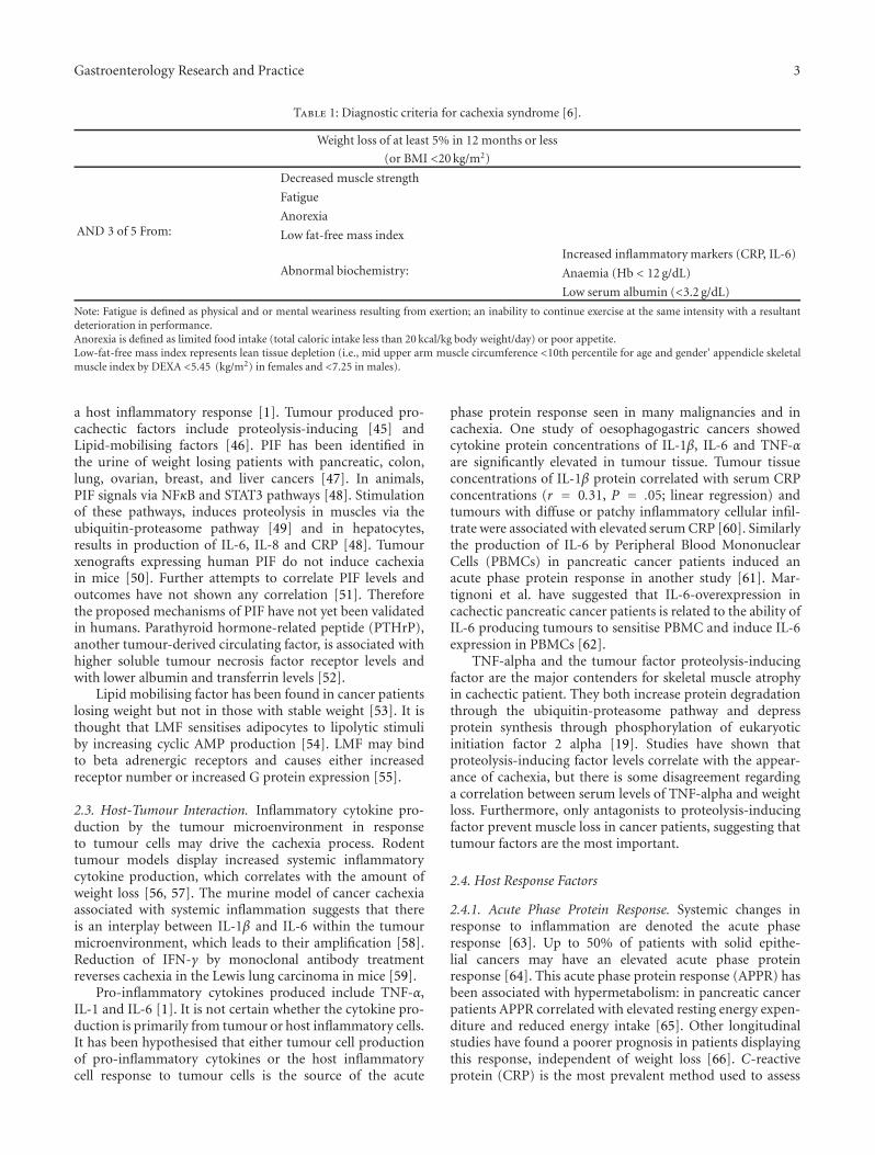
Decreased muscle strength may help distinguish cachexiafrom other causes of anorexia and fatigue in cancer patients[11]. Decreased muscle strength could be used as a diagnosticcriterion with greater sensitivity and specificity for cancercachexia. Cancer patients who are losing weight and havea systemic inflammatory response have poorer performancestatus [4]. Until a clear definition with well-defined cut-offsemerges, identification and treatment of cachectic patientsas well as research in the area will remain limited. Anew consensus definition for diagnostic purposes has beensuggested and is outlined in Table 1 [6].
2. Pathophysiology
Pathophysiological changes and clinical consequences ofcachexia are summarised in Figure 1.
2.1. Metabolic Changes. The metabolic changes found incachexia resemble those of infection rather than starvation[12] and are multifactorial and complex. Weight loss ofcancer cachexia is due to loss of both skeletal muscleand adipose tissue mass, whereas weight loss is mainlyfrom adipose tissue stores in starvation [13]. In cachexiathere is an increase in muscle protein catabolism leadingto net loss of muscle mass. The ATP ubiquitin-dependentproteolytic pathway is the greatest contributor to proteolysisin cachexia [14, 15]. Other proteolytic pathways such aslysosomal cathepsins B, H, D, and L [16] and activity ofthe calcium/calpain pathway have also been implicated [17].Increased intracellular proteolytic activity usually manifestsas loss of body weight. This proteolysis has been shown tooccur even in the absence of weight loss in cancer patients.Activation of proteolysis is an early event during tumour
Host-tumour
interaction
•Tumour factors
ProinflammatoryPro-cachectic•
••
Host responseAcute phase protein responseNeuroendocrine dysregulation
•Host-tumour interaction
Systemic inflammation
•Protein metabolism
Proteolysis
•Lipid metabolism
Lipolysis
Increased resting energy expenditure
Weight loss: decreased lean body mass and
fat deposits
Anorexia
Reduced overall survival
Decreased quality of life
Reduced physical activity
Cachexia
Clinicalendpoints
Metabolic
dysregulation
Figure 1: Clinical consequences of cancer cachexia.
growth and it may be present for a long time prior to itsclinical manifestation. Protein synthesis may be increased orunchanged [18].
Loss of adipose tissue mass is due to lipolysis [5]. Thisprocess is driven by lipid mobilising factor (LMF) andtumour (and host) factor zinc-alpha-2 glycoprotein whichhas a direct lipolytic effect and sensitises adipocytes to lipoly-tic stimuli and shows increased expression in cachexia [19].A further compounding factor is the increased resting energyexpenditure due to the dysregulation of energy metabolism.Cancer patients have a higher resting energy expenditurethan noncancer controls [20]. It has been speculated that thisis due to altered gene expression of mitochondrial membraneuncoupling proteins which uncouple respiration from ATPproduction resulting in loss of energy as heat [5].
The metabolic changes seen in cachexia are a result of theinterplay of tumour factors, host factors, and the interactionbetween the two.
2.2. Tumour Factors. Tumour cells produce both pro-inflammatory and procachectic factors, which stimulate
Gastroenterology Research and Practice 3
Table 1: Diagnostic criteria for cachexia syndrome [6].
Weight loss of at least 5% in 12 months or less
(or BMI <20 kg/m2)
AND 3 of 5 From:
Decreased muscle strength
Fatigue
Anorexia
Low fat-free mass index
Abnormal biochemistry:Increased inflammatory markers (CRP, IL-6)
Anaemia (Hb < 12 g/dL)
Low serum albumin (<3.2 g/dL)
Note: Fatigue is defined as physical and or mental weariness resulting from exertion; an inability to continue exercise at the same intensity with a resultantdeterioration in performance.Anorexia is defined as limited food intake (total caloric intake less than 20 kcal/kg body weight/day) or poor appetite.Low-fat-free mass index represents lean tissue depletion (i.e., mid upper arm muscle circumference <10th percentile for age and gender’ appendicle skeletalmuscle index by DEXA <5.45 (kg/m2) in females and <7.25 in males).
a host inflammatory response [1]. Tumour produced pro-cachectic factors include proteolysis-inducing [45] andLipid-mobilising factors [46]. PIF has been identified inthe urine of weight losing patients with pancreatic, colon,lung, ovarian, breast, and liver cancers [47]. In animals,PIF signals via NFκB and STAT3 pathways [48]. Stimulationof these pathways, induces proteolysis in muscles via theubiquitin-proteasome pathway [49] and in hepatocytes,results in production of IL-6, IL-8 and CRP [48]. Tumourxenografts expressing human PIF do not induce cachexiain mice [50]. Further attempts to correlate PIF levels andoutcomes have not shown any correlation [51]. Thereforethe proposed mechanisms of PIF have not yet been validatedin humans. Parathyroid hormone-related peptide (PTHrP),another tumour-derived circulating factor, is associated withhigher soluble tumour necrosis factor receptor levels andwith lower albumin and transferrin levels [52].
Lipid mobilising factor has been found in cancer patientslosing weight but not in those with stable weight [53]. It isthought that LMF sensitises adipocytes to lipolytic stimuliby increasing cyclic AMP production [54]. LMF may bindto beta adrenergic receptors and causes either increasedreceptor number or increased G protein expression [55].
2.3. Host-Tumour Interaction. Inflammatory cytokine pro-duction by the tumour microenvironment in responseto tumour cells may drive the cachexia process. Rodenttumour models display increased systemic inflammatorycytokine production, which correlates with the amount ofweight loss [56, 57]. The murine model of cancer cachexiaassociated with systemic inflammation suggests that thereis an interplay between IL-1β and IL-6 within the tumourmicroenvironment, which leads to their amplification [58].Reduction of IFN-γ by monoclonal antibody treatmentreverses cachexia in the Lewis lung carcinoma in mice [59].
Pro-inflammatory cytokines produced include TNF-α,IL-1 and IL-6 [1]. It is not certain whether the cytokine pro-duction is primarily from tumour or host inflammatory cells.It has been hypothesised that either tumour cell productionof pro-inflammatory cytokines or the host inflammatorycell response to tumour cells is the source of the acute
phase protein response seen in many malignancies and incachexia. One study of oesophagogastric cancers showedcytokine protein concentrations of IL-1β, IL-6 and TNF-αare significantly elevated in tumour tissue. Tumour tissueconcentrations of IL-1β protein correlated with serum CRPconcentrations (r = 0.31, P = .05; linear regression) andtumours with diffuse or patchy inflammatory cellular infil-trate were associated with elevated serum CRP [60]. Similarlythe production of IL-6 by Peripheral Blood MononuclearCells (PBMCs) in pancreatic cancer patients induced anacute phase protein response in another study [61]. Mar-tignoni et al. have suggested that IL-6-overexpression incachectic pancreatic cancer patients is related to the ability ofIL-6 producing tumours to sensitise PBMC and induce IL-6expression in PBMCs [62].
TNF-alpha and the tumour factor proteolysis-inducingfactor are the major contenders for skeletal muscle atrophyin cachectic patient. They both increase protein degradationthrough the ubiquitin-proteasome pathway and depressprotein synthesis through phosphorylation of eukaryoticinitiation factor 2 alpha [19]. Studies have shown thatproteolysis-inducing factor levels correlate with the appear-ance of cachexia, but there is some disagreement regardinga correlation between serum levels of TNF-alpha and weightloss. Furthermore, only antagonists to proteolysis-inducingfactor prevent muscle loss in cancer patients, suggesting thattumour factors are the most important.
2.4. Host Response Factors
2.4.1. Acute Phase Protein Response. Systemic changes inresponse to inflammation are denoted the acute phaseresponse [63]. Up to 50% of patients with solid epithe-lial cancers may have an elevated acute phase proteinresponse [64]. This acute phase protein response (APPR) hasbeen associated with hypermetabolism: in pancreatic cancerpatients APPR correlated with elevated resting energy expen-diture and reduced energy intake [65]. Other longitudinalstudies have found a poorer prognosis in patients displayingthis response, independent of weight loss [66]. C-reactiveprotein (CRP) is the most prevalent method used to assess

4 Gastroenterology Research and Practice
Table 2: Modified Glasgow Prognostic Score (mGPS): an inflam-mation-based prognostic score [21].
Biochemical measure Score
C-reactive protein ≤10 mg/L + Albumin ≥35 g/L 0
C-reactive protein ≤10 mg/L + Albumin <35 g/L 0
C-reactive protein >10 mg/L 1
C-reactive protein >10 mg/L + Albumin <35 g/L 2
the magnitude of the systemic inflammatory response [63].The modified Glasgow prognostic score (mGPS) (Table 2)combines CRP and albumin concentrations to create a sim-ple scoring system which is a prognostic factor independentof stage and treatment and predicts survival [21, 67].
Raised CRP concentrations at the time of admissionto hospital are indicative of an increased risk for all-causemortality; there is a 22.8-fold increase in cancer mortal-ity in patients with highly elevated CRP concentrations(>80 mg/L) [68]. This response appears to be prevalentamongst cancer patients with elevated CRP measured inalmost 80% of 106 patients with inoperable nonsmall celllung cancer (NSCLC), 40% of whom had >5% weight loss[69]. In patients without weight loss, those who displayedevidence of a systemic inflammatory response reported morefatigue (P < .05) [69]. In patients with gastro-oesophagealcancer, the rate of weight loss correlates with serum con-centrations of C-reactive protein [70]. Elevated CRP levelsat the time of diagnosis has been found to be a predictorof poor prognosis in pancreatic, lung, melanoma, multiplemyeloma, lymphoma, ovarian, renal, and gastrointestinaltumours [71].
The exact mechanisms linking cachexia, APPR, andpoor outcomes is not known. It may be that this systemicalteration in protein metabolism drives the proteolysis ofskeletal muscle to fuel the switch to acute phase reactantproduction. The APPR requires large amounts of essentialamino acids: 2.6 g of muscle protein must be catabolised toproduce 1 g of fibrinogen [72].
2.4.2. Neuroendocrine Factors. A number of neuroendocrinefactors appear to be dysregulated in the cancer state resultingin insulin resistance, reduced anabolic activity, and elevatedcortisol [47]. This dysregulation may be driven by thesystemic inflammatory response associated with cancer.Inflammatory cytokines such as TNF-α and IL-6 have beenimplicated in insulin resistance [73]. The endogenous pro-duction of or response to anabolic growth factors in patientsmay be affected either by the tumour or the host response tothe tumour and may contribute to cachexia. Testosterone orderivatives have been shown to increase protein synthesis andmuscle mass [74]. Emerging evidence implicates reduction ininsulin-like growth factor 1 in cachectic states [75].
2.5. Anorexia and Cachexia: An Interdependent Relationship?Whilst loss of appetite and resultant decrease in energy intakeundoubtedly contribute to weight loss associated with cancercachexia, whether anorexia occurs by an independent processor is a result of the inflammatory process of cachexia is
not fully understood. Anorexia itself may have a numberof components—nausea, altered taste sensation, swallowingdifficulties, or depression. The failure of aggressive supple-mentary nutritional regimes to reverse weight loss in manypatients points to primacy of the cachexia disease process [5]and in fact, this disease process may act to establish anorexia.It is thought that lack of appetite is secondary to factorsproduced by the tumour or the immune response to thetumour. Specifically, cytokines may inhibit the neuropeptideY pathway or mimic negative feedback action of leptin on thehypothalamus, leading to anorexia [76, 77].
In a study of patients with gastro-oesophageal malig-nancy (n = 220), 83% of whom had weight loss, multipleregression identified dietary intake (estimate of effect: 38%),serum CRP concentration (estimate of effect: 34%), andstage of disease (estimate of effect: 28%) as independentvariables in weight loss in these patients [70]. If serum CRPis taken as a proxy measure of systemic inflammation due tocancer cachexia, this indicates that weight loss in cancer isnot merely due to reduced calorie intake.
Recently, understanding of the physiological mechanismsof appetite regulation has been increasing. There are two setsof neurons within the arcuate nucleus of the hypothalamusidentified to be involved: the melanocortin system andthe neuropeptide Y system. Neuropeptide Y stimulatesappetite on its own or via release of other orexigenic pro-teins [78]. Neurons which release α-melanocyte-stimulatinghormone (α-MSH) and signal via melanocortin-3 and 4receptors (MC3R, MC4R) result in decrease in food-seekingbehaviour, increased basal metabolic rate and decreasedlean body mass [79, 80]. These neurons are constitutivelyactive as mutation in the MC4R results in childhood obesity[81]. Agouti-related protein (AgRP) is produced by neurons(which also produce neuropeptide Y) and counteracts theaction of MC4R-stimulating proteins promoting appetite[82]. These “appetite neurons” also express receptors forcirculating leptin [83] and interleukin-1β (IL-1β) [84], bothof which downregulate appetite and receptors for ghrelin(the orexigenic protein, which increases AgRP) [85].
3. Consequences
Cachexia results in a state of active inflammation wherebytumour-derived factors and the aberrant host responseto these factors result in a catabolic state. Whether thiscatabolic state is the ultimate cause of death in somepatients is unknown although a substantial proportion ofcancer patients die with symptoms of advanced cachexia [9].Cachexia directly impacts overall survival, quality of life, andphysical activity.
3.1. Survival. Weight loss has been indicated as an importantprognostic factor for cancer patients. A classic study byDeWys and colleagues underscores the impact and outcomeof weight loss in cancer patients [2]. Using retrospectiveevaluation in a multicentre study of more than 3000 patientswith different tumour types, these researchers reportedmoderate to severe weight loss in 30% to 70% of patients,depending on the tumor type. The amount of weight loss

Gastroenterology Research and Practice 5
depends upon tumor site, size, type, and stage. Age andtreatment type also play a role. The greatest incidence ofweight loss was seen among patients with solid tumours, forexample, gastric, pancreatic, lung, colorectal, and head andneck. Patients with solid tumours are often likely to lose 10%or more of their usual body weight. There is a lower risk ofweight loss in patients with breast and hematological cancers.Within each tumour type, survival times were shorter forpatients who had experienced weight loss than in those whodid not. Not only did weight loss predict overall survival,but it also indicated a trend towards lower chemotherapyresponse rates.
In more recent studies, similar findings of reducedsurvival have been reported. Buccheri and Ferrigno (2001)[86] reported in 388 NSCLC cases that total weight loss wasthe best indicator of prognosis. In ovarian cancer Hess et al.(2007) [87] found a significant relationship between weightchange and survival—on multivariate analysis the risk ofdeath increased by 7% for each 5% drop of body weight. InGastro-oesophageal cancer Deans and Wigmore (2009) [71]reported that patients with the lowest rate of weight loss hada median survival of 30.2 months versus 7.5 months in thosewith the highest rate of weight loss. Similar findings have alsobeen reported in pancreatic cancer [88].
One proposed mechanism to explain why patientswith weight loss have a poorer survival is the increasedincidence of complications from surgical, radiotherapeutic,and chemotherapeutic treatments. In a study by Andreyevet al. [89], 1555 patients with a number of differentgastrointestinal tumour types were analysed to examinewhether weight loss affected prognosis. In patients withweight loss: chemotherapy doses were lower; they developedmore frequent and more severe dose limiting toxicity andreceived, on average, one month less chemotherapy (P <.001 in all). Weight loss correlated with shorter failure-freesurvival, overall survival, decreased response, quality of life,and performance status (P < .001 in all) [89]. Whetherreduced survival is due to a more aggressive tumour profilein patients with weight loss or due to suboptimal treatmentrelated to weight loss, remains unknown.
3.2. Quality of Life. Cachexia contributes substantially tomorbidity in cancer patients. It is associated with symptomssuch as fatigue, weakness, poor physical performance, andthus leads to a lower self-rated quality of life. Indeed, whenthe impact of various factors is related to self-rated qualityof life scores, the proportion determined by weight loss is30% and by nutritional intake 20%, compared to cancerlocation (30%), disease duration (3%), and stage (1%)[90]. Patients who continue to lose weight while receivingpalliative chemotherapy have reduced global quality of lifeand performance scores when compared to those whoseweight loss stabilises [91].
3.3. Physical Activity. Physical activity has been describedas a novel, objective, and robust functional outcome mea-sure that is frequently impaired in cachectic states [92].Activity levels are influenced by several conventional qualityof life domains. Measurement of physical activity has
long represented a challenge for researchers using time-consuming and expensive tools such as doubly labelledwater and indirect calorimetry. However research usingthese methods has revealed that although resting energyexpenditure may be elevated in cachectic patients, totalenergy expenditure is reduced because weight-losing cancerpatients reduce the magnitude of their energy deficit throughreductions in physical activity. This reduction in physicalactivity can be significant—in one study the measured meanphysical activity rate was equivalent to that of spinal cordinjury patients living at home and greatly reduced versusnormal controls [93]. In a more recent study by Daheleet al. (2007) [94] using advanced ambulatory pedometertechnology, cancer patients receiving palliative chemother-apy were shown to spend significantly more time lyingand sitting, and significantly less time in quiet standingor stepping compared with controls, taking on average43% less steps than healthy controls. It is known that bedrest leads to a decrease in skeletal muscle mass in healthypatients, due to reduced protein synthesis [95]. Thus, loss ofphysical function results in decreases in performance status,ability to perform activities of daily living, decreased socialinteractions, and alterations in body image, all of whichmanifest as reduced quality of life [96]. Interventions whichincrease physical activity would be anticipated to be highlybeneficial.
Antineoplastic therapies such as surgery, radiotherapyand chemotherapy, may also impact on the developmentof systemic inflammation and particularly may impact onswallowing difficulties and anorexia due to nausea [97].
4. Therapeutic Approaches
4.1. Goals of Therapy. Clearly since cancer cachexia isassociated with a poor prognosis, the aim of management isoften to improve symptoms and quality of life. It is notedthat a response to chemotherapeutic treatment by shrinkageof the tumour burden often leads to improvement in thecachectic state. The primary endpoints of optimal treatmentof cancer cachexia are improvements in lean body mass,resting energy expenditure, fatigue, anorexia, quality of life,performance status, and a reduction in pro-inflammatorycytokines.
A greater understanding of the process of inflammationand its fundamental role in the development of cachexiahas led to new avenues opening up in the approach tomanagement of the condition. The hypothesis is that effec-tive treatment of cancer cachexia will improve performancestatus and quality of life and by inhibiting the processdriving cachexia, survival may be improved. In patientswho stop losing weight while receiving chemotherapy forgastrointestinal cancers, median survival is improved (15.7months versus 8.1 months, P = .0004) [89]. Animal modelsare generally unsatisfactory models for assessing the efficacyof intervention due to the larger proportional size and theaggressive doubling rate of tumours: thus the biologicalbehaviour is different to that seen in the clinical setting [98].
There has been recent progress in producing trials of highclinical quality for licensing purposes but these trials may

6 Gastroenterology Research and Practice
Table 3: Endpoints for evaluating interventions in cancer cachexia.
Clinical Functional Biochemical
Nutritional status Performance score (ECOG; Karnofsky) Plasma fatty acid composition
Tolerance of diet Quality of life scores Pro-inflammatory cytokines
GI symptoms Appetite Acute phase protein reactants
Infections Fatigue
Survival Physical activity as measured electronically [22]
Muscle strength
be beset by difficulties in adequate endpoint analysis due tothe numbers lost to followup or patients being unable tocomply with therapy due to their poor overall condition, thuslimiting their duration, power, or generalisability [99, 100].In addition there is a degree of heterogeneity in definingrelevant end points for analysis of intervention in cancercachexia. Table 3 summarises the range of endpoints whichmay be used. One study of 388 nonsmall cell lung cancerpatients found that total weight loss was the best predictor ofprognosis rather than speed of weight loss [101]. However,weight loss alone does not identify the full effect of cachexiaon physical function [8]. It is the loss of fat-free mass(FFM) that is responsible for the reduced functional status,increased mortality, and other negative outcomes associatedwith malnutrition [102]. Body fat is easier to gain than FFM,so studies that show improved body weight may not translateinto reductions in morbidity or improvements in functionalstatus. To improve functional ability and hence quality of lifepatients need not only to become weight stable but regain thelean tissue lost in the cachectic process. Thus, interventionswhich lead to improvements in functional status would beexpected to cause increases in lean body mass rather thanfat mass, however, this distinction is often not reported ininterventions.
The strong impact that cancer cachexia has on cancerpatients’ outcome and quality of life suggests that nutritionalissues should be taken into consideration from the beginningof the natural history of cancer, a concept termed the parallelpathway [103]. Indeed studies of nutritional interventionthat have reported a better weight maintenance in patientsare in those who are treated in the “precachexia” phase,that is, prior to loss of >10% of body weight and prior toelevations of CRP. Dietary counselling with or without oralnutritional supplements has proven efficacy in stabilisingnutritional status in pre-cachectic patients [104, 105]. Anutritional assessment to seek reversible causes of weightloss is the first step in management in cachectic patients.Approximately 40% of cancer patients eat less than the34 kcal/kg/day required to maintain weight [106]. The Euro-pean Society of Parenteral and Enteral Nutrition (ESPEN)report in a consensus statement that there is Grade Aevidence for intensive dietary counselling with food plus orminus oral nutritional supplements in preventing therapy-associated weight loss, preventing treatment interruptionsand increasing dietary intake in gastrointestinal or head andneck cancer patients undergoing radio- or chemotherapy[107].
For patients with advanced cachexia (>10% weight loss,systemic inflammation and poor appetite) studies seeking toassess the effect of targeted nutritional advice and supple-ments have generally reported no significant improvementin nutritional status. Standard enteral or parenteral supple-ments do not appear to result in lean mass weight gain forthe typical cancer patient [5, 98, 108]. The largest evaluationof the literature regarding nutritional supplementation (NS)(oral or tube) in cancer patients was the systematic reviewby Elia et al. (2006) showing no difference in mortality inpatients undergoing chemotherapy/radiotherapy (4 RCTs)or surgery (4 RCTs) [109]. A systematic review of par-enteral nutrition in cancer patients showed no difference inmortality (19 RCTs), increase in total complication rates inthose given parenteral nutrition (8 RCTs), and significantlylower tumour response rate in patients receiving parenteralnutrition (15 RCTs) [110].
This is likely because the inflammatory response ofcachexia prevents anabolism. In many cases an attempt isbeing made to reverse or halt a rapidly advancing catabolicprocess and it is unrealistic to expect a reversal with caloriesand protein alone.
The poor results observed with conventional nutritionsupport in cachectic patients led to the emergence of so-called nutraceuticals or immunonutrition supplements, inan attempt to nutritionally modify the metabolic milieu byproviding anti-inflammatory substances, such as eicosapen-taenoic acid (EPA), at levels much higher than that typicallyfound in the diet.
4.2. Eicosapentaenoic Acid. Eicosapentaenoic acid (EPA), along-chain polyunsaturated fatty acid (PUFA) of the omega-3 (n-3) family, has been studies in relation to cancer cachexiafor over 15 years. It is of interest in the context of cancercachexia as it has potential to impact on both the underlyingmetabolic abnormalities of tumour-induced weight loss, aswell as modulation of immune function. When EPA isconsumed at levels above that normally found in the diet,it replaces arachidonic acid (AA), an n-6 PUFA, in cellmembrane phospholipids. It then acts as a substrate forthe production of the 3 series prostaglandins and the 5series leukotrienes. Eicosanoids synthesized from the n-3PUFAs (i.e., EPA) rather than the n-6 PUFAs (i.e., AA) havelower potential for promoting inflammation. Modulationof dietary fatty acids can therefore have an impact onmany immune processes such as proliferation, phagocytosis,cytotoxicity, and cytokine production [111].

Gastroenterology Research and Practice 7
Table 4: Pharmacological options for management of cachexia.
Agent Clinical effect (RCT)# Hypothetical mechanism of action
Anabolic agents Corticosteroids
Improves anorexia and weakness;no improvement in weight orcalorie intake [23–25]; welltolerated; effects short lasting
Not established. May inhibitprostaglandin metabolism andcentral euphoric effect
Nandrolone decanoate Decrease in weight loss [26]Not established. Promote proteinnitrogen accumulation
OxandroloneNo published randomised clinicaltrials in cancer cohort
Not established
InsulinIncreases whole body fat andcarbohydrate intake [27]
Not established
Adenosine Triphosphate (ATP)Stabilises weight loss and increasesenergy intake[28]
Not established
Appetite stimulantsProgesterones: Megestrol acetate (MA)
Medroxyprogesterone (MP)
Improves appetite, calorie intakeand weight (not lean body mass)[29]
MA: may increase the centralappetite stimulant neuropeptideYMP: reduces serotonin andcytokine production by PBMCs[30]
Cannabinoids: Dronabinol
No benefit when added to MA;inferior to MA when used alone[31]. No increase in appetite orQoL [32]
May act on endorphin receptors,reduce prostaglandin synthesis orinhibit IL-1 secretion [33]
Cytokine inhibitors CyproheptadineNo improvement in weight gain[34]
Serotonin antagonist withantihistaminic properties
ThalidomideAttenuates weight loss, increaseslean body mass [35]
Immunomodulatory:downregulates TNF-α (bydestabilising mRNA [36]), NFκB,pro-inflammatory cytokines,COX2 [37]
PentoxifyllineNo improvement in appetite orweight in cachectic patients [38]
Phosphodiesterase inhibitor:inhibits TNF gene transcription
Eicosapentaenoic acid (EPA)
Cochrane meta-analysis:insufficient evidence to establishwhether EPA is better thanplacebo [39]
In vitro attenuates increased cAMPactivity and lipolysis by LMF [40]
Melatonin
Improves cachexia (term notdefined) and one year survivalincreased in advanced NCSC lungcancer [41]
Immunomodulatory [42],Downregulates TNF production[43]
Anti-inflammatories Non-steroid anti-inflammatory drugs
Reduced inflammatory markers,reduced resting energyexpenditure, preservation of totalbody fat [44]
Not established. Maydownregulate systemicinflammatory response to tumour
#Results from randomised controlled trials (RCTs) are cited.
Despite initial studies showing anabolic effects, princi-pally gains of lean body mass, improvements in grip strength,quality of life, and reductions in IL-6 and PIF could beachieved in a variety of cancers [99], including pancreaticcancer [112, 113], lung cancer [114], and colorectal cancer[115], analysis of RCTs only, using the Cochrane approach,did not show any differences between EPA supplementationand placebo [39]. Whether this is a true representationor a reflection of the advanced cachexia of participants orinherent differences in EPA metabolism between individuals(with only a proportion of patients able to respond toEPA) needs further examination. On subgroup analysis,
patients who comply with EPA supplementation seem tohave improved lean body mass [116].
EPA-enriched oral nutritional supplements (ONSs) havebeen compared to megestrol acetate in the North CentralCancer Treatment Group trial of 421 patients with weightloss, poor intake, and anorexia [117]. In a 3-month inter-vention period, patients were randomized to either EPA-enriched ONS plus placebo liquid suspension, standardONS plus megestrol acetate suspension, or EPA-enrichedONS plus megestrol acetate suspension. Weight gain washighest in the megestrol acetate group but unfortunatelybody composition was not assessed and so changes in water

8 Gastroenterology Research and Practice
weight cannot be controlled for. There was no difference insurvival, appetite, or quality of life scores between the groups,however patients on megestrol acetate reported higher ratesof impotence. The fact that an EPA enriched ONS scoredas well as drug therapy on certain clinical endpoints (e.g.,survival and global quality of life) underscores the limitationsof each treatment.
β-hydroxyl β-methyl butyrate (HMB), glutamine, andarginine supplementation have been combined in the hopeof a synergistic effect of HMB (a modulator of proteinturnover) and the amino acids (immunomodulatory) wouldincrease weight. A phase III RCT of this combination did notshow any difference in lean body mass between control andintervention groups [100].
4.3. Pharmacological Agents. Pharmacological options aresummarised in Table 4. Among orexigenic agents, megestrolacetate is by far the most widely prescribed and at least 15randomised controlled clinical trials have demonstrated thatthis drug, at doses ranging from 160–1600 mg/d significantlyimproves appetite with respect to placebo [118]. A recentCochrane meta-analysis reported that it improves weightgain and appetite in cancer patients [29]. Although thisincrease in appetite is very desirable for both patients andtheir carers, in most of these trials no definitive improvementin global quality of life was observed [29].
Anti-inflammatory agents (COX inhibitors) can reduceweight loss and aid maintenance of performance status inadvanced cancer [119]. The COX-2 inhibitor, meloxicamshowed activity against PIF-induced proteolysis, prior toits withdrawal from the market [120]. Beta-adrenoreceptorblockade can reduce resting energy expenditure in patientswith cancer (n = 10) but have not been trialled in larger-scale studies [121]. They are thought to inhibit proteolysisand lipolysis [122] and have been shown to downregulatecatecholamine-induced catabolism in burns patients [123].Agents which reduced cytokine levels such as thalidomideand pentoxifylline have only shown modest or minimalactivity. At RCT, thalidomide has been shown to attenuateweight loss and lead to improved physical function [35].Pentoxifylline did not have any clinical benefit. Specificantitumour necrosis factor- (TNF-)α agents, etanercept andinfliximab, did not show any positive effect on appetite orbody weight in RCTs [124, 125]. Corticosteroids, althoughwidely used, have significant side effects including proteinbreakdown, insulin resistance, water retention, and adrenalsuppression and tend to be used during the preterminalphase of patient illness [23, 126]. Anabolic steroid derivativessuch as nandrolone and oxandrolone have not been studiedin clinical trials in a cancer cohort. Insulin [27], ATPinfusions [28], and melatonin [41] have produced modestpositive effects in small clinical trials and require furthersubstantiation.
4.4. Combination Therapy. In unresectable cancer cases,there is currently no goal standard treatment that canattenuate catabolism and inflammation, stimulate appetiteand intake and consequently promote anabolism (specificallyof lean body mass). A multimodal approach has therefore
been advocated in the treatment of cancer cachexia. Man-tovani (2010) randomised 332 patients with cancer-relatedanorexia/cachexia syndrome to one of five arms of treatment:(1) medroxyprogesterone 500 mg/d or megestrol acetate320 mg/d; (2) oral supplementation with eicosapentaenoicacid (EPA); (3) L-carnitine 4 g/d; (4) thalidomide 200 mg/d;(5) a combination of the above for a total of 4 months [127].Results showed the superiority of arm 5 over the othersfor all primary endpoints. Significant improvements wereobserved in arm 5 in LBM, fatigue scores, appetite, and totalenergy and active energy expenditure with REE decreasingsignificantly. Toxicity was negligible and comparable betweentreatment arms.
4.5. Potential Therapeutic Targets. Due to the lack of clinicalefficacy of agents which seemed promising in the laboratorysetting, ongoing research has continued to explore newtherapeutic targets and to develop new agents. Much of thishas focussed on manipulation of the melanocortin system ofappetite regulation [128]. Activation of the Melanocortin-4-receptor (MC4R) in murine models decreases food-seekingbehaviour, increases basal metabolic rate, and decreaseslean body mass [80]. Treatment with a MC4R antago-nist attenuated these responses [79]. Ghrelin induces therelease of growth hormone, regulates appetite, and has anti-inflammatory properties [129, 130]. Initial human studies inPhase I open trials have confirmed safety and show someincrease in appetite and body weight [131]. Myostatin is agrowth factor involved in the normal regulation of musclemass [132]. Myostatin inhibitors and IL-6 antagonists arecurrently at Phase I RCT stage in development [131].
5. Conclusions
A consensus definition incorporating clinical, functional,and biochemical parameters is necessary in order to ade-quately identify and treat patients with cancer cachexia. Agreater understanding of the pathophysiology, particularly interms of the processes which drive cachexia will lead to newtherapeutic target development. A number of issues remainto be resolved including whether inflammation drives theprocess or is a byproduct of the process. Does reversal ofweight loss alone result in improved survival? By improvingcachexia (i.e., leading to improved physical and physiologicalfunction) in cachexia, can patients become better able totolerate anticancer therapies such as chemotherapy?
Composite endpoints which measure clinically relevantoutcomes such as physical activity and quality of life arerequired in order to best assess the impact of interventions oncancer cachexia patients. Objective measures of function (asrepresented by physical activity) using advance ambulatorytechnology and integrated subjective quality of life parame-ters are likely to become standard practice in the clinical trialsetting.
Acknowledgment
This paper is funded through an Irish Cancer SocietyResearch Scholarship.

Gastroenterology Research and Practice 9
References
[1] M. J. Tisdale, “Mechanisms of cancer cachexia,” PhysiologicalReviews, vol. 89, no. 2, pp. 381–410, 2009.
[2] W. D. Dewys, C. Begg, P. T. Lavin et al., “Prognostic effect ofweight loss prior to chemotherapy in cancer patients. EasternCooperative Oncology Group,” American Journal of Medicine,vol. 69, pp. 491–497, 1980.
[3] M. Maltoni, A. Caraceni, C. Brunelli et al., “Prognosticfactors in advanced cancer patients: evidence-based clinicalrecommendations—a study by the steering committee of theeuropean association for palliative care,” Journal of ClinicalOncology, vol. 23, no. 25, pp. 6240–6248, 2005.
[4] P. O’Gorman, D. C. McMillan, and C. S. McArdle, “Longi-tudinal study of weight, appetite, performance status, andinflammation in advanced gastrointestinal cancer,” Nutritionand Cancer, vol. 35, no. 2, pp. 127–129, 1999.
[5] M. J. Tisdale, “Cachexia in cancer patients,” Nature ReviewsCancer, vol. 2, no. 11, pp. 862–871, 2002.
[6] W. J. Evans, J. E. Morley, J. Argiles et al., “Cachexia: a newdefinition,” Clinical Nutrition, vol. 27, no. 6, pp. 793–799,2008.
[7] N. MacDonald, A. M. Easson, V. C. Mazurak, G. P. Dunn,and V. E. Baracos, “Understanding and managing cancercachexia,” Journal of the American College of Surgeons, vol.197, no. 1, pp. 143–161, 2003.
[8] K. C. Fearon, A. C. Voss, and D. S. Hustead, “Definitionof cancer cachexia: effect of weight loss, reduced foodintake, and systemic inflammation on functional status andprognosis,” American Journal of Clinical Nutrition, vol. 83, no.6, pp. 1345–1350, 2006.
[9] E. Bruera, “ABC of palliative care: anorexia, cachexia, andnutrition,” British Medical Journal, vol. 315, no. 7117, pp.1219–1222, 1997.
[10] K. M. Fox, J. M. Brooks, S. R. Gandra, R. Markus, and C. F.Chiou, “Estimation of cachexia among cancer patients basedon four definitions,” Journal of Oncology, vol. 2009, Article ID693458, 2009.
[11] F. Strasser, “Diagnostic criteria of cachexia and their assess-ment: decreased muscle strength and fatigue,” Current Opin-ion in Clinical Nutrition and Metabolic Care, vol. 11, no. 4,pp. 417–421, 2008.
[12] J. M. Argiles, R. Moore-Carrasco, G. Fuster, S. Busquets,and F. J. Lopez-Soriano, “Cancer cachexia: the molecularmechanisms,” International Journal of Biochemistry and CellBiology, vol. 35, no. 4, pp. 405–409, 2003.
[13] J. F. Moley, R. Aamodt, and W. Rumble, “Body cell mass incancer-bearing and anorexic patients,” Journal of Parenteraland Enteral Nutrition, vol. 11, no. 3, pp. 219–222, 1987.
[14] J. Khal, A. V. Hine, K. C. H. Fearon, C. H. C. Dejong, andM. J. Tisdale, “Increased expression of proteasome subunitsin skeletal muscle of cancer patients with weight loss,”International Journal of Biochemistry and Cell Biology, vol. 37,no. 10, pp. 2196–2206, 2005.
[15] C. H. DeJong, S. Busquets, A. G. Moses et al., “Systemicinflammation correlates with increased expression of skeletalmuscle ubiquitin but not uncoupling proteins in cancercachexia,” Oncology Reports, vol. 14, no. 1, pp. 257–263, 2005.
[16] A. Bosutti, G. Toigo, B. Ciocchi, R. Situlin, G. Guarnieri,and G. Biolo, “Regulation of muscle cathepsin B proteolyticactivity in protein-depleted patients with chronic diseases,”Clinical Nutrition, vol. 21, no. 5, pp. 373–378, 2002.
[17] S. Busquets, C. Garcıa-Martınez, B. Alvarez, N. Carbo, F. J.Lopez-Soriano, and J. M. Argiles, “Calpain-3 gene expressionis decreased during experimental cancer cachexia,” Biochim-ica et Biophysica Acta, vol. 1475, no. 1, pp. 5–9, 2000.
[18] D. C. McMillan, T. Preston, K. C. H. Fearon, H. J. G. Burns, C.Slater, and A. Shenkin, “Protein synthesis in cancer patientswith inflammatory response: Investigations with [N]glycine,”Nutrition, vol. 10, no. 3, pp. 232–240, 1994.
[19] M. J. Tisdale, “Cancer cachexia,” Current Opinion in Gas-troenterology, vol. 26, no. 2, pp. 146–151, 2010.
[20] A. Hyltander, C. Drott, U. Korner, R. Sandstrom, and K.Lundholm, “Elevated energy expenditure in cancer patientswith solid tumours,” European Journal of Cancer, vol. 27, no.1, pp. 9–15, 1991.
[21] D. C. McMillan, “Systemic inflammation, nutritional statusand survival in patients with cancer,” Current Opinion inClinical Nutrition and Metabolic Care, vol. 12, no. 3, pp. 223–226, 2009.
[22] G. Mantovani, A. Maccio, C. Madeddu et al., “Randomizedphase III clinical trial of five different arms of treatment forpatients with cancer cachexia: interim results,” Nutrition, vol.24, no. 4, pp. 305–313, 2008.
[23] J. C. Willox, J. Corr, and J. Shaw, “Prednisolone as an appetitestimulant in patients with cancer,” British Medical Journal,vol. 288, no. 6410, p. 27, 1984.
[24] C. G. Moertel, A. J. Schutt, R. J. Reitemeier, and R. G. Hahn,“Corticosteroid therapy of preterminal gastrointestinal can-cer,” Cancer, vol. 33, no. 6, pp. 1607–1609, 1974.
[25] E. Bruera, E. Roca, and L. Cedaro, “Action of oral methyl-prednisolone in terminal cancer patients: a prospectiverandomized double-blind study,” Cancer Treatment Reports,vol. 69, no. 7-8, pp. 751–754, 1985.
[26] R. T. Chlebowski, J. Herrold, and I. Ali, “Influence onnandrolone decanoate on weight loss in advanced non-smallcell lung cancer,” Cancer, vol. 58, no. 1, pp. 183–186, 1986.
[27] K. Lundholm, U. Korner, L. Gunnebo et al., “Insulintreatment in cancer cachexia: effects on survival, metabolism,and physical functioning,” Clinical Cancer Research, vol. 13,no. 9, pp. 2699–2706, 2007.
[28] H. J. Agteresch et al., “Beneficial effects of adenosinetriphosphate on nutritional status in advanced lung cancerpatients: a randomized clinical trial,” Nutrition in ClinicalPractice, vol. 19, no. 4, p. 413, 2004.
[29] E. G. Berenstein and Z. Ortiz, “Megestrol acetate for the treat-ment of anorexia-cachexia syndrome,” Cochrane Database ofSystematic Reviews, no. 2, Article ID CD004310, 2005.
[30] G. Mantovani, A. Maccio, S. Esu et al., “Medroxyproges-terone acetate reduces the In vitro production of cytokinesand serotonin involved in anorexia/cachexia and emesisby peripheral blood mononuclear cells of cancer patients,”European Journal of Cancer Part A, vol. 33, no. 4, pp. 602–607, 1997.
[31] A. Jatoi, H. E. Windschitl, C. L. Loprinzi et al., “Dronabinolversus megestrol acetate versus combination therapy forcancer-associated anorexia: a North Central Cancer Treat-ment Group study,” Journal of Clinical Oncology, vol. 20, no.2, pp. 567–573, 2002.
[32] F. Strasser, D. Luftner, K. Possinger et al., “Compari-son of orally administered cannabis extract and delta-9- tetrahydrocannabinol in treating patients with cancer-related anorexia-cachexia syndrome: a multicenter, phaseIII, randomized, double-blind, placebo-controlled clinicaltrial from the Cannabis-In-Cachexia-Study-Group,” Journalof Clinical Oncology, vol. 24, no. 21, pp. 3394–3400, 2006.

10 Gastroenterology Research and Practice
[33] J. M. Argils, H. Meijsing, J. Pallars-Trujillo, X. Guirao, and F.J. Lpez-Soriano, “Cancer cachexia: a therapeutic approach,”Medicinal Research Reviews, vol. 21, no. 1, pp. 83–101, 2001.
[34] C. G. Kardinal, C. L. Loprinzi, D. J. Schaid et al., “A controlledtrial of cyproheptadine in cancer patients with anorexiaand/or cachexia,” Cancer, vol. 65, no. 12, pp. 2657–2662,1990.
[35] J. N. Gordon, T. M. Trebble, R. D. Ellis, H. D. Duncan, T.Johns, and P. M. Goggin, “Thalidomide in the treatment ofcancer cachexia: a randomised placebo controlled trial,” Gut,vol. 54, no. 4, pp. 540–545, 2005.
[36] A. L. Moreira, E. P. Sampaio, A. Zmuidzinas, P. Frindt, K.A. Smith, and G. Kaplan, “Thalidomide exerts its inhibitoryaction on tumor necrosis factor alpha by enhancing mRNAdegradation,” The Journal of Experimental Medicine, vol. 177,no. 6, pp. 1675–1680, 1993.
[37] J. N. Gordon and P. M. Goggin, “Thalidomide and itsderivatives: emerging from the wilderness,” PostgraduateMedical Journal, vol. 79, no. 929, pp. 127–132, 2003.
[38] R. M. Goldberg, C. L. Loprinzi, J. A. Mailliard et al., “Pen-toxifylline for treatment of cancer anorexia and cachexia? Arandomized, double-blind, placebo-controlled trial,” Journalof Clinical Oncology, vol. 13, no. 11, pp. 2856–2859, 1995.
[39] A. Dewey, C. Baughan, T. Dean, B. Higgins, and I. Johnson,“Eicosapentaenoic acid (EPA, an omega-3 fatty acid from fishoils) for the treatment of cancer cachexia,” Cochrane Databaseof Systematic Reviews, no. 1, Article ID CD004597, 2007.
[40] S. A. Price and M. J. Tisdale, “Mechanism of inhibition ofa tumor lipid-mobilizing factor by eicosapentaenoic acid,”Cancer Research, vol. 58, no. 21, pp. 4827–4831, 1998.
[41] P. Lissoni, F. Paolorossi, A. Ardizzoia et al., “A randomizedstudy of chemotherapy with cisplatin plus etoposide versuschemoendocrine therapy with cisplatin, etoposide and thepineal hormone melatonin as a first-line treatment ofadvanced non-small cell lung cancer patients in a poorclinical state,” Journal of Pineal Research, vol. 23, no. 1, pp.15–19, 1997.
[42] P. Lissoni, “Is there a role for melatonin in supportive care?”Supportive Care in Cancer, vol. 10, no. 2, pp. 110–116, 2002.
[43] D. P. Kotler, “Cachexia,” Annals of Internal Medicine, vol. 133,no. 8, pp. 622–634, 2000.
[44] K. Lundholm, P. Daneryd, U. Korner, A. Hyltander, and I.Bosaeus, “Evidence that long-term COX-treatment improvesenergy homeostasis and body composition in cancer patientswith progressive cachexia,” International Journal of Oncology,vol. 24, no. 3, pp. 505–512, 2004.
[45] P. Todorov, P. Cariuk, T. McDevitt, B. Coles, K. Fearon, andM. Tisdale, “Characterization of a cancer cachectic factor,”Nature, vol. 379, no. 6567, pp. 739–742, 1996.
[46] K. Hirai, H. J. Hussey, M. D. Barber, S. A. Price, and M. J.Tisdale, “Biological evaluation of a lipid-mobilizing factorisolated from the urine of cancer patients,” Cancer Research,vol. 58, no. 11, pp. 2359–2365, 1998.
[47] R. J. E. Skipworth, G. D. Stewart, C. H. C. Dejong, T. Preston,and K. C. H. Fearon, “Pathophysiology of cancer cachexia:much more than host-tumour interaction?” Clinical Nutri-tion, vol. 26, no. 6, pp. 667–676, 2007.
[48] T. M. Watchorn, I. Waddell, N. Dowidar, and J. A. Ross,“Proteolysis-inducing factor regulates hepatic gene expres-sion via the transcription factors NF-(kappa)B and STAT3,”The FASEB Journal, vol. 15, no. 3, pp. 562–564, 2001.
[49] A. S. Whitehouse and M. J. Tisdale, “Increased expressionof the ubiquitin—proteasome pathway in murine myotubesby proteolysis-inducing factor (PIF) is associated with acti-vation of the transcription factor NF-κB,” British Journal ofCancer, vol. 89, no. 6, pp. 1116–1122, 2003.
[50] C. L. Monitto, S. M. Dong, J. Jen, and D. Sidransky,“Characterization of a human homologue of proteolysis-inducing factor and its role in cancer cachexia,” ClinicalCancer Research, vol. 10, no. 17, pp. 5862–5869, 2004.
[51] B. M. Wieland, G. D. Stewart, R. J. E. Skipworth et al.,“Is there a human homologue to the murine proteolysis-inducing factor?” Clinical Cancer Research, vol. 13, no. 17, pp.4984–4992, 2007.
[52] C. Deans, S. Wigmore, S. Paterson-Brown, J. Black, J. Ross,and K. C. H. Fearon, “Serum parathyroid hormone-relatedpeptide is associated with systemic inflammation and adverseprognosis in gastroesophageal carcinoma,” Cancer, vol. 103,no. 9, pp. 1810–1818, 2005.
[53] P. T. Todorov, T. M. McDevitt, D. J. Meyer, H. Ueyama, I.Ohkubo, and M. J. Tisdale, “Purification and characteriza-tion of a tumor lipid-mobilizing factor,” Cancer Research, vol.58, no. 11, pp. 2353–2358, 1998.
[54] S. Khan and M. J. Tisdale, “Catabolism of adipose tissueby a tumour-produced lipid-mobilising factor,” InternationalJournal of Cancer, vol. 80, no. 3, pp. 444–447, 1999.
[55] B. Islam-Ali, S. Khan, S. A. Price, and M. J. Tisdale,“Modulation of adipocyte G-protein expression in cancercachexia by a lipid-mobilizing factor (LMF),” British Journalof Cancer, vol. 85, no. 5, pp. 758–763, 2001.
[56] J. Gelin, L. L. Moldawer, C. Lonnroth, B. Sherry, R.Chizzonite, and K. Lundholm, “Role of endogenous tumornecrosis factor α and interleukin 1 for experimental tumorgrowth and the development of cancer cachexia,” CancerResearch, vol. 51, no. 1, pp. 415–421, 1991.
[57] G. Strassmann, M. Fong, J. S. Kenney, and C. O. Jacob, “Evi-dence for the involvement of interleukin 6 in experimentalcancer cachexia,” Journal of Clinical Investigation, vol. 89, no.5, pp. 1681–1684, 1992.
[58] K. Yasumoto, N. Mukaida, A. Harada et al., “Molecularanalysis of the cytokine network involved in cachexia in colon26 adenocarcinoma-bearing mice,” Cancer Research, vol. 55,no. 4, pp. 921–927, 1995.
[59] P. Matthys, H. Heremans, G. Opdenakker, and A. Billiau,“Anti-interferon-γ antibody treatment, growth of Lewis lungtumours in mice and tumour-associated cachexia,” EuropeanJournal of Cancer, vol. 27, no. 2, pp. 182–187, 1991.
[60] D. A. C. Deans, S. J. Wigmore, H. Gilmour, S. Paterson-Brown, J. A. Ross, and K. C. H. Fearon, “Elevated tumourinterleukin-1beta is associated with systemic inflammation:a marker of reduced survival in gastro-oesophageal cancer,”British Journal of Cancer, vol. 95, no. 11, pp. 1568–1575, 2006.
[61] M. G. O’Riordain, J. S. Falconer, J. Maingay, K. C. Fearon,and J. A. Ross, “Peripheral blood cells from weight-losingcancer patients control the hepatic acute phase response bya primarily interleukin-6 dependent mechanism,” Interna-tional Journal of Oncology, vol. 15, no. 4, pp. 823–827, 1999.
[62] M. E. Martignoni, P. Kunze, W. Hildebrandt et al., “Role ofmononuclear cells and inflammatory cytokines in pancreaticcancer-related cachexia,” Clinical Cancer Research, vol. 11, no.16, pp. 5802–5808, 2005.
[63] C. Gabay and I. Kushner, “Acute-phase proteins and othersystemic responses to inflammation,” The New EnglandJournal of Medicine, vol. 340, no. 6, pp. 448–454, 1999.

Gastroenterology Research and Practice 11
[64] J. S. Falconer, K. C. H. Fearon, J. A. Ross et al., “Acute-phase protein response and survival duration of patients withpancreatic cancer,” Cancer, vol. 75, no. 8, pp. 2077–2082,1995.
[65] J. S. Falconer, K. C. H. Fearon, C. E. Plester, J. A. Ross, and D.C. Carter, “Cytokines, the acute-phase response, and restingenergy expenditure in cachectic patients with pancreaticcancer,” Annals of Surgery, vol. 219, no. 4, pp. 325–331, 1994.
[66] P. O’Gorman, D. C. McMillan, and C. S. McArdle, “Prognos-tic factors in advanced gastrointestinal cancer patients withweight loss,” Nutrition and Cancer, vol. 37, no. 1, pp. 36–40,2000.
[67] D. C. McMillan, “An inflammation-based prognostic scoreand its role in the nutrition-based management of patientswith cancer,” Proceedings of the Nutrition Society, vol. 67, no.3, pp. 257–262, 2008.
[68] C. Marsik, L. Kazemi-Shirazi, T. Schickbauer et al., “C-reactive protein and all-cause mortality in a large hospital-based cohort,” Clinical Chemistry, vol. 54, no. 2, pp. 343–349,2008.
[69] H. R. Scott, D. C. McMillan, D. J. F. Brown, L. M. Forrest, C.S. McArdle, and R. Milroy, “A prospective study of the impactof weight loss and the systemic inflammatory response onquality of life in patients with inoperable non-small cell lungcancer,” Lung Cancer, vol. 40, no. 3, pp. 295–299, 2003.
[70] D. A. C. Deans, B. H. Tan, S. J. Wigmore et al., “The influenceof systemic inflammation, dietary intake and stage of diseaseon rate of weight loss in patients with gastro-oesophagealcancer,” British Journal of Cancer, vol. 100, no. 1, pp. 63–69,2009.
[71] C. Deans and S. J. Wigmore, “Systemic inflammation,cachexia and prognosis in patients with cancer,” CurrentOpinion in Clinical Nutrition and Metabolic Care, vol. 8, no.3, pp. 265–269, 2005.
[72] P. J. Reeds, C. R. Fjeld, and F. Jahoor, “Do the differencesbetween the amino acid compositions of acute-phase andmuscle proteins have a bearing on nitrogen loss in traumaticstates?” Journal of Nutrition, vol. 124, no. 6, pp. 906–910,1994.
[73] R. Feinstein, H. Kanety, M. Z. Papa, B. Lunenfeld, andA. Karasik, “Tumor necrosis factor-α suppresses insulin-induced tyrosine phosphorylation of insulin receptor and itssubstrates,” Journal of Biological Chemistry, vol. 268, no. 35,pp. 26055–26058, 1993.
[74] R. Orr and M. Fiatarone Singh, “The anabolic androgenicsteroid oxandrolone in the treatment of wasting and catabolicdisorders: review of efficacy and safety,” Drugs, vol. 64, no. 7,pp. 725–750, 2004.
[75] P. Costelli, M. Muscaritoli, M. Bossola et al., “IGF-1 isdownregulated in experimental cancer cachexia,” AmericanJournal of Physiology, vol. 291, no. 3, pp. R674–R683, 2006.
[76] E. J. B. Ramos, S. Suzuki, D. Marks, A. Inui, A. Asakawa,and M. M. Meguid, “Cancer anorexia-cachexia syndrome:cytokines and neuropeptides,” Current Opinion in ClinicalNutrition and Metabolic Care, vol. 7, no. 4, pp. 427–434, 2004.
[77] A. Inui, “Cancer anorexia-cachexia syndrome: are neuropep-tides the key?” Cancer Research, vol. 59, no. 18, pp. 4493–4501, 1999.
[78] W. T. Chance, A. Balasubramaniam, H. Thompson, B.Mohapatra, J. Ramo, and J. E. Fischer, “Assessment of feedingresponse of tumor-bearing rats to hypothalamic injectionand infusion of neuropeptide Y,” Peptides, vol. 17, no. 5, pp.797–801, 1996.
[79] S. Markison, A. C. Foster, C. Chen et al., “The regulationof feeding and metabolic rate and the prevention of murinecancer cachexia with a small-molecule melanocortin-4 recep-tor antagonist,” Endocrinology, vol. 146, no. 6, pp. 2766–2773,2005.
[80] D. L. Marks, N. Ling, and R. D. Cone, “Role of the centralmelanocortin system in cachexia,” Cancer Research, vol. 61,no. 4, pp. 1432–1438, 2001.
[81] I. S. Farooqi, J. M. Keogh, G. S. H. Yeo, E. J. Lank, T.Cheetham, and S. O’Rahilly, “Clinical spectrum of obesityand mutations in the melanocortin 4 receptor gene,” The NewEngland Journal of Medicine, vol. 348, no. 12, pp. 1085–1095,2003.
[82] M. M. Ollmann, B. D. Wilson, Y. K. Yang et al., “Antagonismof Central Melanocortin receptors in vitro and in vivo byagouti-related protein,” Science, vol. 278, no. 5335, pp. 135–138, 1997.
[83] R. J. Seeley, K. A. Yagaloff, S. L. Fisher et al., “Melanocortinreceptors in leptin effects,” Nature, vol. 390, no. 6658, p. 349,1997.
[84] K. W. Whitaker and T. M. Reyes, “Central blockade ofmelanocortin receptors attenuates the metabolic and loco-motor responses to peripheral interleukin-1β administra-tion,” Neuropharmacology, vol. 54, no. 3, pp. 509–520, 2008.
[85] J. Kamegai, H. Tamura, T. Shimizu, S. Ish II, H. Sugihara,and I. Wakabayashi, “Chronic central infusion of ghrelinincreases hypothalamic neuropeptide Y and agouti-relatedprotein mRNA levels and body weight in rats,” Diabetes, vol.50, no. 7–12, pp. 2438–2443, 2001.
[86] G. Buccheri and D. Ferrigno, “Importance of weight lossdefinition in the prognostic evaluation of non-small-cell lungcancer,” Lung Cancer, vol. 34, no. 3, pp. 433–440, 2001.
[87] L. M. Hess, R. Barakat, C. Tian, R. F. Ozols, and D. S.Alberts, “Weight change during chemotherapy as a potentialprognostic factor for stage III epithelial ovarian carcinoma: aGynecologic Oncology Group study,” Gynecologic Oncology,vol. 107, no. 2, pp. 260–265, 2007.
[88] J. Bachmann, M. Heiligensetzer, H. Krakowski-Roosen, H.Friess, and M. E. Martignoni, “Cachexia worsens prognosisin patients with resectable pancreatic cancer,” Journal ofGastrointestinal Surgery, vol. 12, no. 7, pp. 1193–1201, 2008.
[89] H. J. N. Andreyev, A. R. Norman, J. Oates, and D. Cun-ningham, “Why do patients with weight loss have a worseoutcome when undergoing chemotherapy for gastrointesti-nal malignancies?” European Journal of Cancer, vol. 34, no. 4,pp. 503–509, 1998.
[90] P. Ravasco, I. Monteiro-Grillo, P. Marques Vidal, and M. E.Camilo, “Cancer: disease and nutrition are key determinantsof patients’ quality of life,” Supportive Care in Cancer, vol. 12,no. 4, pp. 246–252, 2004.
[91] C. Persson and B. Glimelius, “The relevance of weight lossfor survival and quality of life in patients with advancedgastrointestinal cancer treated with palliative chemotherapy,”Anticancer Research, vol. 22, no. 6, pp. 3661–3668, 2002.
[92] M. Dahele and K. C. H. Fearon, “Research methodology:cancer cachexia syndrome,” Palliative Medicine, vol. 18, no.5, pp. 409–417, 2004.
[93] A. W. G. Moses, C. Slater, T. Preston, M. D. Barber, and K. C.H. Fearon, “Reduced total energy expenditure and physicalactivity in cachectic patients with pancreatic cancer can bemodulated by an energy and protein dense oral supplementenriched with n-3 fatty acids,” British Journal of Cancer, vol.90, no. 5, pp. 996–1002, 2004.

12 Gastroenterology Research and Practice
[94] M. Dahele, R. J. E. Skipworth, L. Wall, A. Voss, T. Preston,and K. C. H. Fearon, “Objective physical activity andself-reported quality of life in patients receiving palliativechemotherapy,” Journal of Pain and Symptom Management,vol. 33, no. 6, pp. 676–685, 2007.
[95] G. Biolo, B. Ciocchi, M. Stulle et al., “Metabolic consequencesof physical inactivity,” Journal of Renal Nutrition, vol. 15, no.1, pp. 49–53, 2005.
[96] M. Fouladiun, U. Korner, L. Gunnebo, P. Sixt-Ammilon, I.Bosaeus, and K. Lundholm, “Daily physical-rest activities inrelation to nutritional state, metabolism, and quality of lifein cancer patients with progressive cachexia,” Clinical CancerResearch, vol. 13, no. 21, pp. 6379–6385, 2007.
[97] F. Bozzetti, “Basics in clinical nutrition: nutritional supportin cancer,” e-SPEN, vol. 5, no. 3, pp. e148–e152, 2010.
[98] F. Bozzetti, C. Gavazzi, L. Mariani, and F. Crippa, “Artificialnutrition in cancer patients: which route, what composi-tion?” World Journal of Surgery, vol. 23, no. 6, pp. 577–583,1999.
[99] E. Bruera, F. Strasser, J. L. Palmer et al., “Effect of fish oilon appetite and other symptoms in patients with advancedcancer and anorexia/cachexia: a double-blind, placebo-controlled study,” Journal of Clinical Oncology, vol. 21, no. 1,pp. 129–134, 2003.
[100] L. Berk, J. James, A. Schwartz et al., “A randomized, double-blind, placebo-controlled trial of a β-hydroxyl β-methylbutyrate, glutamine, and arginine mixture for the treatmentof cancer cachexia (RTOG 0122),” Supportive Care in Cancer,vol. 16, no. 10, pp. 1179–1188, 2008.
[101] G. Buccheri and D. Ferrigno, “Importance of weight lossdefinition in the prognostic evaluation of non-small-cell lungcancer,” Lung Cancer, vol. 34, no. 3, pp. 433–440, 2001.
[102] N. S. Tchekmedyian, D. Zahyna, C. Halpert, and D. Heber,“Clinical aspects of nutrition in advanced cancer,” Oncology,vol. 49, supplement 2, pp. 3–7, 1992.
[103] M. Muscaritoli, P. Costelli, Z. Aversa, A. Bonetto, F. M.Baccino, and F. R. Fanelli, “New strategies to overcomecancer cachexia: from molecular mechanisms to the ’ParallelPathway’,” Asia Pacific Journal of Clinical Nutrition, vol. 17,supplement 1, pp. 387–390, 2008.
[104] P. Ravasco, I. Monteiro-Grillo, P. M. Vidal, and M. E.Camilo, “Dietary counseling improves patient outcomes:a prospective, randomized, controlled trial in colorectalcancer patients undergoing radiotherapy,” Journal of ClinicalOncology, vol. 23, no. 7, pp. 1431–1438, 2005.
[105] P. Ravasco, I. Monteiro-Grillo, P. M. Vidal, and M. E.Camilo, “Impact of nutrition on outcome: a prospectiverandomized controlled trial in patients with head and neckcancer undergoing radiotherapy,” Head and Neck, vol. 27, no.8, pp. 659–668, 2005.
[106] J. L. Hutton, L. Martin, C. J. Field et al., “Dietary patternsin patients with advanced cancer: implications for anorexia-cachexia therapy,” American Journal of Clinical Nutrition, vol.84, no. 5, pp. 1163–1170, 2006.
[107] J. Arends, G. Bodoky, F. Bozzetti et al., “ESPEN guidelines onenteral nutrition: non-surgical oncology,” Clinical Nutrition,vol. 25, no. 2, pp. 245–259, 2006.
[108] W. K. Evans, R. Makuch, and G. H. Clamon, “Limited impactof total parenteral nutrition on nutritional status duringtreatment for small cell lung cancer,” Cancer Research, vol.45, no. 7, pp. 3347–3353, 1985.
[109] M. Elia, M. A. Van Bokhorst-de van der Schueren, J. Garveyet al., “Enteral (oral or tube administration) nutritionalsupport and eicosapentaenoic acid in patients with cancer:
a systematic review,” International Journal of Oncology, vol.28, no. 1, pp. 5–23, 2006.
[110] R. L. Koretz, T. O. Lipman, and S. Klein, “AGA technicalreview on parenteral nutrition,” Gastroenterology, vol. 121,no. 4, pp. 970–1001, 2001.
[111] K. Fritsche, “Fatty acids as modulators of the immuneresponse,” Annual Review of Nutrition, vol. 26, no. 1, pp. 45–73, 2006.
[112] M. D. Barber, J. A. Ross, A. C. Voss, M. J. Tisdale, and K.C. H. Fearon, “The effect of an oral nutritional supplementenriched with fish oil on weight-loss in patients withpancreatic cancer,” British Journal of Cancer, vol. 81, no. 1,pp. 80–86, 1999.
[113] S. J. Wigmore, M. D. Barber, J. A. Ross, M. J. Tisdale, and K.C. H. Fearon, “Effect of oral Eicosapentaenoic acid on weightloss in patients with pancreatic cancer,” Nutrition and Cancer,vol. 36, no. 2, pp. 177–184, 2000.
[114] M. Guarcello, S. Riso, R. Buosi, and F. D’Andrea, “EPA-enriched oral nutritional support in patients with lungcancer: effects on nutritional status and quality of life,”Nutritional Therapy and Metabolism, vol. 24, pp. 168–175,2007.
[115] J. A. Read, P. J. Beale, D. H. Volker, N. Smith, A. Childs, and S.J. Clarke, “Nutrition intervention using an eicosapentaenoicacid (EPA)-containing supplement in patients with advancedcolorectal cancer. Effects on nutritional and inflammatorystatus: a phase II trial,” Supportive Care in Cancer, vol. 15,no. 3, pp. 301–307, 2007.
[116] K. C. H. Fearon, M. F. Von Meyenfeldt, A. G. W. Moses et al.,“Effect of a protein and energy dense n-3 fatty acid enrichedoral supplement on loss of weight and lean tissue in cancercachexia: a randomised double blind trial,” Gut, vol. 52, no.10, pp. 1479–1486, 2003.
[117] A. Jatoi, K. Rowland, C. L. Loprinzi et al., “An eicosapen-taenoic acid supplement versus megestrol acetate versus bothfor patients with cancer-associated wasting: a North CentralCancer Treatment Group and National Cancer Institute ofCanada collaborative effort,” Journal of Clinical Oncology, vol.22, no. 12, pp. 2469–2476, 2004.
[118] A. P. Lopez, M. Roque I Figuls, G. U. Cuchi et al.,“Systematic review of megestrol acetate in the treatment ofanorexia-cachexia syndrome,” Journal of Pain and SymptomManagement, vol. 27, no. 4, pp. 360–369, 2004.
[119] C. Gridelli, C. Gallo, A. Ceribelli et al., “Factorial phaseIII randomised trial of rofecoxib and prolonged constantinfusion of gemcitabine in advanced non-small-cell lungcancer: the GEmcitabine-COxib in NSCLC (GECO) study,”Lancet Oncology, vol. 8, no. 6, pp. 500–512, 2007.
[120] H. J. Hussey and M. J. Tisdale, “Effect of the specificcyclooxygenase-2 inhibitor meloxicam on tumour growthand cachexia in a murine model,” International Journal ofCancer, vol. 87, no. 1, pp. 95–100, 2000.
[121] A. Hyltander, P. Daneryd, R. Sandstrom, U. Korner, andK. Lundholm, “β-adrenoceptor activity and resting energymetabolism in weight losing cancer patients,” EuropeanJournal of Cancer, vol. 36, no. 3, pp. 330–334, 2000.
[122] J. G. Ryall and G. S. Lynch, “The potential and the pitfallsof β-adrenoceptor agonists for the management of skeletalmuscle wasting,” Pharmacology and Therapeutics, vol. 120,no. 3, pp. 219–232, 2008.
[123] D. N. Herndon, D. W. Hart, S. E. Wolf, D. L. Chinkes, andR. R. Wolfe, “Reversal of catabolism by beta-blockade aftersevere burns,” The New England Journal of Medicine, vol. 345,no. 17, pp. 1223–1229, 2001.

Gastroenterology Research and Practice 13
[124] A. Jatoi, S. R. Dakhil, P. L. Nguyen et al., “A placebo-controlled double blind trial of etanercept for the canceranorexia/weight loss syndrome: results from NOOC1 fromthe North Central Cancer Treatment Group,” Cancer, vol.110, no. 6, pp. 1396–1403, 2007.
[125] A. Jatoi, H. L. Ritter, A. Dueck et al., “A placebo-controlled,double-blind trial of infliximab for cancer-associated weightloss in elderly and/or poor performance non-small cell lungcancer patients (N01C9),” Lung Cancer, vol. 68, no. 2, pp.234–239, 2010.
[126] C. L. Loprinzi, J. W. Kugler, J. A. Sloan et al., “Random-ized comparison of megestrol acetate versus dexametha-sone versus fluoxymesterone for the treatment of canceranorexia/cachexia,” Journal of Clinical Oncology, vol. 17, no.10, pp. 3299–3306, 1999.
[127] G. Mantovani, A. Maccio, C. Madeddu et al., “Randomizedphase III clinical trial of five different arms of treatment in332 patients with cancer cachexia,” Oncologist, vol. 15, no. 2,pp. 200–211, 2010.
[128] M. D. DeBoer, “Update on melanocortin interventions forcachexia: progress toward clinical application,” Nutrition, vol.26, no. 2, pp. 146–151, 2010.
[129] M. Nakazato, N. Murakami, Y. Date et al., “A role for ghrelinin the central regulation of feeding,” Nature, vol. 409, no.6817, pp. 194–198, 2001.
[130] M. D. DeBoer, X. Z. Xin, P. Levasseur et al., “Ghrelintreatment causes increased food intake and retention of leanbody mass in a rat model of cancer cachexia,” Endocrinology,vol. 148, no. 6, pp. 3004–3012, 2007.
[131] T. Kung, J. Springer, W. Doehner, S. D. Anker, and S. VonHaehling, “Novel treatment approaches to cachexia andsarcopenia: highlights from the 5th Cachexia Conference,”Expert Opinion on Investigational Drugs, vol. 19, no. 4, pp.579–585, 2010.
[132] T. A. Zimmers, M. V. Davies, L. G. Koniaris et al., “Inductionof cachexia in mice by systemically administered myostatin,”Science, vol. 296, no. 5572, pp. 1486–1488, 2002.

Submit your manuscripts athttp://www.hindawi.com
Stem CellsInternational
Hindawi Publishing Corporationhttp://www.hindawi.com Volume 2014
Hindawi Publishing Corporationhttp://www.hindawi.com Volume 2014
MEDIATORSINFLAMMATION
of
Hindawi Publishing Corporationhttp://www.hindawi.com Volume 2014
Behavioural Neurology
EndocrinologyInternational Journal of
Hindawi Publishing Corporationhttp://www.hindawi.com Volume 2014
Hindawi Publishing Corporationhttp://www.hindawi.com Volume 2014
Disease Markers
Hindawi Publishing Corporationhttp://www.hindawi.com Volume 2014
BioMed Research International
OncologyJournal of
Hindawi Publishing Corporationhttp://www.hindawi.com Volume 2014
Hindawi Publishing Corporationhttp://www.hindawi.com Volume 2014
Oxidative Medicine and Cellular Longevity
Hindawi Publishing Corporationhttp://www.hindawi.com Volume 2014
PPAR Research
The Scientific World JournalHindawi Publishing Corporation http://www.hindawi.com Volume 2014
Immunology ResearchHindawi Publishing Corporationhttp://www.hindawi.com Volume 2014
Journal of
ObesityJournal of
Hindawi Publishing Corporationhttp://www.hindawi.com Volume 2014
Hindawi Publishing Corporationhttp://www.hindawi.com Volume 2014
Computational and Mathematical Methods in Medicine
OphthalmologyJournal of
Hindawi Publishing Corporationhttp://www.hindawi.com Volume 2014
Diabetes ResearchJournal of
Hindawi Publishing Corporationhttp://www.hindawi.com Volume 2014
Hindawi Publishing Corporationhttp://www.hindawi.com Volume 2014
Research and TreatmentAIDS
Hindawi Publishing Corporationhttp://www.hindawi.com Volume 2014
Gastroenterology Research and Practice
Hindawi Publishing Corporationhttp://www.hindawi.com Volume 2014
Parkinson’s Disease
Evidence-Based Complementary and Alternative Medicine
Volume 2014Hindawi Publishing Corporationhttp://www.hindawi.com