reviewtranscriptional regulation of bone formation by the
TRANSCRIPT

Zhang Journal of Orthopaedic Surgery and Research 2010, 5:37http://www.josr-online.com/content/5/1/37
Open AccessR E V I E W
ReviewTranscriptional regulation of bone formation by the osteoblast-specific transcription factor OsxChi Zhang
AbstractBone formation is a complex developmental process involving the differentiation of mesenchymal stem cells to osteoblasts. Osteoblast differentiation occurs through a multi-step molecular pathway regulated by different transcription factors and signaling proteins. Osx (also known as Sp7) is the only osteoblast-specific transcriptional factor identified so far which is required for osteoblast differentiation and bone formation. Osx knock-out mice lack bone completely and cartilage is normal. This opens a new window to the whole research field of bone formation. Osx inhibits Wnt pathway signaling, a possible mechanism for Osx to inhibit osteoblast proliferation. These reports demonstrate that Osx is the master gene that controls osteoblast lineage commitment and the subsequent osteoblast proliferation and differentiation. This review is to highlight recent progress in understanding the molecular mechanisms of transcriptional regulation of bone formation by Osx.
IntroductionBone formation takes place through two distinct pro-cesses: endochondral ossification involving a cartilagemodel and intramembranous ossification by which bonesform directly from condensations of mesenchymal cellswithout a cartilage intermediate. Bone formation is ahighly regulated process involving the differentiation ofmesenchymal stem cells to osteoblasts. Osteoblasts pro-duce a characteristic extracellular collagenous matrix thatsubsequently becomes mineralized after hydroxyapatitecrystals deposition. Much progress has been made inunderstanding the factors that control the gene expres-sion program through the osteoblast induction, prolifera-tion, differentiation, and maturation. Osteoblastdifferentiation occurs through a multistep molecularpathway regulated by different transcription factors andsignaling proteins (Table 1). Indian hedgehog (Ihh) isrequired for endochondral but not for intramembranousbone formation [1] and is needed for the establishment ofthe osteogenic portion of the perichondrium/periosteumand for the initial activation of the gene for Runx2. Runx2is needed for the formation of both endochondral andmembranous skeletal elements. In Runx2-null mutants,
no endochondral and no membranous bones form [2].Runx2 is required for the differentiation of mesenchymalcells into preosteoblasts. As a downstream gene ofRunx2, Osx is required for the differentiation of preosteo-blasts into mature osteoblasts. Osx is specificallyexpressed in all osteoblasts. In Osx-null embryos, carti-lage is formed normally, but the embryos completely lackbone formation [3]. Wnt signaling is also essential toosteoblast differentiation during embryonic develop-ment. Conditional inactivation of β-catenin in either skel-etal progenitor cells or at a later stage of osteoblastdevelopment in mouse embryos blocks osteoblast differ-entiation [4-7]. Other transcription factors involved inosteoblast differentiation include Twist1, ATF4, SatB2,Shn3, and Dlx5 [8-12]. This review focuses mainly on themolecular mechanisms of transcriptional regulation ofbone formation by Osx.
Osx is an osteoblast-specific transcription factorOsx was discovered as a bone morphogenic protein-2(BMP2) induced gene in mouse pluripotent mesenchymalcells, encoding a transcription factor that is highly spe-cific to osteoblasts [3]. Osx is also expressed at low levelin pre-hypertrophic chondrocytes. The Osx gene islocated in chromosome 15 in mouse and in chromosome12 in human. There are only two exons in the Osx gene.Exon 1 sequence encodes the seven N-terminal aminoacids of Osx, and exon 2 contains the remaining open
* Correspondence: [email protected] Bone Research Laboratory, Texas Scottish Rite Hospital for Children, Department of Orthopedic Surgery, University of Texas Southwestern Medical Center at Dallas, Texas, USAFull list of author information is available at the end of the article
© 2010 Zhang; licensee BioMed Central Ltd. This is an Open Access article distributed under the terms of the Creative Commons Attri-bution License (http://creativecommons.org/licenses/by/2.0), which permits unrestricted use, distribution, and reproduction in anymedium, provided the original work is properly cited.

Zhang Journal of Orthopaedic Surgery and Research 2010, 5:37http://www.josr-online.com/content/5/1/37
Page 2 of 8
reading frame (ORF) and 3-prime UTR. The mouse Osxprotein is a 428 amino acid polypeptide with a molecularmass of about 46 kDa as shown in Figure1. The DNA-binding domain of Osx is located at its C terminus andcontains three C2H2-type zinc finger domains that sharea high degree of identity with a similar motif in Sp1, Sp3,and Sp4. There is a proline-rich region (PRR) close to theN-terminus. Osx binds to functional GC-rich sequencessimilar to the consensus binding sites of erythroid Krüpp-el-like factor (EKLF) and Sp1. The subcellular localiza-tion of Osx is restricted to the nucleus. The PRR region isresponsible for the Osx inhibitory effect on the Wnt sig-naling pathway [13].
During mouse embryogenesis, Osx transcripts are notdetected before embryonic stage E13 [3]. Osx firstappears in differentiating chondrocytes, the surrounding
perichondrium, and mesenchymal condensations offuture membranous bones of E13.5 embryos. After E15.5,Osx is strongly expressed in cells that are associated withall bone trabeculae and bone collar formation. Weakexpression of Osx is observed in the prehypertrophiczone. Osx is highly expressed in bone trabeculae and insecondary ossification centers after birth. Osx is onlyexpressed in cells in the bone matrix and the inner(endosteum) and outer (periosteum) bone surfaces.
Osx is required for bone formation and osteoblast differentiationIt has been demonstrated that Osx is necessary for boneformation and mineralization in vivo [3]. The Osx genewas inactivated in the mouse embryonic stem (ES) cellsusing homologous recombination to understand Osxfunction. Most of the exon2 coding sequence was deleted.As a result, the Osx gene was inactivated. HeterozygousOsx mutant mice were normal and fertile. HomozygousOsx mutant mice were lethal and these mice had diffi-culty in breathing, rapidly became cyanotic, and diedwithin 15 min of birth. Newborn homozygous mutantmice showed severe inward bending of forelimbs andhindlimbs [3]. Although Osx-null embryos have normalcartilage development, they completely lack bone forma-tion, so neither endochondral nor intramembranousbone formation occurs. The mesenchymal cells in Osx-
Figure1 Domain structure of osteoblast-specific transcription factor Osx. The DNA-binding domain of Osx is located at its C termi-nus containing three Z-finger domains and there is a proline-rich re-gion (PRR) close to N terminus in Osx.
Table 1: Transcription factors and mouse models associated with osteoblast differentiation
Gene Phenotype on osteoblasts (OB) in knock-out mice Role citation
Ihh reduced chondrocyte proliferation, maturation of chondrocytes at inappropriate position, and failure of OB development in endochondral bones
required for endochondral but not for intramembranous bone formation
1
Runx2 devoid of OB and impaired chondrocyte differentiation
required for OB differentiation of mesenchymal cells into preosteoblasts
2
Osx completely lack bone formation and cartilage is normal
required for differentiation of preosteoblasts into mature OB
3
β-catenin block OB differentiation and develop into chondrocyte
important for OB differentiation, and prevent transdifferentiation of OB into chondrocyte
4-7
Twist1 leads to premature OB differentiation antiosteogenic function by inhibiting Runx2 function during skeletogenesis
8
ATF4 delayed bone formation during embryonic development and low bone mass throughout postnatal life
critical regulator of OB differentiation and function
9
SatB2 both craniofacial abnormalities and defects in OB differentiation and function
a molecular node in a transcriptional network regulating skeletal development and OB differentiation
10
Shn3 adult-onset osteosclerosis with increased bone mass due to augmented OB activity
a central regulator of postnatal bone mass 11
Dlx5 delayed ossification of the roof of the skull and abnormal osteogenesis
positive regulator in OB differentiation 12

Zhang Journal of Orthopaedic Surgery and Research 2010, 5:37http://www.josr-online.com/content/5/1/37
Page 3 of 8
null mice do not deposit bone matrix, and cells in theperiosteum and the condensed mesenchyme of membra-nous skeletal elements cannot differentiate into osteo-blasts. In the endochondral skeletal elements of Osx-nullmutants, a dense mesenchyme emerges from the per-ichondrium/periosteum and invades the zone of hyper-trophic chondrocytes along with blood vessels. However,cells in this mesenchyme are arrested during differentia-tion. A similar, dense mesenchyme is also found in themembranous skeletal elements. Bone trabeculae are com-pletely absent in all skeletal elements. Although mineral-ization does not occur in membranous skeletal elements,it does in the endochondral skeleton because of the phys-iological mineralization of the zone of hypertrophicchondrocytes. No mineralization occurs in the perios-teum, suggesting that bone collars do not form.
In Osx-null mutant embryos, expression of type I colla-gen (Col1a1) in the condensed mesenchyme of the mem-branous skeleton and the periosteum and mesenchyme ofthe endochondral skeleton is severely reduced. Expres-sions of the osteoblast-specific markers such as osteonec-tin, osteopontin and bone sialoprotein (BSP) cannot bedetected in these mesenchymes. In E18.5 Osx-nullembryos, osteocalcin, a late, highly specific osteoblastmarker, is not expressed in endochondral and membra-nous skeletal elements. Despite a lack of osteoblast mark-ers expression, Runx2 expression in Osx-null mutantsremains comparable to that of wild-type osteoblasts inthe dense mesenchyme of both membranous and endo-chondral skeletal elements. Thus, osteoblast differentia-tion is completely arrested in Osx-null embryos, eventhough similar expression of Runx2 remains compared towild-type embryos. On other hand, over-expressed Osxin vitro has been shown to induce expression of osteocal-cin and collagen type 1a1.
In the skeletal elements of E18.5 Osx-null embryos thenumber of TRAP-positive cells appear to be reducedcompared to wild-type embryos. In long bones of Osx-null embryos cells from the periosteum invade the zoneof the hypertrophy chondrocyte as a wedge-shapedexpansion of the periosteum in which osteoblast precur-sors are arrested in their differentiation. These observa-tions are supported by the evidence that expressions ofboth OPG and RANKL are downregulated, but the ratioof OPG/RANKL increases in E18.5 Osx-null calvarialcells [13]. Expression of the osteoclast marker TRAP isalso downregulated. Thus, it is possible that the inhibi-tion of Wnt signaling by Osx also reduces osteoclast dif-ferentiation and function. It is speculated that theinhibition of Wnt signaling by Osx, which itself has anessential role in osteoblast differentiation, insures anoptimal bone formation rate.
Osx inhibits osteoblast proliferation during bone developmentIt has been demonstrated that canonical Wnt signaling isrequired for normal osteoblast proliferation. A markedincrease in osteoblast proliferation occurs when β-catenin is stabilized in osteoblasts during mouse embry-onic development [6]. Moreover Lrp5-null mice, whichphenocopy the osteoporosis-pseudoglioma syndrome inhumans [14], develop a phenotype with low bone massdue to decreased osteoblast proliferation [15]. In con-trast, gain-of-function mutants of Lrp5 lead to high bonemass syndrome in patients [16] and in mice [17]. TheWnt signaling antagonist Dkk1 prevents the activation ofWnt signaling by binding to LRP5/6. It has been shownthat the bone formation and bone mass of heterozygousDkk1 mutant mice increase with an increased number ofosteoblasts [18]. In contrast, the overexpression of Dkk1in osteoblasts causes severe osteopenia with decreasedosteoblast numbers [19]. These data indicate that Wntsignaling stimulates osteoblast proliferation.
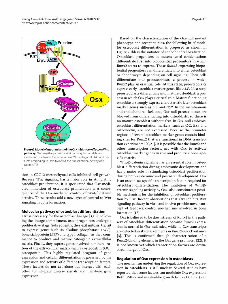
Recent studies in our research group have provided evi-dences showing that the osteoblast-specific transcriptionfactor Osx is able to inhibit Wnt pathway activity duringosteoblast differentiation [13]. In calvarial cells of E18.5Osx-null embryos, expression of the Wnt antagonistDkk1 was abolished, and that of Wnt target genes c-Mycand cyclin D1 was increased. It has been demonstratedthat Osx binds to and activates the Dkk1 promoter. Osx isshown to inhibit β-catenin-induced Topflash reporteractivity and also inhibit β-catenin-induced secondary axisformation in Xenopus embryos. Moreover, this studyshowed that in calvaria of E18.5 Osx-null embryos har-boring the TOPGAL reporter transgene, β-galactosidaseactivity was increased, suggesting that Osx inhibited theWnt pathway in osteoblasts in vivo [13]. Osx can disruptTcf binding to DNA, providing a likely mechanism for theinhibition by Osx of β-catenin transcriptional activity.The transcription factor Tcf is known to interact with β-catenin to form a functional complex in promoter regionof Wnt signaling targets to activate gene expression.
The PRR region of Osx is responsible for disruption ofTcf1 binding to DNA, and for inhibition of β-catenintranscriptional activity. These findings indicate that Osxnegatively controls the activity of β-catenin in two differ-ent mechanisms shown in Figure2: first, by being neededfor the expression of a major Wnt antagonist and second,by inhibiting the transcriptional activity of β-catenin/Tcf.
We have shown that Osx decreases osteoblast prolifera-tion [13]. E18.5 Osx-null calvaria showed greater BrdUincorporation than wild-type calvaria, and primary calva-rial cells from Osx-null E18.5 embryos also grew fasterthan wild-type cells. On the other hand, Osx over-expres-
Zhang Journal of Orthopaedic Surgery and Research 2010, 5:37http://www.josr-online.com/content/5/1/37
Page 4 of 8
sion in C2C12 mesenchymal cells inhibited cell growth.Because Wnt signaling has a major role in stimulatingosteoblast proliferation, it is speculated that Osx-medi-ated inhibition of osteoblast proliferation is a conse-quence of the Osx-mediated control of Wnt/β-cateninactivity. These results add a new layer of control to Wntsignaling in bone formation.
Molecular pathway of osteoblast differentiationOsx is necessary for the osteoblast lineage [3,13]. Follow-ing the lineage commitment, osteoprogenitors undergo aproliferative stage. Subsequently, they exit mitosis, transitto express genes such as alkaline phosphatase (ALP),bone sialoprotein (BSP) and type I collagen, as they com-mence to produce and mature osteogenic extracellularmatrix. Finally, they express genes involved in mineraliza-tion of the extracellular matrix such as osteocalcin (OC),osteopontin. This highly regulated program of geneexpression and cellular differentiation is governed by theexpression and activity of different transcription factors.These factors do not act alone but interact with eachother to integrate diverse signals and fine-tune geneexpression.
Based on the characterization of the Osx-null mutantphenotype and recent studies, the following brief modelfor osteoblast differentiation is proposed as shown inFigure3. Ihh is the initiator of endochondral ossification.Osteoblast progenitors in mesenchymal condensationsdifferentiate first into biopotential progenitors in whichRunx2 starts to express. These Runx2-expressing biopo-tential progenitors can differentiate into either osteoblastor chondrocyte depending on cell signaling. Then cellsdifferentiate into preosteoblasts, a process in whichRunx2 play an essential role. At this stage, preosteoblastsexpress early osteoblast marker genes like ALP. Next step,preosteoblasts differentiate into mature osteoblast, a pro-cess in which Osx plays a critical role. Mature functioningosteoblasts strongly express characteristic later osteoblastmarker genes such as OC and BSP. In the membranousand endochondral skeletons, Osx-null preosteoblasts areblocked from differentiating into osteoblasts, so there isno mature osteoblast without Osx. In Osx-null embryos,osteoblast differentiation markers, such as OC, BSP andosteonectin, are not expressed. Because the promoterregions of several osteoblast marker genes contain bind-ing sites for Runx2 that are functional in DNA transfec-tion experiments [20,21], it is possible that the Runx2 andother transcription factors, act with Osx to activateosteoblast marker genes in vivo and produce a bone-spe-cific matrix.
Wnt/β-catenin signaling has an essential role in osteo-blast differentiation during embryonic development andhas a major role in stimulating osteoblast proliferationduring both embryonic and postnatal development. Osxis an osteoblast-specific transcription factor, required forosteoblast differentiation. The inhibition of Wnt/β-catenin signaling activity by Osx, also constitutes a possi-ble mechanism for the inhibition of osteoblast prolifera-tion by Osx. Recent observations that Osx inhibits Wntsignaling pathway in vitro and in vivo provide novel con-cept of feedback control mechanisms involved in boneformation [13].
Osx is believed to be downstream of Runx2 in the path-way of osteoblast differentiation because Runx2 expres-sion is normal in Osx-null mice, while no Osx transcriptsare detected in skeletal elements in Runx2-knockout mice[3]. This is confirmed through characterization of aRunx2-binding element in the Osx gene promoter [22]. Itis not known yet which transcription factors are down-stream target of Osx.
Regulation of Osx expression in osteoblastsThe mechanism underlying the regulation of Osx expres-sion in osteoblasts is still unclear. Several studies havereported that some factors can modulate Osx expression.Both BMP-2 and insulin-like growth factor-1 (IGF-1) can
Figure2 Model of mechanisms of the Osx inhibitory effect on Wnt pathway. Osx negatively controls Wnt pathway by two different mechanisms: activates the expression of Wnt antagonist Dkk1 and dis-rupts Tcf binding to DNA to inhibit the transcriptional activity of β-catenin/Tcf.

Zhang Journal of Orthopaedic Surgery and Research 2010, 5:37http://www.josr-online.com/content/5/1/37
Page 5 of 8
induce Osx expression in undifferentiated mesenchymalstem cells [23]. IGF-I-mediated Osx expression requiredall three MAPK components (Erk, p38, and JNK),whereas BMP-2 required p38 and JNK signaling. Block-ing Runx2 activity inhibited the BMP-2-mediated induc-tion of Osx, suggesting a Runx2-dependent pathway.However, another research group showed that BMP-2induced Osx expression through a Runx2-independentpathway [24]. Even if Osx has been suggested as a down-stream target of Runx2, the results of this study indicatedthat Osx expression was still induced by BMP-2 treat-ment in Runx2 null cells but not induced by Runx2 over-expression in C2C12 cells. Regulatory mechanisms ofBMP-2 on Osx are not yet fully understood. Ascorbic acidand 1,25(OH)2 vitamin D3, which have positive roles inosteoblast function, have also been shown to up-regulateOsx expression [25,26]. It was demonstrated that Ascor-bic acid induced Osx expression via a novel mechanisminvolving Nrf1 nuclear translocation and Nrf1 binding toan antioxidant-responsive element to activate genes criti-cal for cell differentiation.
Some studies indicate that negative regulators of osteo-blastogenesis can inhibit Osx expression. TNF inhibitedOsx mRNA in pre-osteoblastic cells without affectingOsx mRNA half-life [27,28]. Inhibitors of MEK1 andERK1, but not of JNK or p38 kinase, abrogated TNF inhi-
bition of Osx mRNA and promoter activity. In vivo stud-ies provide genetic evidence that p53 tumor suppressorblocks osteoblast differentiation and bone development[27,28]. Prolonged exposure to parathyroid hormone(PTH) negatively regulates Osx expression in osteoblastsby a transcriptional mechanism mediated by cAMP sig-naling [29]. PTH inhibited Osx mRNA and proteinexpression, and this effect could be mimicked by forsko-lin, 8-bromo-cAMP, or expression of constitutively activeGsalpha. On the other hand, some other researchersfound that systemic PTH treatments accelerated fracturehealing in mice concomitantly with increased Osxexpression in the PTH treated fracture calluses, suggest-ing a mechanism for PTH-mediated fracture healing pos-sibly via Osx induction [30]. Recently studies indicatedthat intermittent PTH increased in vivo Osx expressionin osteoblasts through a pathway requiring activatingtranscription factor 4 (ATF4) [31]. ATF4-responsive ele-ment has been identified in the proximal Osx promoter.
Despite these interesting findings, the details concern-ing the regulation and function of Osx are incompletelyunderstood.
Osteoporosis and OsxOsteoporosis is characterized by reduced bone mass,alterations in the microarchitecture of bone tissue,
Figure3 The proposed model of coordinated regulation of osteoblast differentiation and proliferation during bone formation by Osx and Wnt/β-catenin signaling. Ihh is the initiator of endochondral ossification. The Runx2-expressing biopotential progenitors can differentiate into either osteoblast or chondrocyte. Then cells differentiate into preosteoblasts, in which Runx2 play an essential role. In the next step, preosteoblasts differen-tiate into mature osteoblast, a process in which Osx plays a critical role. Wnt/β-catenin signaling has an essential role in osteoblast differentiation and osteoblast proliferation. The inhibition of Wnt/β-catenin signaling activity by Osx constitutes a possible mechanism for the inhibition by Osx of osteo-blast proliferation.

Zhang Journal of Orthopaedic Surgery and Research 2010, 5:37http://www.josr-online.com/content/5/1/37
Page 6 of 8
reduced bone strength, and an increased risk of fracture[32]. Osteoporosis is a common condition that affects upto 30% of women and 12% of men at some point in life.The prevalence of osteoporosis increases with age due toan imbalance in the rate at which bone is removed andreplaced during the bone remodeling, which is an impor-tant physiological process essential for healthy skeletonmaintenance. Many factors influence the risk of osteopo-rosis--including diet, physical activity, medication use,and coexisting diseases--but one of the most importantclinical risk factors is a positive family history, emphasiz-ing the importance of genetics in the pathogenesis ofosteoporosis. Genetic factors have been recognized toplay important roles in the pathogenesis of osteoporosis.Evidence from twin and family studies suggests thatbetween 50% and 85% of the variance in peak bone massis genetically determined [33].
Recent study has indicated that genetic variants in thechromosomal region of Osx are associated with bonemineral density (BMD) in children and adults probablythrough primary effects on growth [34]. A genome-wideassociation study of BMD and related traits in 1518 chil-dren from the Avon Longitudinal Study of Parents andChildren (ALSPAC) was carried out to identify geneticvariants affecting BMD. This research group identifiedassociations with BMD in an area of chromosome 12 con-taining the Osx (SP7) locus. A meta-analysis of theseexisting studies revealed strong association betweenSNPs in the Osx region and adult lumbar spine BMD. Inlight of these findings, this research group genotyped afurther 3692 individuals from ALSPAC who had wholebody BMD and confirmed the association in children aswell.
Although Osx has been identified to be associated withosteoporosis-related phenotypes, further investigationneeds to be done to determine whether Osx will repre-sent a useful diagnostic index of osteoporosis or molecu-lar target for therapeutic manipulation.
Possible clinical application of OsxOsx is indispensable for the commitment of the osteo-blast lineage and the expression of the osteoblast-specificmatrix proteins, including type I collagen, bone sialopro-tein, osteonectin, and osteocalcin. No pharmacologicalapproach to target Osx in osteoblasts has been reported.Heterozygous mutations in Runx2 , which is an upstreamof Osx, have been shown to be the cause of the humangenetic disease cleidocranial dysplasia [35]. There is noevidence so far that any Osx mutation leads to any clinicalhuman disease.
The extensive studies by many laboratories to explorehow to control the Wnt signaling pathway in osteoblastsstems from the realization that this pathway has an essen-tial role in bone mass determination in the adult skeleton.
There is also an expectation that efforts to pharmacologi-cally target this pathway should yield promising agents totreat bone diseases such as osteoporosis. Results in ourgroup showing that Osx inhibits Wnt/β-catenin signalingadd an important new layer of control to the complexregulation of the Wnt pathway in osteoblasts [13].
It was observed that the Osx expression was decreasedin two mouse osteosarcoma cell lines and in three humanosteosarcoma cell lines [36]. Transfection of the Osx geneinto the mouse osteosarcoma cells inhibited tumor cellgrowth in vitro and in vivo and significantly reducedtumor incidence, tumor volume, and lung metastasis fol-lowing intratibial injection. Using an in vitro migrationassay, Osx suppressed the migration of tumor cells tolung extracts. These results suggest that Osx expressionmay play a role in osteosarcoma tumor growth andmetastasis, and that osteolytic activity of tumor cells maybe regulated by Osx via down-regulation of interleukin-1gene transcription [36]. It is relatively consistent with therecent mechanism studies that Osx inhibits osteoblastproliferation through controlling the Wnt pathway [13].
Bone formation is essential for maintenance and heal-ing of the skeleton following injury and operative inter-ventions, such as osteotomies and limb lengthening. Innumerous orthopedic conditions, such as congenitalpseudoarthrosis of tibia, femoral head osteonecrosis, andlarge bone lengthening, bone healing and regenerationremain challenging goal to achieve. Most therapy for skel-etal diseases with less bone such as osteoporosis andosteonecrosis is aimed at inhibiting bone resorption, butto cure these diseases, it is also critically important tostimulate new bone formation. Therefore, there is cur-rently great interest in understanding the regulation ofosteoblast differentiation and activity to guide the devel-opment of anabolic therapies. Although no pharmacolog-ical approach to target Osx in osteoblasts has identifiedyet, an interesting future research direction is to look forupstream genes or molecules which can selectively targetOsx expression and activity. We speculate that Osx couldbecome a therapeutic target in efforts to stimulate theanabolic pathway of bone synthesis.
ConclusionsBone formation is a complex process regulated by multi-ple factors and pathways; it is clearly shown that Osx isrequired for the final commitment of osteoblast lineage.Although recent molecular and genetic studies usinggene targeting in mice have established Osx as a masterregulator of osteoblast differentiation during bone forma-tion, the mechanisms of Osx regulation of osteoblast dif-ferentiation and function are still under investigation.Future studies to decipher the Osx direct upstream ordownstream molecular targets, Osx expression regula-tion and Osx functional partners are required to clarify

Zhang Journal of Orthopaedic Surgery and Research 2010, 5:37http://www.josr-online.com/content/5/1/37
Page 7 of 8
the detailed mechanism of the temporal and spatial regu-lation of Osx for bone formation and homeostatic regula-tion of skeletal system. The need to develop novel drugsthat stimulate bone formation and thereby elevate bonemass (anabolic agents) has opened new research areas fortherapeutic intervention in the treatment of bone-relateddiseases.
AbbreviationsOsx: Osterix; OB: osteoblast; E18.5: embryonic day 18.5; Ihh: Indian hedgehog;Col1a1: type I collagen; OC: osteocalcin; BSP: bone sialoprotein; ALP: alkalinephosphatase; PRR: proline-rich region; BMP2: bone morphogenic protein-2;BMD: bone mineral density.
Competing interestsThe authors declare that they have no competing interests.
Authors' contributionsThe author contributed to the article. The author has read and approved thefinal manuscript.
AcknowledgementsWe would like to thank Benoit de Crombrugghe for his help. Work in Bone Research Laboratory is supported by Research Grant from Arthritis Foundation (To Chi Zhang) and RAP01 grant from Texas Scottish Rite Hospital for Children (To Chi Zhang).
Author DetailsBone Research Laboratory, Texas Scottish Rite Hospital for Children, Department of Orthopedic Surgery, University of Texas Southwestern Medical Center at Dallas, Texas, USA
References1. St-Jacques B, Hammerschmidt M, McMahon AP: Indian hedgehog
signaling regulates proliferation and differentiation of chondrocytes and is essential for bone formation. Genes Dev 1999, 13:2072-2086.
2. Komori T, Yagi H, Nomura S, Yamaguchi A, Sasaki K, Deguchi K, Shimizu Y, Bronson RT, Gao YH, Inada M, Sato M, Okamoto R, Kitamura Y, Yoshiki S, Kishimoto T: Targeted disruption of Cbfa1 results in a complete lack of bone formation owing to maturational arrest of osteoblasts. Cell 1997, 89:755-764.
3. Nakashima K, Zhou X, Kunkel G, Zhang Z, Deng JM, Behringer RR, de Crombrugghe B: The novel zinc finger-containing transcription factor osterix is required for osteoblast differentiation and bone formation. Cell 2002, 108:17-29.
4. Day TF, Guo X, Garrett-Beal L, Yang Y: Wnt/beta-catenin signaling in mesenchymal progenitors controls osteoblast and chondrocyte differentiation during vertebrate skeletogenesis. Dev Cell 2005, 8:739-750.
5. Hill TP, Spater D, Taketo MM, Birchmeier W, Hartmann C: Canonical Wnt/beta-catenin signaling prevents osteoblasts from differentiating into chondrocytes. Dev Cell 2005, 8:727-738.
6. Hu H, Hilton MJ, Tu X, Yu K, Ornitz DM, Long F: Sequential roles of Hedgehog and Wnt signaling in osteoblast development. Development 2005, 132:49-60.
7. Rodda SJ, McMahon AP: Distinct roles for Hedgehog and canonical Wnt signaling in specification, differentiation and maintenance of osteoblast progenitors. Development 2006, 133:3231-3244.
8. Bialek P, Kern B, Yang X, Schrock M, Sosic D, Hong N, Wu H, Yu K, Ornitz DM, Olson EN, Justice MJ, Karsenty G: A twist code determines the onset of osteoblast differentiation. Dev Cell 2004, 6:423-435.
9. Yang X, Matsuda K, Bialek P, Jacquot S, Masuoka HC, Schinke T, Li L, Brancorsini S, Sassone-Corsi P, Townes TM, Hanauer A, Karsenty G: ATF4 is a substrate of RSK2 and an essential regulator of osteoblast biology; implication for Coffin-Lowry Syndrome. Cell 2004, 117:387-398.
10. Dobreva G, Chahrour M, Dautzenberg M, Chirivella L, Kanzler B, Farinas I, Karsenty G, Grosschedl R: SATB2 is a multifunctional determinant of craniofacial patterning and osteoblast differentiation. Cell 2006, 125:971-986.
11. Jones DC, Wein MN, Oukka M, Hofstaetter JG, Glimcher MJ, Glimcher LH: Regulation of adult bone mass by the zinc finger adapter protein Schnurri-3. Science 2006, 312:1223-1227.
12. Acampora D, Merlo GR, Paleari L, Zerega B, Postiglione MP, Mantero S, Bober E, Barbieri O, Simeone A, Levi G: Craniofacial, vestibular and bone defects in mice lacking the Distal-less-related gene Dlx5. Development 1999, 126:3795-3809.
13. Zhang C, Cho K, Huang Y, Lyons JP, Zhou X, Sinha K, McCrea PD, de Crombrugghe B: Inhibition of Wnt signaling by the osteoblast-specific transcription factor Osterix. Proc Natl Acad Sci USA 2008, 105:6936-6941.
14. Gong Y, Slee RB, Fukai N, Rawadi G, Roman-Roman S, Reginato AM, Wang H, Cundy T, Glorieux FH, Lev D, Zacharin M, Oexle K, Marcelino J, Suwairi W, Heeger S, Sabatakos G, Apte S, Adkins WN, Allgrove J, Arslan-Kirchner M, Batch JA, Beighton P, Black GC, Boles RG, Boon LM, Borrone C, Brunner HG, Carle GF, Dallapiccola B, De Paepe A, et al.: LDL receptor-related protein 5 (LRP5) affects bone accrual and eye development. Cell 2001, 107:513-523.
15. Kato M, Patel MS, Levasseur R, Lobov I, Chang BH, Glass DA, Hartmann C, Li L, Hwang TH, Brayton CF, Lang RA, Karsenty G, Chan L: Cbfa1-independent decrease in osteoblast proliferation, osteopenia, and persistent embryonic eye vascularization in mice deficient in Lrp5, a Wnt coreceptor. J Cell Biol 2002, 157:303-314.
16. Little RD, Carulli JP, Del Mastro RG, Dupuis J, Osborne M, Folz C, Manning SP, Swain PM, Zhao SC, Eustace B, Lappe MM, Spitzer L, Zweier S, Braunschweiger K, Benchekroun Y, Hu X, Adair R, Chee L, FitzGerald MG, Tulig C, Caruso A, Tzellas N, Bawa A, Franklin B, McGuire S, Nogues X, Gong G, Allen KM, Anisowicz A, Morales AJ, et al.: A mutation in the LDL receptor-related protein 5 gene results in the autosomal dominant high-bone-mass trait. Am J Hum Genet 2002, 70:11-19.
17. Babij P, Zhao W, Small C, Kharode Y, Yaworsky PJ, Bouxsein ML, Reddy PS, Bodine PV, Robinson JA, Bhat B, Marzolf J, Moran RA, Bex F: High bone mass in mice expressing a mutant LRP5 gene. J Bone Miner Res 2003, 18:960-974.
18. Morvan F, Boulukos K, Clement-Lacroix P, Roman Roman S, Suc-Royer I, Vayssiere B, Ammann P, Martin P, Pinho S, Pognonec P, Mollat P, Niehrs C, Baron R, Rawadi G: Deletion of a single allele of the Dkk1 gene leads to an increase in bone formation and bone mass. J Bone Miner Res 2006, 21:934-945.
19. Li J, Sarosi I, Cattley RC, Pretorius J, Asuncion F, Grisanti M, Morony S, Adamu S, Geng Z, Qiu W, Kostenuik P, Lacey DL, Simonet WS, Bolon B, Qian X, Shalhoub V, Ominsky MS, Zhu Ke H, Li X, Richards WG: Dkk1-mediated inhibition of Wnt signaling in bone results in osteopenia. Bone 2006, 39:754-766.
20. Ducy P, Zhang R, Geoffroy V, Ridall AL, Karsenty G: Osf2/Cbfa1: a transcriptional activator of osteoblast differentiation. Cell 1997, 89:747-754.
21. Thirunavukkarasu K, Halladay DL, Miles RR, Yang X, Galvin RJ, Chandrasekhar S, Martin TJ, Onyia JE: The osteoblast-specific transcription factor Cbfa1 contributes to the expression of osteoprotegerin, a potent inhibitor of osteoclast differentiation and function. J Biol Chem 2000, 275:25163-25172.
22. Nishio Y, Dong Y, Paris M, O'Keefe RJ, Schwarz EM, Drissi H: Runx2-mediated regulation of the zinc finger Osterix/Sp7 gene. Gene 2006, 372:62-70.
23. Celil AB, Campbell PG: BMP-2 and insulin-like growth factor-I mediate Osterix (Osx) expression in human mesenchymal stem cells via the MAPK and protein kinase D signaling pathways. J Biol Chem 2005, 280:31353-31359.
24. Lee MH, Kwon TG, Park HS, Wozney JM, Ryoo HM: BMP-2-induced Osterix expression is mediated by Dlx5 but is independent of Runx2. Biochem Biophys Res Commun 2003, 309:689-694.
25. Maehata Y, Takamizawa S, Ozawa S, Kato Y, Sato S, Kubota E, Hata R: Both direct and collagen-mediated signals are required for active vitamin D3-elicited differentiation of human osteoblastic cells: roles of osterix, an osteoblast-related transcription factor. Matrix Biol 2006, 25:47-58.
26. Xing W, Singgih A, Kapoor A, Alarcon CM, Baylink DJ, Mohan S: Nuclear factor-E2-related factor-1 mediates ascorbic acid induction of osterix
Received: 13 March 2010 Accepted: 15 June 2010 Published: 15 June 2010This article is available from: http://www.josr-online.com/content/5/1/37© 2010 Zhang; licensee BioMed Central Ltd. This is an Open Access article distributed under the terms of the Creative Commons Attribution License (http://creativecommons.org/licenses/by/2.0), which permits unrestricted use, distribution, and reproduction in any medium, provided the original work is properly cited.Journal of Orthopaedic Surgery and Research 2010, 5:37
Zhang Journal of Orthopaedic Surgery and Research 2010, 5:37http://www.josr-online.com/content/5/1/37
Page 8 of 8
expression via interaction with antioxidant-responsive element in bone cells. J Biol Chem 2007, 282:22052-22061.
27. Lu X, Gilbert L, He X, Rubin J, Nanes MS: Transcriptional regulation of the osterix (Osx, Sp7) promoter by tumor necrosis factor identifies disparate effects of mitogen-activated protein kinase and NF kappa B pathways. J Biol Chem 2006, 281:6297-6306.
28. Zambetti GP, Horwitz EM, Schipani E: Skeletons in the p53 tumor suppressor closet: genetic evidence that p53 blocks bone differentiation and development. J Cell Biol 2006, 172:795-797.
29. Hong SH, Lu X, Nanes MS, Mitchell J: Regulation of osterix (Osx, Sp7) and the Osx promoter by parathyroid hormone in osteoblasts. J Mol Endocrinol 2009, 43:197-207.
30. Kaback LA, Soung do Y, Naik A, Geneau G, Schwarz EM, Rosier RN, O'Keefe RJ, Drissi H: Teriparatide (1-34 human PTH) regulation of osterix during fracture repair. J Cell Biochem 2008, 105:219-226.
31. Yu S, Franceschi RT, Luo M, Fan J, Jiang D, Cao H, Kwon TG, Lai Y, Zhang J, Patrene K, Hankenson K, Roodman GD, Xiao G: Critical role of activating transcription factor 4 in the anabolic actions of parathyroid hormone in bone. PLoS One 2009, 4:e7583.
32. Kanis JA, Melton LJ, Christiansen C, Johnston CC, Khaltaev N: The diagnosis of osteoporosis. J Bone Miner Res 1994, 9:1137-1141.
33. Smith DM, Nance WE, Kang KW, Christian JC, Johnston CC Jr: Genetic factors in determining bone mass. J Clin Invest 1973, 52:2800-2808.
34. Timpson NJ, Tobias JH, Richards JB, Soranzo N, Duncan EL, Sims AM, Whittaker P, Kumanduri V, Zhai G, Glaser B, Eisman J, Jones G, Nicholson G, Prince R, Seeman E, Spector TD, Brown MA, Peltonen L, Smith GD, Deloukas P, Evans DM: Common variants in the region around Osterix are associated with bone mineral density and growth in childhood. Hum Mol Genet 2009, 18:1510-1517.
35. Mundlos S, Otto F, Mundlos C, Mulliken JB, Aylsworth AS, Albright S, Lindhout D, Cole WG, Henn W, Knoll JH, Owen MJ, Mertelsmann R, Zabel BU, Olsen BR: Mutations involving the transcription factor CBFA1 cause cleidocranial dysplasia. Cell 1997, 89:773-779.
36. Cao Y, Zhou Z, de Crombrugghe B, Nakashima K, Guan H, Duan X, Jia SF, Kleinerman ES: Osterix, a transcription factor for osteoblast differentiation, mediates antitumor activity in murine osteosarcoma. Cancer Res 2005, 65:1124-1128.
doi: 10.1186/1749-799X-5-37Cite this article as: Zhang, Transcriptional regulation of bone formation by the osteoblast-specific transcription factor Osx Journal of Orthopaedic Surgery and Research 2010, 5:37