revised role of glycosaminoglycans in tat protein transduction domain-mediated cellular
TRANSCRIPT

Revised Role of Glycosaminoglycans in TAT ProteinTransduction Domain-mediated Cellular Transduction□S
Received for publication, May 16, 2009, and in revised form, October 12, 2009 Published, JBC Papers in Press, October 26, 2009, DOI 10.1074/jbc.M109.021964
Jacob M. Gump, Ronald K. June, and Steven F. Dowdy1
From the Howard Hughes Medical Institute, Department of Cellular and Molecular Medicine, School of Medicine, University ofCalifornia, San Diego, La Jolla, California 92093-0686
Cellular uptake of the human immunodeficiency virus TATprotein transduction domain (PTD), or cell-penetrating pep-tide, has previously been surmised to occur in a manner depen-dent on the presence of heparan sulfate proteoglycans that areexpressed ubiquitously on the cell surface. These acidic polysac-charides form a large pool of negative charge on the cell surfacethat TAT PTD binds avidly. Additionally, sulfated glycans havebeen proposed to aid in the interaction of TAT PTD and otherarginine-rich PTDs with the cell membrane, perhaps aidingtheir translocation across the membrane. Surprisingly, how-ever, TAT PTD-mediated induction of macropinocytosis andcellular transduction occurs in the absence of heparan sulfateand sialic acid. Using labeledTATPTDpeptides and fusion pro-teins, in addition to TAT PTD-Cre recombination-based phe-notypic assays, we show that transduction occurs efficiently inmutant Chinese hamster ovary cell lines deficient in glycosami-noglycans and sialic acids. Similar results were obtained in cellswhere glycans were enzymatically removed. In contrast, enzy-matic removal of proteins from the cell surface completelyablatedTATPTD-mediated transduction.Our findings supportthe hypothesis that acidic glycans form a pool of charge thatTAT PTD binds on the cell surface, but this binding is indepen-dent of the PTD-mediated transduction mechanism and theinduction of macropinocytotic uptake by TAT PTD.
Cationic peptide-mediated cellular transduction representsa cell entrymodality with enormous potential for the delivery ofmacromolecular therapeutic agents. The human immunodefi-ciency virus TAT protein basic domain (RKKRRQRRR) andother protein/peptide transduction domains (PTDs)2 havebeen used to deliver a wide variety of bioactive cargo into cellsin culture and preclinical models in vivo (1–3). Efficient deliv-ery of peptide, nucleic acid, and full-length protein cargoes bycationic PTDs has been demonstrated by many groups, and
several clinical trials are currently under way using PTD-medi-ated delivery. Despite a number of studies on the mechanismused by PTDs to enter cells, many questions about their uptakeremain unanswered (4–7). We and others (8–10) have previ-ously shown that TAT PTD and other cationic PTDs induce aubiquitous form of fluid-phase endocytosis termedmacropino-cytosis and enter cells in this manner. However, many mecha-nistic aspects of TATPTD transduction remain poorly defined.For instance, we have yet to identify the molecules that TATPTD binds on the cell surface to induce macropinocytosis, andwe do not know how escape across the endosomal lipid bilayerinto the cytoplasm is accomplished.Using fluorescently labeled peptides, it has previously been
reported that TAT PTD avidly binds to sulfated glycans andthat the presence of acidic polysaccharides like heparin andchondroitin sulfate in solution can compete with TAT PTD forbinding to the cell surface and inhibit transduction (8, 11, 12).Based on these observations, several studies argue for a signifi-cant role for glycans in the transduction of cationic PTDs, withsome researchers concluding that transduction is dependent onheparan sulfate (HS) proteoglycans (13–20). However, thesestudies relies predominantly on both flow cytometry andmicroscopic visual observations of fluorescently labeled PTDpeptides and are therefore did not necessarily assay for theamount of PTD cargo that has escaped from endocytic vesiclesinto the cytoplasm.We refer to these observations as “cell asso-ciation” because they do not sufficiently distinguish betweencells that have taken up fluorescently labeled PTDs in endocyticvesicles (or bound to the cell surface) and PTD peptide andcargo that have actually escaped endosomes into the cytoplasm.This distinction is critical because the inside of the endosome isessentially the outside of the cell; no biological response will beelicited simply by uptake into vesicles. Indeed, endosomalescape is requisite for transduction of cargo into the cytoplasmto induce phenotypic responses, and the majority of labeledTAT PTD taken up by cells is present in endosomes, with onlya small fraction actually escaping into the cytoplasm.Here, we sought to explore the role of glycans in TAT PTD-
mediated transduction using Chinese hamster ovary (CHO)glycan-deficient cell lines that are genetically deficient for cell-surface expression of heparan and chondroitin sulfate glycos-aminoglycans or sialic acids (SAs) (21–24). Although previousstudies using fluorescently labeled PTD peptides indicated arequisite role for HS in transduction (13, 16, 17), surprisingly,we found, via a phenotypic assay, that cells lackingHS are in factfully functional for TAT PTD-mediated transduction. More-over, transduction occurs efficiently in cells completely lacking
Author’s Choice—Final version full access.□S The on-line version of this article (available at http://www.jbc.org) contains
supplemental Fig. S1.1 Supported by the Howard Hughes Medical Institute, the Leukemia and Lym-
phoma Society, and the Pardee Foundation. To whom correspondenceshould be addressed: Howard Hughes Medical Inst., Dept. of Cellular andMolecular Medicine, School of Medicine, University of California, SanDiego, 9500 Gilman Dr., GPL 231B, MC 0686, La Jolla, CA 92093-0686.E-mail: [email protected].
2 The abbreviations used are: PTD, protein transduction domain; HS, heparansulfate; CHO, Chinese hamster ovary; SA, sialic acid; EGFP, enhanced greenfluorescent protein; FITC, fluorescein isothiocyanate; TMR, tetramethylrho-damine; LSL, LoxP-STOP-LoxP; PBS, phosphate-buffered saline; FACS, fluo-rescence-activated cell sorting.
THE JOURNAL OF BIOLOGICAL CHEMISTRY VOL. 285, NO. 2, pp. 1500 –1507, January 8, 2010Author’s Choice © 2010 by The American Society for Biochemistry and Molecular Biology, Inc. Printed in the U.S.A.
1500 JOURNAL OF BIOLOGICAL CHEMISTRY VOLUME 285 • NUMBER 2 • JANUARY 8, 2010
by guest on Novem
ber 16, 2018http://w
ww
.jbc.org/D
ownloaded from

all glycosaminoglycans, including HS, in cells lacking SAs, andin cells depleted of both together. Our findings support thehypothesis that acidic glycans form a pool of negative chargethat TAT PTD binds on the cell surface but are dispensable forcationic PTD-mediated transduction into cells.
EXPERIMENTAL PROCEDURES
Generation of Stable Cell Lines—Parental CHO-K1 cells andglycosaminoglycan mutant pgsA (745) cells lacking xylosyl-transferase necessary for glycosaminoglycan chain initiationwere a gift from Dr. Jeffrey Esko (University of California, SanDiego). Lec2 cells, which lack activity of the SA Golgi trans-porter SLC35A1 and are extremely deficient in expression ofcell-surface SA, were a gift from Dr. Ajit Varki (Universityof California, San Diego). Cells were maintained in Ham’sF-12 medium (Invitrogen) containing 10% fetal bovine serum(Sigma). Stable clones were made using a construct (pZ/EG)that contains a Lox-STOP-Lox-EGFPmotif, resulting in strongEGFP expression only after Cre-mediated recombination. Cellswere transfected with pZ/EG and selected with G418. Multiplesubclones were analyzed by flow cytometry for Cre-inducibleEGFP expression following transfection with a Cre expressionconstruct and subsequently by treatment with TAT PTD-Cre.The clones chosen for use in this study had low backgroundEGFP expression and showed comparable induction of EGFPexpression after expression of Cre.Peptide Synthesis and Purification of Recombinant Proteins—
Peptides were synthesized by SPPS on a Symphony Quar-tet peptide synthesizer (Rainin) using Fmoc (N-(9-fluorenyl)me-thoxycarbonyl)-protected L-amino acids on an amidatedsupport (EMD). Fluorescein isothiocyanate (FITC) or tetrameth-ylrhodamine (TMR) labeling was performed by reaction of thepeptide with carboxyfluorescein (Sigma) while still on solid-phase support. Crude peptides were purified by reverse-phase high pressure liquid chromatography and confirmedby matrix-assisted laser desorption ionization time-of-flightmass spectroscopy.Recombinant TAT-Cre and Cre proteins were expressed in
BL21-CodonPlus(DE3)-RIPL cells (Stratagene) using pET28.2protein expression constructs (Novagen). Both the TAT PTD-Cre and Cre constructs contain a His6 motif at the C terminus,with TAT at the N terminus in the TAT-Cre expression con-struct. Cells were transformed and grown overnight at 37 °Cunder kanamycin and chloramphenicol selection. Expressionwas induced with 500 �M isopropyl �-D-thiogalactopyranosidefor 4 h at 37 °C. Bacteria were lysed by sonication in the pres-ence of protease inhibitors, lysozyme, RNase A, and DNase I.Recombinant Cre was purified from cleared lysate first by nick-el-nitrilotriacetic acid (Qiagen) chromatography followed byion exchange chromatography using a Mono S column on anAKTAFPLC system (GEHealthcare). Purity and concentrationof proteins were confirmed by SDS-PAGE, and Cre activity wasconfirmed by in-cell transduction and in vitro recombinationassays. Recombinant Cre and TAT PTD-Cre were labeled withAlexa Fluor 546 by reaction with the succinimidyl ester(Invitrogen) for 1 h under slightly basic conditions according tothe company’s protocol. Labeled protein was concentrated and
repurified by ion exchange fast protein liquid chromatographyand confirmed by gel electrophoresis.TAT-Cre Transduction and Recombination Assays—Stable
CHO-K1, pgsA, and Lec2 LoxP-STOP-LoxP (LSL)-GFP cloneswere seeded overnight onto 24-well plates at a density of 20,000cells/well. Cells were washed and incubated in serum-freemedium for 1 h at 37 °C. When pharmacological inhibitors(Sigma) were used, these were added with the serum-free me-dium and added again with recombinant protein. Cells werethen treated with Cre/TAT PTD-Cre for 1 h in serum-freemedium at 37 or 4 °C as indicated. Cells were washed withphosphate-buffered saline (PBS), trypsinized, and replated. At24 h after Cre addition, cells were trypsinized and assayed forGFP expression on an LSRII flow cytometer equipped withFACSDiva acquisition and analysis software (BD Biosciences).The live cell population was gated by forward scatter/side scat-ter, and 10,000 live cellswere used for each analysis.Histogramswere generated using FlowJo software (Tree Star).For trypsin depletion of cell-surface proteins, cells were
treated with trypsin/EDTA (Invitrogen) or EDTA-based celldissociation solution (Sigma) for 15 min at 37 °C, followed bywasheswith PBS and 1� soybean trypsin inhibitor (10mg/ml inPBS; Sigma). Cells were then treatedwith TATPTD-Cre for 1 hat 37 °C in serum-free medium with 1� soybean trypsin inhib-itor, followed by trypsinization and replating. Cells were ana-lyzed for recombination at 24 h as described above.TAT PTD-FITC and Labeled TAT PTD-Cre Cell Association
and Extracellular Binding—CHO-K1, pgsA, and Lec2 cells(pZ/EG LSL-GFP clones were used interchangeably with iden-tical results) were seeded overnight onto 24-well plates at adensity of 75,000 cells/well. Cells werewashed and incubated inserum-free medium for 1 h at 37 °C. For cell association, cellswere then treated with TAT PTD-FITC, TAT PTD-TMR, orTAT PTD-Cre-Alexa Fluor 546 for 1 h in serum-free mediumat 37 or 4 °C as indicated. Cells were placed on ice, washedseveral timeswith PBS, washedwith 0.5mg/ml heparin (Sigma)in PBS, trypsinized, and placed on ice for FACS.Cell associationwas quantitated by FACS as described above. For extracellularbinding, cells were placed on ice and incubated for 15 min withlabeled TAT PTD peptide or TAT PTD-Cre in serum-freemedium. Cells were then washed several times with ice-coldPBS, washed with 0.5 mg/ml heparin in PBS, removed from theplate with cell dissociation solution, and placed on ice forimmediate FACS analysis. For microscopic visualization of cellassociation, cells were plated on glass coverslips and treated asdescribed above with 0.5 �M TAT PTD-Cre-Alexa Fluor 546for 1 h in serum-free medium at 37 °C. Cells were washed sev-eral timeswith PBS,washedwith 0.5mg/ml heparin in PBS, andvisualized on an Axiovert 200M microscope equipped with alive cell incubation chamber and CCD camera (Zeiss). Imageswere acquired using Axiovision 4.5 software (Zeiss) and pro-cessed for publication using Photoshop CS2 (Adobe).MacropinocytoticUptake ofNeutralDextran—For flow cyto-
metric analysis, CHO-K1, pgsA, and Lec2 cells (LSL-GFPclones were used interchangeably with identical results) wereseeded overnight onto 24-well plates at a density of 50,000 cells/well. Cells were washed and incubated in serum-free mediumfor 1 h at 37 °C. Cells were then incubated for 1 h at 37 °C with
Glycans and TAT PTD Transduction
JANUARY 8, 2010 • VOLUME 285 • NUMBER 2 JOURNAL OF BIOLOGICAL CHEMISTRY 1501
by guest on Novem
ber 16, 2018http://w
ww
.jbc.org/D
ownloaded from

0.5 mg/ml 70-kDa neutral dextran-Texas Red with the indi-cated concentrations of unlabeled TAT PTD peptide or TATPTD-Cre protein. Cells were placed on ice, washed severaltimes with PBS, washed with 0.5 mg/ml heparin in PBS,trypsinized, and placed on ice for FACS. Cells were analyzed byFACS as described above. For microscopy, cells were plated onglass coverslips overnight and incubated in serum-free Ham’sF-12 medium for 1 h at 37 °C. Cells were then incubated for 1 hat 37 °Cwith 0.5mg/ml 70-kDa neutral dextran-TMR and 1�M
unlabeled TAT PTD peptide, followed by washes with PBS and0.5 mg/ml heparin/PBS and microscopy as described above.Heparinase and Sialidase Depletion of Glycans—CHO-K1,
pgsA, and Lec2 LSL-GFP cells or Tex-LoxP-EGFP cells wereseeded overnight on 24-well plates and treated with the indi-cated concentrations of heparinase-3 (Sigma) or sialidase(neuraminidase from Arthrobacter ureafaciens; E-Y Laborato-ries) in serum-free medium for 1 h at 37 °C. After washing withPBS, cells were assayed for TAT PTD-Cre recombination, cellassociation, or dextran uptake as described above. To deter-mine the efficiency ofHS depletion, cells were placed on ice andtreated with 1 �g/ml biotinylated fibroblast growth factor 2(from the laboratory of Jeffrey Esko) for 15 min, followed bywashes with ice-cold PBS, a 15-min incubation with streptavi-din-phycoerythrin-Cy5 (BD Biosciences), and washes withPBS. Following removal from the plate with cell dissociationsolution, cells were analyzed by FACS. To assay SA depletion,cells were incubated with FITC-conjugated wheat germ agglu-tinin (Sigma) on ice for 15 min, followed by PBS washing andFACS.
RESULTS
Fluorescently Labeled TAT PTD Actively Associates withCells Lacking Glycans—To obtain a quantitative measure oftransduction efficiency and to reconcile our preliminary datawith those of previous researchers, we measured, via flowcytometry, the cell association of fluorescently labeled TATPTD peptide and TAT PTD-Cre in wild-type and glycan-defi-cient CHO cells. Cells treated with fluoresceinated TAT PTDpeptide (TAT PTD-FITC) exhibited differences in cell associa-tion, with less TAT peptide association in the glycan-deficientcells, particularly at 5�M, where there was a 30–40% reductionin cell association (Fig. 1A). Also notable were differences in thedynamics of TAT PTD-TMR uptake (Fig. 1B). Incubation ofcells at 4 °C is known to inhibit macropinocytotic uptake ofTAT PTDs (8–10).We detected differences between wild-typeCHO cells and HS- and SA-deficient cells in the cell-surfacebinding of TAT PTD peptides at 4 °C (Fig. 1C) that correlatedwith differences in TAT PTD-FITC cell association, indicatinga reduction in extracellular binding capacity in glycan-deficientcells. We also examined uptake differences of a TAT PTDfusion protein, namely TAT PTD-Cre (37 kDa). Consistentwith the TAT PTD peptide results above, cells lacking HS andSA also had reduced TAT PTD-Cre-Alexa Fluor 546 cell asso-ciation relative towild-type glycan-positiveCHOcells (Fig. 2A).Cells treated with TAT PTD-Cre-Alexa Fluor 546 also exhibitpunctate macropinocytotic staining as visualized by live cellmicroscopy (Fig. 2B). Extracellular binding of labeled TATPTD-Cre at 4 °C (Fig. 2C) revealed similar differences in the
FIGURE 1. TAT PTD peptide cell association and cell-surface binding inglycan-deficient cells. A, TAT PTD peptide cell association in parentalCHO-K1 and derivative glycan mutant pgsA and Lec2 cell lines. Cells weretreated with the indicated concentrations of fluoresceinated TAT PTD pep-tide (TAT PTD-FITC) at 37 °C for 1 h, followed by washes with PBS, heparin, andtrypsin to remove extracellular peptide. Cells were placed on ice and imme-diately analyzed by flow cytometry. B, time course of TAT PTD cell associationin parental and glycan mutant cell lines. Cells were treated with 2.5 �M TATPTD-TMR as described for A for the indicated times. C, TAT PTD peptide cell-surface binding at 4 °C. Cells were treated for 15 min with TAT PTD-FITC pep-tide, followed by washes with PBS, nonenzymatic cell dissociation, and flowcytometry. WT, wild-type.
Glycans and TAT PTD Transduction
1502 JOURNAL OF BIOLOGICAL CHEMISTRY VOLUME 285 • NUMBER 2 • JANUARY 8, 2010
by guest on Novem
ber 16, 2018http://w
ww
.jbc.org/D
ownloaded from
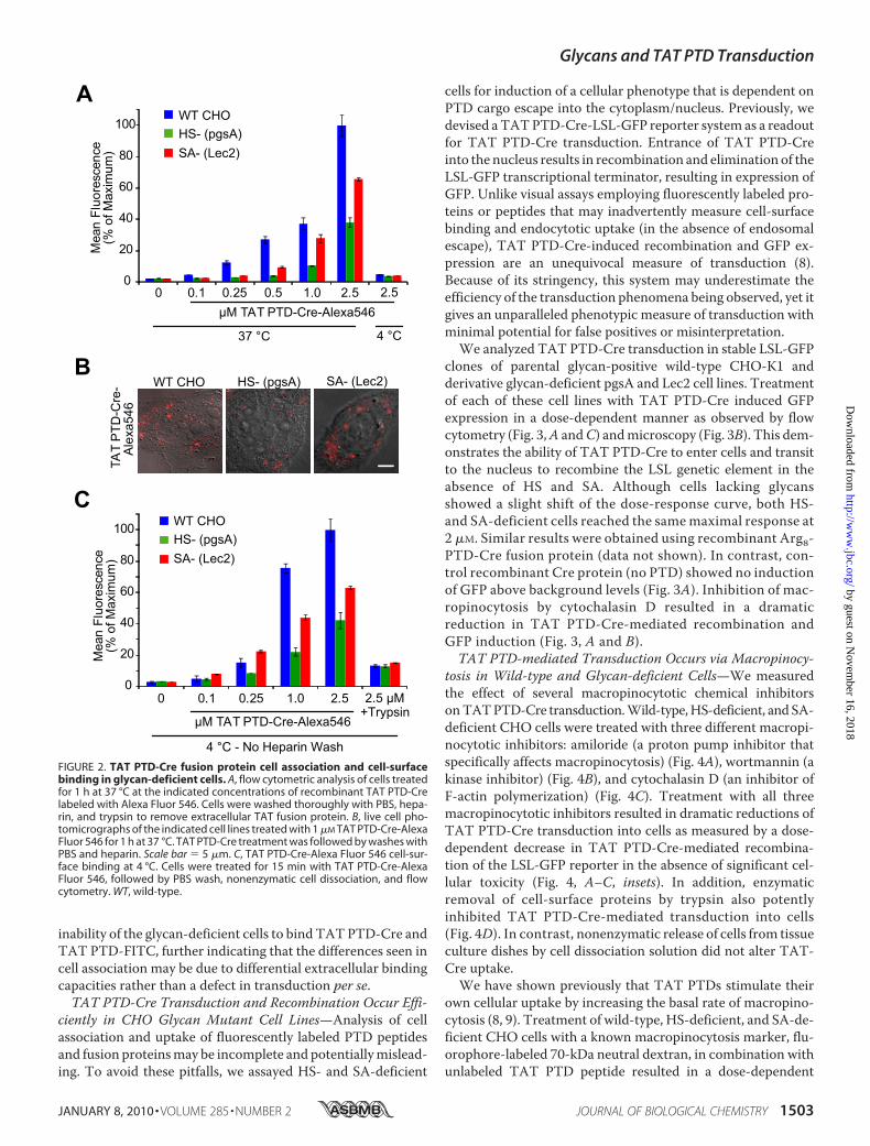
inability of the glycan-deficient cells to bind TATPTD-Cre andTAT PTD-FITC, further indicating that the differences seen incell association may be due to differential extracellular bindingcapacities rather than a defect in transduction per se.TAT PTD-Cre Transduction and Recombination Occur Effi-
ciently in CHO Glycan Mutant Cell Lines—Analysis of cellassociation and uptake of fluorescently labeled PTD peptidesand fusion proteinsmay be incomplete and potentiallymislead-ing. To avoid these pitfalls, we assayed HS- and SA-deficient
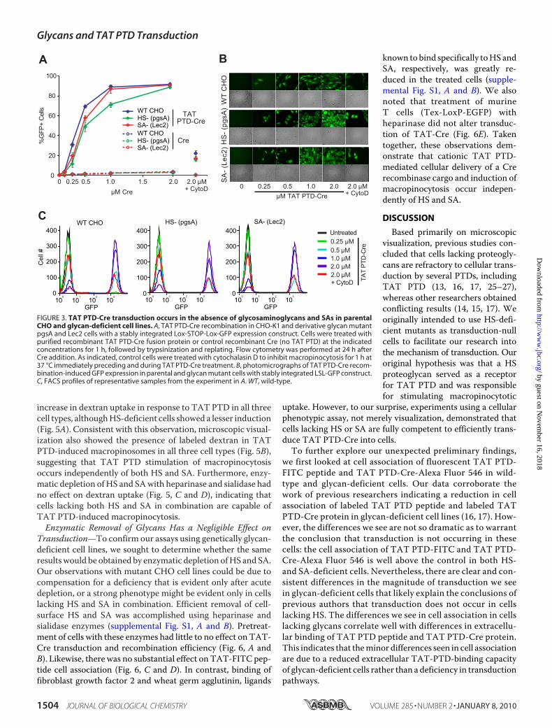
cells for induction of a cellular phenotype that is dependent onPTD cargo escape into the cytoplasm/nucleus. Previously, wedevised aTATPTD-Cre-LSL-GFP reporter system as a readoutfor TAT PTD-Cre transduction. Entrance of TAT PTD-Creinto the nucleus results in recombination and elimination of theLSL-GFP transcriptional terminator, resulting in expression ofGFP. Unlike visual assays employing fluorescently labeled pro-teins or peptides that may inadvertently measure cell-surfacebinding and endocytotic uptake (in the absence of endosomalescape), TAT PTD-Cre-induced recombination and GFP ex-pression are an unequivocal measure of transduction (8).Because of its stringency, this system may underestimate theefficiency of the transduction phenomena being observed, yet itgives an unparalleled phenotypic measure of transduction withminimal potential for false positives or misinterpretation.We analyzed TAT PTD-Cre transduction in stable LSL-GFP
clones of parental glycan-positive wild-type CHO-K1 andderivative glycan-deficient pgsA and Lec2 cell lines. Treatmentof each of these cell lines with TAT PTD-Cre induced GFPexpression in a dose-dependent manner as observed by flowcytometry (Fig. 3,A andC) andmicroscopy (Fig. 3B). This dem-onstrates the ability of TAT PTD-Cre to enter cells and transitto the nucleus to recombine the LSL genetic element in theabsence of HS and SA. Although cells lacking glycansshowed a slight shift of the dose-response curve, both HS-and SA-deficient cells reached the samemaximal response at2 �M. Similar results were obtained using recombinant Arg8-PTD-Cre fusion protein (data not shown). In contrast, con-trol recombinant Cre protein (no PTD) showed no inductionof GFP above background levels (Fig. 3A). Inhibition of mac-ropinocytosis by cytochalasin D resulted in a dramaticreduction in TAT PTD-Cre-mediated recombination andGFP induction (Fig. 3, A and B).TAT PTD-mediated Transduction Occurs via Macropinocy-
tosis in Wild-type and Glycan-deficient Cells—We measuredthe effect of several macropinocytotic chemical inhibitorsonTATPTD-Cre transduction.Wild-type,HS-deficient, andSA-deficient CHO cells were treated with three different macropi-nocytotic inhibitors: amiloride (a proton pump inhibitor thatspecifically affects macropinocytosis) (Fig. 4A), wortmannin (akinase inhibitor) (Fig. 4B), and cytochalasin D (an inhibitor ofF-actin polymerization) (Fig. 4C). Treatment with all threemacropinocytotic inhibitors resulted in dramatic reductions ofTAT PTD-Cre transduction into cells as measured by a dose-dependent decrease in TAT PTD-Cre-mediated recombina-tion of the LSL-GFP reporter in the absence of significant cel-lular toxicity (Fig. 4, A–C, insets). In addition, enzymaticremoval of cell-surface proteins by trypsin also potentlyinhibited TAT PTD-Cre-mediated transduction into cells(Fig. 4D). In contrast, nonenzymatic release of cells from tissueculture dishes by cell dissociation solution did not alter TAT-Cre uptake.We have shown previously that TAT PTDs stimulate their
own cellular uptake by increasing the basal rate of macropino-cytosis (8, 9). Treatment of wild-type, HS-deficient, and SA-de-ficient CHO cells with a known macropinocytosis marker, flu-orophore-labeled 70-kDa neutral dextran, in combination withunlabeled TAT PTD peptide resulted in a dose-dependent
FIGURE 2. TAT PTD-Cre fusion protein cell association and cell-surfacebinding in glycan-deficient cells. A, flow cytometric analysis of cells treatedfor 1 h at 37 °C at the indicated concentrations of recombinant TAT PTD-Crelabeled with Alexa Fluor 546. Cells were washed thoroughly with PBS, hepa-rin, and trypsin to remove extracellular TAT fusion protein. B, live cell pho-tomicrographs of the indicated cell lines treated with 1 �M TAT PTD-Cre-AlexaFluor 546 for 1 h at 37 °C. TAT PTD-Cre treatment was followed by washes withPBS and heparin. Scale bar � 5 �m. C, TAT PTD-Cre-Alexa Fluor 546 cell-sur-face binding at 4 °C. Cells were treated for 15 min with TAT PTD-Cre-AlexaFluor 546, followed by PBS wash, nonenzymatic cell dissociation, and flowcytometry. WT, wild-type.
Glycans and TAT PTD Transduction
JANUARY 8, 2010 • VOLUME 285 • NUMBER 2 JOURNAL OF BIOLOGICAL CHEMISTRY 1503
by guest on Novem
ber 16, 2018http://w
ww
.jbc.org/D
ownloaded from
increase in dextran uptake in response to TAT PTD in all threecell types, althoughHS-deficient cells showed a lesser induction(Fig. 5A). Consistent with this observation, microscopic visual-ization also showed the presence of labeled dextran in TATPTD-induced macropinosomes in all three cell types (Fig. 5B),suggesting that TAT PTD stimulation of macropinocytosisoccurs independently of both HS and SA. Furthermore, enzy-matic depletion ofHS and SAwith heparinase and sialidase hadno effect on dextran uptake (Fig. 5, C and D), indicating thatcells lacking both HS and SA in combination are capable ofTAT PTD-induced macropinocytosis.Enzymatic Removal of Glycans Has a Negligible Effect on
Transduction—To confirm our assays using genetically glycan-deficient cell lines, we sought to determine whether the sameresultswould be obtained by enzymatic depletion ofHS and SA.Our observations with mutant CHO cell lines could be due tocompensation for a deficiency that is evident only after acutedepletion, or a strong phenotype might be evident only in cellslacking HS and SA in combination. Efficient removal of cell-surface HS and SA was accomplished using heparinase andsialidase enzymes (supplemental Fig. S1, A and B). Pretreat-ment of cells with these enzymes had little to no effect on TAT-Cre transduction and recombination efficiency (Fig. 6, A andB). Likewise, there was no substantial effect on TAT-FITC pep-tide cell association (Fig. 6, C and D). In contrast, binding offibroblast growth factor 2 and wheat germ agglutinin, ligands
known to bind specifically toHS andSA, respectively, was greatly re-duced in the treated cells (supple-mental Fig. S1, A and B). We alsonoted that treatment of murineT cells (Tex-LoxP-EGFP) withheparinase did not alter transduc-tion of TAT-Cre (Fig. 6E). Takentogether, these observations dem-onstrate that cationic TAT PTD-mediated cellular delivery of a Crerecombinase cargo and induction ofmacropinocytosis occur indepen-dently of HS and SA.
DISCUSSION
Based primarily on microscopicvisualization, previous studies con-cluded that cells lacking proteogly-cans are refractory to cellular trans-duction by several PTDs, includingTAT PTD (13, 16, 17, 25–27),whereas other researchers obtainedconflicting results (14, 15, 17). Weoriginally intended to use HS-defi-cient mutants as transduction-nullcells to facilitate our research intothe mechanism of transduction. Ouroriginal hypothesis was that a HSproteoglycan served as a receptorfor TAT PTD and was responsiblefor stimulating macropinocytotic
uptake. However, to our surprise, experiments using a cellularphenotypic assay, not merely visualization, demonstrated thatcells lacking HS or SA are fully competent to efficiently trans-duce TAT PTD-Cre into cells.To further explore our unexpected preliminary findings,
we first looked at cell association of fluorescent TAT PTD-FITC peptide and TAT PTD-Cre-Alexa Fluor 546 in wild-type and glycan-deficient cells. Our data corroborate thework of previous researchers indicating a reduction in cellassociation of labeled TAT PTD peptide and labeled TATPTD-Cre protein in glycan-deficient cell lines (16, 17). How-ever, the differences we see are not so dramatic as to warrantthe conclusion that transduction is not occurring in thesecells: the cell association of TAT PTD-FITC and TAT PTD-Cre-Alexa Fluor 546 is well above the control in both HS-and SA-deficient cells. Nevertheless, there are clear and con-sistent differences in the magnitude of transduction we seein glycan-deficient cells that likely explain the conclusions ofprevious authors that transduction does not occur in cellslacking HS. The differences we see in cell association in cellslacking glycans correlate well with differences in extracellu-lar binding of TAT PTD peptide and TAT PTD-Cre protein.This indicates that theminor differences seen in cell associationare due to a reduced extracellular TAT-PTD-binding capacityof glycan-deficient cells rather than a deficiency in transductionpathways.
FIGURE 3. TAT PTD-Cre transduction occurs in the absence of glycosaminoglycans and SAs in parentalCHO and glycan-deficient cell lines. A, TAT PTD-Cre recombination in CHO-K1 and derivative glycan mutantpgsA and Lec2 cells with a stably integrated Lox-STOP-Lox-GFP expression construct. Cells were treated withpurified recombinant TAT PTD-Cre fusion protein or control recombinant Cre (no TAT PTD) at the indicatedconcentrations for 1 h, followed by trypsinization and replating. Flow cytometry was performed at 24 h afterCre addition. As indicated, control cells were treated with cytochalasin D to inhibit macropinocytosis for 1 h at37 °C immediately preceding and during TAT PTD-Cre treatment. B, photomicrographs of TAT PTD-Cre recom-bination-induced GFP expression in parental and glycan mutant cells with stably integrated LSL-GFP construct.C, FACS profiles of representative samples from the experiment in A. WT, wild-type.
Glycans and TAT PTD Transduction
1504 JOURNAL OF BIOLOGICAL CHEMISTRY VOLUME 285 • NUMBER 2 • JANUARY 8, 2010
by guest on Novem
ber 16, 2018http://w
ww
.jbc.org/D
ownloaded from

To further explore transduction in the absence of glycans, wesought to generate a transduction data set based on a pheno-typic assay rather than using cell association of labeled TAT asan approximation of transduction. To accomplish this, we gen-erated stable Cre-responsive cell lines from CHO-K1, pgsA,and Lec2 cells using a LSL-GFP reporter gene. As we haveshown previously (8), cell lines with an integrated LSL-GFPconstruct do not express GFP in the absence of Cre recombi-nase. However, after treatment of LSL-GFP cells with exoge-nous recombinant TAT PTD-Cre protein, GFP expression isinduced. Using this system, we can determine the relative effi-ciency of TAT PTD transduction without the caveats of visual-ization based on labeled peptides and proteins. Assays that relyon fluorescent tags to measure cellular uptake cannot discrim-inate between endosomal and cytosolic fractions of the cell andmay grossly overestimate the actual fraction of peptide andcargo that has transduced into the cell (escaped from the endo-some). We prefer to refer to these measures as cell associationbecause they do not sufficiently distinguish cells that have fluo-
rescent label in endocytic vesicles or bound to the cell surfacefrom cells where the peptide and cargo have actually escapedendosomes into the cytoplasm. This distinction is importantbecause endosomal escape is requisite for transduction; themajority of labeled TAT PTD taken up by cells is present inendosomes, andmost researchers would agree that only a smallproportion escapes to the cytoplasm. In addition, for the pur-poses of most therapeutic interventions, the inside of the endo-some is essentially the outside of the cell; no biological responsewill be elicited simply by uptake into vesicles. The importanceof escape is further illustrated by the increase in transductionefficiency seen with endosome-disrupting peptides (8, 28).Treatment of Cre-responsive cells with TAT PTD-Cre
revealed a trivial difference between wild-type and HS- or SA-deficient cells. If transduction were not occurring in these cells,we would expect to see a reduction to the base line as seen withrecombinant Cre lacking TAT PTD or treatment with cytocha-lasin D. The efficient manner with which TAT PTD-Cre entersglycan-deficient cells is strong and convincing evidence that
FIGURE 4. Macropinocytotic inhibitors and TAT PTD-Cre transduction in glycan-deficient cells. A, CHO-K1, pgsA, and Lec2 cells with a stably integratedLSL-GFP construct treated with the indicated concentrations of amiloride, an inhibitor of macropinocytosis, for 1 h. Cells were then treated with TAT PTD-Creprotein in the presence of amiloride for 1 h, followed by trypsinization and replating. GFP expression was assayed by flow cytometry at 24 h after TAT PTD-Creaddition. Inset, cell viability as assayed by flow cytometry immediately following TAT PTD-Cre treatment. B, treatment with wortmannin, a kinase inhibitor, asdescribed for A. C, treatment with cytochalasin D, an inhibitor of F-actin and macropinocytosis, as described for A. D, depletion of cell-surface proteins bytrypsin. CHO-K1 LSL cells were treated with trypsin or cell dissociation solution (CDS) for 30 min, followed by washes with PBS and trypsin inhibitor andtreatment with TAT PTD-Cre for 1 h. Cells were then washed, trypsinized, and replated. GFP expression was assayed by FACS at 24 h following TAT PTD-Cretreatment. WT, wild-type.
Glycans and TAT PTD Transduction
JANUARY 8, 2010 • VOLUME 285 • NUMBER 2 JOURNAL OF BIOLOGICAL CHEMISTRY 1505
by guest on Novem
ber 16, 2018http://w
ww
.jbc.org/D
ownloaded from

transduction occurs in the absence of extracellular glycosami-noglycans and SAs.Further evidence that transduction is occurring in these cells
is the existence of TATPTD-inducedmacropinocytotic uptakeof 70-kDa neutral dextran in glycan-deficient pgsA and Lec2cells. If these cells were refractory to transduction or if therewere another pathway for entry, we would not expect to seedextran uptake increased in response to TAT PTD. Further-more, the mode of uptake is macropinocytosis in wild-type andglycan-deficient cell lines, as evidenced by the similar reduc-tions in transduction efficiency seen after treatment withchemical inhibitors of macropinocytosis.We have shown here, in an extensive exploration, that TAT
PTD-mediated transduction does not require HS or SAs; itoccurs efficiently in their absence. As shown by our very strin-gent Cre-LoxP system and by more conventional fluorescentvisualization and cytometry, TAT PTD enters cells lackingHS or SA and cells that have been depleted of both in combina-tion. Furthermore, TAT PTD-inducedmacropinocytotic fluid-phase uptake is intact in glycan-deficient cells, indicating thatTAT PTD does not require these glycans to stimulate endocy-tosis. By contrast, depletion of cell-surface proteins with prote-ase results in drastically reduced transduction efficiency, indi-
FIGURE 5. TAT PTD-induced macropinocytotic fluid-phase uptake isintact in glycan-deficient cells. A, parental CHO and glycan mutant cellstreated with unlabeled TAT PTD peptide in the presence of 70-kDa neutraldextran-Texas Red for 1 h at 37 °C. After washing and trypsinization, cells wereassayed for dextran uptake by FACS. B, live cell photomicrographs of theindicated cell lines following treatment with 1 �M TAT PTD in the presence of70-kDa dextran-TMR. C, TAT PTD-induced uptake of 70-kDa dextran-TexasRed following treatment with 200 milliunits of heparinase for 1 h at 37 °C. TheTAT PTD concentration was 1 �M. D, cells treated with 200 milliunits of siali-dase followed by dextran and TAT PTD as described for C. WT, wild-type.
FIGURE 6. Enzymatic depletion of HS and SAs does not impair TAT PTDtransduction into parental or glycan-deficient cells or murine T cells.A and B, the indicated Lox-STOP-Lox-GFP stable cell lines were treated for 1 hwith heparinase or sialidase as indicated at 37 °C, followed by 1 �M TAT PTD-Cre for 1 h at 37 °C, trypsinization, and replating. GFP expression was assayedby flow cytometry 24 h after TAT PTD-Cre treatment. C and D, the indicatedcell lines were treated with heparinase and sialidase enzymes as described forA and B, followed by treatment with 1 �M TAT PTD-FITC peptide for 1 h at37 °C, immediately followed by PBS, heparin, and trypsin washes and flowcytometry. E, heparinase treatment does not affect transduction efficiency inthe Tex-LoxP-EGFP murine thymoma cell line. WT, wild-type.
Glycans and TAT PTD Transduction
1506 JOURNAL OF BIOLOGICAL CHEMISTRY VOLUME 285 • NUMBER 2 • JANUARY 8, 2010
by guest on Novem
ber 16, 2018http://w
ww
.jbc.org/D
ownloaded from

cating that a cell-surface protein is necessary for TAT PTDtransduction (although not necessarily as a receptor). Thesedata suggest that cationic peptides like TAT PTD may inducemacropinocytotic uptake and transduction via binding to a pro-tein on the cell surface rather than through interactions withglycans or direct interaction with the membrane.Together, our findings are consistent with the hypothesis
that the dense forest of extracellular glycans forms a pool ofnegative charge that TAT PTD binds on the cell surface. Dif-ferences in this charge pool affect the efficiency of TAT trans-duction to varying degrees but are independent of the ability ofPTDs to transduce cells or to induce macropinocytotic uptake.Further study to determine the extracellular proteins requiredfor transduction will offer important insights into the mecha-nism of transduction of TAT PTD as well as other PTDs.
REFERENCES1. Meade, B. R., andDowdy, S. F. (2007)Adv. DrugDelivery Rev. 59, 134–1402. El-Andaloussi, S., Holm, T., and Langel, U. (2005) Curr. Pharm. Des. 11,
3597–36113. Goun, E. A., Pillow, T. H., Jones, L. R., Rothbard, J. B., and Wender, P. A.
(2006) ChemBioChem 7, 1497–15154. Gump, J. M., and Dowdy, S. F. (2007) Trends Mol. Med. 13, 443–4485. Nakase, I., Takeuchi, T., Tanaka, G., and Futaki, S. (2008) Adv. Drug De-
livery Rev. 60, 598–6076. Fischer, R., Fotin-Mleczek, M., Hufnagel, H., and Brock, R. (2005) Chem-
BioChem. 6, 2126–21427. Heitz, F., Morris, M. C., and Divita, G. (2009) Br. J. Pharmacol. 157,
195–2068. Wadia, J. S., Stan, R. V., and Dowdy, S. F. (2004) Nat. Med. 10, 310–3159. Kaplan, I. M., Wadia, J. S., and Dowdy, S. F. (2005) J. Controlled Release
102, 247–25310. Nakase, I., Niwa, M., Takeuchi, T., Sonomura, K., Kawabata, N., Koike, Y.,
Takehashi, M., Tanaka, S., Ueda, K., Simpson, J. C., Jones, A. T., Sugiura,Y., and Futaki, S. (2004)Mol. Ther. 10, 1011–1022
11. Hakansson, S., Jacobs, A., and Caffrey, M. (2001) Protein Sci. 10,2138–2139
12. Rusnati, M., Coltrini, D., Oreste, P., Zoppetti, G., Albini, A., Noonan, D.,d’Adda di Fagagna, F., Giacca,M., and Presta,M. (1997) J. Biol. Chem. 272,11313–11320
13. Tyagi, M., Rusnati, M., Presta, M., and Giacca, M. (2001) J. Biol. Chem.276, 3254–3261
14. Silhol, M., Tyagi, M., Giacca, M., Lebleu, B., and Vives, E. (2002) Eur.J. Biochem. 269, 494–501
15. Violini, S., Sharma, V., Prior, J. L., Dyszlewski,M., and Piwnica-Worms, D.(2002) Biochemistry 41, 12652–12661
16. Nakase, I., Tadokoro,A., Kawabata,N., Takeuchi, T., Katoh,H.,Hiramoto,K., Negishi, M., Nomizu, M., Sugiura, Y., and Futaki, S. (2007) Biochemis-try 46, 492–501
17. Console, S., Marty, C., García-Echeverría, C., Schwendener, R., andBallmer-Hofer, K. (2003) J. Biol. Chem. 278, 35109–35114
18. Richard, J. P., Melikov, K., Brooks, H., Prevot, P., Lebleu, B., and Cherno-mordik, L. V. (2005) J. Biol. Chem. 280, 15300–15306
19. Poon, G. M., and Gariepy, J. (2007) Biochem. Soc. Trans. 35, 788–79320. Brooks, H., Lebleu, B., and Vives, E. (2005) Adv. Drug Delivery Rev. 57,
559–57721. Esko, J. D. (1992) Adv. Exp. Med. Biol. 313, 97–10622. Esko, J. D., Stewart, T. E., and Taylor, W. H. (1985) Proc. Natl. Acad. Sci.
U.S.A. 82, 3197–320123. Esko, J. D., Weinke, J. L., Taylor, W. H., Ekborg, G., Roden, L., Ananthara-
maiah, G., and Gawish, A. (1987) J. Biol. Chem. 262, 12189–1219524. Deutscher, S. L., Nuwayhid, N., Stanley, P., Briles, E. I., and Hirschberg,
C. B. (1984) Cell 39, 295–29925. Nascimento, F. D., Hayashi, M. A., Kerkis, A., Oliveira, V., Oliveira, E. B.,
Radis-Baptista, G., Nader, H. B., Yamane, T., Tersariol, I. L., and Kerkis, I.(2007) J. Biol. Chem. 282, 21349–21360
26. Elson-Schwab, L., Garner, O. B., Schuksz, M., Crawford, B. E., Esko, J. D.,and Tor, Y. (2007) J. Biol. Chem. 282, 13585–13591
27. Sandgren, S., Cheng, F., and Belting, M. (2002) J. Biol. Chem. 277,38877–38883
28. El-Sayed, A., Futaki, S., and Harashima, H. (2009) AAPS J. 11, 13–22
Glycans and TAT PTD Transduction
JANUARY 8, 2010 • VOLUME 285 • NUMBER 2 JOURNAL OF BIOLOGICAL CHEMISTRY 1507
by guest on Novem
ber 16, 2018http://w
ww
.jbc.org/D
ownloaded from
Jacob M. Gump, Ronald K. June and Steven F. DowdyDomain-mediated Cellular Transduction
Revised Role of Glycosaminoglycans in TAT Protein Transduction
doi: 10.1074/jbc.M109.021964 originally published online October 26, 20092010, 285:1500-1507.J. Biol. Chem.
10.1074/jbc.M109.021964Access the most updated version of this article at doi:
Alerts:
When a correction for this article is posted•
When this article is cited•
to choose from all of JBC's e-mail alertsClick here
Supplemental material:
http://www.jbc.org/content/suppl/2009/10/26/M109.021964.DC1
http://www.jbc.org/content/285/2/1500.full.html#ref-list-1
This article cites 28 references, 9 of which can be accessed free at
by guest on Novem
ber 16, 2018http://w
ww
.jbc.org/D
ownloaded from