revisiting diabetes and retinopathy hand out€¦ · revisiting diabetes and retinopathy: ......
TRANSCRIPT

1/6/15
1
Revisiting Diabetes and Retinopathy: Advanced Diagnosis and Treatment
Options Michael D. Gerstner, O. D.
Louisiana Optometric Association February 7, 2015 COPE #43351-PS
Dr. Michael Gerstner
• Associate Professor @ Southern College of Optometry
• Chief, Advanced Care Ocular Disease @ The Eye Center
• Private Practice and Partner @ Midtown Eye Care
• Diplomate, American Board of Optometry
Correspondence C
901.921.7832
The Eye Center at SCO 1225 Madison Ave
Memphis, TN 38104
Commercial Disclosure
• The content of this course was prepared independently by Dr. Gerstner without any input from members of the ophthalmic industry.
• Dr. Gerstner does not have financial interests in any companies, products, or services mentioned in this presentation.
Course Description
• This course explore imaging technology utilized for improved diagnosis and progression analysis of ocular disease associated with diabetes mellitus. An in-depth discussion will familiarize the optometric physician with emerging and changing treatment protocols for the treatment of diabetic retinopathy. Case based presentations will illustrate imaging interpretation and techniques. Systemic diabetes mellitus and levels of diabetic retinopathy will be thoroughly reviewed. Evidence based medicine and clinical trials will be discussed.
Clinical Decision Making
• Who needs what treatment, and when do they need it? • Subjectivity in every patient encounter • Data collection and constructing an argument based on “facts” • Perhaps there is more than one way to apply diagnostic or therapeutic
options • Elements of uncertainty • Technology • Still no substitute for your own judgment and experience

1/6/15
2
Evidence Based Medicine
• Occam’s Razor, or “common things happen commonly”
• Simple: take the path that gives the best outcome
• Follow the studies that help us make better decisions
• Use the GPS, not the map…
DCCT and EDIC
• Diabetes Control and Complications Trial and Epidemiology of Diabetes Interventions and Complications
• Type 1 diabetes mellitus: 50% will have retinopathy 10 years after diagnosis
• Type 2 diabetes mellitus: 95% will have retinopathy 10 years after diagnosis
• Intense blood glucose control reduces risk by 76%
Diabetes – Control to Prevent Complications
• Intense blood glucose control (DCCT)
• Hemoglobin A1c (HbA1c) to monitor control • 6.0% or less for diabetics
• 40% reduction of complications if A1c is reduced one percentage point
Diabetes – Control to Prevent Complications
• Blood pressure control • 33% to 50% reduction in heart disease or CVA
• Reduction of diastolic pressure from 90 mmHg to 80 mmHg decreases a major cardiovascular event by 50%
• Lipid control • Strict LDL control can reduce cardiovascular complications 20% - 50%
Metabolic Syndrome
• Abdominal obesity • Dyslipidemia
• Elevated blood pressure
• Diabetes or pre-diabetes
• Pro-thrombotic state
• Pro-inflammatory state
Clinical Studies and the Standard of Care
• Early Treatment Diabetic Retinopathy Study (ETDRS) facts on macular edema and laser photocoagulation: • Generally stabilizes visual acuity but often does not improve it
• Consists of directly treating focal areas of leakage and placing a grid in areas of diffuse capillary leakage – guided by IVFA
• Should be avoided in presence of significant loss of perifoveal capillaries
• May take months to show resolution of thickening and resolution may take longer for exudates

1/6/15
3
Clinical Studies and the Standard of Care – PRP Facts
• Visual acuity will not improve • Macular edema may actually worsen
• Significant night and peripheral vision loss
• Neovascularization will not always regress
• Indicated for neovascularization of the iris
Clinical Studies and the Standard of Care
Overview of Diabetes Mellitus
• Diabetes is a group of metabolic disorders defined by elevated blood glucose resulting from insulin production defects, impaired insulin action, or both
• The 1997 International Expert Committee on Diabetes Mellitus changed the classification of diabetes, criteria for the diagnosis of diabetes, and control guidelines
• Revised guidelines were published in 2003
Overview of Diabetes Mellitus
• Type 1 (no longer considered IDDM or Type I) • Pancreatic beta cell destruction
• Viral/environmental insult and autoimmune injury
• 5% of all diagnosed cases
Overview of Diabetes Mellitus
• Type 2 (no longer considered NIDDM or Type II) • Pancreatic beta cell inefficiency or insulin resistance
• Age, obesity, and family history
• 90% - 95% of all diagnosed cases
• Pre-diabetes (formally known as impaired glucose tolerance)
Overview of Diabetes Mellitus
• Gestational diabetes • 2% to 10% of all pregnancies
• 5% to 10% will have diabetes immediately following pregnancy
• 35% to 60% will develop diabetes in the next 10 – 20 years
• Other types • MODY 1 – 6
• 1% to 5% of all diagnosed cases

1/6/15
4
Diagnostic Ranges of Diabetes
Revised guidelines 2003
Diagnostic Ranges of Pre-Diabetes
Hemoglobin A1C or Random Plasma Glucose?
• Which one do we do in clinic? • Better understanding of the over all blood glucose control
• Why? • Did you check your BG today Mrs. Jones?
• When do we do this in the clinic? • When we have time…
• Who? • Not engaged in care
Pharmacological Treatment for Diabetes
• Insulin • Rapid acting
• Short acting
• Intermediate acting
• Long acting
Pharmacological Treatment for Diabetes – Oral
• Insulin secretagogues • Insulin sensitizers
• Metformin
• α-glucosidase inhibitors • Dipeptidyl peptidase IV inhibitors • Incretin mimetics • Sodium glucose transport inhibitor

1/6/15
5
How Many People Are Affected?
• Total: 25.8 million children and adults or 8.3% of the population • Diagnosed: 18.8 million people
• Undiagnosed: 7.0 million people
• Pre-diabetes: 79 million people • New Cases: 1.9 million
How Many People Are Affected? Total prevalence of DM, US 2013
Under 20 years of age 215,000 1.9 million newly diagnosed 2 million aged 12-19 are pre-diabetic (1 in 6 are overweight)
Age 20 years or older 25.6 million or 11.3% of all people in this age group
Age 65 or older 10.9 million or 26.9% of all people in this age group
Men 13.0 million or 11.8% of all men 20 years of age or older
Women 12.6 million or 10.8% of all women 20 years of age or older
The Diabetes “Belt” Diabetes Diagnosis
Diabetes – Lack of Activity ILM NFL GCL
IPL INL OPL ONL
ELM IS RPE
OS Choroid
It is impossible to discuss retinopathy without knowing this slide

1/6/15
6
Retina Anatomy Review: Structure and Function
• Inner retina – what we are mostly reviewing today • Outer retina – external limiting membrane and posterior
• Photoreceptors
• Retinal pigmented epithelium
• Bruch’s membrane
• Choriocapillaris
• Choroid
Retinal Vasculature
• Arteries and arterioles v. veins and venules
• Location – located in the nerve fiber or ganglion cell layers
• Function – supplies the inner retina with blood, nutrients, and O2
Retinal Anatomy Review: Capillary Networks
• Superficial or inner network • Located within the ganglion cell layer
• Think post-arteriole and affected in artery based disease
• Deep or outer network • Located in the inner nuclear layer
• Think pre-venule and affected in venous based disease
Inner Capillary Network Compromise
• Flame-shaped hemorrhages
• Cotton wool spots
Outer Capillary Network Compromise
• Intra-retinal hemorrhages
• Exudative changes
Microaneurysm
1/6/15
7
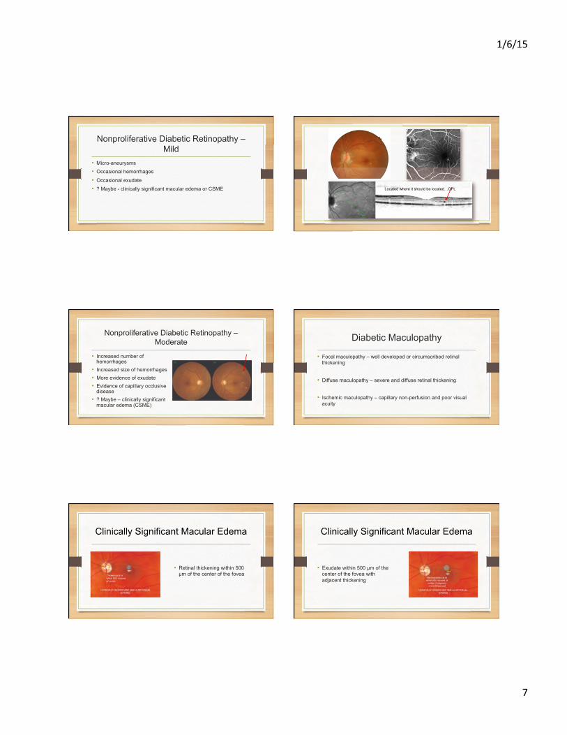
Nonproliferative Diabetic Retinopathy – Mild
• Micro-aneurysms • Occasional hemorrhages
• Occasional exudate
• ? Maybe - clinically significant macular edema or CSME Located where it should be located…OPL
Nonproliferative Diabetic Retinopathy – Moderate
• Increased number of hemorrhages
• Increased size of hemorrhages • More evidence of exudate • Evidence of capillary occlusive
disease • ? Maybe – clinically significant
macular edema (CSME)
Diabetic Maculopathy
• Focal maculopathy – well developed or circumscribed retinal thickening
• Diffuse maculopathy – severe and diffuse retinal thickening
• Ischemic maculopathy – capillary non-perfusion and poor visual acuity
Clinically Significant Macular Edema
• Retinal thickening within 500 µm of the center of the fovea
Clinically Significant Macular Edema
• Exudate within 500 µm of the center of the fovea with adjacent thickening

1/6/15
8
Clinically Significant Macular Edema
• Thickening of at least one disc diameter within one disc diameter of the center of the fovea
Nonproliferative Diabetic Retinopathy – Severe
4-2-1 RULE Severe retinal hemorrhages in 4 quadrants
Venous beading in 2 quadrants
IRMA in 1 quadrant
50% risk of developing PDR within one year
Worsening of everything with macular edema
4 – 2 – 1 Proliferative Diabetic Retinopathy
• NVD – neovascularization of the disc • Neovascularization development within one disc diameter of the optic nerve
• NVE – neovascularization elsewhere • Neovascularization anywhere within the retina that is not NVD • Junction between perfused and non-perfused retina
• NVI – neovascularization of the iris • Threatening sign of neovascular glaucoma
• Vitreous hemorrhage • Bleeding from NVD or NVE
Case Presentation
• 53-year-old male • “My vision has been blurry in both eyes for awhile”
• HTN
• Type 2 diabetes
Recent Clinical Trials for Diabetic Macular Edema
• READ • Ranibizumab 0.5 mg • Focal and grid laser photocoagulation • Ranibizumab 0.5 mg followed by laser photocoagulation
• RESTORE • Ranibizumab 0.5 mg with sham laser photocoagulation • Focal and grid laser photocoagulation with sham injection • Ranibizumab 0.5 mg followed by laser photocoagulation

1/6/15
9
Recent Clinical Trials for Diabetic Macular Edema
• RESOLVE • Ranibizumab 0.5 mg v. 0.3 mg v. sham
• RISE • Ranibizumab 0.5 mg v. sham
• RIDE • Ranibizumab 0.5 mg v. sham
Diabetic Macular Edema – Local Treatment Trends
• Lucentis or Avastin or Eylea • Combination of focal and grid laser photocoagulation
• Possible needed for sustained effect
• Laser at one week after injection if continued marked edema
• Lower intensity and shorter duration laser photocoagulation
Diabetic Macular Edema – Local Treatment Trends
• NSAIDS and topical steroids?
• Implantable steroids?
• Injectable steroids?
• PPV
Proliferative Diabetic Retinopathy – Local Treatment Trends
• Pan-retinal photocoagulation is still the standard of care • Less intense energy and shorter duration of burn, or “light” photocoagulation
• Smaller spot size with more spots, or “moderate” treatment
• 30 day follow-up
• Combination anti-VEGF injection if poor response to PRP at 30 day follow-up
Proliferative Diabetic Retinopathy – Local Treatment Trends
• Lucentis or Avastin or Eylea followed by PRP • Not approved as initial therapy
• Pre-vitrectomy to reduce bleeding
• Vitreous hemorrhage with neovascularization followed by PRP
• NVI or neovascular glaucoma
A Brief History of OCT
Time domain (old) verses spectral domain (new)

1/6/15
10
CIRRUS HD – OCT 5000
• Macular cube 512 x 128 • Macular cube 200 x 200
• HD 5 - line raster
• Optic disc cube 200 x 200
• Anterior segment 5 - line raster
• Anterior segment cube 512 x 128
CIRRUS HD – OCT 5000
Macular analysis • Macular thickness • Ganglion cell analysis
• Macular change analysis
• Advanced RPE analysis
• 3-D and advanced visualization
5 – line raster • High definition image
Case Presentation
• 50-year-old male
• Type 2 diabetes x 20 years
• HTN and kidney disease
• Blurred vision OD for one day
Case Presentation
• 59-year-old female • “I have noticed blurred vision OD for a few weeks” • Type 2 diabetes for 20 years • Well controlled • Compliant patient that is followed annually at TEC • No previous retinopathy identified • Her vision has dropped OD compared to previous
Optos Daytona
• Pros • Wide field
• Magnification
• Patient education
• Cons • You be the judge…

1/6/15
11
Billing and Coding
• Coding and Medicare allowable fee structure 2014 – Tennessee
• 92134: Retina = 42.79
• 92250: Fundus Photography = 74.43
Billing and Coding – Pitfalls
• Vision insurance, medical insurance, or both? • ICD-9 codes and “baseline” testing
• Photography and OCT
• Calendar year and OCT
• The eye is considered to be a single organ
• Clear documentation and interpretation
Case Presentation
• 54-year-old male • Type 2 diabetes x 24 years
• HTN
• Blurred and fluctuating vision OU
• New patient – vision benefit