revisiting the cd14: epitope mapping by phage display
TRANSCRIPT

Accepted Manuscript
Title: Revisiting the CD14: epitope mapping by Phage Display
Author: Patrıcia Terra Alves Patrıcia Tiemi Fujimura LeaDuarte da Silva Morais Luiz Ricardo Goulart
PII: S0171-2985(14)00115-6DOI: http://dx.doi.org/doi:10.1016/j.imbio.2014.07.002Reference: IMBIO 51159
To appear in:
Received date: 28-6-2013Revised date: 25-4-2014Accepted date: 14-7-2014
Please cite this article as: Alves, P.T., Fujimura, P.T., Morais, L.D.S., Goulart,L.R.,Revisiting the CD14: epitope mapping by Phage Display, Immunobiology (2014),http://dx.doi.org/10.1016/j.imbio.2014.07.002
This is a PDF file of an unedited manuscript that has been accepted for publication.As a service to our customers we are providing this early version of the manuscript.The manuscript will undergo copyediting, typesetting, and review of the resulting proofbefore it is published in its final form. Please note that during the production processerrors may be discovered which could affect the content, and all legal disclaimers thatapply to the journal pertain.

Page 1 of 27
Accep
ted
Man
uscr
ipt
Title: Revisiting the CD14: epitope mapping by Phage Display
Authorship
Patrícia Terra Alves1, Patrícia Tiemi Fujimura1, Léa Duarte da Silva Morais1
Luiz Ricardo Goulart1,2*
Affiliation
1Laboratory of Nanobiotechnology, Institute of Genetics and Biochemistry,
Federal University of Uberlandia, Uberlandia, MG, Brazil.
2Department of Medical Microbiology and Immunology, University of California
Davis, Davis, CA, USA.
Alves, P.T. [email protected]
Fujimura, P.T. [email protected]
Morais, L. D.S. [email protected]
Goulart, L.R. [email protected]
*Corresponding author: Luiz Ricardo Goulart, Laboratory of
Nanobiotechnology, Institute of Genetics and Biochemistry, Federal University
of Uberlandia, Campus Umuarama, Bloco 2E, Sala 248, 38400-902,
Uberlandia, MG, Brazil. Phone: (55+34)3218-2478. [email protected] /
Manuscript

Page 2 of 27
Accep
ted
Man
uscr
ipt
ABSTRACT
The cluster of differentiation antigen 14 (CD14) is a key molecule of the innate
immunity. This pattern recognition receptor binds mainly to lipopolysaccharide
(LPS), lipotechoic acid (LTA), arachidonic acid, and thus induces the releases
various cytokines, as a defense mechanism. Several studies suggest that
different regions of the amino-terminal portion of the molecule may be involved
in the LPS binding; however, controversial results on the recognition sequence
still persist. In this work, functional epitopes of the CD14 molecule were mapped
through Phage Display by using a 7-mer conformational constrained random
peptide library against a monoclonal antibody anti-soluble CD14-fraction ST and
a polyclonal anti-CD14. In silico and empirical analyzes were performed to map
the selected peptides into the CD14 3D structure. Immunoreactivity tests of
peptides against bacterial components of Gram+ and Gram- bacteria were
performed in order to demonstrate their functional recognition. All peptides
strongly reacted against all bacteria, and besides the recognition of the amino-
terminal region, we were able to demonstrate a second epitope site in the
middle of the receptor. Additional in silico analysis suggests a possible role of
CD14 epitopes as natural antimicrobial peptides.
Keywords: CD14, epitope mapping, LPS receptor, Phage Display
Abreviations used in this article:
CD14: Cluster differentiation 14; LPS: Lipopolysaccharide; GPI:
Glycosilphophatidylinositol; PD: Phage Display

Page 3 of 27
Accep
ted
Man
uscr
ipt
INTRODUCTION
Cluster differentiation antigen 14, CD14, plays a crucial role in the
immune system acting in the detection and clearance of pathogens on host
organism (Kitchens and Thompson, 2005). CD14 is a pattern recognition
receptor that enhances innate immune responses to infection by sensitizing
host cells to bacterial LPS (endotoxin), lipoproteins, lipoteichoic acid, and other
acylated microbial products (Kelley, et al., 2013). This molecule also recognizes
endogenous ligands such as the human heat shock protein 60 (Kol, et al.,
2000), ceramide, phospholipids, modified lipoproteins and opsonized particles
(Schmitz and Orso, 2002). After recognizing its ligands, CD14 initiates the
activation of the downstream complex inflammatory network.
The CD14 is a glycoprotein composed of 375 amino acids, which are
encoded by a gene located at 5q31.1 region of the human chromosome
(Camussi, et al., 1995, Ulevitch and Tobias, 1995). Myeloid lineage cells can
express this protein in two forms: bound to the plasma membrane (mCD14) via
a glycosylphosphatidylinositol anchor (GPI), and the soluble form (sCD14),
which are found in the blood and body fluids (Ulevitch and Tobias, 1995)
The mCD14 has 55 kDa and is expressed at high levels of monocytes,
macrophages and neutrophils, but can also be found in basophils, Kupffer cells
and lymphocytes (Li, et al., 2003, Dai, et al., 2003, Iida, et al., 1994). The
sCD14 has 48kDa and may be generated by secretion (Austenaa, et al., 2012)
or through a proteolytic cleavage by phospholipase D action on the GPI anchor.
Both mechanisms will induce the release of the soluble form of CD14 (Sharom
and Radeva, 2004).

Page 4 of 27
Accep
ted
Man
uscr
ipt
The sCD14 and mCD14 are the same protein except the GPI anchor,
which is found only in mCD14 (Viriyakosol, et al., 2000). The sCD14 is of
extreme importance to the body because enables the CD14 signaling pathway
in cells that do not express this molecule in the membrane, such as epithelial
and endothelial cells (Lloyd-Jones, et al., 2008).
The ability to bind different targets, the high binding specificity, and the
incomplete information about the CD14-triggered signaling, make its epitopes
excellent targets for investigations. Epitope mapping has been performed
elsewhere by site directed mutagenesis (Kim, et al., 2005, Shapiro, et al.,
1997), synthetic peptides (Voss, et al., 2006), and neutralizing antibodies
(Kirkland, et al., 1993) in order to identify structural elements of the CD14
responsible for the recognition and signaling to the immune response.
Among CD14 ligands, LPS is the main molecule investigated, which
binds to the N-terminal region comprising amino acids 1–152, a site present in
both soluble and membrane-bound forms that is sufficient for binding and
enabling cellular responses (Kim, et al., 2005); however, controversies about
the recognition sequence still persist. Some authors claimed that the N-terminal
region is responsible for LPS recognition (Voss, et al., 2006, Juan, et al., 1995,
Viriyakosol and Kirkland, 1996), but others have shown that only few critical
amino acids are necessary, since sCD14 individual mutations may prevent the
formation of the complex CD14-LPS (Kelley, et al., 2013, Kim, et al., 2005).
The sCD14 is found in high concentrations in the bloodstream of
individuals with several pathologies associated with systemic inflammation.
Among them, it is believed that the collapse of the immune response in the

Page 5 of 27
Accep
ted
Man
uscr
ipt
presence of infection, as demonstrated in sepsis, is closely correlated to the
action of CD14.
In this investigation we have revisited the CD14 receptor and its
functional epitopes for bacterial recognition through Phage Display using a 7-
mer conformational constrained random peptide library to identify mimetic
peptides (mimotopes) of CD14 3D structure with bacterial detection function.
This strategy helped us to map a new epitope site besides the N-terminal
region, and all mimotopes presented a broad recognition of bacterial
components with a possible relevance to the immune response of inflammatory-
associated diseases.
MATERIALS AND METHODS
Targeted CD14-antibody
Human monoclonal and polyclonal antibodies anti-CD14 used in this
study were obtained from the PATHFASTTM Presepsin kit diagnosis (Mitsubishi
Chemical Medience, Japan). The monoclonal antibody anti- soluble CD14-
fraction ST was already coupled to magnetic beads, and the polyclonal antibody
anti-CD14 was coupled with protein G-conjugated magnetic beads, as
recommended by the manufacturer (Dynabeads Protein G, Life Technology,
Carlsbad, CA, USA).
Peptide selection through phage display
For the peptide selection, a PhD-C7C phage library (New England
Biolabs, Beverly, MA, USA) was used. This is a 7-mer random peptide library
fused to the minor coat protein (pIII) of the M13 bacteriophage, with a peptide

Page 6 of 27
Accep
ted
Man
uscr
ipt
diversity of 1.9X10⁹. A sample of the library containing 2x10¹¹ infectious phage
particles was subjected to three rounds of selection and amplification.
The selection was carried out using 20 µL of magnetic beads previously
coupled with the anti-CD14, which was incubated with 2x10¹¹ phage particles
from the PhD-C7C library in 180 µL of TBS solution (Tris Buffered Saline) at
room temperature, under slight shaking with gentle rocking for 1 hour. Unbound
phages were removed by ten washing steps with TBS-T 0.05% (Tris Buffered
Saline plus 0.05% of Tween 20), followed by elution of bound phages with
100µL of elution buffer (0.2M Glycine-HCl, pH2.2) for 10min at room
temperature. After elution, beads were captured from the supernatant with the
aid of a magnet and the supernatant was transferred to a new microtube
containing 20µL of 1M Tris-HCl (pH9.1) for neutralization.
Eluated phages were amplified in E. coli ER2738 strain (New England
Biolabs, Beverly, MA, USA), purified using PEG-NaCl precipitation, and after
each selection cycle, individual bacterial colonies containing amplified phages
were grown in a microtiter plate and titrated, as described elsewhere (Barbas,
2001).
DNA sequencing
Phagemid DNA was isolated from 1 mL overnight cultures, and the
sequencing reactions were carried out by using the DyEnamic ET Dye
Terminator Cycle sequencing Kit (GE Healthcare) with the primer -96 M13 (59-
CCCTCATTAGTTAGCGCGTAA-CG-39), according to the manufacturer’s
instructions, and detection was performed in a MegaBace 1000 Genetic
Analyzer (Amersham Biosciences) automatic capillary sequencer.

Page 7 of 27
Accep
ted
Man
uscr
ipt
Bioinformatics analysis
DNA sequences were analyzed through bioinformatics online softwares.
(http://www.bioinformatics.org/sms/rev_comp.html). Amino acid sequences
were deduced based on the nucleotide sequences through the Expasy translate
program (http://web.expasy.org/translate/). The three-dimensional structure of
the CD14 was obtained from the Protein Data Bank (PDB:1WWL) (Kim, et al.,
2005). The antigenicity degree of the CD14 molecule was calculated for its
entire three-dimensional structure by the Epitopia Server program
(http://epitopia.tau.ac.il/) (Rubinstein, et al., 2009). The location of epitopes at
the CD14 molecule was pointed out by 3D-conformational alignment between
the molecule and selected peptides using the PEPSURF online software
(http://pepitope.tau.ac.il/) (Mayrose, et al., 2007, Mayrose, et al., 2007).
Physicochemical properties of the peptide (Molecular weight and p.I) were
predicted using the Compute pI/Mw tool expasy Server
(http://web.expasy.org/compute_pi/). To evaluate the potential role of CD14
epitopes as antimicrobial peptides (AMPs), hydrophobicity and net charge of the
peptides were predicted using the Antimicrobial peptide database Server
(http://aps.unmc.edu/AP/prediction/prediction_main.php) (Wang, et al., 2009).
The sequences of α-helical peptides are presented according to the Shiffer-
Edmundson wheel projection (http://cti/it.virginia.edu/~cmg/Demo/wheel/
wheelApp.html).
Bead-ELISA and reactivity to the anti-CD14 antibody
Enzyme-linked immunosorbent assay (ELISA) was performed to verify
the reactivity and binding specificity of selected phage clones. An irrelevant
Page 8 of 27
Accep
ted
Man
uscr
ipt
peptide fused to a bacteriophage obtained from the discarded phase during
selection of the peptide library was used as a negative control. Reactions were
performed in 96-well microtiter plates by mixing 1x10¹¹pfu/well in 100µL of TBS
and 1µL the magnetics beads coupled with anti-CD14, which were incubated at
37ºC under 100 rpm agitation for one hour. Plates were washed three times
with TBST 0.1% followed by an additional incubation with HRP-conjugated anti-
M13 (Roche Applied Science) diluted (1:2500) in TBS with 3% BSA for 1h at
37°C. The beads were washed four times in TBS-T, and revealed with OPD
SigmaFastTM (Sigma-Aldrich) and read at 492nm
Competitive Bead-ELISA Assay between CD14 and M.H2 clones for
binding to the anti-CD14 antibody
Reactions were performed as previously described in the Bead-ELISA
procedure, but the competitive reactions were assembled in 96-well microtiter
plates by mixing 1x10¹⁰pfu/well of M.H2 clone in 100µL of TBS with 0, 10, 100
or 1000 ng/well of sCD14 human recombinant (Sigma-Aldrich).
Detection of bacterial components
Endotoxin assay detection
A 96-well MaxisorpTM microtiter plate (NUNC, NY, USA) was coated with
100 to 2000 ng/well of Lipopolysaccharides (LPS) from Escherichia coli
0127:B8 (Sigma-Aldrich) in 50µL of PBS (phosphate-buffered saline). The
phage ELISA assay was performed as described elsewhere (Matsumoto, et al.,
2010).

Page 9 of 27
Accep
ted
Man
uscr
ipt
Bacterial detection
The ability to recognize bacterial structures was verified by ELISA. All
bacteria samples were obtained at the Clinics’ Hospital of the Federal University
of Uberlândia. Colonies were isolated from cultures of patients’ biological
samples that were further diluted to a concentration of 1x 10⁸ cfu/mL in saline
buffer, with turbidity equivalent to 0.5 McFarland (Soloaga, et al., 2000).
Detection of bacterial lysate
Isolated colonies of gram-negative (S. marcenses, K.pneumoniae, P.
aeruginosa and E. coli) and gram-positive (S. epidermidis, S. haemolytics and
E. faecalis) bacteria were diluted in PBS, and the bacterial cell membrane was
lysed by physical mechanisms (heat shock and vortex). Ninety-six-well
MaxisorpTM microtiter plates (NUNC, NY, USA) were coated with 1µg mL-1 of
lysed bacteria in 50mL of carbonate buffer (0.1 M NaHCO3, pH 8.6) and
incubated overnight at 4°C. Antigen-coated wells were emptied and washed
once with 200µL PBS, and then blocked with 300µL of PBS containing 5% BSA
(Bovine Serum Albumin) (w/v) for 1hr at 37°C. Wells were emptied and washed
once with 200µL PBS. We then added 50µL of 1x 10¹¹ pfu /well diluted in PBS
and incubated at 37°C for one hour. Excess phage particles were removed with
three washes of 200µL PBST 0.05%. The anti-M13-HRP conjugate (Roche
Applied Science) was added at a 1:5000 dilution for 1h in 5% PBS/BSA at 37°C.
The plate was washed four times in PBS-T, revealed with OPD SigmaFastTM
(Sigma-Aldrich) and read at 492 nm.
Detection of whole bacteria
The ability to recognize native bacteria was measured by ELISA, which
was performed as described for bacterial lysates. However, the plates were

Page 10 of 27
Accep
ted
Man
uscr
ipt
coated with 50µL/well containing 10µL of whole bacteria (1x 10⁸ cfu/mL) diluted
in 40µL of carbonate buffer. The Gram-negative bacteria tested were:
Pseudomonas aeruginosa, Proetus mirabilis, Klebsiella pneumoniae,
Escherichia coli and Citrobacter youngae. The Gram-positive bacteria were:
Staphylococcus hominis, Staphylococcus haemolyticus, Staphylococcus
epidermidis, Streptococcus agalactiae, Stenotrophomonas maltophilia.
Statistical analysis
The ELISA Index was calculated using the ratio between the absorbance
of the reaction and three times of the average reactivity of irrelevant clone. The
GraphPad Prism 5 was used to determine reactivity average differences among
phage clones. A two-way ANOVA with Bonferroni post-test was used to
determine significance. Values were considered significant when p<0.05.
RESULTS
Mimetic peptides to CD14 selected by Phage Display
Thirty-one randomly selected CD14 mimotopes were obtained after three
rounds of biopanning using a phage displayed 7-mer random peptide library
(Ph.D-7C7) against both anti-CD14 antibodies. Five clones were selected
against the monoclonal antibody (M.A2, M.A3, M.E1, M.H1 and M.H2) and nine
against the polyclonal anti-CD14 antibody (P.B11, PC10, P.C12, P.D8, P.F4.
P.F11, P.G6, P.G8 and P.H7). The selected clones showed different reactivities
against each target (Figure 1). The competitive ELISA assay between M.H2 clone and
the recombinant sCD14 for binding to the polyclonal anti-CD14 antibody demonstrated

Page 11 of 27
Accep
ted
Man
uscr
ipt
its specificity to the target, as evidenced by the increased inhibition of M.H2 binding to
the pAb as the CD14 concentration was increased (Figure 2).
The CD14 molecule was highly antigenic and presented different
degrees of immunogenicity, which are depicted in its three-dimensional
structure (Figure 3).
Conformational analyzes were performed with the 7-mer peptide
sequence and the surrounded pIII phage amino acid sequences
(ACxxxxxxxCGGGS). Previous molecular modeling and immunoassay results
showed that for correct exposure of the peptide to the target recognition, one
must include part of the fused pIII sequence (data not shown).
The selected mimotopes of the CD14 molecule were aligned with its 3D
structure through the Epitope Server software (Figure 4), and the alignment
identified two major epitope core regions: the N-terminal region from 8 to 46
residues, and the central region from 159 to 214 residues. The functional
epitopes mapped by mimotopes matched the predicted antigenic sites of
putative epitopes of CD14 (Figure 3).
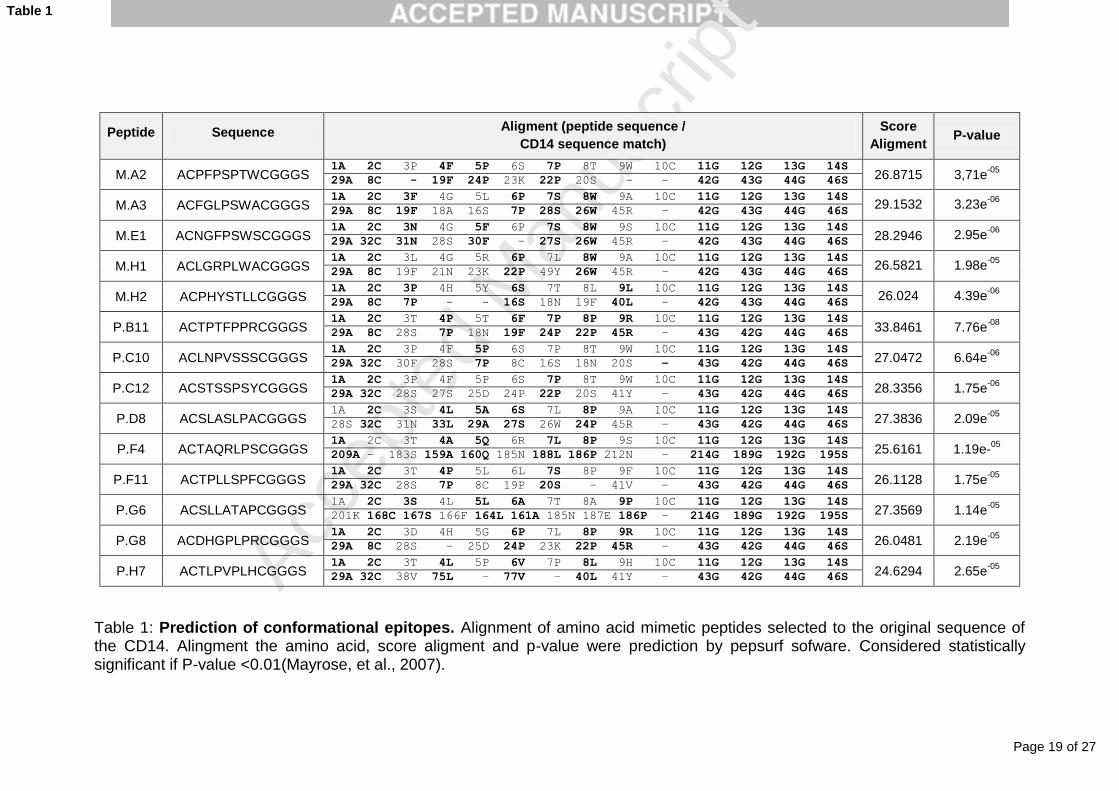
The similarity analysis between the selected peptides and the CD14
protein using the Pepitope Server program showed statistically significant
correspondence (Table 1), evidenced by very low P-values for all peptides.
Peptide characterization
The mimotope characteristics were predicted (Table 2) and
demonstrated to present zero or positive net changes, and hydrophobicity ratio
lower than 50% for almost all peptides. All selected peptides presented partial
similarity (37 to 40%) to the natural antibiotic proteins, with activity against gram
negative, gram positive and fungi, which was verified by alignment with the

Page 12 of 27
Accep
ted
Man
uscr
ipt
Antimicrobial Peptide Database (APD)
(http://aps.unmc.edu/AP/prediction/actionInput.php).
Additional evidences that suggest antimicrobial function to mimotopes
are provided by simulations of helical wheel projections for mimotopes, and the
positive net charges, especially observed for M.H1, P.F4 and P.B11. The
sequences adopt a α-helical amphipathic conformation, as subtended by
hydrophilic (positively charged) and hydrophobic sectors (Figure 5).
Ability to recognize bacterial structures
The recognition of bacterial molecules by the CD14 is the main feature
described in the literature. To assess the functionality of the CD14 mimotopes
that mimic the two epitope regions, we have selected three reactive peptides,
two for the N-terminal (M.H2 and P.F4) and one for the central portion (P.F11),
and tested them against different bacterial species by ELISA. An irrelevant
peptide was used as negative control. Statistical differences were observed
between clones and the irrelevant phage (p <0.0001), demonstrating that the
peptide fused to the phage protein III specifically recognizes the target. The
sequence of the irrelevant phage clone is NMSDFLRIQLRS.
LPS detection
The reactivity of clones M.H2, P.F4 and P.F11 was evaluated with
different concentrations of LPS. All clones recognized the endotoxin of Gram-
bacteria (LPS) (data not shown).
Detection of bacterial lysates and whole bacteria
ELISA assays were carried out to mimic the in vivo function of CD14,
which may bind to bacterial lysates and whole bacteria. All clones were able to

Page 13 of 27
Accep
ted
Man
uscr
ipt
recognize molecules present in both bacterial lysates and whole bacteria;
however, none of them were statistically different in the recognition of different
bacterial species, demonstrating that clones that mimic CD14 epitopes may
recognize common bacterial structures present in both Gram- and Gram+
bacteria (Figures 6).
DISCUSSION
In this investigation, we have used Phage Display (PD) to select peptides
that mimic CD14 epitopes (mimotopes) in order to map functional regions in the
molecule using as targets a polyclonal antibody anti-CD14 and a monoclonal
antibody anti-sCD14ST. The selected mimotopes recognized Gram- and Gram+
bacterial components and could mimic CD14 binding affinity to LPS.
Our PD approach has never been used for CD14 epitope mapping
before, which has been intensively investigated by site direct mutagenesis,
overlapping synthetic peptides, and neutralizing anti-CD14 antibody (Kim, et al.,
2005, Shapiro, et al., 1997). These previous studies have reached the
consensus that the CD14 N-terminal region was responsible for the recognition
of LPS and immune activation of associated receptors.
Due to the great similarity and conserved sequences between human
and mouse CD14 proteins (Kelley, S.L. et al., 2013), our conformational
mimotopes were mapped into the mouse 3D CD14 crystallographic structure
based on in silico and functional analyses. All selected peptides could recognize
LPS, and were mapped inside the previously described amino acid region 1-152
(Kim, et al., 2005), with specific reactivity sequences that include amino acids 3
to 11, 26 to 32, 41 to 44, 56 and 64, and 78 to 83, which are located near the
sheet inside the β1 loop preceding α1 between β3 and within α3, respectively

Page 14 of 27
Accep
ted
Man
uscr
ipt
(Juan, et al., 1995). There is much discrepancy between authors on the LPS-
binding sites. The selected peptides were aligned in specific points of the CD14
molecule encompassing the amino acids 8-214. The peptides were able to
recognize LPS, bacterial lysates and whole bacteria, demonstrating that all
epitopes can bind to PAMPs with different degrees of sensitivity. Suggesting
that some amino acids are essential for LPS recognition in different regions of
the molecule.
The ability of CD14 in recognizing different molecules of microorganisms
may not only be associated with PAMPs’ recognition, pro-inflammatory
signaling, or pathogens’ clearance, but also, our in silico analysis provided
evidences that some peptides may function as antimicrobial peptides (AMPs),
with action in both bacteria and fungi.
Anti-microbicide effects as a result of the sCD14 action and of a synthetic
peptide (amino acids 81-100) of the CD14 showed significant inhibition of E. coli
growth in culture (Voss, et al., 2006, Ohnishi, et al., 2010). Interestingly, clones
P.F4, P.B11 and M.H1 also presented characteristics of cationic and
hydrophobic AMPs. It is believed that the cationic charge of peptides
electrostatically attracts them to negatively charged microbial membranes, and
the hydrophobic properties enable peptides subsequently to insert and disrupt
membranes (Eliasson and Egesten, 2008, Shai, 2002, Shai, 2002).
However, in sepsis, there is an increasing amount of CD14 subtype
(sCD14-ST), which is generated by cleavage of CD14 in the N-terminal region
by an unclear mechanism (Shozushima, et al., 2011), but probably mediated by
neutrophil serine proteases (neutrophil elastase and cathepsin G) (Pham,

Page 15 of 27
Accep
ted
Man
uscr
ipt
2008). The role of sCD14 cleavage in the inflammatory response followed by
immunosuppression, or as anti-microbicides is yet to be defined.
It is important to emphasize that our mimotopes have shown a dominant
recognition site in the N-terminal sequence of the CD14 for both monoclonal
antibody anti-sCD14-ST and polyclonal anti-sCD14, specifically targeting
residues 8 to 46, coinciding with the LPS recognition epitope region(Kim, et al.,
2005). However, a second antigenic site mapped by 20% of reactive mimotopes
to the polyclonal antibody was also demonstrated, which may explain the low
frequency of detection of the entire sCD14 in sepsis patients (FUKUSAKO,
2004). Interestingly, high sCD14 levels are associated with the severity of
sepsis (Aalto, et al., 2007), as well as the sCD14-ST (Shozushima, et al., 2011),
and this molecular event seems to play a detrimental role in sepsis yet to be
clarified. The broad recognition of many molecules by CD14 is also
unexplained, but our work provides novel evidences that may improve our
understanding of the CD14 role and its relevance to the immune response in
inflammatory-associated diseases.
ACKNOWLEDGEMENTS
The authors would like to thank the staff of Clinical Hospital of Federal
University of Uberlandia for providing the bacterial samples.
GRANT SUPPORT
The authors would like to thank to the financial support from CNPq, CAPES and
FAPEMIG.
DECLARATION OF INTEREST
The authors have declared that no competing interests exist.

Page 16 of 27
Accep
ted
Man
uscr
ipt
REFERENCES
Kitchens, R.L., Thompson, P.A. 2005. Modulatory effects of sCD14 and LBP on LPS-host cell interactions. J Endotoxin Res 11, 225.
Kelley, S.L., Lukk, T., Nair, S.K., Tapping, R.I. 2013. The crystal structure of human soluble CD14 reveals a bent solenoid with a hydrophobic amino-terminal pocket. J Immunol 190, 1304.
Kol, A., Lichtman, A.H., Finberg, R.W., Libby, P., Kurt-Jones, E.A. 2000. Cutting edge: heat shock protein (HSP) 60 activates the innate immune response: CD14 is an essential receptor for HSP60 activation of mononuclear cells. J Immunol 164, 13.
Schmitz, G., Orso, E. 2002. CD14 signalling in lipid rafts: new ligands and co-receptors. Curr Opin Lipidol 13, 513.
Camussi, G., Mariano, F., Biancone, L., De Martino, A., Bussolati, B., Montrucchio, G., Tobias, P.S. 1995. Lipopolysaccharide binding protein and CD14 modulate the synthesis of platelet-activating factor by human monocytes and mesangial and endothelial cells stimulated with lipopolysaccharide. J Immunol 155, 316.
Ulevitch, R.J., Tobias, P.S. 1995. Receptor-dependent mechanisms of cell stimulation by bacterial endotoxin. Annu Rev Immunol 13, 437.
Li, X.H., Gong, J.P., Shi, Y.J., Liu, C.A., Peng, Y. 2003. In vitro expression of CD14 protein and its gene in Kupffer cells induced by lipopolysaccharide. Hepatobiliary Pancreat Dis Int 2, 571.
Dai, L.L., Gong, J.P., Zuo, G.Q., Wu, C.X., Shi, Y.J., Li, X.H., Peng, Y., Deng, W., Li, S.W., Liu, C.A. 2003. Synthesis of endotoxin receptor CD14 protein in Kupffer cells and its role in alcohol-induced liver disease. World J Gastroenterol 9, 622.
Iida, M., Hirai, K., Shinohara, S., Yamaguchi, M., Takaishi, T., Sakamoto, Y., Ito, K., Morita, Y. 1994. Lipopolysaccharide primes human basophils for enhanced mediator release: requirement for plasma co-factor and CD14. Biochem Biophys Res Commun 203, 1295.
Austenaa, L., Barozzi, I., Chronowska, A., Termanini, A., Ostuni, R., Prosperini, E., Stewart, A.F., Testa, G., Natoli, G. 2012. The histone methyltransferase Wbp7 controls macrophage function through GPI glycolipid anchor synthesis. Immunity 36, 572.
Sharom, F.J., Radeva, G. 2004. GPI-anchored protein cleavage in the regulation of transmembrane signals. Subcell Biochem 37, 285.
Viriyakosol, S., Mathison, J.C., Tobias, P.S., Kirkland, T.N. 2000. Structure-function analysis of CD14 as a soluble receptor for lipopolysaccharide. J Biol Chem 275, 3144.
Lloyd-Jones, K.L., Kelly, M.M., Kubes, P. 2008. Varying importance of soluble and membrane CD14 in endothelial detection of lipopolysaccharide. J Immunol 181, 1446.
Kim, J.I., Lee, C.J., Jin, M.S., Lee, C.H., Paik, S.G., Lee, H., Lee, J.O. 2005. Crystal structure of CD14 and its implications for lipopolysaccharide signaling. J Biol Chem 280, 11347.
Shapiro, R.A., Cunningham, M.D., Ratcliffe, K., Seachord, C., Blake, J., Bajorath, J., Aruffo, A., Darveau, R.P. 1997. Identification of CD14 residues involved in specific lipopolysaccharide recognition. Infect Immun 65, 293.
Voss, S., Welte, S., Fotin-Mleczek, M., Fischer, R., Ulmer, A.J., Jung, G., Wiesmuller, K.H., Brock, R. 2006. A CD14 domain with lipopolysaccharide-binding and -neutralizing activity. Chembiochem 7, 275.
Kirkland, T.N., Finley, F., Leturcq, D., Moriarty, A., Lee, J.D., Ulevitch, R.J., Tobias, P.S. 1993. Analysis of lipopolysaccharide binding by CD14. J Biol Chem 268, 24818.
Juan, T.S., Kelley, M.J., Johnson, D.A., Busse, L.A., Hailman, E., Wright, S.D., Lichenstein, H.S. 1995. Soluble CD14 truncated at amino acid 152 binds lipopolysaccharide (LPS) and enables cellular response to LPS. J Biol Chem 270, 1382.
Viriyakosol, S., Kirkland, T.N. 1996. The N-terminal half of membrane CD14 is a functional cellular lipopolysaccharide receptor. Infect Immun 64, 653.

Page 17 of 27
Accep
ted
Man
uscr
ipt
Barbas, C.F.B., D. R.; Scott, J. K.; Silverman, G. J. . 2001. Phage display: a laboratory manual. Cold Spring Harbor Laboratory Press.
Rubinstein, N.D., Mayrose, I., Martz, E., Pupko, T. 2009. Epitopia: a web-server for predicting B-cell epitopes. BMC Bioinformatics 10, 287.
Mayrose, I., Shlomi, T., Rubinstein, N.D., Gershoni, J.M., Ruppin, E., Sharan, R., Pupko, T. 2007. Epitope mapping using combinatorial phage-display libraries: a graph-based algorithm. Nucleic Acids Res 35, 69.
Mayrose, I., Penn, O., Erez, E., Rubinstein, N.D., Shlomi, T., Freund, N.T., Bublil, E.M., Ruppin, E., Sharan, R., Gershoni, J.M., Martz, E., Pupko, T. 2007. Pepitope: epitope mapping from affinity-selected peptides. Bioinformatics 23, 3244.
Wang, G., Li, X., Wang, Z. 2009. APD2: the updated antimicrobial peptide database and its application in peptide design. Nucleic Acids Res 37, D933.
Matsumoto, M., Horiuchi, Y., Yamamoto, A., Ochiai, M., Niwa, M., Takagi, T., Omi, H., Kobayashi, T., Suzuki, M.M. 2010. Lipopolysaccaride-binding peptides obtained by phage display method. J Microbiol Methods 82, 54.
Soloaga, R., Defain, V., Blanco, M., Buchovsky, A., Fernandez, A., Gutfraind, Z., Nagel, C., Russo, M., Tokumoto, M. 2000. [Blood cultures: use of presumptive antiobiograms]. Rev Argent Microbiol 32, 149.
Ohnishi, T., Muroi, M., Tanamoto, K. 2010. Inhibitory effects of soluble MD-2 and soluble CD14 on bacterial growth. Microbiol Immunol 54, 74.
Eliasson, M., Egesten, A. 2008. Antibacterial chemokines--actors in both innate and adaptive immunity. Contrib Microbiol 15, 101.
Shai, Y. 2002. Mode of action of membrane active antimicrobial peptides. Biopolymers 66, 236. Shai, Y. 2002. From innate immunity to de-novo designed antimicrobial peptides. Curr Pharm
Des 8, 715. Shozushima, T., Takahashi, G., Matsumoto, N., Kojika, M., Okamura, Y., Endo, S. 2011.
Usefulness of presepsin (sCD14-ST) measurements as a marker for the diagnosis and severity of sepsis that satisfied diagnostic criteria of systemic inflammatory response syndrome. J Infect Chemother 17, 764.
Pham, C.T. 2008. Neutrophil serine proteases fine-tune the inflammatory response. Int J Biochem Cell Biol 40, 1317.
FUKUSAKO, S.S., KAMON.; HIROSE, JIRO. 2004. NOVEL SOLUBLE CD14 ANTIGEN, Tokyo. Aalto, H., Takala, A., Kautiainen, H., Siitonen, S., Repo, H. 2007. Monocyte CD14 and soluble
CD14 in predicting mortality of patients with severe community acquired infection. Scand J Infect Dis 39, 596.

Page 18 of 27
Accep
ted
Man
uscr
ipt
Figure Legends
Figure 1: Reactivity of the CD14 mimetic peptides to the selection target. Interaction of phage clones with the commercially available anti-CD14 antibody (M. selected by monoclonal antibody and P. selected by polyclonal antibody).
Figure 2: Competition Bead-ELISA of CD14 and M.H2 to the paratope of anti-CD14. The high reactivity of M.H2 to the polyclonal anti-CD14 antibody with decreased sCD14 dose showed the specific bind of clone to the paratope of the antibody.
Figure 3: Predict immunogenic regions in CD14 three-dimensional structure by Epitopia Server software (http://epitopia.tau.ac.il/) (Rubinstein, et al., 2009).
Figure 4: Epitope Mapping of CD14. The figure represent regions in CD14 are localized the peptide selected. A and B are mirrored representation of the location of the selected peptide. (1) Represent the binding site of LPS in CD14 (amino acids 1-151), it was determined according to the literature (Kelley, et al., 2013, Kim, et al., 2005) and predicted in the PyMol server software. The molecules shown in Figures 2nd to 15th were predicted in Pepsurf program and representing selected epitopes by phage display. Epitopes are (2) M.A2, (3) M.A3, (4) M.E1, (5) M.H1, (6) M.H2, (7) P.B11, (8) P.C10, (9) P.C12, (10) P.D8, (11) P.F4, (12) P.F11, (13) P.G6, (14) P.G8 and (12) P.H7.
Figure 5: Helical Wheel projection for M.H1, P.F4 and PB11 mimetic peptides to CD14. The sequences of α-helical peptides are presented according to the Shiffer-Edmundson wheel projection. Dashed lines divide the hydrophilic and hydrophobic sectors.
Figure 6: Phage-ELISA with peptides that represent different regions of the
molecule CD14 and the recognition bacterial components (Gram+:
Staphylococcus hominis, Staphylococcus haemolyticus, Staphylococcus
epidermidis, Streptococcus agalactiae, Stenotrophomonas maltophilia; and
Gram-: Pseudomonas aeruginosa, Proetus mirabilis, Klebsiella pneumoniae,
Escherichia coli and Citrobacter younga). Graphs A and C show the reactivity in
the recognition of bacteria lysed while B and D represent entire bacteria. A and
B are Gram- and C and D, Gram+ bacteria
Page 19 of 27
Accep
ted
Man
uscr
ipt
Peptide Sequence Aligment (peptide sequence /
CD14 sequence match)
Score
Aligment P-value
M.A2 ACPFPSPTWCGGGS 1A 2C 3P 4F 5P 6S 7P 8T 9W 10C 11G 12G 13G 14S
26.8715 3,71e-05
29A 8C - 19F 24P 23K 22P 20S - - 42G 43G 44G 46S
M.A3 ACFGLPSWACGGGS 1A 2C 3F 4G 5L 6P 7S 8W 9A 10C 11G 12G 13G 14S
29.1532 3.23e-06
29A 8C 19F 18A 16S 7P 28S 26W 45R – 42G 43G 44G 46S
M.E1 ACNGFPSWSCGGGS 1A 2C 3N 4G 5F 6P 7S 8W 9S 10C 11G 12G 13G 14S
28.2946 2.95e-06
29A 32C 31N 28S 30F – 27S 26W 45R – 42G 43G 44G 46S
M.H1 ACLGRPLWACGGGS 1A 2C 3L 4G 5R 6P 7L 8W 9A 10C 11G 12G 13G 14S
26.5821 1.98e-05
29A 8C 19F 21N 23K 22P 49Y 26W 45R – 42G 43G 44G 46S
M.H2 ACPHYSTLLCGGGS 1A 2C 3P 4H 5Y 6S 7T 8L 9L 10C 11G 12G 13G 14S
26.024 4.39e-06
29A 8C 7P - - 16S 18N 19F 40L - 42G 43G 44G 46S
P.B11 ACTPTFPPRCGGGS 1A 2C 3T 4P 5T 6F 7P 8P 9R 10C 11G 12G 13G 14S
33.8461 7.76e-08
29A 8C 28S 7P 18N 19F 24P 22P 45R – 43G 42G 44G 46S
P.C10 ACLNPVSSSCGGGS 1A 2C 3P 4F 5P 6S 7P 8T 9W 10C 11G 12G 13G 14S
27.0472 6.64e-06
29A 32C 30F 28S 7P 8C 16S 18N 20S – 43G 42G 44G 46S
P.C12 ACSTSSPSYCGGGS 1A 2C 3P 4F 5P 6S 7P 8T 9W 10C 11G 12G 13G 14S
28.3356 1.75e-06
29A 32C 28S 27S 25D 24P 22P 20S 41Y – 43G 42G 44G 46S
P.D8 ACSLASLPACGGGS 1A 2C 3S 4L 5A 6S 7L 8P 9A 10C 11G 12G 13G 14S
27.3836 2.09e-05
28S 32C 31N 33L 29A 27S 26W 24P 45R – 43G 42G 44G 46S
P.F4 ACTAQRLPSCGGGS 1A 2C 3T 4A 5Q 6R 7L 8P 9S 10C 11G 12G 13G 14S
25.6161 1.19e-05
209A – 183S 159A 160Q 185N 188L 186P 212N – 214G 189G 192G 195S
P.F11 ACTPLLSPFCGGGS 1A 2C 3T 4P 5L 6L 7S 8P 9F 10C 11G 12G 13G 14S
26.1128 1.75e-05
29A 32C 28S 7P 8C 19P 20S – 41V – 43G 42G 44G 46S
P.G6 ACSLLATAPCGGGS 1A 2C 3S 4L 5L 6A 7T 8A 9P 10C 11G 12G 13G 14S
27.3569 1.14e-05
201K 168C 167S 166F 164L 161A 185N 187E 186P – 214G 189G 192G 195S
P.G8 ACDHGPLPRCGGGS 1A 2C 3D 4H 5G 6P 7L 8P 9R 10C 11G 12G 13G 14S
26.0481 2.19e-05
29A 8C 28S – 25D 24P 23K 22P 45R – 43G 42G 44G 46S
P.H7 ACTLPVPLHCGGGS 1A 2C 3T 4L 5P 6V 7P 8L 9H 10C 11G 12G 13G 14S
24.6294 2.65e-05
29A 32C 38V 75L – 77V – 40L 41Y – 43G 42G 44G 46S
Table 1: Prediction of conformational epitopes. Alignment of amino acid mimetic peptides selected to the original sequence of the CD14. Alingment the amino acid, score aligment and p-value were prediction by pepsurf sofware. Considered statistically significant if P-value <0.01(Mayrose, et al., 2007).
Table 1

Page 20 of 27
Accep
ted
Man
uscr
ipt
Clone Sequence Net charge Hydrophobicity Mw1 % similarity AMP2 Target of AMPs3
M.A2 ACPFPSPTWCGGGS 0 35% 1348.542 37,5% (APD:AP01305) Gram +, Gram- and fungi
M.A3 ACFGLPSWACGGGS 0 50% 1306.494 40% (APD:AP01764) Gram + and Gram -
M.E1 ACNGFPSWCGGGS 0 35% 1236.359 36,8% (APD:AP02231) Gram -
M.H1 ACLGRPLWACGGGS +1 50% 1341.587 40% (APD:AP01595) Gram+
M.H2 ACPHYSTLLCGGGS 0 35% 1359.555 37,5% (APD:AP01935) Gram+ and Gram-
P.B11 ACTPTFPPRCGGGS +1 28% 1332.544 40% (APD:AP00303) Gram+ and Gram-
P.C10 ACLNPVSSSCGGGS 0 35% 1232.366 37,5% (APD:AP00605) Gram+ and cancer cells
P.C12 ACSTSSPSYCGGGS 0 21% 1257.328 38% (APD:AP01912) Gram+
P.D8 ACSLASLPACGGGS 0 50% 1187.369 37% (APD:AP01512) Gram+, mammalian and cancer cells
P.F4 ACTAQRLPSCGGGS +1 35% 1301.476 35% (APD:AP01206) Gram+
P.F11 ACTPLLSPFCGGGS 0 42% 1297.532 40% (APD:AP00820) Gram+
P.G6 ACSLLATAPCGGGS 0 50% 1201.396 38% (APD:AP01677) Fungi
P.G8 ACDHGPLPRCGGGS 0 28% 1314.482 37,5% (APD:AP01803) Fungi
P.H7 ACTLPVPLHCGGGS 0 42% 1299.551 40% (APD:AP01935) Gram+ and Gram-
Table 1: Characteristics mimetics peptides of CD14 identified by phage-display.
1MW : Molecular weight (theoretical)
2AMP: Antimicrobial Peptide
3Target of AMPs: Based in the action of peptides described in Antimicrobial Peptide database (APD)
Table 2

Page 21 of 27
Accep
ted
Man
uscr
ipt
Figure 1

Page 22 of 27
Accep
ted
Man
uscr
ipt
Figure 2

Page 23 of 27
Accep
ted
Man
uscr
ipt
Figure 3

Page 24 of 27
Accep
ted
Man
uscr
ipt
Figure 4

Page 25 of 27
Accep
ted
Man
uscr
ipt
Figure 5

Page 26 of 27
Accep
ted
Man
uscr
ipt
Figure 6

Page 27 of 27
Accep
ted
Man
uscr
ipt
DECLARATION OF INTEREST
The authors have declared that no competing interests exist.
*Conflict of Interest