rhabdosomes - polish academy of sciences
TRANSCRIPT

ACT A
Vol. 23
PAL A EON T 0 LOG I C A, 'p 0 LON I C 'A
1978 No.4
DENIS EDWIN BEECHING BATES and NANCY HARTSHORNE KIRK
CONTRASTING MODES OF CONSTRUCTION OF RETIOLITE-TYPERHABDOSOMES
Abstract. - Investigation of retiolite-type rhabdosomes has so far revealed twocontrastin~'typesof construction:1. In a morphological series' including Reteograptus geinitzianu8, Gothograptus nassa,Holoret~oZites spp. and 'RetioUtes' sp. it is possible to demonstrate that sclerotizationof theperider»t secreted between the zooid and its mantle evagination becameprogressively localized, culminating in the formation of the highly specialized andsparse framework of Holoietiolites and 'Retiolites' sp.2. In Retiolites geinitzianus (Barrande) the same kind of peridermal secretion combin-
I ,
ed with localized sc1erotization to produce a superficially similar type of rhabdosometo some of those in the above morphological series, but with a quite different relationship to the secretary tissues of the zooids., Secretion and. sclerotization of fibrous strips or 'bandages' seems to have occurredin many normal graptoloids, but resulting in the formation of a continuous cortex.The restriction of the sc1erotized 'bandages' in the retiolites, so as to produce a sparsebut resilient framework, suggests that their function was mechanical rather thanprotective.
PART 1. CONSTRUCTION OF RETEOGRAPTU,S GEINITZIANUSGOTHOGRAPTUS !lASSA, HOLORETIOLITES AND 'RETIOLITES' SP.
In 1972 and 1973 Kirk attempted,to explain the construction of normaland retiolite-type graptbloid rhabdosomes in terms of a model for graptoloid secretion. This was based on the conventional view that graptoliteperiderm was dual, mad~ of fuseHar increments overlaid by corticallaminae. With Urbanek and Towe'~ revelation (1974, 19,75) that theperiderm was formed of 'successive ",single increments, arch-shaped inlongitudinal section, it became necessary to modify some of the details ofrhabdosome construction (Kirk 1974, 19'75) though the model invoked forsecretion was essentially correct.' _
Revision of the ideas on retiolite 'construction was difficult because, at first only flattened and carbonized material was available for ex
amination by SE¥. Nevertheless it' was found possible to work out most ;'

428' DENisE.B. B:""T:£S '~nd NANCylI. kIRK'
of the details of construction of Retiolites geinizianus (Barrande) bysystematically chipping away at this material. These conclusions were'confirmed and amplified when we were able to examine an isolated distalfragment of this species given to us by Dr. A. C. Lenz. Our understanding of retiolite structure was further increased by the study of isolatedmaterial of Holoretiolites and Gothbgraptus sent to us by Professor Dr.G. F. Lutze and Dr. H. Jaeger, of Reteograptus geinitzianus Hall sent byDr. S. Finney, ,and .of a species of Retiolites ,se,nt by Dr, R. B. Rickards.We would like t~ take this opportunity,of~xpre~sirlgour 'deep indebtedness and gratitude for the gift of these specimens which have providedthe key to a truer understanding of retioliteconstruction. ' '
In Reteograptus geinitzianus the sicularperiderm is preserved evenin young rhabdosomes, and shows good incremental, structure in themetasicular part ,even though this is overlaid by strips or 'bandages', ofcortical material showing strong parallel fibres (pI. 4:1). "Thise stripsare mainly longitudinal extending without -interruption along the thicklongitudinal ribs of the prosicuia onto the metasicula. In the other direction they exten~ along the nema: Some of these longitudiaal sti'ipsaremore than half the length of th.ei>icula, -:- about 0.4 mm. There. are alsoa few diagonal strips, and others swing into, parallelism w"ith the meta,sicular rim (pI. 4:2). There is no sign of a prosicular rim; The metasicularrim is' also lined with fibrous strips, but these do not seem to extend farinto the interior which is line~ by a finer fabric. The fibrous'strips edgingthe metasic~lar rim turn along the virgella, on its obver:seand reversesides, but its inner face seems to be covered by fiI}e fabric.
x • 2
DISTAL GROOVE
Fig; 1."Retiol'ites--geinitzianus.' Ideogram ,showing ',incremental membranes of threei,ntert~c.alsepta.passing into grooves in the- fI;amew<;>rkof, ,list,. S.,B.X.----;-l?~ptal-1:)~r
"', of zooid X, R.R.X. +2 - transverse rod of' zooid -X+2. " _'

.,:R.ETIOI.,ITE-T,Y!;'J!: RHA:eI;>0SOMES.,'
The hood of I 1 has co~rsely,fi.brousstrips ,on ·its.outer surfa~e, but itis' lined .by fine fabric which 'also extends as a' broad banQ.lacrosS' t.hereverse fa€e -of 'the metasicula, to the· origin ·of :the ,.antral list of 12
, (pI.1:2; pI. 4:3). It covers' and m~sks the coarsely,ribbed longitudinal stripsof the metasicula and so is later, though the fibrous strips are themselvesclearly later than the 'fusellar' increments. 'j
,The .ventral lists of II, and J2 eaC!h -receive 2 or· 3 ribbed fibrous stripsfrom' the metasicula and these can extend,' without interruption, to theaper.tures. They cover the ventral and rather more obverse, faceo! eachlist, and are separated .by a revetse seam and an ohver'se, seam fr,om thefine fabric covering the distal and rather, more reverse face (pI. 4:2).This fine fabric links with the hood~lining of 11, with the band of finefabric crossing, the sicula: to 12
, and With a hoop-shaped list exte;ndingfroin the ventral'. list of.J1 to the' dorsal list. ' .' .'
The obverse and reverse prosicular lists arise, on the:' ab-apical' sideof the thin areas between the prosicularridges. Coarsely fibrousst~ips
\ '
extend from the prosicula along the outet, obverse .face, of the .obverselist, with 2 seams separating them from fine fabric on its r~ver:se face(pI. 5:2). ' " 1 {l:',
In young spedme,ns the reverse pr0sicular list is also seen to receivecoarsely fibrouS strips from the prosicula, 'but, iIi other,specimeIlS this 11stand the adjacent'prosicula are overlaid by a fine .fabric;. The ,other ,mainlists' of the juvenile. rhabdosome -,- the. ventral longitudinals, .the dorsalzig-zag and the horiz'ontal lists; all have, ~oarsely ·fibrous, strips OD: theirouter faces' and fine fabric. on' their inner, faces,. a seam ,on 'each ·sideseparating. the two fabrics.-
It is C'lear, from older specimens, that the windows in the frameworkof lists were once closed by -incremental periderm' (pI. ,5:5). Over thisperiderm was secreted sclerotized 'sheeting, ,possibly" made, up in ;part ofcoarsely fibrous strips rather like that covering:'thesicula.T.hese tend,torun along and parallel to the lists, -but locally. they crOss.,a list and'runout over the incremental' p,eriderm (pI. 5:3). ,The!. resulting membraneseems to have been attached to' the lists in such a way that'the·;outersurface of the rhabdosome is very:;;mooth, while the lists project :;;troIlglyon the inner surface (pI. 5:4). ' , '
The reverse p'r0sicular list is wholly internal 'in. the adult colony. Thecoarsely fibrous strips covering it in very youngrhabdosomes, could perhaps be related to, a stage in astogeny when it was exposed. The Qbverse,face oithe ob"erse prosicular list temains.exposed,on the obvers-e side ofthe colony.
It seems reasonable to conclude frbm this that. the. thecae as well asthesicula were formed initially of norm{l1.overlapping peridermal in-:crements; arch shaped in 'longitudinal 'Section. ,Evert in 'Very young'rhabdosomes the peridermal increments. of -the .sicula were overl,aid by cortex

430 DENIS E.B. BATES and NANCY H. KIRK
in the form of coarsely fibrous strips following certain selected pathsmainly longitudinal- down nema, prosicular ribs and. 'metasicula, butswinging into parallelism with the metasicular rim, and into the basesof the sicular and prosicular lists~ It is not possible, from examination ofour material by 8EM, to say -whether' these strips 'of fibrous mate'rialwere directly continuous with arch-shaped increments secreted in theevagination of extrathecal tissue at the growing extremity. of the rhabdosome. There is, however, little doubt that they were secreted by thelining of that mantle of evaginated tissue and therefore had potentialcontinuity with the contemporaneously secreted distal increments.
In the sicula of Reteograptus the ,sclerotized fibrous strips were addedearly, before the sicular mantle was withdrawn prior to the secretionof fine internal fabric over them by the crossing canal of P. They werealso added at the time of the growth of the meta- and prosicular lists,serving to stiffen them and bind them to the sicula and give them tensilestrength. This early secretion of the fibrous strips led to the preservationof the initial incremental periderm of the sicula even in very young rhabdosomes.
Beyond the sicula the sclerotized fibrous strips were again added earlyto the outside of the initial incremental periderm, but eviden~ly onlyalong selected paths to form the lis.ts. On the inside of the' lists a finetextured rather soft fabric seems to have been secreted by the zooidalepithelium so that the lists <ieveloped an almost circular cross section.Beyond these lists the incremental periderm itself was not sufficientlysclerotized and was not overlaid by sclerotized layers or fibrous stripssoon enough to be preserved in young fossilized rhabdosomes. Traces ofit can be seen as ragged edges along the seams separating thecoaT'Selyfibrous external fabric from the fine internal fabric of the lists.
Probably considerably later the secretion of sclerotized fibrous stripsappears to have spread from the lists over the initial incremental membrane covering the theace. This process started at the oldest, proximalend of the rhabdosome and spread distally, with the result that in oldcolonies the proximal windows between the lists are closed, while increasingly large unsclerotized areas remained over the more distal thecaeand appear as holes in the fossils.
Since traces of parallel fibres are only occasionally visible on t~e
external surface of the periderm, usually adjacent to the underlying lists,it would appear possible that the secretion of fibrouspandages was succeeded by a more general fine textured sheeting which resulted ina smooth outer surface over the mature rhabdosome. .
On the inside a fine textured sheeting also smooths over the junctionbetween lists and periderm, but the lists project strongly on the insidesuggesting that the sheeting was thick only where it lined the lists butattenuated rapidly where it passed onto the incremental periderm of the

RB:TIOLITE~TYPE BHABDOSOMES' 431
windows. The tendency to deposit thick cortex on the outside of the incremental periderm aJ:ld a thmcortical lining on the. inside recalls thesecretion of 'normal' graptoloipg. . .
The ventral walls of the I.th~·t:ae arise froQm a .c;tista.l ~eaIn in the postapertural list. A proximal seam on this list can be followed back downthe inside of .the adjacent longitudinal and horizontal lists and bearstraces of an interthecal septum. A. 'seam and ragged fringe can also betraced distally on the inside of the zigzag list from a point between thecae31 and 41
• This suggests the existence of a median septum which maynever have been overlaid by sufficient sclerotized layers for preservation.Proximal to this, the zigzag list is smooth on its inside Sind the virgulaappears' to lie free within· the rhabdosome.
In Gothograptus nassa (Holm) there are no preserved traces of normalincremental periderm, and the interpretation of the structure is thereforemore difficult.
In this retiolite the sicula is preserved as a network of lists, but itsform and situation within the proximal end of the colony leave no doubt
Fig. 2. Retiolites geinitzianus. Ideogram showing relationship or reticular threadsto septal bars:

432 DENIS E.B. BATES and NANCY H. KIRK
of' its identity (pI. 6:1). The lists were clearly secreted, from the outside;the earliest component strips being visible on the inside (pI. 7:2). -Thelongitudinal strips were earliest - probably serving the same mechanicalpurpose as the. longitudinal fibrous strips in Reteograptus. Later transverse strips -were deposited outside them. The longitudinal lists extendfrom what wereptesuinably the prosicular rods to a fairly' well definedaperturalrim.The 'principal longitudinal list projects beyond this r·imand probably corresponds to the, virgella. It forks to form the· so calledancora. In our specimen, which may be a rather old colony, the ancorais extremely massive (pI. 6:2). Although: it appears to fork ~gain ratherirregularly, this may. be a somewhat tendentious way of describing whatcould also be described as a network of, ancora lists over the proximalend of 'the colony (pI. 2). These lists were clearly secreted from theinside~ the opposite of' the lists, forming the sicular· reticulum. Where.the ancora lists joip. the apertures of 11 and 12 their smooth innermostand, youngest layer is continuous with the last formed covering of theapei'tural list (pI, 6:4).
The ancora network lies inside an outer reticular network. This outernetwork closely resembles that of the sicula and was clearly secretedfrom the outside~.Where it joins the apertures of 11 and 12 th~ youngest,outermost, slightly pustulose layer is continuous with the covering of theapertural list (pI. 6:3). The ancora and reticular lists therefore cometogether at '~he apertural lists formed in the 'armpits' of the mantleevagination, but otherwise they do not coincide. It is presumed thatG. nassa, as in Retograptus geinitzianus, the fibrous strips forming theancora and reticular lists were secreted onto the inside and the outsideof an initial incremental periderm.
The ancora network is restricted to the proximal end of the rhabdosome where the initial incremental periderm must have been thick, fillingthe gap between ancora and reticulum. More distally, just proximal toaperture 31, some of the more important reticular lists show evidence ofan innermost ornamented layer secreted from the inside in contact withthe layers secrete'd from the outside (pI. 7:4). Traces of a seam betweenthe two could mark the insertion of the incremental periderm whichbecomes thin away from the proximal end.
Where no strips secreted from the inside are preserved one has tosuppose that the original incremental periderm underlay the innermostlayer of the reticulum, and that the edges Of the strips forming the listsonce extended as unsclerotized layers on to it (pI. 8:2;' pI. 9:1). Evidencefor this is provided by a fine, late-formed list which crosses a window ata 'high level'. It was evidently not secreted directly onto the initial incremental periderm but onto a thickness of unsclerotized'layers extendingover it from the edges of the strips forming the older reticular lists forming the window (pI. 7:1, 3; pI. 8:4, 5). More thin unsclerotized layers may

. ·RETIOX.ITE-T,YJ:'E .RHABDOSOMES 433
have lined the incr:emental per~dex:m_on the inside.The apertural ~laps were. two sjq.ed, secreted lwithin a fold of extra.,.
thecal tissue. The Qutermost layers. were clearly secreted in, continuitywith the outermost layers of the adjoining x~ticular lists ,and have .thesame kind of pustulose ribbeddecQration (pI. 8;3). This follows. somewhatirregular,· curved paths suggestingt that it. was l~id down in strips orbandages on both the inner and olilter surfaces which were completelycovered,. Where the fibrous 'strips ,are. thin a fine concentric ridging isvisible which seems to r~present the original, increment~l, possibly plicrofusellar, construction of the flap(p~.8:1). This seelJls to be the only traceof the initial.incremental periderm.in G. nassa.
In Holoretiolites mancki (Munch) the sicula is not preserved, only thevirgella may be represented by a thin rod. From .it there extends a forkingancora - clearly secreted from the inside as i.n Gothograptus(pI. 9:2, andpI. 3). The ancora continued to be thickened while the virgeUa did 'not so the sclerotized layers of the anc()ra end abruptly at th~. base. of th~
virgella, forming a scar (pI. 9:3).In this, Hol.oretiolites there seems ·to be no overlaying of an ancora
network by a reticular network such as occurred in Gothog.rap~us; insteadthe 'ends' .of the ancora lists appear ,to be gl"afted onto .the ~beginnings'
of the reticular lists (pI. 10:1 apd ,3),The reticu;lar lis~, .as in Gotho.graptus, were secreted from the outside, with the' youngest,: ridged andslightly pustulose layer on the 'outside. If, as seems. probable .from Reteo·graptus, the lists were sclerotized strips laid down along certainpfeferredpaths over an incremental periderm, one would expect to find traces ofthis periderm betw;een the outer reticular strips and the in.ner ancora
.strips at the grafts. Such traces can be seen, and there is a .tendency forthe ancora and reticular strips to: part along the ragged junction re'presenting this unsclerotized periderm.
As in Gothograptus, the apert:u,ral lists were .presumably formed inthe armpit of the extrathecal evagination or mantle. In Balticograptus,viewed by transmitted light, apertural flaps somewhat resembling thoseof Gothograptus occasionally preserve a translucent membrane betwe~m
concentric and radial thickening (pI. 10:2). It .wouldseem that here the'fibrous strips following the concentric, and radial 'paths' e.xtended, overthinner, slightly sclerotized' la~ers.,in the windows between them. Thismight be regarded as intermediate between the stage seen in the Gotho-:graptus apertural· naps where the incremental peride.rm was,' preservedbetween the fibrous strips; ,and the stage ,seen in the thecal walls of Holoretiolites and GothQgraptus where, in the windQwsbetween the reticularlists, the original. incremental peri~erm seems never to have,: been reinforced by sclerotized add,itions though it undoubtedly e~,isted and thin. . . .
unsclerotized membranes almost certainly. extended on to. it..- from theedges of the sclerotized strips forming the reticular lists (pI. '10:4)"

434 DENIS E.B. BATES and NANCY H. KIRK
In G. nassa and Holoretiolites secretion from the inside by the zooidal'epithelium produced the ancora lists, and the lining occasionally develop-ed on the inside of some reticular lists. It seems doubtful if unsclerotizedlayers extended from the strips forming th~se structures over the inside
. of the incremental periderm. If they did so extend, it s~ems likely thatthey were thin, as in the late lining of· the incremental periderm inReteograptus geinitzianits and in the eqrticallining of normal graptoloids.
In another retiolite - provisionally called'Retiolites' sp. the structuralnetwork is even more sparse, but it seems that the virgulacontinued tobe thickened in continuity with the virgella to form a stout axial supportto the colony (pI. 10:5). The ancora grew in continuity with it, so thereis no scar surrounding its base (pI. 11:2). There is, however, a seam on eachside of the virgella, which passes into a seam on the proximal and distalsides of a hoop arising from it. It is suggested that the urtsclerotizedincremental wall of the sicula extended from these seams, the virgellaand hoop representing local sclerotized fibrous additions to the insideand outside of this initial wall.
The ancora network is like a wheel with 4 spokes, the lists formingthe rim and spokes all being secreted from the inside - i.e. the sicularside. Presumably the quadrant in line with the hoop would have served·as an aperture for the sicular zooid but it seems in no way to differ fromthe other quadrants. The ventral lists of thecae 11 and 11 arise from therim' and extend distally till each forks to form an aperture.
In this species of 'Retiolites' the seam postulated as the remnant ofthe incremental periderm of the sicula ends on the virgella, and does not-extend down any of the spokes of the ancora network. This suggeststhat the common body of the sicular zooid and the daughter zooids 11 andP budding from it, extended,beyond the sicular aperture and communallysecreted the ancora network from the inside. In Gothograptus the reticularsicula had a sclerotized apertural rim, though the sicular zooid mustlater have extended beyond it to emerge through a window in the ancoranetwork. In Holoretiolites the sicula was unsclerotized and one can onlyguess at the window in the ancora through which the siculozooid ultimat--ely emerged. . . .
In the •Retiolites' specimen the rim of the ancora network has itsoldest layer facing away from the colony.and continuous with the oldeststrip-like layer on the spokes (pI. 11:1). The ventral lists of 11 and P onthe other hand,' have the oldest strip facing the colony and the youngeston the outside~ like the 'reticular network of Holoretiolites mancki and,Gothograpttis. The ventral lists of 11 and II appear to have arisen from
, the outer side of the 'rim, possibly at first as q short projecting spineformed in the mantle armpits of zooids 11 and 11. Later stripsof sclerotizedmaterial were added on the outside· forming typical reticular lists. Theseultimately forked presumably. at the thecal apertures.

RETIOLITE-TYPE 'RHABDOSOMES 435
At the origin of the spokes an irregular fibrous network is seen onthe outward-facing surface of the oldest, first formed layer (,pI. 11:3). Itwas presumably secreted onto' the incremental periderm which is notpreserved. It could represent the -fu~ellar;core 'of fan increment addeddistally, though here it represents only that part ef its proximal extensionwhich iIl normal graptoloids constitutes the cortical lining. It se~msdoubtful if the strips' composing· the ancora extended as unsclerotizedlayers over, the inside of the incremental periderm in the windows ofthe ancora network. Whether the sclerotized strips forming the ventrallists of 11 and 11 extended as unsclerotized layers over the outside of theincremental periderm is also' in doubt as the lists do not have the markedly flexed selvedges such as occurred in G. nassa and Holoretiolites. Infact, in 'Retiolites' sp. the ancora and reticular lists are rather similar,somewhat cylindrical structures which presumably gave rigidity to thesparse rhabdosome. .
Why then should the colony have secreted fibrous strips from theinside on to the incremental periderm' to form the ancora network, andfrom the outside to foqn the lists of the reticulum? The ariswer may besuggested by the early development of Reteograptus, the most normal ofthis morphological series. When zooids 11 and .1 1 grew across the sicula,the mantle of sicula and zooids 11 and II was withdrawn allowingthe zooidal epithelium of zooids 11 and P to secrete internal fabric acrossthe sicula. In Gothograptus nassa this seems to have been accompaniedby some extension of the siculozooid, and zooids 11 and 11 secretal internalfabric in continuity with part of the sicular aperture to form the first twobranches of the ancora. It would seem, moreover, that in this example,zooid II passed round the 'obverse' side of the sicula.
In Holoretiolites and the 'Retiolites' sp. the virgella was the only partof the sicula to be sclerotized, but the ancora was presumably againsecreted from the inside by the communal zooidal epithelium of thesiculozooid and 11 and 11.
Why was ~here then a change to secretion of sclerotized fibrous' stripsfrom the outside? One has to remember that all periderm is 2 sided,
•connected, at least potentially,' by the incremental arches formed in thearmpit of the evaginated extrathecal tissue at the growing ends of thecolony. Thickening of the outer surface by secretion of sclerotized corticalsheets was t~e :rp.ethod adopted by most graptoloids - presumably because it best protected the soft fusellar core of the increments, becauseit did not reduce the·livi~g space of the zooids, and because the cortex,
wlike the extrathecal tissue secreting' it, extended without interruptionover the whole surface of the rhabdosome.So, after secreting the ancoranetwork, these graptoloids returned to the more orthodox mode of secretingcortex from the outside to produce the reticular network. This requir-

436 DENJS E:B. B~TES and NANCY H. KIRK
.ed, .in the morphologica,l series ~escribed above, a v:ar~l;!ty of, devices to:weld the two,networks, together. <"
Secret~on from the outside, by ,the, lining of the communal mantle inwhich thecal bouI:ldaries had d,isappeared,proyided the opportunity toconstruct ·a reticular networkjndependent of growth, increments an,dthecal boundaries, purely to meet the mechanical requirements of the colony. It is interesting that different combinations of longitudinal, horizontal and diagonaUists have been emlpoyed by different lfetiolites. Presumably all of these combinations successfully enabled the rhabdoso:tne toresist distortion by, the stress resulting from the. coordinated ciliaryaction of the zooids. But the uns~lerotized initial incremental periderm,and the unsclerotized layers E7xtending over it from thE:! reticular lists,must have been adequatE:! to afford protection to the zooids.' Evidentlysclerotization was not essential for protection, in graptolites ---- after, allthe. vital mantle lay outside the periderm - sclerotizationmust .haveserved a primarily ~echanical function. This CQuld ac;cQunt ~or the..-factthat overall secretion, of ,sclerotizedc9rt.i~afl,ayers,such. as, may haveoccurred, in Didymograptus (U!ba~ek and: To~e 1975;' pI. 18).,_ seems tohave been replac~d in ma~y graptoloi~s b! se,<;:re,tion of corti~~l str~ps
or bandages aloI:lg mechan~cally sel~~ted paths leading eventually to ,theretiolite condition. ' ,' , " .
• PART. 2. CONSTRUCTION OF RETIOLITES GEINITZIANUS'
A number of specimens pre~eryed in shale; two, specirt;ll;!ns, in limestone, and the isolated specimen kindly sent to us by Dr. A.. C. Lenzformed the material for an investigation which began in 1974. '
One of the spedmens in lim~stone' showl;!d interth'ecal septa. Thesewere senii-tra~sparent when' viewed by the light micr~scope, and boreclear traces of incremental boundaries adjacent 'to the virgula, zigzag listand septal bars (pI. i2:2). Examination by SEM of the zigzag 'list on theisolated specifien'showed the incremental boundaries to be' arch-shapedclosures of fibrous sheeting which passed into each of the lateral grooves.Where the distal-facing angle between Closure and zigzag list ,was obtusethe groove looked rather smooth, the closure being almost parallel to thesurface of the list. But where the distal-facing angle was acute the closures were more clearly seen (pI. 12:1). On the opposite side of the zigzaglist the same relationship wa; observed with the increments of the oppos-ite interthecal septum. ' , "
A broken end of the zigzag list showed the increments making theobtuse angle to be attached to the outer layers of the list, while thoseof the preceding interthecal septum entering the opposite side at an acute

437
distal~fadng',angle; Were c'ontinuQus with the"innermost layers of the:Zigzag list (pI. 12:3"11).',.' ,',
,'I1he ,increments',of', the interthecal 'septa' OIi' ,the' opposite 'side of' therhabdosome turned-distally into the virgula'which: grew ahead of thethecae, as in other biserial graptoloids.' The' inctements passed intoa groove on,each side, of .the virgula and were·cleady continuous with itsbut;er layers (pI. 13:1)..Suc~essive.incremental closures were: inserted alongthe virgular and· zigzag ,grooves, and .along the septal bari grooves whichextended from them. The se,ptaLbar ,turned"at -right, angles into theapertural list, and' here; the incremental·closures became. parallel to thelist and formeii the, lining, of. its groove (pI. 14:1), ,'. '" ','
What do these relationships imply? It would seem that· the zigzag listactually fo~med, as locally:-thickened contin\lation~ of the, closures '9f, theinc::r~m~nt~ of the interthecal septum. The.,septal:parandaperturql,.listwere, a continuation of. this .structure uptothe. ,thecal aperture. Theincrements of ,the interthecal sept,ll,J;ll of the nex.tzqoid OI;l the oppositesifle of. the rhabdosoJIle, (call it X+ 1) Were, added, to the ,outside of thezigzag list jnitiated by the prece;ding :z;opid X. The inter,thecal septum, of
'z90id X+.l beg~n;a,t th,e transy~tse,rodatits proximal end(fig~1).The relationship of transverse rod to zigzag list and virgula:, could
b~ seen at the conflue~ce ,of their grooves. ., $e~n frpm th~ ,pro~imal side",the tra~sverse rod appears to have
.qeveloped f.ro~,the more ~xi~l wall of the groo~e extending f~om thez'igzagli~t or .fr~m. v.iIg1.lla t,o th~ ~el?tal.bar. rhe transverse rod appearsto have ,arisen fr6~ tJ1is wall ,at first as a protuber~nce, a kind of shortspiJ?e. Later increments presurpably e~tend~d the reverse spine so thatit, m~t a ~imiiar spi.?e exte~ding,from, the obverse side of the rhabdosome.This formed a tube whiCll,usually.appears as ~n open groove on theproximal, si~e though later increine~ts can b~ s'een to 4ave closed it adjacent tothe zigzag li~t (pl. 13:Z). "" " ," , ,
On the distal ~ide of the transverse ,rod' a somewhat similar grooveo~ctm;ed, co~fh~entwith ,t'he, gro~ve~' ~n ~h~ 'virgula a~d zjgzag list. This~istal 'g'J;'~Qve'on the transvei:se rc;>d~ever;be~am~dosed, and it is concluded' 'that' it .rep~esented' the ~ttachm:e:~t'of ,t'he' ~e~bran~us interth'ecalseptum: The' ~emb~ane appears to have b~en continu~us ~ith ihe ,outermost layer of the transverse rod, tl1e distal groove being floored by anapparently tihbrc;>keri cylinder' of fibrous sheeting' (pI. 1~:'3 and 4). Thetransver~~ rod e~identlyi'co~n:!spo'nds'to 'the' ab-apertural list figured byU~banek'and To~e (1975: pI. 21}:Tlle' tliin'inte'tthetal septum into whichit' p~sses' 'se'ems to be 'represent~a'by"a 'frirlge 'Of fibriis 'perpendicular to,"'. , ,'... ' , ." 1\ '.,.. " . - • T. -/. I . •
the, ro,d: All these lists were' clearly secretedfronifhe outside by envelop-ing 'secretory epithelium: :The' groove's in 'the'lists; and' the' inSides Of the,arch-shaped intrements; enteririg' the gr06ves; we're probably !once filledwith a 'spongy' ·fusellar fabric; but'thiS is not'presetved 'in 'OUr specimens

'l38 DENIS E.B. BATES and NANCY H. KIRK
and it may never have been fully sclerotized.,lf"he. incremental arches~
and the concentric cylinders into which they pass, to form the lists, were.evidently the ,fibrous lamellae of the successive increments and must haveformed .initiaUy dn"ther ,armpits .of the eyaginated extrathecal-tissueormantle of the extending zooids.' , 1
In our spedmen which is a distal fragment, the network 'or reticulumcovering the rhabdosome is only att~ched to the septal bars. The threadsof this network were clearly secreted from the inside, the outertnostlayers being the first formed - the oldest, the innermost layers being thelast formed. The retiCular threads joined the septal bars at a number ofnodes between the angles and the apertural list. Each node had in ita hole (pI. 14:2-4).
Fractures through the holes show that the uppermost and oldest layerof the reticular thread passed down the hole to become confluent withan older inner layer of the septal bar. Successively formed lower layersof the reticular thread grew in continuity with successively added layers,of the septal bar, and the lowest reticular layer was continuous withthe outermost covering of the septal bar. Very late-formed reticularthreads can be frequently seen 'passing 'into the outermost layer on theflank of a septal bar.
How did the growing colony of zooids achieve these relationshipsbetween the 'framework of lists and the threads of the reticulum? Youngcolonies preserved in shale show the most 'distal septal bar and aperturaIlist to have formed a 100p, with fine reticular threads extending to itfrom the previous septal bar of the same thecal series. No reticularthreads are visible on the distal side of the youngest septalbar. This relationship suggests that the septal bar' and aperturallist were formed by local thickening of the incremental closures of theinterthecal septum, the interthecal septum being at, first the dorsal wall'of theca X and formed under its evaginated mantle of extrathecal tissue.At this stage the sides of zooid X overlapped it, the theca being stronglyconcavo-convex in horizontal section. This allowed the zooidal epitheliumof the sides to secrete reticular threads from the inside in coritinuitywiththe increments and septal bar formed as the dorsal wall of the samezooid X. . ,"
At a somewhat later stage zooid X+2 wO\lld have extended along thedorsal wall of X (now a true interthecal, septum) and the mantle of extrathecal tissue would have been withdrawn from between them. This, '
would have brought the zooidal epithelium of X and of X+2 into contactwith the s~ptal bar so that the further thickening of the septal bar wouldhave been by the z?oidal epithelia of the two,adjacent zooids. Zooid X+2would by now have been secreting its own dorsal waH edged by an~w
septal bar,. and its sides would have been secreting reticular threads in.

·~IOLITE~TYPE .RHABDOSOMES 4:39
continuity with the outer layers ofthe septal bar of X proximal to it andwith the inner layers its own septal bar X+2 along its distal edge.
The bodi~s of zooids X and X+2 were undoubtedly separat.ed by theinterthecal septum extending proximally and· ,inwards .fr:oIfi~-thegroovein the septal bar. They were probably als~ separated outside the 'septalbar because no fibrous increments are,seen to pass over, the bar from X toX+2. Instead the fibrous. increments. formed two series, overlapping oneanother from the proximal and distal sides (pI. 15:1). .
In contrast, where the septal bars bend inwards to meet the ends ofthe transverse rods, the fibrous increments occasionally pass over theouter face of the septal bar showing that the zooidal ~pithe1ia were hereconfluent, covering a kind of common canal external to the frameworkof axial lists (pI. 15:2). The order in which the reticular. threads weresecreted can be determined by an examination of the relationship of theoldest fibrous strips on the outward surface (fig. 2). Thus it is found that
. the first reticular thread to be secreted by zooid X extended from theoldest hole on septal bar X - 2, to the oldest hole on septal bar X-I.The second reticular thread secreted by zooid X crossed its flank fromthe second hole in septal bar X - 2 to the oldest hole in its own, septalbar X, reflecting the beginning of. the. extension, of the theca from thecommon canal. The third reticular thread crossed its flank from the thirdhole in X - 2 to the 2nd hole in septal bar X and soon. Probably aboutthis time zooid X+1 secreted its first thread diagonally from the oldesthole in septal bar X-I to the oldest hole in. septal bar X. And a littlelater still zooid X+2 secreted its first, diagonal' from the o).dest .hole inseptal 'bar X to the oldest hole in septal' bar X + L .
Of course while new threads were being formed across the axial partof the rhabdosome, and successively away from the axial region alongthe septal bars towards the apertures, the older· reticular threads werebeing thickened by addition from below in continuity. with successiveaditions to the outside of the septal bars.
The order of secretion of the reticular threads probably reflected theextension of the thecal walls surrounding the budding and growing zboids.As in most biserial graptoloids, the youngest thecae developing in thea~ial part of the rhabdosome seem to have formed a fairly even termination with the older thecae flanking them. The side walls appear to havebeen unsclero,tized at first, apart from the fibrous reticular threads secret..,edacross them. The paths chosen for this secretion would most effectively
. have supported the side walls as they grew, and the linkage of the successive septal bars by diagonals and longitudinals would also have. contributed greatly to the resilience of the rhabdosome-as Ii whole. The longitudinalthreads were later jointed by transversals to give the reticular networlt.
At the apertures the extension of each theca beyond that of the preceding· one of the same· series; led toa slight modification of this patter;n.

440 DENIS'E.B,"BATESrandiNANCY H. KIRK
Transverse reticular threads appear to have· grown out from -the lastcomplete']ol'lgitudinal ~nd then to' have curved round tQ become a :Iong-'itudinaJ passing int(}cthe next"hole, in the septal" bar distal to it.A transveI1sal from this. thread may have repeated the process,' passinginto the next· hole; and then yet another may have extended from this topass into :the last hole of the septal bar. As a result the ventral edge ofthe rhabdosome became· straight from theca to theca, and approximatelyparallel to the long axis ofthe'colony.
Ina'specimenpreserved in shale the reticulum is seen to extend acrossthe' distal parts of the proximal 3 or 4 apertures (pI. 1:1). This seems tohave been the consequence of an even greater ventral extension 'of thedistal parts of the thecal walls. This resulted in ventral fusion, andreticular threads were secreted across from the reverse to the bbverseface. In .later' thecae the ventral extension became progressively less andthe reticular network formed progressively smaller lobes until in thefully adult part of the rhabdosome the extension was only enough toproduce the cl)aracteristic straight ventral edges.
Although the. apertural list probably originated as the thickeneddosure of the increment completing the dorsal wall of zooid X, as thezooid X+2 extended ~long it, it became the proximal apertural list ofthat zooid and it continued to be thickened in the mantle armpit of zooidX+2. The last longitudinal reticular thread adjacent to the proximal partof the aperture of X +2 was also formed in the same mantle-armpit andshared in the prolonged thickening. This produced an outwardly turnedflange which framed the more proximal part of the aperture.
In an older etched specimen on limestone some of the reticular threadsnear to the aperture also show an overturning of the fibrous· strips inan axial-proximal direction. This suggests that these, and perhaps allthe more-regula,r, longitudinal,' reticular threads, could have originatedin the 'mantle-armpit of. the growing zooids.
Viewed from the outside, the fibrous strips composing the reticular.threads sometimes show indications of layering. A thin layer of irregularly anastomosing. fibrils passes down into closely 'packed, parallel fibrils(pI. 16:1, and 2)•.
. Viewed from the inside, the reticular threads and closed windows appear only' to be formed, of successive strips of parallel fibrils, and 'similarstrips are seen to enwrap the framework of lists. The oldest strips, seenon the outside of the reticular thread&" are 20-30"", in width and theseincrease to a width of r 50 or80p.· in the case, of the youngest, innermoststrips,and to 1001-1 over the lists of the framework. Viewed frQm theinside the strips end abruptly at their sides where they .overlie .earlierstrips. They terminate longitudinally ina rather irregular fringe of fibrils(pI. 16:3).
The strips' of parallel fibriJs tend 'to run parallel to' the ·reticular

RETIOLITE-TYPE RHABDOSOMES, 441
threads and to the lists of the framework, but they can also run obliquelyacross the latter and .may form strongly curved wrappings - especiallyover junctions. A single fibrous strip can also be followed for up to 1000fJ.along a very irregular path on the inside.of the reticulum. This couldcertainly not have been secreted within a mantle armpit.
The reason for the different appearance of the reticular strip's, whenviewed from the inside and from the outside, seems to lie in the incremental mode of graptolite secretion. In graptolite periderm each distallyadded increment consists of a core of fusellar fabric bounded by a lamellaof parallel fibrils. The fusellar core is arch-shaped in longitudinal sectionand the lamella extends back over. it to form the limbs of the arch. Moreproximally the increments on the inside and on the outside of the thecalwall could consist of an attenuated layer of fusellar fabric separatedfrom the secretory epithelium by a lamella of parallel fibrils, though moreusually the fusellar layer is omitted.
In Retiolites geinitzianus the sclerotized fibrous secretion which formed the reticulum and thickened the framework of lists was secreted bythe epithelium covering the zooids. It therefore corresponded to the corticallining of normal graptolites. It was presumably secreted onto the insideof an incremental periderm which was not sufficiently, sclerotized forpreservation.
, Seen from the outside there is some suggestion that each, fibrousstrip of the reticulum passed laterally into a thin.membrane which onceadhered to tlie inside of the incremental periderm. In the angles of thereticular windows, gaps between successive fibrous strips show signs ofhaving been occupied by loose fusellar fabric (pI. 17:1, 2 and 3): Wherethe reticular windows eventually became closed by extension of thefibrous strips secreted from the inside, this fusellar fabric thinned outallowing the fibrou,s strips to form a dense sheeting lining the bulged-outperiderm in the windows. The fusellar fabric also thinned out under thewindow frame so that the late fibrous strips increasingly thickened thedense reticular threads. Being formed as a kind of cortical lining, thestrips forming the"reticulum and thickening it from the inside would haveincreasingly reduced the living space of the zooids, causing the incremental periderm to bulge out through the reticlilar windows. It seemslikely therefore that unsclerotized extensions from the fibrous strips on tothe incremental periderm would have been reduced to very narrow stripsof attachment.
Over the septal bars, between the nodes fOli the entry of the reticularthreads, the fibrous strips overlapped alternately from the proximal anddistal sides. It seems impossible that these could have extended as unsclerotized layers lining the incremental periderm. The later formed layerscovering the septal bars do not overlap, and there is evidence that thestrips composing them extended as a lining. to the periderm bulging' out
3 Acta Palaeontologica Polonlca nr 4f18

442 DENIS E.B. BATES and NANCY H. KIRK
of the windows on either side of the septal bar. Over the apertural listsand adjacent flanges the fibrous strips were secreted in the, mantle..;'armpit and therefore represent true cortex. They are, -of course, con"'"tinuous with ~nd indistinguishable, from those of the cortical lining.
The flange framing. the more proximal part of the, apertu.re is formedof incremental. strips which end in unusually ragged edges (pI. 17:4). Itis suggested that during life these extended as 'unsclerotized layers overthe incremental periderm. They would correspond to the outer corticallayers of normal graptoloids, and would have been secreted from theoutside by the lining of the mantle. In one of our specimens an unusuallyextended frange covers the adjacent reticulum, and seemS" to representthe spread of sclerotized strips on the outside comparable to that whichclosed the windows on the inside of the incremental periderm.
According to Holm (1890) old specimens of Stromatograptus werecovered by an outer sclerotized layer masking the outlines of the reticul..;urn. The further spread of sclerotized strips from the extended flange inour specimen, over a thickness of unsclerotized layers extending fromthe ragged edges of the earlier increments of the flange, could also havehad this effect. So it is suggested that in Retiolites geinitzianus, as inReteograptus geinitzianus described in the first part of this paper, theunsclerotized incremental periderm 'glazing' the windows of the reticulum, ,eventually became coated on the inside and on the outside bysclerotized material secreted as cortical lining and cortex by the zooidalepithelium and lining of the mantle.
In the case of the cortical lining there would seem to have been littleor no thickness of unsclerotized layers intervening between the increJ;nental periderm and the sclerotized strips' which later lined the windows.In the case of the outer cortex there may have been a considerablethickness of unsclerotized layers extending from the ~agged edges of theincrements of the flange over the incremental periderm, (compare G. nassa and Holoretiolites). These unsclerotized layers and the underlying in~
cremental periderm would -have been increasingly bulged-out by thepressure of the reticular threads as these became progressively thickened
. from the inside. They would therefore have needed to remain elasticwhile this thickening continued. Pr'esumably only in old rhabdosomeswould secretion of sclerotized strips have extended over these layers,masking the outlines of the reticulum as' shown in Holm's illustration,and preventing further bulging.
This could have been a gerontic effect, like the massive thickening oftheancora lists in old specimens of G. nassa. It could hardly have beena resp~nse to mechanical requirements since the lists of framework andreticulum satisfied/these in the earlier growth stages of the colony. Andit could hardly have been a response to a need to protect the zooids sincethe incremental periderm and unsclerotized layers over the windows had:

: RETIOlJITE-TYPERH'ABDOSOMES 443
previously afforded adequate ,Protection. Also,· as emphasized in theprevious section, it has to be remembered that in all graptolites the extrathecal mantle lay outside all peridermal str';lctures. and presumablyescaped serious damage by some invisible, possibly chemical, protectivedevice and was able to heal minor damage by regeneration of the secretory epithelium prep~ratory to regene~ation of the periderm.
Secretion of. cortex and cortical lining in the form of strips of parallelfibrils was not peculiar to retiolite graptoloids. In many normal graptoloids it seems to have taken' the place of more continuous sheeting(Crowther and Rickards 1977). It could have been the secretion of suchstrips which led to the frequency of 'unconformities' between corticalincrements as seen in longitudinal section, and to their apparent separat~
ion from the, distal increments. Many of the strips were very 'probablynot secreted .in direct continuity with distally added increments, butpotential continuity was always ensured by the continuous secretorvepithelium covering the zooids and .lining the mantle.
ACKNOWLEDGEMENTS
"In addition to the colleagues who have kindly sent us specimens, wewish to thank the Royal Society, and the University College of Wales,for the provision of travel grants which enabled the authors to travelto Warsaw for the presentation. of this paper.
Geology DepaTtment, U.C.W.Penglais, AbeTystwyth
Dyfed SY24 5BGU.K.
DecembeT 19'17
REFERENCES
Geology DepaTtment, U.C.W.Penglais, AbeTystwyth
Dtifed SY24 5BGUX
CROWTHER, P. and RICKARDS, B. 1977 Cortical Bandages and the GraptoliteZooid. - Geol. Palaeont., 11, 9-46.
HOLM, G. 1890. Gotlands Graptoliter: Svenska Vetenskaps. - Akad. Handl., Bihang,16, ser. 4, no. 7, 1-34.
KIRK, N.· H. 1972. Some thoughts an the construction of the rhabdosome in theGraptolithina, with special reference to extrathe~al tissue and its bearing onthe theory of automobility. - Geol. Dept. Publ., Univ. Coll. Wales, AbeTystwyth,1,1-21.1973. Some thoughts on the construction and functioning of the rhabdosomein the Retiolitidae. - Ibidem, 3, 1-26.1974. More thoughts on the construction of the rhabdosome in the Dendroidea,in the light of the ultrastructure of the Dendreidea and of MastigogTaptus.Ibidem" 6, 1-11.1975. More thoughts on the construction and functioning of the rhabdosome inthe Graptoloidea in the light of their ultrastructure. - Ibidem, 7, 1-21.
3·

444 DENIS E.B. BATES and NANCY H. KIRK
URBANEK, A. and TOWE, K. M. 1974. Ultrastructural studies on graptolites., I. Theperiderm and its derivatives in Dendroidea and in Mastigograptus. - Smith.Contr. Paleobiol., 20, 1---:20.and - 1975. Ultrastructural studies on graptolites. 2. The periderm and itsderivatives in the Graptoloidea. - Ibidem, 22, 1-24.
DISCUSSION
P. R. Crowther:
It should be emphasized that there is no a priori reason for assuming thata retolitid skeletal structure must have been secreted within folds of soft tissue andnot by the use of a simple mortaring scheme like that' employed by the recentpterobranch hemicllordates.. The versality of the pterobranch mode of secretion isoften underestimated. Even in our present woefully inadequate state of knowledgeconcerning details of the method )Jsed to construct their coenecia, it is clear fromJohn's' (1931) description of the subgenus Cephalodiscus (Acoelothecia)' that a swarmof free living zooids is perfectly capable of building a coeDEicium from a frameworkof rods. John's illustration of a fragment of distal periderm bears a striking resemb":lence to the reticulum of Retiolites geinitzianus denstreticulatus (note, their relativescale is different). He also described a gradual rounding off of 'list' junctions andfilling in of the orifices with age, similar to the retiolitidskeleton. 'Of course,' thisis not strictly evidence in favour of a similar mode of secretion for retiolid graptolites. But it must be realised that Cephalodiscus ,manages to build a similar structure,without the aid of a covering of soft tissue, using only its cephalic shield (and possiblythe tentacles). Thus, it seems reasonable to admit the possibility that retiolitids hada similar capability.
Evidence in favour of a pterobranch mode of secretion for other graptolites ismounting (see Crowther herein; Crowther and Rickards 1977) and is particularly<;on~lusive f6r the diplograptids, where bandaged cortex is most strikingly observed.Bandaging also occurs.,.on the proximal, sclerotized thecae of Orthoretiolites and canbe seen on Kirk's micrographs of Reteograptus. These observations are in accordancewith the generally accepted 'view that retiolitids originated (polyphyletically) fromdiplograptid stock. The surface of lists on Retiolites geinitzianus densireticulatus areconstructed from arrays, of parallel fibrils. The arrays often exhibit compiex unconformity patterns near list junctions where they overlap, similar to the arrangementof cortical bandages on normal graptolite periderm. Thus, it seems more likely thatretiolitid graptolites adapted the secretionary scheme of their ancestors, the diplograptids, to produce a scaffolding-like structure of rods and lists, just as Acoelothecia redUl;ed the normal, continuous periderm of other Cephalodiscus subgenerato a similar meshwork.
More work must be done on other retolitid species, in conjuction with furtherresearch into the ultrastructure and mode of, secretion of the cephalodiscan skeletonif the tempting comparison outlined above is to be proved meaningful.
REFERENCES
JOHN, P. 19311. Cephalodiscus. - Discovery Reports, 3, 223-260.'CROWTHER, P. and RICKARDS, R. B. 1977. Cortical bandages and the .graptolite
zooid. - Geol. Palaeont., 11, 9--46.

RETIOLITE-TYPE RHABDOSOMES
EXPLANATION OF THE PLATEs 1-17
445
All photographs taken with a Cambridge Stereoscan 600 unless stated otherwise.
Plate 1
ijeteograptus geinitzianus
1. Proximal part of rhabdosome, drawn from a specimen preserved in shale.2. Diagram of young rhabdosome showing sicula and framework of lists seen from
reverse side. Bandages of parallel fibres shown by stipple, fine internal fabricshown uncoloured. 'H - hood, I. - hoop-shaped list.
Plate 2
Gothograptus nassa
Diagram of proximal end of rhabdosome, (a) seen from the inside, (b) seen fromthe outside. Lists of ancora shown white, lists of the reticulum shown stippled. .S - window in ancora-network which may have served as the aperture for the extended siculozooid.
Plate 3
Holoretiolites mancki
Diagram of proximal end of rhabdosome, (a) seen from the inside, (b) seen from theoutside, with outside of reticular lists shown stippl~d.
S - window which may have served as the aperture for the extended siculozooid.
Plate 4
Reteograptus geinitzianus (Upper Ordovician, USA)
1. Sicula of young specimen showing incremental periderm overlain by bandageswith strongly developed parallel fibrils.
2. Apertural part of sicula. Note the fibrous strips running parallel to the apertureand swinging diagonally into the ventral list of theca 12•
3. Abapertural edge of band of fine fabric extending across the reverse face of themetasicula and partly masking the coarsely fibrous strips underneath.
Plate 5
Reteograptus geinitzianus (Upper Ordovician, USA)
1. External cortical tissue on sicula, showing the end of a later strip overlying anearlier one. An earlier NNW fibrous strip is overlain by a younger ENE stripwith an irregular termination.
2. Obverse prosicular list with seam between the ribbed fabric on the obverse faceand fine fabric on' the reverse face.

446 DENIS E.B. BATES and NANCY H. KIRK
3. 01,lter surface of mature specimen.. The ribbed strips tend to run parallel to thelists but also cross them onto the increrp.ental periderm.
4. Cross-sections of list and membrane of' mature specimen outer surface uppermost.5. Traces of incremental boundaries in periderm filling all windows in the frame
'York of lists in a 'Z?ature. specimen.
Plate 6
.,Gothograptus nassa (Erratic boulder, FRG)
1. Proximal end of incomplete mature specimen, with sicula partly preserved a~
-a reticulum of liste secreted from the outside.2. Oblique view of specimen of 1, viewed from the other side, showing thickened
ancora secreted frQm the Inside.3,4. External and internal views of the aperture of theca P, showing its attachment
to the reticulum (R) and to the ancora (A).
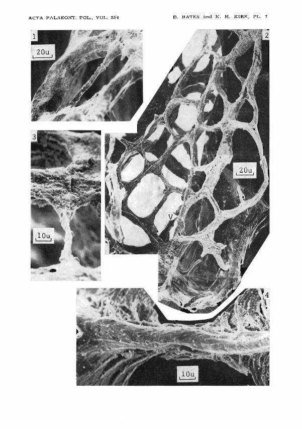
Plate 7-
Gothograptus nassa (Erratic boulder, FRG)
1,3. Two external views of a late reticular list, crossing an aperture at a· high level.Compare with pI. 8:4, 5 which show internal views of the same list.
2. Close up of the siculaseen in pl. 6:1; V - the main virgellar list.4. Internal view of reticular list, with additional tissue added internally. Note its
rounded surface, and pustulose ornament. Compare with pI. 9:1. .
Plate 8
Gothograptus nassa (figs 1 and 3 from Central Wales, figs 2, 4, 5 fromerratic boulder, FRG)
1,3. Latex replicas of the internal and external surfaces of apertural flaps, withgrowth lines on the internal surface and bands of regular pustules on the externalsurface.
2. Internal view of junction of ventral reticular list with apertural flap. Note theinturned edges of the strips forming the list.
4, 5. Internal views of the late reticular list, seen in 1 and 3.
Plate 9
Gothograptus nassa (Erratic boulder, FRG)
1. Oblique internal view of reticulum, showing tissue added from the inside (comparewith pl. 7:4).
Holoretiolites mancki (Erratic boulder, FRG)
2. Proximal end of rhaWosome, seen from the outside. Reticulum has pustuloseornament facing outwards, ancora has a smooth surface with an external seamin which earlier layers are visible.
3. Close-up of junction of ancora and virgella.

.RETIOLITE"TYPE RHABDOSOMES
Plate 10
Holoretiolites mancki (Erratic boulder, FRG). .
447
1,3. Junction' of ancora (lower). and reticular list rimming the thecal aperture, external and internal views. The seam between ancora and pustulose reticulum in 1probably represents the insertion of the incremental periderm.
4. Cross-section of reticular list, showing inwardly turned edges of the componentstrips.
BaltiCograptus sp. (Erratic boulder, FRG)
2. Light microscope photograph of apertural flap of theca showing translucentmembrane between ·concentric thickenings or lists.
"Retiolites" sp. (Lower Silurian, Sweden)
:;. Proximal end of rhabdosome, seen from the outside;A - ancora, V - virgella, H - hoop.
Plate 1.1
"Retiolites" sp. (Lower Silurian, Sweden)\
1. Rim of ancora seen from outside looking towards sicular apex, with ventral listof theca 11 (VL) at top teft, and ancora 'spoke' (AS) to lower right. •
2. Ancora and base of virgella (V) seen from inside looking towards sicular aperture.3. Same,' seen from outside showing the first forIil~d layer.
Plate 12
Retiolites geinitzianus (1, 3 and 4 Silurian, Canadian Artie; 2 erraticboulder, FRG)
1. Zig-zag list, looking distally across an angle between "zig" and "zag". Proximallyto this angle tht;! groove apwsrs smooth with closure of increments almost parallelto the list. Distally the closures make a higher angle with the list and are moreclearly seen.
2. Light microscope photograph of zig-zag list (ZZ) and junctions with septal bar(SB). The increments of three interthecal septa appear as dark lines curving intothe zig-zag list and septal bars.
3,4. Broken end of zig-zag list shown in a composite photograpl;l taken from above,(4) and below (3). On the upper side, the increments of ITS X preceded the growthof the list and form its innermost l~yers. On the, lower side increments of ITSX +1 form its outermost layers.
Plate 13 .
Retiolites geinitzianus (Silurian, Canadian Arctic)I
1. ' View looking distally along the virgula and showing increments of an interthecalseptum continuous with the outer layers of the virgula'.

448 DENIS E.B. BATES and NANCY H. KIRK
2. Junction between transverse rod (TR), zig-zag list (ZZ) and septal bar (SB).A fibrous 'bandage' covers the groove on the proximal side of the transverse bar.
3. Distal face of transverse rod, with a distal groove representing the origin of theinterthecal septum late in the development of the rod.
4. Distal side of junction between transverse rod (TR), virgula (V) and septal bar· (SB).•
Plate 14
Retiolites geinitzianus (Silurian, Canadian Arctic)
1. Groove on septal bar (SB) and apertural list (AL) with traces of incrementalclosures of the interthecal septum.
2. A node on an 'E-W' septai bar is joined by reticular threads from 'N' and'S'.3. Septal bar (SB) with reticular threads entering a node from SW and NE. Later
transverse threads cross these from NW to SE, and a very late thread joins anouter layer on the septal bar.
4. Fractured septal bar and reticulum junction. The oldest layers of the reticularthread pass down the hole to become confluent with the innermost layers of 'theseptal bar. The reticular thread entering from the right has been broken off.
Plate 15
Retiolites geinitzianus (Silurian, Canadian Arctic)
1. Outer face of septal bar, between reticular junctions or nodes. The two zooidsseparated by it have secreted alternately overlapping fibrous' layers of whichit is constructed.
2. Outer face of internal portion of septal bar between the innermost reticularjunction and the junction with the zig-zag list (top). The fibrous bandagesoccasionally pass right over the bar.
Plate 16
Retiolites geinitzianus (Silurian, Arctic)
1. Junction of reticular threads in external view showing succession of fibrous strips.2. Close-up of I, showing irregular anastomosing fibrils above more closely packed
parallel fibrils within a fibrous strip.3. Internal view of reticular junction. The side (A) of one strip of parallel fibrils.
and the end (B) of another,can be seen.
Plate 17
Retiolites geinitzianus (1, 3, 4 Silurian, Canadian Arctic;2 erratic boulder, FRG)
1. Ex~ernal view of reticular window, with anastomosing fusellar fabric in the gapsbetween successive strips of packed parallel fibrils.
2. External view of reticular windows closed strips secreted from the inside.3. Close-up. offusellar fabric of I, showing the anastomosing fibrils underlain by
packed parallel ,fibrils.4. Oblique external view of the flange at the side of a thecal aperture..

ACTA PALAEONT. POL., VOL. 23'4 B ATES and N. H. KIRK, PL. 1D.

ACTA PALAEONT. POL., VOL. 23/4 D. BATES and N. H. KIRK, PL. 2

ACTA PALAEO]',"'!'. POL., VOL. 23/4 D. BATES and N. H. KIRK, PL.

ACTA PALAEONT. POL., VOL. 23/4 D. BATES and N. H. KIRK, PL. 4

ACTA PALAEONT. POL., VOL. 23/4 D. BATES and N. H. KIRK, PL. 5

ACTA PALAEOl'o"T. POL., VOL. 2314 D. BATES and N. H. KIRK, PL. 6
--'--2
ACTA PALAEONT. POL., VOL. 23/4 D. BATES and N. H. KIRK, PL. 7

ACTA PALAE01'o'T. POL., VOL. 23'4 D. BATES and N. H. KIRK, PL. 8

ACTA PALAEOJll"'T. POL., VOL. 23'4 D. BATES and N. H. KIRK, PL. 9

ACTA PALAEONT. POL., VOL. 23/4 D. BATES and N. H. KIRK, PL. 10

ACTA PALAEOl'o"T. POL., VOL. 23/4 D. BATES and N. H. KIRK, PL. 11

ACTA PALAEON'T. POL., VOL. 23'4 D. BATES and N. H. KIRK, PL. 12

ACTA PALAEON'T. POL., VOL. 23'4 D. BATES and N. H. KIRK, PL. 13

ACT A P ALAEOr.--r. POL., VOL. 23/4 D. BATES and N. H. KIRK, PL. 14

ACTA PALAEONT. POL., VOL. 23/4 D. BATES and N. H. KIRK, PL. 15

ACTA PALAEONT. POL., VOL. 23'4
5~.\....-....:--..- ..
D. BATES and N. H. KIRK, PL. 16

ACTA PALAEONT. POL., VOL. 2314 D. BATES and N. H. KIRK, PL. 17