rheumatoid arthritis manal al mashaleh. rheumatoid arthritis (ra) is a chronic systemic...
TRANSCRIPT

Rheumatoid Arthritis
Manal Al Mashaleh
Rheumatoid arthritis (RA) is a chronic systemic inflammatory disease of unknown cause.
An external trigger (eg, cigarette smoking, infection, or trauma) that triggers an autoimmune reaction, leading to synovial hypertrophy and chronic joint inflammation along with the potential for extra-articular manifestations, is theorized to occur in genetically susceptible individuals.

History
The hallmark feature of rheumatoid arthritis (RA) is persistent symmetric polyarthritis (synovitis) that affects the hands and feet, although any joint lined by a synovial membrane may be involved.
The severity of RA may fluctuate over time, but chronic RA most commonly results in the progressive development of various degrees of joint destruction, deformity, and a significant decline in functional status.
Extra-articular involvement of organs such as the skin, heart, lungs, and eyes can also be significant.

Signs and symptoms
In most patients with RA, onset is insidious, often beginning with fever, malaise, arthralgias, and weakness before progressing to joint inflammation and swelling.
Signs and symptoms of rheumatoid arthritis may include the following:
Persistent symmetric polyarthritis (synovitis) of hands and feet (hallmark feature)
Progressive articular deterioration Extra-articular involvement Constitutional symptoms

The physical examination
Important to assess the following: Stiffness Tenderness Pain on motion Swelling Deformity Limitation of motion Extra-articular manifestations Rheumatoid nodules

The boutonniere deformity
nonreducible flexion at the PIP joint along with hyperextension of the distal interphalangeal (DIP) joint of the finger.

Swan-neck deformity of the finger
Hyperextension at the PIP joint with flexion of the DIP joint

Rheumatoid nodules at the elbow

Diagnosis
No test results are pathognomonic The diagnosis is made by using a
combination of clinical, laboratory, and imaging features
Potentially useful laboratory studies in suspected RA include the following:
Erythrocyte sedimentation rate C-reactive protein level Complete blood count

Rheumatoid factor assay Antinuclear antibody assay Anti−cyclic citrullinated peptide and
anti−mutated citrullinated vimentin assays

Images
Radiography (first choice): look for osteopenia, joint space narrowing and erosions
Magnetic resonance imaging: Primarily cervical spine
Ultrasonography of joints: Joints, as well as tendon sheaths, changes and degree of vascularization of the synovial membrane, and even erosions

Joint aspiration and analysis of synovial fluid may be considered, including the following:
Gram stain Cell count Culture Assessment of overall appearance
Differential Diagnoses
Fibromyalgia Lyme Disease Myelodysplastic Syndrome Osteoarthritis Paraneoplastic Syndromes Polychondritis Polymyalgia Rheumatica Psoriatic Arthritis Sarcoidosis Sjogren Syndrome Systemic Lupus Erythematosus

Treatment
Nonsteroidal anti-inflammatory drugs (NSAIDs)
Corticosteroids DMARDS Biologic TNF-inhibiting DMARDs Biologic non-TNF DMARDs

Surgical treatments
Synovectomy Tenosynovectomy Tendon realignment Reconstructive surgery or
arthroplasty Arthrodesis
Prognosis
Outcome in RA is compromised when diagnosis and treatment are delayed
The clinical course of RA is generally one of exacerbations and remissions

Prognostic factors
It has been shown that intervention with DMARDs in very early RA (symptom duration < 12 weeks at the time of first treatment) gives the best opportunity for attempting to achieve disease remission
HLA-DRB1*04/04 genotype High serum titer of autoantibodies (eg, RF and ACPA) Extra-articular manifestations Large number of involved joints Age younger than 30 years Female sex Systemic symptoms Insidious onset

RA affects several organ systems, as follows:
Cutaneous: Subcutaneous nodules (rheumatoid nodules) ,vasculitic lesions as purpura or skin ulceration , palmar erythema and pyoderma gangrenosum
Cardiac: Myocardial infarction, myocardial dysfunction, and asymptomatic pericardial effusions are common; symptomatic pericarditis and constrictive pericarditis are rare. Myocarditis, coronary vasculitis, valvular disease, and conduction defects are occasionally observed
Pulmonary: pleural effusions, interstitial fibrosis, nodules, and bronchiolitis obliterans organizing pneumonia. Methotrexate therapy can induce interstitial fibrosis that may be difficult to distinguish from that which naturally occurs in patients with RA.

Hematologic : anemia of chronic disease, normochromic-normocytic anemia, thrombocytosis. Leukopenia is a finding in patients with Felty syndrome.
Neurologic :Nerve entrapment is common, mononeuritis multiplex, and cervical myelopathy
Ocular: Keratoconjunctivitis sicca is common , episcleritis, uveitis, and nodular scleritis that may lead to scleromalacia
Renal: not directly affected by RA. Secondary involvement is common, including that due to medications (eg, nonsteroidal anti-inflammatory drugs [NSAIDs], gold, and cyclosporine), inflammation (eg, amyloidosis), and associated diseases (eg, Sjögren syndrome with renal tubular abnormalities).
Gastrointestinal (GI): secondary to s medication effects, inflammation, and other diseases. The liver may be affected in patients with Felty syndrome (ie, RA, splenomegaly, and neutropenia).
Vascular: Vasculitic lesions as palpable purpura, skin ulcers, or digital infarcts.
2010 ACR/EULAR Diagnostic Criteria
Historically, the diagnosis of RA was based on the 1987 American College of Rheumatology

The ACR/EULAR classification system
is a score-based algorithm for RA that incorporates the following 4 factors:
Joint involvement Serology test results Acute-phase reactant test results Patient self-reporting of the duration of
signs and symptoms

The maximum number of points possible is 10. A classification of definitive RA requires a score of 6/10 or higher. Patients with a score lower than 6/10 should be reassessed over time. If patients already have erosive changes characteristic of RA, they meet the definition of
RA, and application of this diagnostic algorithm is unnecessary. Joint involvement consists of swelling or tenderness upon examination. The presence of
synovitis may be confirmed on imaging studies. Points are allocated as follows: 1 large joint (ie, shoulders, elbows, hips, knees, ankles) = 0 points 2-10 large joints = 1 point 1-3 small joints (with or without involvement of large joints), such as MCP, PIP, second
to fifth MTP, thumb interphalangeal (IP), and wrist joints = 2 points 4-10 small joints (with or without involvement of large joints) = 3 points More than 10 joints (at least 1 small joint, plus any combination of large and additional
small joints or joints such as the temporomandibular, acromioclavicular, or sternoclavicular) = 5 points

At least 1 serology test result is needed for RA classification. Points are allocated as follows:
Negative rheumatoid factor (RF) and negative anti−citrullinated protein antibody (ACPA; in the ACR/EULAR criteria set, tested as anti−cyclic citrullinated peptide [anti-CCP]) = 0 points
Low-positive RF or low-positive ACPA = 2 points
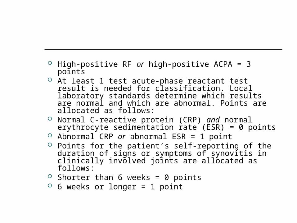
High-positive RF or high-positive ACPA = 3 points At least 1 test acute-phase reactant test result is
needed for classification. Local laboratory standards determine which results are normal and which are abnormal. Points are allocated as follows:
Normal C-reactive protein (CRP) and normal erythrocyte sedimentation rate (ESR) = 0 points
Abnormal CRP or abnormal ESR = 1 point Points for the patient’s self-reporting of the
duration of signs or symptoms of synovitis in clinically involved joints are allocated as follows:
Shorter than 6 weeks = 0 points 6 weeks or longer = 1 point
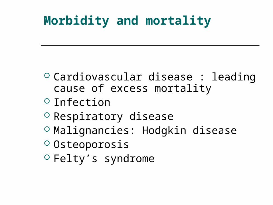
Morbidity and mortality
Cardiovascular disease : leading cause of excess mortality
Infection Respiratory disease Malignancies: Hodgkin disease Osteoporosis Felty’s syndrome

Seronegative Arthritis
Refers to a diverse group of musculoskeletal syndromes linked by common clinical features and common immunopathologic mechanisms.

The entities include
Psoriatic arthritis Reiter’s syndrome Enteropathic arthritis Reactive arthritis Ankylosing spondylitis Undifferentiated seronegative arthritis Whipple’s disease Arthritis associated with pustular acne Post-intestinal bypass arthritis HIV associated arthritis

NOSOLOGY
Spondylarthropathy Seronegative spondylarthropathy Seronegative arthritis BASE syndrome (B27,arthritis,
spondylitis, enthesopathy)
The disease is often incorrectly referred to as seronegative rheumatoid arthritis. This is incorrect because the demographics and clinical presentations of the diseases within this group differ markedly from rheumatoid arthritis.
KEY CLINICAL FEATURES
In contrast to rheumatoid arthritis, seronegative arthritis is male predominant (with exception of psoriatic arthritis where M=F).
The age of onset is variable and disease may start in the teens or early twenties.
The arthritis tends to run a waxing and waning course with spontaneous exacerbations and remissions.
Inflammation occurs not only in the joints, but in the spine and at tendon attachment points (entheses).

The arthritis is often asymmetric and has a tendency to involve the large joints of the lower extremities and the feet and ankles.
Sometimes an entire digit is swollen, producing what is known as sausage digit or dactylitis.
Uncontrolled inflammation tends to result in stiffening and loss of motion as well as in damage to cartilage.
What distinguishes this group of diseases from one another and from rheumatoid arthritis are a wide range of non-articular features which include psoriasis and nail changes, inflammatory eye disease, inflammatory bowel disease, canker sores, and urethritis.
The disease frequently runs in families where it is linked to HLA genes including but not limited to HLA B27.

TREATMENT
Non-steroidal anti-inflammatory agents (NSAIDS)
Non-biologic DMARDS (methotrexate, leflunomide, azulfidine)
Biologic agents (Enbrel, Humira, Remicade).
Topical treatments for skin and eye disease
Physical therapy to maintain range of motion in joints, tendons and the spine.