rho gtpases and signaling networks
TRANSCRIPT

REVIEW
Rho GTPases and signaling networksLinda Van Aelst1,3 and Crislyn D’Souza-Schorey2
1Cold Spring Harbor Laboratory, Cold Spring Harbor, New York 11724 USA; 2Department of Cell Biology, WashingtonUniversity School of Medicine, St. Louis, Missouri 63110 USA
The Rho GTPases form a subgroup of the Ras superfam-ily of 20- to 30-kD GTP-binding proteins that have beenshown to regulate a wide spectrum of cellular functions.These proteins are ubiquitously expressed across thespecies, from yeast to man. The mammalian Rho-likeGTPases comprise at least 10 distinct proteins: RhoA, B,C, D, and E; Rac1 and 2; RacE; Cdc42Hs, and TC10. Acomparison of the amino acid sequences of the Rho pro-teins from various species has revealed that they are con-served in primary structure and are 50%–55% homolo-gous to each other. Like all members of the Ras super-family, the Rho GTPases function as molecularswitches, cycling between an inactive GDP-bound stateand an active GTP-bound state. Until recently, membersof the Rho subfamily were believed to be involved pri-marily in the regulation of cytoskeletal organization inresponse to extracellular growth factors. However, re-search from a number of laboratories over the past fewyears has revealed that the Rho GTPases play crucialroles in diverse cellular events such as membrane traf-ficking, transcriptional regulation, cell growth control,and development. Consequently, a major challenge hasbeen to unravel the underlying molecular mechanismsby which the Rho GTPases mediate these various activi-ties. Many targets of the Rho GTPases have now beenidentified and further characterization of some of themhas provided major insights toward our understanding ofRho GTPase function at the molecular level. This reviewaims to summarize the general established principlesabout the Rho GTPases and some of the more recentexciting findings, hinting at novel, unanticipated func-tions of the Rho GTPases.
Regulators of the Rho GTPases
Like all members of the Ras superfamily, the activity ofthe Rho GTPases is determined by the ratio of theirGTP/GDP-bound forms in the cell (Boguski and McCor-mick 1993). The ratio of the two forms is regulated bythe opposing effects of guanine nucleotide exchange fac-tors (GEFs), which enhance the exchange of bound GDPfor GTP, and the GTPase-activating proteins (GAPs),which increase the intrinsic rate of hydrolysis of bound
GTP. In addition, the Rho-like GTPases are regulatedfurther by guanine nucleotide dissociation inhibitors(GDIs), which can inhibit both the exchange of GTP andthe hydrolysis of bound GTP.
GEFs
GEFs for Rho-like GTPases belong to a rapidly growingfamily of proteins that share a common motif, desig-nated the Dbl-homology (DH) domain for which the Dbloncogene product is the prototype (Cerione and Zheng1996). The Dbl oncogene was originally discovered by itsability to induce focus formation and tumorigenicitywhen expressed in NIH-3T3 cells (Eva and Aaronson1985). The first clue to Dbl’s function as a GEF camefrom the observation that it contains 29% sequenceidentity with the Saccharomyces cerevisiae cell divisioncycle protein Cdc24, which by genetic analysis wasplaced upstream of the yeast small GTP-binding proteinCdc42 in the bud assembly pathway (Ron et al. 1991).Biochemical analysis has shown that Dbl is indeed ableto release GDP from the human homolog of Cdc42 invitro. Furthermore, deletion analysis of the Dbl proteindemonstrated that the DH domain was essential and suf-ficient for this activity and that this domain was alsonecessary to induce oncogenicity (Hart et al. 1991; Ronet al. 1991; Hart and Roberts 1994). In addition to the DHdomain, Dbl and the yeast Cdc24 share a pleckstrin ho-mology (PH) domain, which is essential for proper cellu-lar localization (Zheng et al. 1996).
Since the discovery of Dbl, a growing list of mamma-lian proteins containing both a DH and a PH domain hasbeen assembled (for review, see Cerione and Zheng1996). The majority have been identified as oncogenes intransfection assays (see Table 1). Tiam, however, wasfirst identified as an invasion-inducing gene using provi-ral tagging in combination with in vitro selection forinvasiveness (Habets et al. 1994). Two other members ofthe DH protein family, Fgd1 and Vav, have been shownto be essential for normal embryonic development (Pas-teris et al. 1994; Tarakhovsky 1995; Zhang et al. 1995a).Proteins containing a PH and a DH domain have alsobeen identified in Drosophila melanogaster and Cae-norhabditis elegans (Benian et al. 1996; Sone et al. 1997).
It has been generally assumed that all proteins thatcontain a DH and PH domain in tandem will be GEFs forRho subtype proteins. This appears to be true for some
3Corresponding author.E-MAIL [email protected]; FAX (516) 367-8381.
GENES & DEVELOPMENT 11:2295–2322 © 1997 by Cold Spring Harbor Laboratory Press ISSN 0890-9369/97 $5.00 2295
Cold Spring Harbor Laboratory Press on April 9, 2018 - Published by genesdev.cshlp.orgDownloaded from

but not all DH/PH-containing proteins (see Table 1).Moreover, some members of the DH protein family(such as Dbl) have been shown to exhibit exchange ac-tivity in vitro for a broad range of Rho-like GTPases,whereas others appear to be more specific. Lbc, for ex-ample, and the more recently discovered oncoproteinsLfc and Lsc, are specific for Rho, whereas Fgd1 is specificfor Cdc42 (Glaven et al. 1996; Zheng 1996). AlthoughVav previously was reported to be an activator of Ras(Gulbins et al. 1993), it has been demonstrated more re-cently to function as a GEF for members of the Rhofamily (Crespo et al. 1997; Han et al. 1997).
Further studies will be required to gain more insightinto the determinants defining the selectivity of GEFsfor specific Rho family members. Recently, however,several studies have been performed to determinewhether the proteins that serve as Rho GEFs in vitro canperform similar functions in vivo. Microinjection experi-ments have demonstrated that Lbc induced stress fiberformation and Fgd1 elicited filopodial extensions selec-
tively in response to Rho and Cdc42, respectively (Olsonet al. 1996). Furthermore, Fgd1 has been shown to acti-vate the stress-activated protein kinase/c-Jun amino-ter-minal kinase (SAPK/JNK) signal transduction pathway,an activity mediated by Cdc42 and/or Rac (Olson et al.1996). Both Dbl and Vav have been shown to trigger theformation of filopodia, lamellipodia, and stress fibersmediated by Cdc42, Rac, and Rho, respectively. Dbl andVav also stimulated SAPK/JNK activity (Olson et al.1996). Overexpression of Tiam in fibroblasts elicited theformation of membrane ruffles and activation of JNK ina Rac-dependent manner (Michiels et al. 1995, 1997).Furthermore, Michiels et al. (1997) demonstrated that anintact amino-terminal PH domain was essential forthese activities.
In addition to the PH and DH domains, many of theexhange factors have other domains that are commonlyfound in signaling molecules, such as a Src homology(SH3) domain and a diacylglycerol-binding zinc butterflymotif, suggesting that they may have additional func-
Table 1. Mammalian GEFs for the Rho subfamily of GTPases
DH/PH-containingproteins
GEF specificity forRho GTPases Biological properties Tissue distribution References
Dbl Cdc42, Rho oncogenic brain, adrenal glands,gonads
Hart et al. (1991)
Lbc Rho oncogenic heart, lung, skeletalmuscle
Zheng et al. (1995)
Lfc Rho oncogenic hematopoietic cells,kidney, lung
Glaven et al. (1996)
Lsc Rho oncogenic hematopoietic cells,kidney lung
Glaven et al. (1996);Aasheim et al. (1997)
Dbs ? oncogenic kidney lungpredominantly brain
Whitehead et al. (1995)
Tiam Rac metastatic and oncogenic brain, testis Habets et al. (1994)Vav Rac, Cdc42, Rho oncogenic, implicated in
lymphocyticproliferation andlymphopenia
hematopoietic cells Crespo et al. (1997);Han et al. (1997)
FGD1 Cdc42 implicated in faciogenitaldysplasia
brain, heart, lung, kidney Olson et al. (1996);Zheng et al. (1996)
Trio Rac and Rho cell migration? ubiquitous Debant et al. (1996)Ost Rho, Cdc42 (binds to
Rac-GTP)oncogenic brain, heart, lung, liver Horii et al. (1994)
Bcr Rac, Cdc42, Rho(GAP for Rac)
implicated in leukemia predominantly brain Chuang et al. (1995)
Abr Rac, Cdc42, Rho(GAP for Cdc42 andRac)
? predominantly brain Chuang et al. (1995)
Ect-2 ?(binds to Rho andRac)
oncogenic testis, kidney, liver,spleen
Miki et al. (1993)
Tim ? oncogenic kidney, liver, pancreas,lung, placenta
Chan et al. (1994)
NET1 ? oncogenic ubiquitous Chan et al. (1996)SOS ?
(GEF for Ras)ubiquitious Bowtell et al. (1992);
Chardin et al. (1993)RasGEF ?
(GEF for Ras)brain Shou et al. (1992)
Van Aelst and D’Souza-Schorey
2296 GENES & DEVELOPMENT
Cold Spring Harbor Laboratory Press on April 9, 2018 - Published by genesdev.cshlp.orgDownloaded from

tions (Cerione and Zheng 1996). The Ras GEF, Sos, hasbeen shown to bind the adaptor molecule GRB2, whichin turn binds to the platelet-derived growth factor(PDGF) receptor in response to growth factors (Down-ward 1996). However, little is known about the signalingcascades coupling the Rho GEFs to elements that func-tion upstream of the GTPases (see below).
GAPs
A prototype GAP protein specific for the Rho familyGTPases was purified by biochemical analysis of cell ex-tracts using recombinant Rho. This protein, designatedp50Rho–GAP, was shown to have GAP activity towardRho, Cdc42, and Rac in vitro (Hall 1990; Lancaster et al.1994). Since then, additional proteins that exhibit GAPactivity for the Rho GTPases have been identified inmammalian cells (Table 1). Also several Rho GAP-con-taining proteins have been discovered in S. cerevisiae,Drosophila, and C. elegans (Agnel et al. 1992; Chen et al.1994, 1996; Zheng et al. 1994; Stevenson et al. 1995;Schmidt et al. 1997). These proteins all share a relatedGAP domain that spans 140 amino acids of the proteinbut bears no significant resemblance to Ras GAP. Thesubstrate specificity of the Rho GAPs toward membersof the Rho subfamily varies with each GAP protein(Table 2). Although some of these proteins exhibit GAPactivity for several Rho GTPases in cell-free assays, theirsubstrate specificities in vivo appear to be more re-stricted. For example, the substrate spectrum of p50Rho–GAP in vitro encompasses Cdc42, Rac, and Rho; how-ever, in vivo, it appears to be restricted to Rho only (Rid-ley et al. 1993). The p190GAP, although first identifiedas a tyrosine-phosporylated Ras GAP-associated proteinin Src-transformed cells and in growth factor-treated
cells, was later shown to possess GAP activity for theRho GTPases (Ellis et al. 1990; Settleman et al. 1992).Microinjection of p190GAP in fibroblasts resulted in aninhibition of Rho-mediated stress fiber formation butnot Rac-induced membrane ruffling (Ridley et al. 1993).The biological implications of a direct link between Ras-and Rho-mediated pathways for signaling remain un-clear. Recently, two tyrosine-containing peptides inp190 have been shown to bind simultaneously to theSH2 domains of Ras GAP upon tyrosine phosphorylationof p190. This interaction appears to induce a conforma-tional change in Ras GAP, resulting in an increased ac-cessibility of the target binding surface of its SH3 do-main. Thus, a role for p190 in the Ras GAP signalingcomplex may be to promote Ras GAP interactions via itsSH3 domain (Hu and Settleman 1997).
Rho GAPs, in addition to accelerating the hydrolysisof GTP, may mediate other downstream functions of theRho proteins in mammalian systems. A role for p190 inregulating Rho function in cells undergoing cytoskeletalrearrangements has been suggested (Chang et al. 1995).The N- and b-chimerins have been demonstrated to ex-hibit GAP activity toward the Rac GTPase, and micro-injection of the chimerin GAP domain into fibroblastsprevented Rac- and Cdc42-induced cytoskeletal rear-rangements (Diekmann et al. 1991; Leung et al. 1993;Manser et al. 1995; Kozma et al. 1996). Unexpectedly,microinjection of full-length N-chimerin as well as a chi-merin mutant lacking GAP activity, resulted in the in-duction of lamellipodia and filopodia formation. Further-more, the formation of the latter structures could be in-hibited by dominant-negative Rac and Cdc42 mutants,suggesting that N-chimerin, in addition to functioningas a GAP, may also function as an effector (Kozma et al.1996).
Table 2. Mammalian GAPs for the Rho subfamily of GTPases
Rho–GAP-containing proteins
GAP specificity forRho GTPasesa Tissue distribution References
p50 Rho–GAP Cdc42*, Rac, Rho ubiquitous Barfod et al. (1993);Lancaster et al. (1994)
Bcr Rac*, Cdc42 predominantly brain Diekmann et al. (1991)Abr Rac, Cdc42 predominantly brain Tan et al. (1993)N-Chimerin Rac brain Diekmann et al. (1991)b-chimerin Rac testis Leung et al. (1993)p190GAP Rho*, Rac, CdcC42 ubiquitous Settleman et al. (1992)p85a ? ubiquitous Otsu et al. (1991)p85b ? ubiquitous Otsu et al. (1991)3BP-1 Rac, Cdc42 spleen, kidney, lung, brain,
heartCicchetti et al., (1992, 1995)
p122 Rho Homma and Emori (1995)Myr5 Rho*, Cdc42 ubiquitous Reinhard et al. (1995)RalBP1/RLIP76/RIP1 Cdc42*, Rac ubiquitous Cantor et al. (1995);
Jullien-Flores et al. (1995);Park and Weinberg (1995)
Graf Rho, Cdc42 ubiquitous; abundant inbrain and liver
Hildebrand et al. (1996)
a(*) Preferred GTPase for GAP.
Rho GTPases and signaling networks
GENES & DEVELOPMENT 2297
Cold Spring Harbor Laboratory Press on April 9, 2018 - Published by genesdev.cshlp.orgDownloaded from
GDIs
The first GDI identified for the members of the Rho fam-ily was the ubiquitously expressed protein Rho GDI,which was isolated as a cytosolic protein that preferen-tially associated with the GDP-bound form of RhoA andRhoB and thereby inhibited the dissociation of GDP (Fu-kumoto et al. 1990; Ueda et al. 1990). Subsequently, RhoGDI was found to be active on Cdc42 and Rac (Abo et al.1991; Leonard et al. 1992). Further studies demonstratedthat Rho GDI also associated weakly with the GTP-bound form of Rho, Rac, and Cdc42 (Hart et al. 1992;Chuang et al. 1993). This weak interaction resulted in aninhibition of the intrinsic and GAP-stimulated GTPaseactivity of the Rho GTPases. Thus, Rho GDI appears tobe a molecule capable of blocking the GTP binding/GTPase cycle at two points: at the GDP/GTP exchangestep and at the GTP hydrolytic step.
The Rho GDIs appear to have a crucial role in thetranslocation of the Rho GTPases between membranesand the cytoplasm. In resting cells, the Rho proteins arefound in the cytosol as a complex with Rho GDIs, whichinhibit their GTP/GDP exchange ratio, but are releasedfrom the GDI and translocated to the membranes duringthe course of cell activation (Takai et al. 1995). Untilrecently, the mechanism by which Rho is released fromRho GDI was largely unknown. However, studies fromTakahashi et al. (1997) have provided a tentative modelby which this may occur. Rho GDI was found to coim-munoprecipitate with moesin, which is a member of theERM (ezrin, radaxin, moesin) family (Tsukita et al. 1994,1997; Hirao et al. 1996). ERM proteins have been shownto bind a membrane-spanning protein, CD44, and F-actinvia their amino- and carboxy-terminal regions, respec-tively (Hirao et al. 1996). Takahashi et al. (1997) inves-tigated whether binding of Rho GDI to ERM affectedRho GDI activity. They observed that the amino-termi-nal region of radixin inhibited Rho GDI activity. More-over, they found that binding of the amino-terminal re-gion of radixin to Rho/Rho GDI complex resulted in therelease of Rho from Rho GDI, and that Rho GEF couldstimulate the GDP/GTP exchange reaction of Rho com-plexed with Rho GDI when radixin was present. Similaractivities were found in the presence of ezrin and moe-sin. These data suggest that members of the ERM familyare involved in the activation of Rho. Interestingly, moe-sin has been identified from porcine brain cytosolic ex-tracts as a component that could reconstitute the forma-tion of stress fibers, focal adhesion, and cortical actinpolymerization in response to activation of Rho and Racin permeabilized fibroblasts (Mackay et al. 1997). Theseresults clearly demonstrated the involvement of mem-bers of the ERM family in the formation of stress fibersand focal adhesions, as well as actin polymerization.Whether the mechanism by which moesin can reconsti-tute this response involves GDI is unclear, as GDI wasnot detected in the purified activity.
More recently, two novel GDIs have been isolated: D4-GDI (also called LY-GDI), which is expressed only inhematopoietic tissues (Lelias et al. 1993; Scherle et al.
1993), and Rho GDIg, which is preferentially expressedin brain and pancreas (Adra et al. 1997). A murine ho-molog of Rho GDIg, designated Rho GDI3, has beencloned independently (Zalcman et al. 1996). In vitrostudies showed that D4-GDI can function as a GDI forRhoA, Rac, and Cdc42 (Adra et al. 1993). The combineddata of Rho GDIg and Rho GDI3 indicate that this thirdmember of GDI proteins binds and functions as GDI forCdc42, RhoB, and RhoG and possibly RhoA. Deletionand mutant analysis demonstrated that the last sixamino acids and, more importantly, one residue near thecarboxyl terminus of the GDI molecule, is critical for theinteraction with and specificity for the GTPase. A singlechange at residue 174 of D4-GDI to the correspondingresidue of Rho GDI imparted nearly full GDI activity onthe D4 molecule (Platko et al. 1995).
Little is known about the physiological function of theRho GDIs in vivo. Microinjection studies have shownthat Rho GDI inhibits several downstream functions ofRho (Nishiyama et al. 1994; Coso et al. 1995). Deletionof the D4-GDI gene in embryonic stem (ES) cells resultedin only a subtle defect of superoxide production in D4-GDI−/− macrophages, a function mediated by Rac (Gu-illemot et al. 1996). This suggest that there may be con-siderable redundancy of function between the Rho GDIs.Recent findings by Na et al. (1996) showed that D4-GDIis a specific substrate of apoptotic proteases. The cleav-age and inactivation of D4-GDI (and perhaps other GDIs)may be a crucial part of the process of programmed celldeath and/or a mechanism for regulation of GDIs. Fur-ther studies in vivo will likely point out the necessity ofRho GDIs in several physiological events in the cell.
Upstream signaling pathways
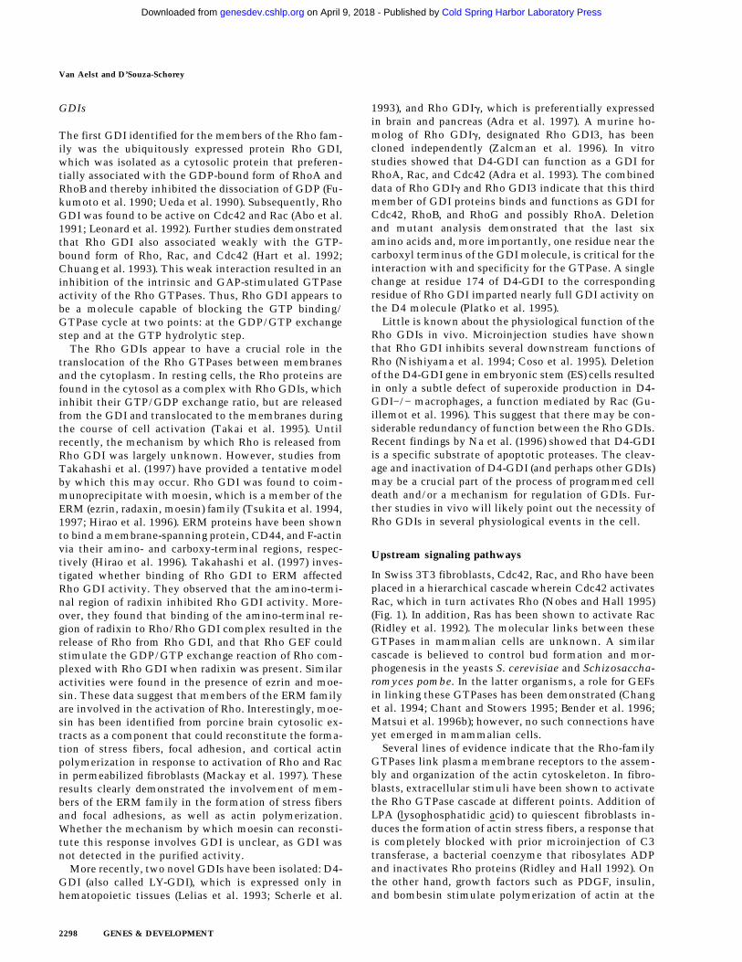
In Swiss 3T3 fibroblasts, Cdc42, Rac, and Rho have beenplaced in a hierarchical cascade wherein Cdc42 activatesRac, which in turn activates Rho (Nobes and Hall 1995)(Fig. 1). In addition, Ras has been shown to activate Rac(Ridley et al. 1992). The molecular links between theseGTPases in mammalian cells are unknown. A similarcascade is believed to control bud formation and mor-phogenesis in the yeasts S. cerevisiae and Schizosaccha-romyces pombe. In the latter organisms, a role for GEFsin linking these GTPases has been demonstrated (Changet al. 1994; Chant and Stowers 1995; Bender et al. 1996;Matsui et al. 1996b); however, no such connections haveyet emerged in mammalian cells.
Several lines of evidence indicate that the Rho-familyGTPases link plasma membrane receptors to the assem-bly and organization of the actin cytoskeleton. In fibro-blasts, extracellular stimuli have been shown to activatethe Rho GTPase cascade at different points. Addition ofLPA (lysophosphatidic acid) to quiescent fibroblasts in-duces the formation of actin stress fibers, a response thatis completely blocked with prior microinjection of C3transferase, a bacterial coenzyme that ribosylates ADPand inactivates Rho proteins (Ridley and Hall 1992). Onthe other hand, growth factors such as PDGF, insulin,and bombesin stimulate polymerization of actin at the
Van Aelst and D’Souza-Schorey
2298 GENES & DEVELOPMENT
Cold Spring Harbor Laboratory Press on April 9, 2018 - Published by genesdev.cshlp.orgDownloaded from
plasma membrane of many cell types to induce lamelli-podia formation and surface membrane ruffling (Ridleyet al. 1992; Nobes et al. 1995). This ruffling response canbe inhibited by the dominant-negative mutant of Rac,RacN17, thereby establishing a Rac-regulated signalingpathway linking growth factor receptors to the polymer-ization of actin at the plasma membrane. Furthermore,the activation of Cdc42 by bradykinin results in the for-mation of filopodia and the subsequent formation oflamellipodia. Filopodia formation was inhibited by adominant-negative mutant of Cdc42, Cdc42N17,whereas RacN17 inhibited only membrane-ruffling for-mation (Kozma et al. 1995; Nobes and Hall 1995).
Progress has been made in establishing the connectionbetween growth factor receptors and Rho-like GTPases.The bradykinin, LPA, and bombesin receptors belong tothe seven-transmembrane-domain heterotrimeric G pro-tein-coupled receptor family. Thus, the trimeric G pro-teins are likely to play a role in the activation of therespective GTPases. Recently, an activated mutant formof the a subunit of the heterotrimeric G protein G12 hasbeen demonstrated to stimulate JNK activity in a Ras-
and Rac-dependent manner (Collins et al. 1996). Further-more, it has been shown that signaling from m1 and m2muscaric receptors (mACHRs) to JNK involves bg sub-units of heterotrimeric G proteins (Coso et al. 1996). In S.cerevisiae, pheromone signaling is mediated by bg sub-units (encoded by STE4 and STE18, respectively) (White-way et al. 1989). Epistasis analysis placed Cdc24 andCdc42 as essential components downstream of Ste4 inthe pheromone signaling pathway. Furthermore Ste4 hasbeen shown to interact with Cdc24 in the yeast two-hybrid system (Zhao et al. 1995). Taken together, theseresults from yeast suggest that a GEF may link the tri-meric G proteins to the low-molecular-weight GTPases.
Several lines of evidence have implicated the involve-ment of phosphoinositide 3 kinase (PI3 kinase) in PDGF-and insulin-induced cytoskeletal rearrangements. Treat-ment of fibroblasts with the drug PI3 kinase inhibitorwortmannin, inhibits membrane ruffling induced byPDGF, epidermal growth factor (EGF), and insulin, al-though not by microinjected Rac protein (Kotani et al.1994; Wennstrom et al. 1994; Nobes et al. 1995). Fur-thermore, PDGF could stimulate the level of Rac GTP byincreasing GEF activity in a PI3 kinase-dependent man-ner (Hawkins et al. 1995). Hence, PI3 kinase appears tofunction upstream of Rac for the induction of membraneruffling in response to extracellular growth factors.Moreover, a constitutively active PI3 kinase mutant hasbeen shown to trigger membrane ruffles and stress fibersin a Rac- and Rho-dependent manner (Reif et al. 1996).Interestingly, this active mutant failed to induce Rac/Rho signaling pathways that regulate gene transcription(Reif et al. 1996). A plausible explanation given for thisobservation is that the Rho GTPases are linked to differ-ent upstream regulatory proteins, which may determinethe interaction with different GTPase effector pathwaysleading to the diverse biological activities. This may ex-plain how the Rho GTPases regulate such a wide varietyof biological activities (see below). The mechanism bywhich PI3 kinase activates Rac is unknown but may in-volve GEFs, GAPs, or GDIs. Interestingly, in S. cerevi-siae, putative phosphatidylinositol kinase homologshave been identified. Among them, TOR2 is required fororganization of the actin cytoskeleton (Schmidt et al.1996) and activates the GTPases RHO1 and RHO2 viatheir exchange factor ROM2 (Schmidt et al. 1997).
As wortmannin does not inhibit the RasV12-inducedmembrane ruffling in Swiss fibroblast cells, it suggeststhat PI3 kinase is not involved in Ras-mediated mem-brane ruffling (Nobes et al. 1995). However, Downwardand coworkers reported recently that wortmannin par-tially blocked RasV12-induced membrane ruffling andthat the inhibition was complete when the RasV12,C40mutant was used in another cell type (Rodriguez et al.1997). RasV12,C40 is a Ras mutant that fails to bind theserine/threonine kinase Raf and RalGDS but can stillbind PI3 kinase and AF6 (Van Aelst et al. 1994; Jonesonet al. 1996b; Khosravi et al. 1996; Rodriguez et al. 1997).Furthermore, Downward and coworkers showed that adominant-negative form of PI3 kinase completelyblocked RasV12-induced membrane ruffling. It is pos-
Figure 1. GTPase cascades involved in cytoskeleton organiza-tion in fibroblasts. Various extracellular stimuli trigger the ac-tivation of Cdc42, Rac, and Rho GTPases and elicit specificshort-term responses such as the formation of filopodia, lamel-lipodia, and stress fibers, respectively. Moreover, Cdc42 appearsto activate Rac, which in turn activates Rho. The direct linksbetween these GTPases remain to be clarified. The Rho GT-Pases can be activated independently by different agonists. Themechanism by which these agonists activate Rho GTPases mayinvolve GEFs, GAPs, or GDIs.
Rho GTPases and signaling networks
GENES & DEVELOPMENT 2299
Cold Spring Harbor Laboratory Press on April 9, 2018 - Published by genesdev.cshlp.orgDownloaded from
sible that the pathways leading to the activation of PI3kinase may vary in different cell types.
More recently, two groups have demonstrated the im-portance of tyrosine phosphorylation of the exchangefactor Vav for its ability to activate members of the Rhofamily both in vitro and in vivo (Crespo et al. 1997; Hanet al. 1997). Incubating Vav with Lck, a member of theSrc family, resulted in an increased GDP/GTP exchangeactivity. Furthermore, coexpression of Lck with Vav en-hanced Vav-transforming activity and its ability to in-duce JNK activation. The mechanism by which LPA ac-tivates Rho also appears to involve a tyrosine kinase, asthe LPA (but not Rho)-induced stress fiber formation canbe blocked by tyrphostin, a tyrosine kinase inhibitor(Nobes and Hall 1995). An alternate mechanism for theregulation of the GTP/GDP ratio of RhoA GTPase re-cently has been described in cytotoxic lymphocytes.Phosphorylation of RhoA by cAMP-dependent proteinkinase A (PKA) increases the affinity of RhoA in its GTP-bound form for GDI, thereby translocating RhoA fromthe membrane to the cytoplasm (Lang et al. 1996).
The identification of additional upstream signalingmolecules will be required to gain more insight into thesignaling pathways that lead to the activation of theRho-like GTPases in response to extracellular stimuli.
Multiple functions mediated by Rho GTPases
Although Rho was initially shown to have a role incytoskeletal remodeling, it is now known that RhoGTPases are involved in several other cellular processessuch as membrane trafficking, transcriptional activa-tion, and cell growth control. The signal transductionpathways mediating these biological phenomena appearto be complex and interwoven. The involvement of theRho family members in the various fundamental cellularprocesses as well as the recent progress made toward abetter understanding of the biochemical nature of thepathways mediating these events is discussed below.
Rho GTPases and cytoskeleton organization
The ability of a eukaryotic cell to maintain or change itsshape and its degree of attachment to the substratum inresponse to extracellular signals is largely dependent onrearrangements of the actin cytoskeleton. Cytoskeletalrearrangements play a crucial role in processes such ascell motility, cytokinesis, and phagocytosis. The actincytoskeleton of animal cells is composed of actin fila-ments and many specialized actin-binding proteins(Stossel 1993; Small 1994; Zigmond 1996). Filamentousactin is generally organized into a number of discretestructures: (1) filopodia—finger-like protrusions thatcontain a tight bundle of long actin filaments in the di-rection of the protrusion. They are found primarily inmotile cells and neuronal growth cones. (2) lamellipo-dia—thin protrusive actin sheets that dominate theedges of cultured fibroblasts and many motile cells.Membrane ruffles observed at the leading edge of the cellresult from lamellipodia that lift up off the substrate and
fold backward. (3) actin stress fibers—bundles of actinfilaments that traverse the cell and are linked to theextracellular matrix (ECM) through focal adhesions. It isimportant, therefore, that the polymerization of corticalactin is tightly regulated. This regulation of actin poly-merization, for the most part, is orchestrated by RhoGTPases.
To date, the GTPase Rho has been shown to be re-quired for many actin-dependent cellular processes, suchas platelet aggregation, lymphocyte and fibroblast adhe-sion, cell motility, contraction, and cytokinesis (Naru-miya et al. 1997). Direct evidence for the involvement ofRho in stress fiber formation was obtained from micro-injection experiments using an activated mutant form ofRho, RhoV14. Expression of RhoV14 in quiescent fibro-blasts resulted in the induction of stress fibers and theappearance of focal adhesions (Ridley and Hall 1992). Fo-cal adhesions are the regions where stress fibers are an-chored to the plasma membrane and where the cell ad-heres most tightly to the substratum. The cytoplasmiccomponents of focal adhesions include cytoskeletal pro-teins such as a-actinin, vinculin, and talin and signalingmolecules such as the focal adhesion kinase, FAK (Burr-idge and Chrzanowska 1996). As mentioned above, LPA-induced stress fiber formation is mediated by Rho (Rid-ley and Hall 1992). Thus, these observations indicatethat Rho regulates signal transduction pathways linkingextracellular stimuli to the reorganization of the actincytoskeleton.
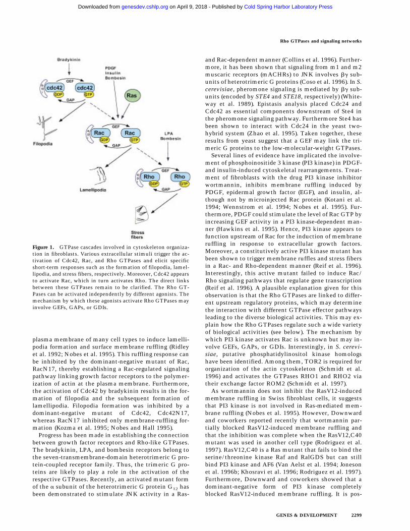
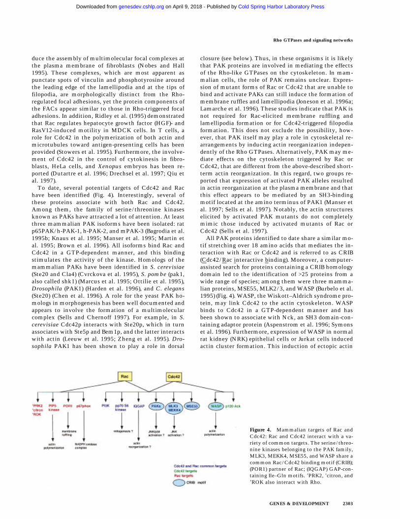
Until recently, little was known about the molecularmechanism(s) by which Rho affects the cytoskeleton.However, over the past year, numerous proteins thatbind Rho in a GTP-dependent manner have been identi-fied (Fig. 2). Characterization of some of these proteinshas provided major insights toward our understanding ofRho action at the molecular level. Of particular interesthas been the serine/threonine kinases, ROKa/Rho ki-nase and its close relative, p160ROCK (also known asROCKII or ROKb) (Leung et al. 1995, 1996; Ishizaki et al.1996; Matsui et al. 1996a; Nakagawa et al. 1996). ROKaand Rho kinase differ only at their amino termini, whereRho kinase is nine amino acids longer. The interaction ofRho with p160ROCK or Rho kinase resulted in a modestincrease of the kinase activity (Leung et al. 1995; Ishizakiet al. 1996; Matsui et al. 1996a). Clues to their functionscame from two lines of experiments. First, Leung et al.(1995) found that expression of full-length ROKa and itsamino-terminal half promotes the formation of stress fi-bers and focal adhesions. Kinase activity, but not theRho-binding domain, was required for this response. Fur-thermore, expression of a kinase-dead mutant or the car-boxy-terminal half of the protein resulted in the disas-sembly of stress fibers and focal adhesions (Leung et al.1996). These results indicate a role for ROKa in the for-mation of stress fibers and focal adhesions. Similar ob-servations were obtained for Rho kinase in other celltypes (Amano et al. 1997). Interestingly, Ishizaki et al.(1997) demonstrated that although a kinase-negative,Rho binding-defective mutant blocked Rho-induced for-mation of stress fibers and focal adhesions in HeLa cells,
Van Aelst and D’Souza-Schorey
2300 GENES & DEVELOPMENT
Cold Spring Harbor Laboratory Press on April 9, 2018 - Published by genesdev.cshlp.orgDownloaded from
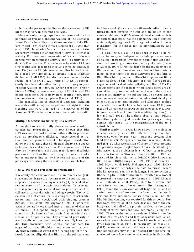
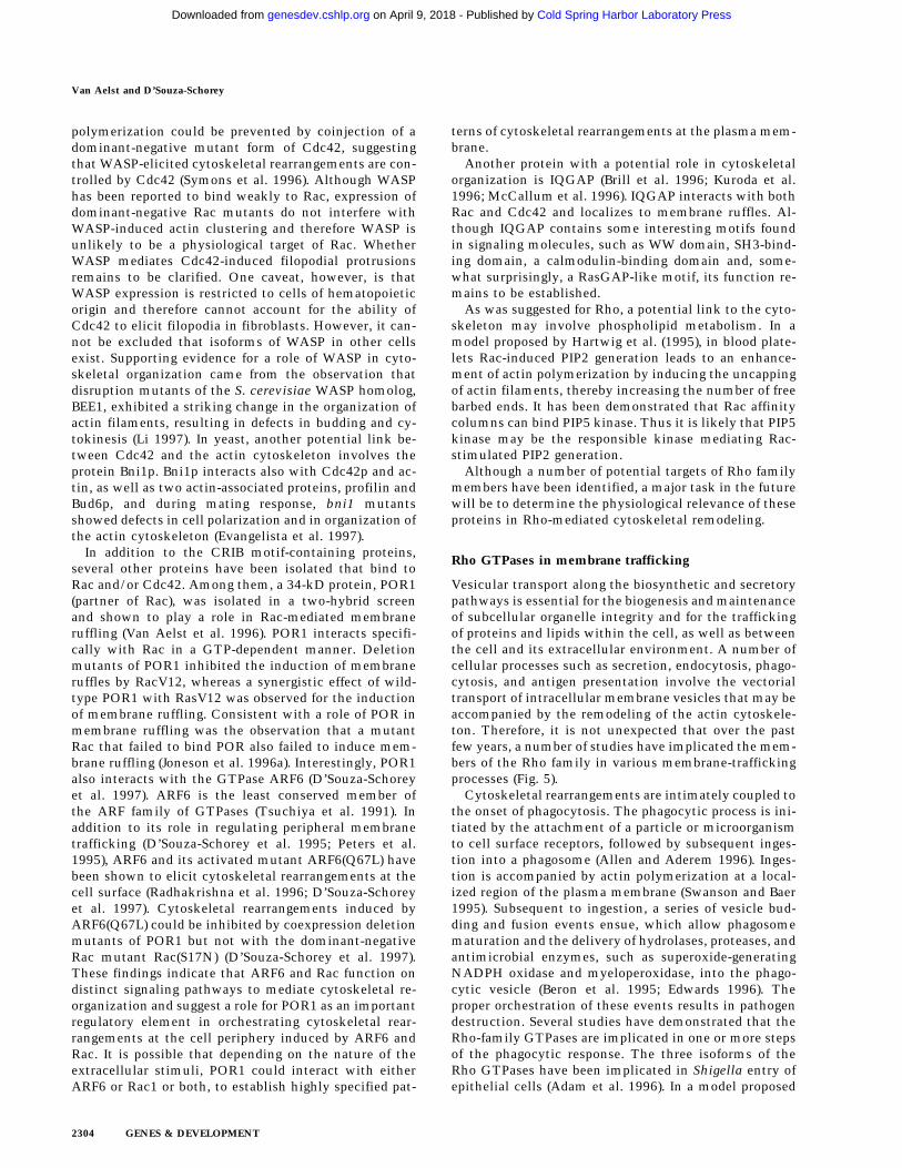
it was still capable of eliciting an enhancement of actinpolymerization. These results suggest that the pathwaysleading to actin polymerization and the formation ofstress fibers and focal adhesions are mediated by distincteffectors. A second contribution toward the elucidationof Rho-kinase function came from the findings that themyosin-binding subunit (MBS) of myosin light-chain(MLC) phosphatase was a substrate for Rho kinase invitro (Kimura et al. 1996; Matsui et al. 1996). It wassubsequently shown that phosphorylation of MBS led toa decrease in MLC phosphatase activity, resulting in anaccumulation of the phosphorylated form of MLC(Kimura et al. 1996). Phosphorylation of MLC has beenshown to induce a conformational change in myosin,thereby increasing its binding to actin filaments and sub-sequently the formation of stress fibers (Tan et al. 1992;Chrzanowska and Burridge 1996). More recently, evi-dence has been provided that Rho kinase stoichiometri-cally phosphorylates MLC at the same site that is phos-phorylated by MLC kinase (Amano et al. 1996a). A mo-lecular model proposed for Rho-induced stress fiberformation is depicted in Figure 3. Notably, it was foundthat overexpression of constitutively activated Rho inNIH-3T3 cells resulted in an increase in MLC phos-phorylation and stress fiber formation (Kimura et al.1996). Also, Rho-stimulated contraction of fibroblastscould be blocked by the MLC kinase inhibitor KT5926,which resulted in a decrease of MLC phosphorylationand the loss of stress fibers and focal adhesions (Chrza-nowska and Burridge 1996). It is likely that a cascade ofevents, as shown in Figure 3, may partly account for themechanism by which Rho regulates cytokinesis, motil-ity, or smooth muscle contraction.
As described above, Rho elicits the formation of focaladhesions. However, it is unknown whether the stressfiber and focal adhesion formation are separate or inter-dependent events. In a model proposed by Burridge andChrzanowski 1996, Rho-mediated stress fiber formationgenerates tension, thereby inducing aggregation of theintegrins on the ventral surface of the cells, which inturn stimulates the formation of focal adhesions and ty-rosine phosphorylation of focal adhesion proteins. A sec-
ond line of investigation, using pharmacological inhibi-tors, demonstrated that the formations of focal adhesion
Figure 3. A potential mechanism for Rho-induced stress fiberformation. Extracellular factors, such as LPA, trigger the acti-vation of Rho, which then binds to and activates Rho kinase.Rho GTP also interacts with MBS; however, the relevance of theinteraction between Rho and MBS remains unclear. ActivatedRho kinase phosphorylates the MBS of the myosin light chain(MLC) phosphatase, leading to the inactivation of the phospha-tase and subsequently an accumulation of the phosphorylatedform of MLC. Direct phosphorylation of MLC by Rho kinasehas also been reported. This increase in the phosphorylatedstate of MLC enhances binding of myosin–actin filaments and,subsequently, stress fiber formation.
Figure 2. Mammalian targets of RhoA. Thekinases PKN and PRK2, and the nonkinasesRhothekin and Rhophilin (REM-1 proteins)contain a homologous Rho-binding motif,whereas ROKh (Rho kinase/ROKa andp160ROCK/ROKb/ROCKII) and citron sharea distinct Rho-binding motif (REM-2). MBS(myosin-binding subunit of myosin lightchain phosphatase). (*) The PIP5 kinase inter-action may not be direct.
Rho GTPases and signaling networks
GENES & DEVELOPMENT 2301
Cold Spring Harbor Laboratory Press on April 9, 2018 - Published by genesdev.cshlp.orgDownloaded from
and stress fibers are separate events (Nobes and Hall1995). In accordance with the latter, Chihara et al. (1997)reported that microinjection of a constitutively activeRho kinase into fibroblasts induced the formation of fo-cal adhesions under conditions wherein actin stress fi-bers were disrupted.
The involvement of phosphoinositide kinases in Rhosignaling has been reported. Rho (and Rac) have beenshown to stimulate the synthesis of phosphatidyl inosi-tol bisphosphate (PIP2) (Chong et al. 1994; Hartwig et al.1995). The exact mechanism by which Rho induces thisevent is not yet known because opposing results havebeen reported as to whether phosphatidyl 4-phosphate-5kinase (PIP5 kinase), which catalyzes the phosphoryla-tion of PI4P to PIP2, interacts directly with Rho (Toliaset al. 1995; Ren et al. 1996). The observation that PIP2binds to and is thought to regulate the function of manyactin-associated proteins, led to the hypothesis that Rho-stimulated PIP2 synthesis may induce actin rearrange-ments. In light of this, Gilmore and Burridge (1996) re-ported that the association of PIP2 with vinculin inducesa conformation change in vinculin, allowing it to inter-act with talin, which binds actin. Furthermore, theyshowed that injection of anti-PIP2 antibodies into fibro-blasts inhibited LPA/Rho-induced stress fiber and focaladhesion formation, suggesting a role for PIP2 in focaladhesion and stress fiber assembly.
Recently, a potential target of the S. cerevisiae Rho1protein, designated Bni1p, was isolated and shown toplay a role in cytoskeleton reorganization (Kohno et al.1996). RHO1 is a homolog of the mammalian RhoA geneand is essential for the budding process (Yamochi et al.1994). Bni1p belongs to a family of formin-related pro-teins, which includes Drosophila diaphanous and cap-puchino, FigA in Aspergillus, and fus1 in S. pombe (Cas-trillon and Wasserman 1994; Emmons et al. 1995; Mar-houl and Adams 1995). These proteins have been shownto be involved in cytokinesis, cell polarity, and cell mor-phology and share two formin homology domains, FH1and FH2. In an elegant study, Imamura et al. (1997) dem-onstrated that Bni1p and its more recent isolated relatedmember, Bnr1p, are mediators of the Rho GTPase effectson actin cytoskeleton reorganization. These investiga-tors observed that these proteins interacted with profilinvia the FH domains. Although originally thought tofunction in the sequestration of unpolymerized actin inthe cytoplasm, profilin recently has been shown to havea promoting effect on actin polymerization (Nishida1985; Goldschmidt et al. 1991; Cao et al. 1992). Takentogether with the above results, Bni1p and Bnr1p arelikely to mediate Rho GTPase effects on the cytoskele-ton through a mechanism involving profilin. More re-cently, a mammalian homolog of Bni1p, p140mDia, wasshown to selectively interact with mammalian Rho in aGTP-dependent manner and also with profilin (Watan-abe et al. 1997). Moreover, p140mDia colocalized withprofilin in polarized and nonpolarized fibroblasts and inphagocytic cells, and overexpression of p140mDia in Coscells enhanced actin filament assembly. Based on thesefindings, a model was proposed in which activated Rho
recruits p140mDia/profilin to a specific site beneath theplasma membrane, which results in a locally increasedconcentration of profilin. This, in turn, may lead to actinpolymerization (Narumiya et al. 1997; Watanabe et al.1997). Such a model points toward a role for p140mDiain mediating the effect of Rho on cytokinesis in mam-malian cells.
In addition to Rho kinase, MBS, and p140mDia, fiveother mammalian proteins have been identified as po-tential Rho targets (Fig. 2). PKN and its homolog PRK2encode leucine zipper-bearing proteins containing a ser-ine/threonine kinase-domain that is highly related tothat of protein kinase C (PKC) (Amano et al. 1996b; Quil-liam et al. 1996; Watanabe et al. 1996). Although it wasreported previously that PKN and PRK2 interact specifi-cally with GTP-bound Rho, it was also reported that theinteraction of PRK2 with Rho is nucleotide-independentand that PRK2 is able to interact with Rac in a GTP-dependent manner (Vincent and Settleman 1997). Theseinvestigators also showed that a kinase-defective form ofPRK2 disrupts actin stress fibers, suggesting a role forPRK2 in actin cytoskeletal organization. A S. cerevisiaePKC homolog, Pkc1, has been isolated and shown to in-teract directly with Rho1 (Nonaka et al. 1995). Further-more, Pkc1 regulates cell wall integrity through the ac-tivation of the mitogen-activated protein kinase(MAPK). Another Rho1 target, glucan synthase (GS),contributes to cell wall remodeling, although in a dis-tinct pathway (Arellano et al. 1996; Drgonova et al. 1996;Qadota et al. 1996). Whereas Rophilin (Watanabe et al.1996) and Rhothekin (Reid et al. 1996) share homologywith PKN in their Rho binding domain, citron (Madauleet al. 1995) has homology with Rho kinase in this do-main. None of the three proteins contains an obviouscatalytic domain and their roles in Rho-mediated cyto-skeletal rearrangements remain to be established.
Rac, Cdc42, and the cytoskeleton
In fibroblasts, Rac has been shown to be a key controlelement in the reorganization of the actin cytoskeletoninduced by growth factors and RasV12 (Ridley et al.1992). Injection of RacV12 is sufficient to induce lamel-lipodia and membrane ruffles and subsequent stress fiberformation, whereas microinjection of RacN17 prior tothe addition of growth factors or together with RasV12abolishes these effects (Ridley et al. 1992). Furthermore,as described above, a role for yet another member of theRho subfamily, Cdc42, in actin remodeling has been es-tablished. Most of our prior understanding of the func-tion of Cdc42 came from studies in the yeast S. cerevi-siae. Much of this work has been reviewed recently byHerskowitz (1995) and hence will not be discussed here.Injection of mammalian Cdc42 into fibroblasts revealeda third distinct signaling pathway linking plasma mem-brane receptors to actin cytoskeletal organization. Ex-pression of Cdc42Hs triggered the formation of filopodialprotrusions at the cell periphery followed by the forma-tion of lamellipodia and membrane ruffling (Kozma et al.1995). Both Rac and Cdc42 have also been shown to in-
Van Aelst and D’Souza-Schorey
2302 GENES & DEVELOPMENT
Cold Spring Harbor Laboratory Press on April 9, 2018 - Published by genesdev.cshlp.orgDownloaded from
duce the assembly of multimolecular focal complexes atthe plasma membrane of fibroblasts (Nobes and Hall1995). These complexes, which are most apparent aspunctate spots of vinculin and phosphotyrosine aroundthe leading edge of the lamellipodia and at the tips offilopodia, are morphologically distinct from the Rho-regulated focal adhesions, yet the protein components ofthe FACs appear similar to those in Rho-triggered focaladhesions. In addition, Ridley et al. (1995) demonstratedthat Rac regulates hepatocyte growth factor (HGF)- andRasV12-induced motility in MDCK cells. In T cells, arole for Cdc42 in the polymerization of both actin andmicrotubules toward antigen-presenting cells has beenprovided (Stowers et al. 1995). Furthermore, the involve-ment of Cdc42 in the control of cytokinesis in fibro-blasts, HeLa cells, and Xenopus embryos has been re-ported (Dutartre et al. 1996; Drechsel et al. 1997; Qiu etal. 1997).
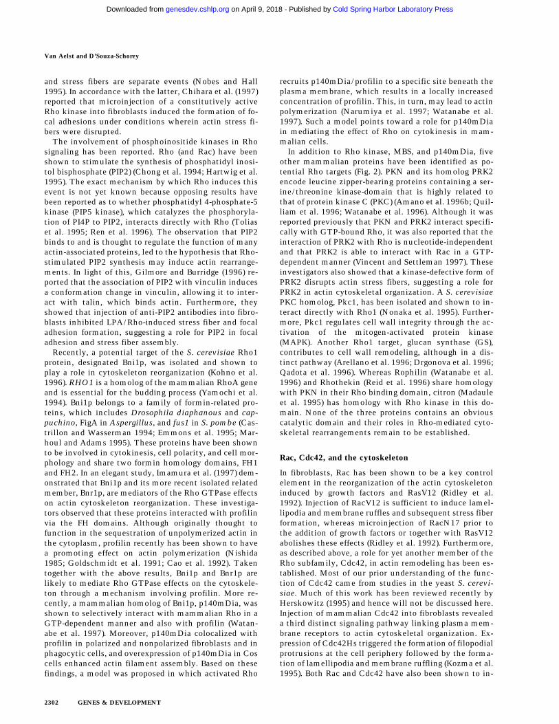
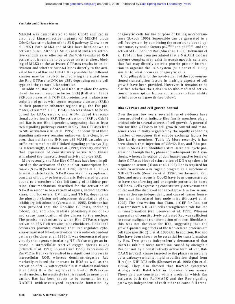
To date, several potential targets of Cdc42 and Rachave been identified (Fig. 4). Interestingly, several ofthese proteins associate with both Rac and Cdc42.Among them, the family of serine/threonine kinasesknown as PAKs have attracted a lot of attention. At leastthree mammalian PAK isoforms have been isolated: ratp65PAK/h-PAK-1, h-PAK-2, and mPAK-3 (Bagrodia et al.1995b; Knaus et al. 1995; Manser et al. 1995; Martin etal. 1995; Brown et al. 1996). All isoforms bind Rac andCdc42 in a GTP-dependent manner, and this bindingstimulates the activity of the kinase. Homologs of themammalian PAKs have been identified in S. cerevisiae(Ste20 and Cla4) (Cvrckova et al. 1995), S. pombe (pak1,also called shk1) (Marcus et al. 1995; Ottilie et al. 1995),Drosophila (PAK1) (Harden et al. 1996), and C. elegans(Ste20) (Chen et al. 1996). A role for the yeast PAK ho-mologs in morphogenesis has been well documented andappears to involve the formation of a multimolecularcomplex (Sells and Chernoff 1997). For example, in S.cerevisiae Cdc42p interacts with Ste20p, which in turnassociates with Ste5p and Bem1p, and the latter interactswith actin (Leeuw et al. 1995; Zheng et al. 1995). Dro-sophila PAK1 has been shown to play a role in dorsal
closure (see below). Thus, in these organisms it is likelythat PAK proteins are involved in mediating the effectsof the Rho-like GTPases on the cytoskeleton. In mam-malian cells, the role of PAK remains unclear. Expres-sion of mutant forms of Rac or Cdc42 that are unable tobind and activate PAKs can still induce the formation ofmembrane ruffles and lamellipodia (Joneson et al. 1996a;Lamarche et al. 1996). These studies indicate that PAK isnot required for Rac-elicited membrane ruffling andlamellipodia formation or for Cdc42-triggered filopodiaformation. This does not exclude the possibility, how-ever, that PAK itself may play a role in cytoskeletal re-arrangements by inducing actin reorganization indepen-dently of the Rho GTPases. Alternatively, PAK may me-diate effects on the cytoskeleton triggered by Rac orCdc42, that are different from the above-described short-term actin reorganization. In this regard, two groups re-ported that expression of activated PAK alleles resultedin actin reorganization at the plasma membrane and thatthis effect appears to be mediated by an SH3-bindingmotif located at the amino terminus of PAK1 (Manser etal. 1997; Sells et al. 1997). Notably, the actin structureselicited by activated PAK mutants do not completelymimic those induced by activated mutants of Rac orCdc42 (Sells et al. 1997).
All PAK proteins identified to date share a similar mo-tif stretching over 18 amino acids that mediates the in-teraction with Rac or Cdc42 and is referred to as CRIB(Cdc42/Rac interactive binding). Moreover, a computer-assisted search for proteins containing a CRIB homologydomain led to the identification of >25 proteins from awide range of species; among them were three mamma-lian proteins, MSE55, MLK2/3, and WASP (Burbelo et al.1995) (Fig. 4). WASP, the Wiskott–Aldrich syndrome pro-tein, may link Cdc42 to the actin cytoskeleton. WASPbinds to Cdc42 in a GTP-dependent manner and hasbeen shown to associate with Nck, an SH3 domain-con-taining adaptor protein (Aspenstrom et al. 1996; Symonset al. 1996). Furthermore, expression of WASP in normalrat kidney (NRK) epithelial cells or Jurkat cells inducedactin cluster formation. This induction of ectopic actin
Figure 4. Mammalian targets of Rac andCdc42: Rac and Cdc42 interact with a va-riety of common targets. The serine/threo-nine kinases belonging to the PAK family,MLK3, MEKK4, MSE55, and WASP share acommon Rac/Cdc42 binding motif (CRIB);(POR1) partner of Rac; (IQGAP) GAP-con-taining Ile–Gln motifs. 8PRK2, 8citron, and8ROK also interact with Rho.
Rho GTPases and signaling networks
GENES & DEVELOPMENT 2303
Cold Spring Harbor Laboratory Press on April 9, 2018 - Published by genesdev.cshlp.orgDownloaded from
polymerization could be prevented by coinjection of adominant-negative mutant form of Cdc42, suggestingthat WASP-elicited cytoskeletal rearrangements are con-trolled by Cdc42 (Symons et al. 1996). Although WASPhas been reported to bind weakly to Rac, expression ofdominant-negative Rac mutants do not interfere withWASP-induced actin clustering and therefore WASP isunlikely to be a physiological target of Rac. WhetherWASP mediates Cdc42-induced filopodial protrusionsremains to be clarified. One caveat, however, is thatWASP expression is restricted to cells of hematopoieticorigin and therefore cannot account for the ability ofCdc42 to elicit filopodia in fibroblasts. However, it can-not be excluded that isoforms of WASP in other cellsexist. Supporting evidence for a role of WASP in cyto-skeletal organization came from the observation thatdisruption mutants of the S. cerevisiae WASP homolog,BEE1, exhibited a striking change in the organization ofactin filaments, resulting in defects in budding and cy-tokinesis (Li 1997). In yeast, another potential link be-tween Cdc42 and the actin cytoskeleton involves theprotein Bni1p. Bni1p interacts also with Cdc42p and ac-tin, as well as two actin-associated proteins, profilin andBud6p, and during mating response, bni1 mutantsshowed defects in cell polarization and in organization ofthe actin cytoskeleton (Evangelista et al. 1997).
In addition to the CRIB motif-containing proteins,several other proteins have been isolated that bind toRac and/or Cdc42. Among them, a 34-kD protein, POR1(partner of Rac), was isolated in a two-hybrid screenand shown to play a role in Rac-mediated membraneruffling (Van Aelst et al. 1996). POR1 interacts specifi-cally with Rac in a GTP-dependent manner. Deletionmutants of POR1 inhibited the induction of membraneruffles by RacV12, whereas a synergistic effect of wild-type POR1 with RasV12 was observed for the inductionof membrane ruffling. Consistent with a role of POR inmembrane ruffling was the observation that a mutantRac that failed to bind POR also failed to induce mem-brane ruffling (Joneson et al. 1996a). Interestingly, POR1also interacts with the GTPase ARF6 (D’Souza-Schoreyet al. 1997). ARF6 is the least conserved member ofthe ARF family of GTPases (Tsuchiya et al. 1991). Inaddition to its role in regulating peripheral membranetrafficking (D’Souza-Schorey et al. 1995; Peters et al.1995), ARF6 and its activated mutant ARF6(Q67L) havebeen shown to elicit cytoskeletal rearrangements at thecell surface (Radhakrishna et al. 1996; D’Souza-Schoreyet al. 1997). Cytoskeletal rearrangements induced byARF6(Q67L) could be inhibited by coexpression deletionmutants of POR1 but not with the dominant-negativeRac mutant Rac(S17N) (D’Souza-Schorey et al. 1997).These findings indicate that ARF6 and Rac function ondistinct signaling pathways to mediate cytoskeletal re-organization and suggest a role for POR1 as an importantregulatory element in orchestrating cytoskeletal rear-rangements at the cell periphery induced by ARF6 andRac. It is possible that depending on the nature of theextracellular stimuli, POR1 could interact with eitherARF6 or Rac1 or both, to establish highly specified pat-
terns of cytoskeletal rearrangements at the plasma mem-brane.
Another protein with a potential role in cytoskeletalorganization is IQGAP (Brill et al. 1996; Kuroda et al.1996; McCallum et al. 1996). IQGAP interacts with bothRac and Cdc42 and localizes to membrane ruffles. Al-though IQGAP contains some interesting motifs foundin signaling molecules, such as WW domain, SH3-bind-ing domain, a calmodulin-binding domain and, some-what surprisingly, a RasGAP-like motif, its function re-mains to be established.
As was suggested for Rho, a potential link to the cyto-skeleton may involve phospholipid metabolism. In amodel proposed by Hartwig et al. (1995), in blood plate-lets Rac-induced PIP2 generation leads to an enhance-ment of actin polymerization by inducing the uncappingof actin filaments, thereby increasing the number of freebarbed ends. It has been demonstrated that Rac affinitycolumns can bind PIP5 kinase. Thus it is likely that PIP5kinase may be the responsible kinase mediating Rac-stimulated PIP2 generation.
Although a number of potential targets of Rho familymembers have been identified, a major task in the futurewill be to determine the physiological relevance of theseproteins in Rho-mediated cytoskeletal remodeling.
Rho GTPases in membrane trafficking
Vesicular transport along the biosynthetic and secretorypathways is essential for the biogenesis and maintenanceof subcellular organelle integrity and for the traffickingof proteins and lipids within the cell, as well as betweenthe cell and its extracellular environment. A number ofcellular processes such as secretion, endocytosis, phago-cytosis, and antigen presentation involve the vectorialtransport of intracellular membrane vesicles that may beaccompanied by the remodeling of the actin cytoskele-ton. Therefore, it is not unexpected that over the pastfew years, a number of studies have implicated the mem-bers of the Rho family in various membrane-traffickingprocesses (Fig. 5).
Cytoskeletal rearrangements are intimately coupled tothe onset of phagocytosis. The phagocytic process is ini-tiated by the attachment of a particle or microorganismto cell surface receptors, followed by subsequent inges-tion into a phagosome (Allen and Aderem 1996). Inges-tion is accompanied by actin polymerization at a local-ized region of the plasma membrane (Swanson and Baer1995). Subsequent to ingestion, a series of vesicle bud-ding and fusion events ensue, which allow phagosomematuration and the delivery of hydrolases, proteases, andantimicrobial enzymes, such as superoxide-generatingNADPH oxidase and myeloperoxidase, into the phago-cytic vesicle (Beron et al. 1995; Edwards 1996). Theproper orchestration of these events results in pathogendestruction. Several studies have demonstrated that theRho-family GTPases are implicated in one or more stepsof the phagocytic response. The three isoforms of theRho GTPases have been implicated in Shigella entry ofepithelial cells (Adam et al. 1996). In a model proposed
Van Aelst and D’Souza-Schorey
2304 GENES & DEVELOPMENT
Cold Spring Harbor Laboratory Press on April 9, 2018 - Published by genesdev.cshlp.orgDownloaded from

by Adam and coworkers, Shigella invasion begins withactin nucleation and RhoA-induced actin polymeriza-tion. This is followed by continued actin polymerizationaround membrane-bound protrusions that fold over thebacterium, coalesce, and engulf it. The observations of acomplete inhibition of Shigella-induced membrane fold-ing by C3 transferase suggests that actin polymerizationis essential for the generation of the surface extensions.Further studies will be required to examine the role ofRho in the induction of membrane alterations that ac-company surface remodeling during Shigella entry andthe interaction of the membrane and the cytoskeleton inthis process. Although Rac has been shown to induceactin polymerization at the cell periphery, Rac-inducedlamellipodia formation and membrane ruffling have notbeen directly linked to the internalization step of phago-cytosis. The observation that Salmonella invasion of ep-ithelial cells leads to the activation of EGF receptors(Galan et al. 1992), a known mechanism for induction ofRac-dependent ruffling (Ridley et al. 1992), suggests arole for Rac in this process. However, there have beenreports that Salmonella phagocytosis occurs indepen-dent of Rho and Rac (Jones et al. 1993). Interestingly,Cdc42 has been shown to play a direct role in Salmonellainternalization (Chen et al. 1996). Although cells ex-pressing an activated mutant of Cdc42 were capable ofphagocytosing an invasion-defective Salmonella mu-tant, the dominant-negative Cdc42 prevented bacterialinternalization. On the other hand, Rac in its GTP-bound form, in addition to two other cytosolic proteins,p47phox and p67phox has been shown to be required forthe activation of NADPH oxidase of phagocytic cells(Abo et al. 1992; Diekmann et al. 1994; Bokoch 1995).Rac antisense oligonucleotides and expression ofRacN17 inhibit superoxide generation (Dorseuil et al.1995; Gabig et al. 1995). Bcr, a Rac GAP, has been shownto down-regulate Rac-induced activation of NADPH oxi-dase in neutrophils, thereby preventing excess tissue
damage (Voncken et al. 1995). Whether these distinctfunctions of Rac, such as NADPH oxidase activation andactin reorganization, are coordinately or sequentiallyregulated awaits further investigation.
Rac and Rho have also been implicated in the regula-tion of endocytosis. In mammalian cells, expression ofconstitutively activated mutants of Rac or Rho de-creased the efficiency of receptor-mediated endocytosisof the transferrin receptor (Lamaze et al. 1996). In a cell-free assay these mutant proteins have been shown toinhibit the formation of clathrin-coated vesicles (Lamazeet al. 1996). It has been proposed that the activation ofPLD and/or PIP5 kinase might control coated pit assem-bly, consistent with the contention that alterations inmembrane lipids might trigger the recruitment of com-ponents required to initiate vesicle budding. In contrast,microinjection of either wild-type or activated Rho intoXenopus oocytes enhanced constitutive endopinocytosis(Schmalzing et al. 1995). Furthermore, injection of C3transferase or ADP-ribosylated RhoA into oocytes pre-vented the uptake of surface sodium pumps and causedthe formation of large membranous folds at the cell sur-face. Interestingly, the dense cortical layer of filamen-tous actin beneath the plasma membrane in oocytes wasinsensitive to C3 treatment. These data suggest a directinvolvement of Rho in endocytic membrane traffickingin Xenopus, independent of its effect on the actin cyto-skeleton. RhoB is localized to early endosomes in rat-2cells, suggesting a potential role for this GTPase in en-docytic trafficking in mammalian systems (Adamson etal. 1992). RhoD, another member of the Rho family, hasbeen identified and also localizes on early endosomes atthe cell surface (Murphy et al. 1996). Upon overexpres-sion, RhoD induced the formation of long thin F-actin-containing membrane processes and a disassembly ofstress fibers and focal adhesions along the cell periphery.This cytoskeletal remodeling was accompanied by an in-crease in endosome fission and a scattering of vesicles
Figure 5. Schematic representation of intra-cellular membrane trafficking in eukaryoticcells and steps where a role for Rho familyGTPases have been implicated.
Rho GTPases and signaling networks
GENES & DEVELOPMENT 2305
Cold Spring Harbor Laboratory Press on April 9, 2018 - Published by genesdev.cshlp.orgDownloaded from

throughout the cell. Time-lapse video microscopy re-vealed that cells expressing the RhoD mutant exhibiteddecreased organelle motility that impeded movementand fusion of endosomes. Thus, the ability of RhoD tocycle between its GTP- and GDP-bound forms appears tobe critical for the regulation of endosomal motility andfor maintaining the equilibrium between endosomal fu-sion and fission (Murphy et al. 1996).
The role for the Rac GTPase in pinocytosis is stillunclear. Membrane ruffling has long been thought to belinked to an increase in pinocytosis. In Swiss 3T3 cells,expression of the activated mutants of Rac stimulatedpinocytosis (Ridley et al. 1992). However, Li et al. (1997)demonstrated more recently that the expression of theactivated Rac mutant had no effect on pinocytosis inbaby hamster kidney fibroblasts. The latter study alsodemonstrated that H-Ras-induced enhancement of pino-cytosis could not be blocked by a dominant-negative mu-tant of Rac but was effectively blocked by the dominantinterfering mutant of Rab5. The latter is a member of theRab subfamily and has been shown to promote fusionamong early endosomes (Bucci et al. 1992; Barbieri et al.1994). Whether these discrepancies reflect differences inpinocytic routes among various cell types remains to beexplored.
In addition to a role in endocytic trafficking, Rac andRho have also been implicated in the regulation of se-cretory vesicle transport. In mast cells, recombinant Racand Rho proteins stimulated the exocytosis of secretorygranules, whereas C3 transferase and the dominant-negative mutants of Rac or Rho inhibited secretion in-duced by GTPgS (Norman et al. 1996). Although the se-cretory granule exocytosis is accompanied by a redistri-bution and polymerization of F-actin, the latter isselectively inhibited by cytochalasin D, whereas mem-brane secretion remains unaffected. Furthermore, consti-tutively activated mutants of Rac and Rho enhance se-cretion in the presence of cytochalasin D. Therefore, thesignaling pathways controlling outward membrane flowand cytoskeletal rearrangements in mast cells are diver-gent and act in parallel rather than in concert. Consis-tent with the above findings, it was independently dem-onstrated that Rho GDI inhibited GTPgS-stimulatedexocytosis in mast cells (Mariot et al. 1996). In S. cerevi-siae, the RhoA homolog Rho1 has been localized to theGolgi and post-Golgi vesicles, supporting the contentionthat Rho may be involved in the secretory response inyeast (McCaffery et al. 1991). On the other hand, yeastCdc42 has been shown to be localized at the plasmamembrane in the vicinity of secretory vesicles that arefound at the site of bud emergence (Ziman et al. 1993). Inmammalian cells however, wild-type Cdc42 has been lo-calized to the Golgi apparatus, and it has been proposedthat activation of Cdc42 and the formation of filopodia atthe cell periphery may be coupled to vesicular transportfrom the trans-Golgi network to the plasma membrane(Erickson et al. 1996). Interestingly in this regard, Cdc42has been shown to regulate polarization of T cells towardantigen-presenting cells, which may indeed be coupledto targeted lymphokine secretion toward the appropriate
antigen-presenting cell (Stowers et al. 1995). Furtherstudies will be required to address the role of Cdc42 onlymphokine secretion in T cells.
Although it is now clear that a number of vesiculartransport processes require coordinated interactions be-tween the membrane and the cytoskeleton, the bio-chemical mechanisms that integrate these cellular pro-cesses are largely unknown. It is possible that the effectsof Rho GTPases on phospholipid metabolism may pro-vide a point of intersection between the coordinated con-trol of membrane flow and cytoskeletal organization.There is mounting evidence that polyphosphoinositidesare implicated in the regulation of vesicular traffic in avariety of systems. PIP2 has been shown to have a role inCa2+-regulated exocytosis in adrenal chromaffin cells(Eberhard et al. 1990). In addition, enzymes involved inPIP2 biosynthesis are required for the priming of secre-tory granules during exocytosis in PC12 cells (Hay andMartin 1993). Mutations in the SEC14 gene of S. cerevi-siae, which codes for PITP (phosphatidyl inositol trans-fer protein), results in defective post-Golgi secretory traf-fic (Bankaitis et al. 1990; Novick et al. 1980). Moreover,wortmannin has been shown to affect fluid-phase endo-cytosis and receptor recycling (Clague et al. 1995; Li etal. 1995; Shepherd et al. 1995; Martys et al. 1996). Also,mutations in the PDGF receptor that inhibit its associa-tion with PI3 kinase result in a defect in postendosomalsorting of the receptor (Kapeller et al. 1993). As describedabove, polyphosphoinositides such as PIP2 and the ki-nases that regulate their turnover have been shown toserve as a link between the Rho GTPases and the actincytoskeleton. Thus, it is tempting to speculate that localchanges in membrane phospholipids induce changes inthe actin-based cytoskeleton and regulate vesicle-targetinteractions. Further studies are required to determine ifand how the Rho/Rac-induced effects on phospholipidmetabolism bridge vesicular transport with the actincytoskeleton.
Rho GTPases and transcriptional activation
Accumulating data points to the involvement of Rhofamily members in regulating nuclear signaling.Whereas Ras has been shown to control the activation ofthe p42/44 MAPK cascade, several groups demonstratedthat in certain cell types, Rac and Cdc42 (but not Rho)regulate the activation of JNK and the reactivating ki-nase p38RK (Seger and Krebs 1995). Expression of con-stitutively active mutants of Rac and Cdc42 in HeLa,NIH-3T3, and Cos cells resulted in a stimulation of JNKand p38 activity (Coso et al. 1995; Minden et al. 1995).Furthermore, the same effects were obtained with onco-genic GEFs for these Rho proteins (Minden et al. 1995).However, Teramoto et al. (1996b) reported that in hu-man kidney 293 T cells, Cdc42 and the Rho protein, butnot Rac, can induce activation of JNK. In contrast to thep44/42 MAPKs, the JNKs and p38 are poorly activated bymitogens but strongly activated by inflammatory cyto-kines, tumor necrosis factor a (TNFa), interleukin-1b(IL-1b), and a diverse array of cellular stresses such as
Van Aelst and D’Souza-Schorey
2306 GENES & DEVELOPMENT
Cold Spring Harbor Laboratory Press on April 9, 2018 - Published by genesdev.cshlp.orgDownloaded from

heat shock, UV, and ionizing radiation (Kyriakis andAvruch 1996). Until now, only a few of these stimuli(e.g., IL-1b) have been demonstrated to exert their effectsthrough Rho GTPases (Bagrodia et al. 1995a; Zhang et al.1995; Whitmarsh et al. 1997). Upon activation, JNKs andp38 translocate to the nucleus where they phosphorylatetranscription factors. Substrates for JNKs include theamino terminus of c-Jun, ATF2, and Elk (Pulverer et al.1991; Derijard et al. 1994; Gille et al. 1995; Gupta et al.1995). Furthermore, Rac has been shown to stimulatethe transcriptional activity of PEA3, a member of the Etsfamily, in a JNK-dependent fashion (O’Hagan et al.1996). Activated p38 phosphorylates ATF2, Elk, Max(Zervos et al. 1995), and the cAMP response element-binding protein–homologous protein/growth arrestDNA damage 153 (CHOP/GADD153) (Wang and Ron1996). In addition to the above transcription factors,MAPKAP-K2 and more recently 3pK have been identi-fied as targets of p38 (Rouse et al. 1994; Ludwig et al.1996). The latter is also activated by JNK and MAPK. Itis important to note that the substrate specificities forthese kinases appear to be cell-type dependent.
The identity of the molecules that link the Rac andCdc42 GTPases to JNK and p38 is still not completelyunderstood. Direct activators of JNK and p38 are eitherthe dual specificity kinase SEK1 (also called MEKK4 orJNKK), which activates both JNK and p38 when overex-pressed, or MKK3 and MKK6 which specifically activatep38 (Sanchez et al. 1994; Derijard et al. 1995; Han et al.1996; Jiang et al. 1996; Raingeaud et al. 1996). Althoughoriginally identified as MEK1/2 kinases (Lange-Carter etal. 1993), MEKK1 has since been shown not to triggeractivation of MEK and MAPK, but to stimulate SEK1 (Xuet al. 1995). Because MEKK1 activates SEK1 and JNK butnot p38 in vivo, it is not clear whether SEK1 functions as
a physiological p38 activator. The family of serine/threo-nine kinases known as PAKs were proposed to act as thefarthest upstream kinases connecting Rho GTPases toJNK and p38 as they bind Rac and Cdc42 in vitro in aGTP-dependent manner and become activated uponbinding to the activated forms of these GTPases (Bagro-dia et al. 1995b; Knaus et al. 1995; Manser et al. 1995;Martin et al. 1995). Furthermore, certain constitutively-activated forms of PAK can stimulate the activity of JNKand p38 (Bagrodia et al. 1995a; Knaus et al. 1995; Zhanget al. 1995b; Brown et al. 1996; Frost et al. 1996). Byanalogy with the Ste20 kinase cascade in S. cerevisiae(Fig. 6), it was suggested that PAK regulates the activityof MEKK. However, direct phosphorylation of any of theidentified MEKKs by PAK has not been demonstratedthus far. Notably, the recent work of Peter et al. (1996)strongly suggests that Cdc42/Ste20 interaction is not re-quired for the activation of the MAPK pathway triggeredby mating pheromones. Also, some groups did not ob-serve an increase in JNK activity upon coexpression ofPAK1 with activated forms of Cdc42 or Rac (Teramoto etal. 1996a). Furthermore, an effector mutant of Rac,which fails to bind to PAK, remains a potent JNK acti-vator (Westwick et al. 1997). Therefore, it is possible thatother kinases, in addition to (or instead of) PAK, partici-pate in signaling from the Rho-like GTPases to JNK. Insupport of this possibility, MLK3 (also called SPRK) andMEKK4 are regulated by Cdc42 and Rac and selectivelyactivate the JNK pathway (Gallo et al. 1994; Teramoto etal. 1996a; Tibbles et al. 1996; Gerwins et al. 1997). Dif-ferent groups reported the ability of Cdc42/Rac to bindto MLK3 both in vitro and in vivo and that coexpressionof activated Cdc42/Rac mutant forms elevated the en-zymatic activity of MLK3 in Cos-7 cells (Gallo et al.1994; Teramoto et al. 1996b; Gerwins et al. 1997).
Figure 6. Signal transduction through members of the Rho subfamily leading to transcriptional activation in yeast and mammals. (A)Pheromone signaling pathway in S. cerevisiae; (B) the JNK and p38 signaling cascades in mammals; (C) the SRF signaling pathway; (D)the pathway leading to NF-kB activation in mammalian cells.
Rho GTPases and signaling networks
GENES & DEVELOPMENT 2307
Cold Spring Harbor Laboratory Press on April 9, 2018 - Published by genesdev.cshlp.orgDownloaded from
MEKK4 was demonstrated to bind Cdc42 and Rac invivo, and kinase-inactive mutants of MEKK4 blockCdc42/Rac stimulation of the JNK pathway (Gerwins etal. 1997). Both MLK3 and MKK4 have been shown toactivate SEK1. Although MLK3 and MEKK4 are attrac-tive candidates as effectors of Rac/Cdc42-induced JNKactivation, it remains to be proven whether direct bind-ing of MLK3 to the activated GTPases results in its ac-tivation and whether MEKK4 binds directly to the acti-vated forms of Rac and Cdc42. It is possible that differentkinases may be involved in mediating the signal fromthe Rho GTPase to JNK (or p38), depending on the celltype and the extracellular stimulus.
In addition, Rac, Cdc42, and Rho stimulate the activ-ity of the serum response factor (SRF) (Hill et al. 1995).SRF complexes with TCF/Elk proteins to stimulate tran-scription of genes with serum response elements (SREs)in their promoter enhancer regions (e.g., the Fos pro-moter) (Treisman 1990, 1994). Rho was shown to be re-quired for LPA-, serum-, and AIF4-induced transcrip-tional activation by SRF. The activation of SRF by Cdc42and Rac is not Rho-dependent, suggesting that at leasttwo signaling pathways controlled by Rho GTPases leadto SRF activation (Hill et al. 1995). The identity of thesesignaling pathways remains unknown. It is clear, how-ever, that neither the JNK nor p38 MAPK cascades aresufficient to mediate SRF-linked signaling pathways (Fig.6). Interestingly, Chihara et al. (1997) recently observedthat expression of constitutively active Rho kinasestimulated the transcriptional activity of c-fos SRE.
More recently, the Rho-like GTPases have been impli-cated in the activation of the nuclear transcription fac-tor-kB (NF-kB) (Sulciner et al. 1996; Perona et al. 1997).In unstimulated cells, NF-kB consists of a cytoplasmiccomplex of homo- or heterodimeric Rel-related proteinsbound to a member of the IkB family of inhibitor pro-teins. One mechanism described for the activation ofNF-kB in response to a variety of agents, including cyto-kines, phorbol esters, UV light, and TNFa, depends onthe phosphorylation and subsequent degradation of theinhibitory IkB subunits (Verma et al. 1995). Evidence hasbeen provided that the Rho-like GTPases, includingCdc42, Rac, and Rho, mediate phosphorylation of IkBand cause translocation of the dimers to the nucleus.The precise mechanism by which Rho GTPases triggeractivation of NF-kB remains to be elucidated. Finkel andcoworkers provided evidence that Rac regulates cyto-kine-stimulated NF-kB activation via a redox-dependentpathway (Sulciner et al. 1996). It was demonstrated pre-viously that agents stimulating NF-kB also trigger an in-crease in intracellular reactive oxygen species (ROS)(Schreck et al. 1991; Lo and Cruz 1995). Expression ofRacV12 in HeLa cells resulted in a significant increase inintracellular ROS, whereas dominant-negative Racmarkedly reduced the increase in ROS as well as theactivation of NF-kB after cytokinin stimulation (Sulcineret al. 1996). How Rac regulates the level of ROS is cur-rently unclear. Interestingly in this regard, as mentionedearlier, Rac has been shown to be essential for theNADPH oxidase-catalyzed superoxide formation by
phagocytic cells for the purpose of killing microorgan-isms (Bokoch 1995). Superoxide can be generated in acell-free system by combining the membrane-bound cy-tochrome, cytosolic factors p47phox and p67phox, and theactivated GTP-bound Rac (Abo et al. 1992; Diekmann etal. 1994). It has been postulated that a NADPH oxidaseenzyme complex may exist in nonphagocytic cells andthat Rac may directly activate protein–protein interac-tion to organize the ROS system (Sulciner et al. 1996),similar to what occurs in phagocytic cells.
Compiling data for the involvement of the above-men-tioned transcription factors in multiple aspects of cellgrowth have been provided. However, it remains to beclarified whether the Cdc42/Rac/Rho-mediated activa-tion of transcription factors contributes to their abilityto influence cell growth (see below).
Rho GTPases and cell growth control
Over the past few years, several lines of evidence havebeen provided that indicate Rho family members play acritical role in several aspects of cell growth. A potentialrole for Rho GTPases in cell growth control and mito-genesis was initially suggested by the rapidly expandingnumber of oncogenes that encode exchange factors forRho family members (Table 1). More recently, it hasbeen shown that injection of Cdc42, Rac, and Rho pro-teins in Swiss 3T3 fibroblasts stimulated cell cycle pro-gression through the G1 phase and subsequent DNA syn-thesis, whereas injection of dominant-negative forms ofthese GTPases blocked stimulation of DNA synthesis inresponse to serum (Olson et al. 1995). Rac is required forv-Abl to activate a mitogenic program in a subclone ofNIH-3T3 cells (Renshaw et al. 1996). Furthermore, Rac,Rho, and more recently Cdc42 have been demonstratedto have transforming and oncogenic potential in somecell lines. Cells expressing constitutively active mutantsof Rac and Rho displayed enhanced growth in low serum,were anchorage independent, and caused tumor forma-tion when inoculated into nude mice (Khosravi et al.1995). The observation that Tiam, a GEF for Rac, canalso transform NIH-3T3 cells strengthens a role for Racin transformation (van Leeuwen et al. 1995). Whereasexpression of constitutively activated Rac was sufficientto cause malignant transformation of rodent fibroblasts,this was not the case for Rho, suggesting that thegrowth-promoting effects of the Rho-related proteins arecell type specific (Qiu et al. 1995a,b). In addition, Rac andRho have been shown to be essential for transformationby Ras. Two groups independently demonstrated thatRacN17 inhibits focus formation caused by oncogenicRas but not by a constitutively active form of Raf, Raf-CAAX (a cRaf1 kinase targeted to the plasma membraneby a carboxy-termanial lipid modification signal fromH-ras) in NIH-3T3 cells (Khosravi et al. 1995; Qiu et al.1995a). They also showed that RacV12 synergizesstrongly with Raf–CAAX in focus-formation assays.These data are consistent with a model in which Rasactivates both the Raf/MAPK and the Rac signalingpathways independent of each other to cause full trans-
Van Aelst and D’Souza-Schorey
2308 GENES & DEVELOPMENT
Cold Spring Harbor Laboratory Press on April 9, 2018 - Published by genesdev.cshlp.orgDownloaded from

formation. In light of this, it was previously well docu-mented that multiple pathways contribute to Ras trans-formation (White et al. 1995; Khosravi et al. 1996). Thesame groups also demonstrated that a dominant-nega-tive form of Rho, RhoN19, inhibits the transformation ofoncogenic Ras and that an activated mutant form of Rho,RhoV14, cooperates with Raf–CAAX for the induction offoci, consistent with a role for Rho in Ras transformation(Khosravi et al. 1995; Qiu 1995b). Somewhat puzzlingwas the observation that focus formation induced byRaf–CAAX was inhibited by coexpression with RhoN19,as RhoV14 failed to activate MAPK in several differentcell systems (Qiu 1995b). A model in which Rho medi-ates an autocrine loop driven by Raf–CAAX was postu-lated to explain this phenomenon (Symons 1995). Byanalogy to the previous studies using the reorganizationof the actin cytoskeleton as readout, it was assumed thatRho acts downstream of Rac in mediating oncogenicRas-induced transformation. However, Qiu et al. (1997)observed a synergistic effect between the activated mu-tant forms of Rac and Rho for the induction of foci, sug-gesting that Rac and Rho may act independently of eachother and downstream of Ras.
More recently, evidence for a role of Cdc42 in cellgrowth has been provided by two groups. Qui et al. (1997)demonstrated that fibroblasts expressing constitutivelyactive Cdc42 were anchorage independent and prolifer-ated in nude mice. Surprisingly, in contrast to RacV12-expressing Rat1 fibroblasts, Cdc42V12 cell lines failed toshow enhanced growth in low serum. Lin et al. (1997)established a role for Cdc42 in transformation, by mak-ing use of a Cdc42 mutant, Cdc42(F28L), which can un-dergo GTP–GDP exchange in the absence of GEF. Theyreported that cells stably transfected with Cdc42(F28L)did not only exhibited anchorage-independent growthbut also lower dependence on serum for growth. In theirinitial experiments using the GTPase defective mutantCdc42(Q61L), Lin et al. (1997) observed that expressionof the latter mutant gave rise to pronounced growth in-hibition. Thus, their results suggest that complete cy-cling is necessary for Cdc42 growth-promoting signal.The discrepancy between the observations made by thetwo groups may be attributable to different levels of ex-pression, induced by the Cdc42V12 and Cdc42(F28L)mutants, with the latter being higher expressed. An al-ternative explanation may be the differences in cell linesand conditions used. Also, a role for Cdc42 in Ras trans-formation has been established (Qiu et al. 1997). Coex-pression of a dominant-negative mutant of Cdc42,Cdc42N17, with oncogenic Ras in Rat1 fibroblasts re-sulted in an inhibition of RasV12-induced focus forma-tion and anchorage-independent growth and reversed themorphology of RasV12-transformed cells. AlthoughRacN17 was able to suppress growth on soft agar inducedby RasV12, it did not inhibit Cdc42-induced anchorageindependency. Furthermore, expression of RacN17caused inhibition of Ras-induced low serum growth,whereas Cdc42N17 had only a small effect. On the otherhand, RacN17 had only a minor effect on the morphol-ogy of RasV12 expressing Rat1 fibroblasts (Qiu et al.
1997). These studies suggest that the pathways mediatedby Rac and Cdc42 in the control of cell growth and Rastransformation are independent of each other and thatthe Rho GTPases may have differential effects on cellgrowth and Ras transformation. Further experimentswill be required to establish the relative contribution ofeach of the Rho GTPases to cell growth control.
A role for Rac in invasiveness was suggested by theobservation that the tumor invasiveness gene Tiam1 en-codes a GEF for Rac (Michiels et al. 1995). As mentionedabove, Tiam1 was identified by combining retroviral in-sertional mutagenesis with efficient in vitro selection forinvasive T lymphoma cell variants (Habets et al. 1994).Invasiveness was acquired either by truncation of Tiam1or by amplification of the normal Tiam1 protein. Fur-thermore, the cell clones that were invasive in vitro pro-duced experimental metastasis in nude mice. Recently,activated Rac, when expressed in lymphoma cells, hasbeen shown to confer an invasive phenotype and poten-tial metastatic potential to cells that could not otherwisepenetrate into surrounding fibroblasts (Michiels et al.1995).
Despite the evidence that Rho GTPases have an im-portant role in processes such as mitogenesis, prolifera-tion, and invasiveness, the mechanism by which theyfunction is not understood. One possible mechanism isthat Rho GTPases, like Ras, might trigger changes ingene expression by modulating the activities of varioustranscription factors. As discussed above, Cdc42, Rac,and Rho have been shown to activate several transcrip-tion activators. However, studies employing Rac andCdc42 effector loop mutants, which separate the abilityof Rac and Cdc42 to interact with different downstreameffectors, indicate that the effects of Rac and Cdc42 onthe JNK–MAPK cascade and cell proliferation are medi-ated by distinct effector pathways that diverge at thelevel of Rac or Cdc42 itself (Joneson et al. 1996a; Lamar-che et al. 1996). Mouse or rat embryo fibroblasts express-ing Cdc42 or Rac mutants that fail to activate PAK andJNK nevertheless retain the ability to stimulate DNAsynthesis or to mediate transformation. On the otherhand, Cdc42 and Rac mutants, although able to activatePAK and JNK, failed to stimulate DNA synthesis or tomediate transformation. Using the same genetic ap-proach, Westwick et al. (1997) demonstrated that theRac-induced SRF activation is dispensable for Racgrowth-promoting activity. As described above, Racregulates the production of ROS by phagocytic and non-phagocytic cells, and in the latter this was correlatedwith NF-kB activation (Sulciner et al. 1996). The mito-genic activity of Ras-transformed NIH-3T3 cells is inhib-ited by agents that block ROS production (Irani et al.1997). Evidence was provided that the superoxide pro-duction in response to Ras was independent of theMAPK pathway but involved a Rac-mediated signalingpathway. Therefore, a model was proposed in which theRac-dependent ROS pathway cooperates with the MAPKpathway downstream of Ras to mediate Ras-induced mi-togenic signaling. Another potential link to mitogenicsignaling pathways may involve S6 kinase, shown to in-
Rho GTPases and signaling networks
GENES & DEVELOPMENT 2309
Cold Spring Harbor Laboratory Press on April 9, 2018 - Published by genesdev.cshlp.orgDownloaded from
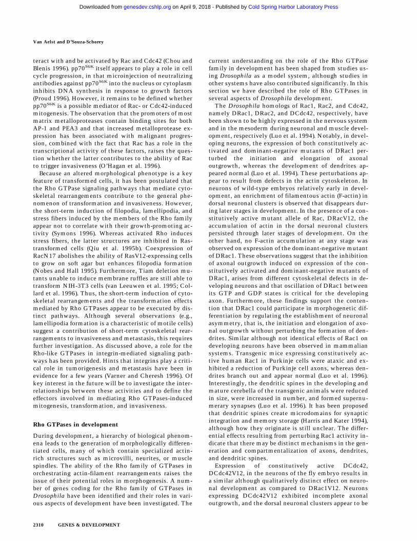
teract with and be activated by Rac and Cdc42 (Chou andBlenis 1996). pp70S6K itself appears to play a role in cellcycle progression, in that microinjection of neutralizingantibodies against pp70S6K into the nucleus or cytoplasminhibits DNA synthesis in response to growth factors(Proud 1996). However, it remains to be defined whetherpp70S6K is a possible mediator of Rac- or Cdc42-inducedmitogenesis. The observation that the promoters of mostmatrix metalloproteases contain binding sites for bothAP-1 and PEA3 and that increased metalloprotease ex-pression has been associated with malignant progres-sion, combined with the fact that Rac has a role in thetranscriptional activity of these factors, raises the ques-tion whether the latter contributes to the ability of Racto trigger invasiveness (O’Hagan et al. 1996).
Because an altered morphological phenotype is a keyfeature of transformed cells, it has been postulated thatthe Rho GTPase signaling pathways that mediate cyto-skeletal rearrangements contribute to the general phe-nomenon of transformation and invasiveness. However,the short-term induction of filopodia, lamellipodia, andstress fibers induced by the members of the Rho familyappear not to correlate with their growth-promoting ac-tivity (Symons 1996). Whereas activated Rho inducesstress fibers, the latter structures are inhibited in Ras-transformed cells (Qiu et al. 1995b). Coexpression ofRacN17 abolishes the ability of RasV12-expressing cellsto grow on soft agar but enhances filopodia formation(Nobes and Hall 1995). Furthermore, Tiam deletion mu-tants unable to induce membrane ruffles are still able totransform NIH-3T3 cells (van Leeuwen et al. 1995; Col-lard et al. 1996). Thus, the short-term induction of cyto-skeletal rearrangements and the transformation effectsmediated by Rho GTPases appear to be executed by dis-tinct pathways. Although several observations (e.g.,lamellipodia formation is a characteristic of motile cells)suggest a contribution of short-term cytoskeletal rear-rangements to invasiveness and metastasis, this requiresfurther investigation. As discussed above, a role for theRho-like GTPases in integrin-mediated signaling path-ways has been provided. Hints that integrins play a criti-cal role in tumorigenesis and metastasis have been inevidence for a few years (Varner and Cheresh 1996). Ofkey interest in the future will be to investigate the inter-relationships between these activities and to define theeffectors involved in mediating Rho GTPases-inducedmitogenesis, transformation, and invasiveness.
Rho GTPases in development
During development, a hierarchy of biological phenom-ena leads to the generation of morphologically differen-tiated cells, many of which contain specialized actin-rich structures such as microvilli, neurites, or musclespindles. The ability of the Rho family of GTPases inorchestrating actin-filament rearrangements raises theissue of their potential roles in morphogenesis. A num-ber of genes coding for the Rho family of GTPases inDrosophila have been identified and their roles in vari-ous aspects of development have been investigated. The
current understanding on the role of the Rho GTPasefamily in development has been shaped from studies us-ing Drosophila as a model system, although studies inother systems have also contributed significantly. In thissection we have described the role of Rho GTPases inseveral aspects of Drosophila development.
The Drosophila homologs of Rac1, Rac2, and Cdc42,namely DRac1, DRac2, and DCdc42, respectively, havebeen shown to be highly expressed in the nervous systemand in the mesoderm during neuronal and muscle devel-opment, respectively (Luo et al. 1994). Notably, in devel-oping neurons, the expression of both constitutively ac-tivated and dominant-negative mutants of DRac1 per-turbed the initiation and elongation of axonaloutgrowth, whereas the development of dendrites ap-peared normal (Luo et al. 1994). These perturbations ap-pear to result from defects in the actin cytoskeleton. Inneurons of wild-type embryos relatively early in devel-opment, an enrichment of filamentous actin (F-actin) indorsal neuronal clusters is observed that disappears dur-ing later stages in development. In the presence of a con-stitutively active mutant allele of Rac, DRacV12, theaccumulation of actin in the dorsal neuronal clusterspersisted through later stages of development. On theother hand, no F-actin accumulation at any stage wasobserved on expression of the dominant-negative mutantof DRac1. These observations suggest that the inhibitionof axonal outgrowth induced on expression of the con-stitutively activated and dominant-negative mutants ofDRac1, arises from different cytoskeletal defects in de-veloping neurons and that oscillation of DRac1 betweenits GTP and GDP states is critical for the developingaxon. Furthermore, these findings support the conten-tion that DRac1 could participate in morphogenetic dif-ferentiation by regulating the establishment of neuronalasymmetry, that is, the initiation and elongation of axo-nal outgrowth without perturbing the formation of den-drites. Similar although not identical effects of Rac1 ondeveloping neurons have been observed in mammaliansystems. Transgenic mice expressing constitutively ac-tive human Rac1 in Purkinje cells were ataxic and ex-hibited a reduction of Purkinje cell axons, whereas den-drites branch out and appear normal (Luo et al. 1996).Interestingly, the dendritic spines in the developing andmature cerebella of the transgenic animals were reducedin size, were increased in number, and formed supernu-merary synapses (Luo et al. 1996). It has been proposedthat dendritic spines create microdomains for synapticintegration and memory storage (Harris and Kater 1994),although how they originate is still unclear. The differ-ential effects resulting from perturbing Rac1 activity in-dicate that there may be distinct mechanisms in the gen-eration and compartmentalization of axons, dendrites,and dendritic spines.
Expression of constitutively active DCdc42,DCdc42V12, in the neurons of the fly embryo results ina similar although qualitatively distinct effect on neuro-nal development as compared to DRac1V12. Neuronsexpressing DCdc42V12 exhibited incomplete axonaloutgrowth, and the dorsal neuronal clusters appear to be
Van Aelst and D’Souza-Schorey
2310 GENES & DEVELOPMENT
Cold Spring Harbor Laboratory Press on April 9, 2018 - Published by genesdev.cshlp.orgDownloaded from
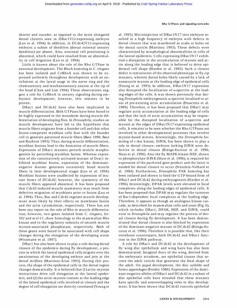
shorter and rounder, as opposed to the more elongateddorsal clusters seen in DRac1V12-expressing embryos(Luo et al. 1994). In addition, in DCdc42V12-expressingembryos a subset of dendrites (dorsal external sensorydendrites) are absent. Also, neuronal cell positioning isabnormal, which could have resulted from an abnormal-ity in cell migration (Luo et al. 1994).
Little is known about the role of the Rho GTPase inneuronal development. The RhoA homolog in C. eleganshas been isolated and CeRhoA was shown to be ex-pressed uniformly throughout development with an en-richment at the larval stage in the nerve ring and thechemosensory and mechanosensory neuron at the tip ofthe head (Chen and Lim 1994). These observations sug-gest a role for CeRhoA in sensory signaling during em-bryonic development; however, this remains to beproven.
DRac1 and DCdc42 have also been implicated inmuscle differentiation. Both GTPases have been found tobe highly expressed in the mesoderm during muscle dif-ferentiation of developing flies. In Drosophila, studies onmuscle development have led to the hypothesis thatmuscle fibers originate from a founder cell and that otherfusion-competent myoblast cells fuse with the foundercell to generate patterned muscle fibers (Bate 1990; Bateand Rushton 1993). Thus, during muscle differentiation,myoblast fusions lead to the formation of muscle fibers.Expression of DRac1 mutants perturb muscle morpho-genesis by perturbing myoblast fusion. Whereas expres-sion of the constitutively activated mutant of Drac1 in-hibited myoblast fusion, expression of the dominant-negative mutant generates excessively fused musclefibers in later developmental stages (Luo et al. 1994).Myoblast fusions were unaffected by expression of mu-tant forms of DCdc42; however, the symmetry of themuscle fibers appeared abnormal. It has been proposedthat Cdc42-induced muscle asymmetry may result fromdefective migration of myoblasts prior to fusion (Luo etal. 1994). Thus, Rac and Cdc42 regulate muscle develop-ment most likely by their effects on membrane fusionand the actin cytoskeleton, respectively. There has notbeen any report on the role of Rho in muscle differentia-tion; however, two genes isolated from C. elegans, let-502 and mel-11, show homology to the mammalian Rhokinase and to the regulatory subunits of smooth musclemyosin-associated phosphatase, respectively. Both ofthese genes were found to be associated with cell shapechanges during the elongation of the developing worm(Wissmann et al. 1997).
DRac1 has also been shown to play a role during dorsalclosure of the epidermis during fly development, a pro-cess in which the lateral epidermal cells migrate over theamnioserosa of the developing embryo and join at thedorsal midline (Martinez-Arias 1993). During this pro-cess, the shape of the epidermal cells in the dorsal regionchanges dramatically. It is believed that (1) actin–myosininteractions drive cell elongation at the lateral epider-mis, and (2) the actin–myosin content at the leading edgeof the lateral epidermal cells involved in closure and thedegree of cell elongation are directly correlated (Young et
al. 1993). Microinjection of DRac1N17 into embryos re-sulted in a high frequency of embryos with defects indorsal closure that was manifested as scabs or holes onthe dorsal cuticle (Martinez 1993). These defects werecharacterized by morphological abnormalities in cells ofthe lateral epidermis. Cells expressing DRac1N17 exhib-ited a disruption in the accumulation of myosin and ac-tin along the leading edge that is believed to drive epi-dermal cell shape (Harden et al. 1995). Such a closuredefect is reminiscent of the observed phenotype in fly zipmutants, wherein dorsal holes likely caused by a lack ofnonmuscle myosin at the leading edge of epidermal cells(Young et al. 1993). In addition, DRac1N17 expressionalso disrupted the localization of a-spectrin at the lead-ing edges of the cells. It was shown previously that dur-ing Drosophila embryogenesis a-spectrin localized to ar-eas of pre-existing actin accumulation (Pesacreta et al.1989). Therefore, it has been proposed that DRac1 mayregulate actin accumulation at the leading edge of cellsand that the lack of actin accumulation may be respon-sible for the disrupted localization of a-spectrin andmyosin at the edges of DRac1N17-expressing epidermalcells. It remains to be seen whether the Rho GTPases areinvolved in other developmental processes that involvemyosin-based motors. Interestingly, the Drosophila ho-molog of c-Jun kinase, DJNK, has been shown to play arole in dorsal closure; embryos lacking DJNK were de-fective in dorsal closure (Riesgo-Escovar et al. 1996;Sluss et al. 1996). Also the fly MAPK kinase HEP, shownto phosphorylate DJNK (Sluss et al. 1996), is required forexpression of the puckered gene product and the latter isneeded for dorsal closure to occur (Nusslein-Volhard etal. 1984). Furthermore, Drosophila PAK homolog hasbeen isolated and shown to bind the GTP-bound form ofDRac1 and DCdc42 during embryogenesis (Harden et al.1996). Interestingly, DPAK levels were elevated in focalcomplexes along the leading edges of epidermal cells. Ithas been proposed that DPAK may regulate the turnoverof Rac1-dependent focal complexes at the leading edge.Therefore, it appears as though an analogous kinase cas-cade, as described for mammalian cells and yeast (Fig. 6),which includes DRac1, DPAK, HEP, and DJNK, couldexist in Drosophila and may regulate the process of dor-sal closure during fly development. It has been demon-strated that dorsal closure is also blocked by expressionof the dominant-negative mutant of DCdc42 (Riesgo-Es-covar et al. 1996). Therefore it is possible that, like theirvertebrate counterparts, both DCdc42 and DRac1 func-tion in the DJNK pathway.
A role for DRac1 and DCdc42 in the development offly wing disc epithelium and wing hairs has also beendemonstrated. Imaginal discs of the wing, derived fromthe embryonic ectoderm, are epithelial tissues that se-crete the adult cuticle that generates the final shape ofthe adult. On pupal development, the disc unfolds andforms appendages (Poodry 1980). Expression of the domi-nant-negative alleles of DRac1 and DCdc42 in a subset ofdisc epithelial cells have revealed that these mutantshave specific and nonoverlapping roles in disc develop-ment. It has been shown that DCdc42 controls epithelial
Rho GTPases and signaling networks
GENES & DEVELOPMENT 2311
Cold Spring Harbor Laboratory Press on April 9, 2018 - Published by genesdev.cshlp.orgDownloaded from

cell shape changes by modulating the basic actin cyto-skeleton during pupal and larval development. Whereasnormal wing disc cells increase in height at least twofoldduring the third instar, cells expressing dominant-nega-tive DCdc42 remain short and unelongated. Expressionof the negative mutant of DRac1 during disc develop-ment did not appear to affect cytoskeletal elements thatregulate cell shape (Eaton et al. 1995). However, DRac1activity appears to be required for actin assembly at theadherens junctions as the latter is disrupted onDRac1N17 expression (Eaton et al. 1995). Failure of ad-herens junction actin assembly was accompanied byincreased cell death and consequently a reduction inwing size. Cell death may have resulted from DRac1N17disrupting another vital cell function such as cytokine-sis, or because adherens junction disassembly may itselfbe lethal. The defects caused by dominant-negativeDRac1 or DCdc42 did not affect epithelial polarity perse, and apical and basolateral proteins remain correctlylocalized in cells expressing these mutants (Eaton et al.1995).
The wing of Drosophila is covered by an array of dis-tally pointing hairs. The wing epithelial cells form winghairs by extending a single process from the apical mem-branes of the hairs (Mitchell et al. 1983; Wong and Adler1993; Fristrom et al. 1994). This latter process is pre-ceded by the accumulation of actin on the distal side ofthe cell and a bundling of microtubules that extend intothe wing hair throughout its elongation. It has beenshown that cells expressing the dominant-negativeDCdc42 lack wing hairs and that at lower levels of mu-tant protein expression deformed and stunted hairs wereformed (Eaton et al. 1996). In cells that did not extendhairs, actin failed to accumulate distally. In hair exten-sions, Cdc42 has been shown to be localized at the distalend, which may be its site of action. On the other hand,in cells expressing DRac1N17, multiple, but structurallynormal, wing hairs were formed (Eaton et al. 1996). Thisfailure to restrict hair outgrowth to a single site suggeststhat DRac1 might function to select the site for hairformation. Whether this function may be related to itsrole in organizing junctional actin is unclear. TheDRac1N17 expressing cells also exhibited a disorganizedmicrotubule network in the apical region of the cell.Thus, the site selection for hair outgrowth may requirean intact microtubule network. It has been proposed thatthe regulation of adherens junction assembly and micro-tubule organization in response to Rac1 activity may bedirectly coupled to the suppression of inappropriate hairgrowth (Eaton et al. 1996). Notably, a multiple wing hairphenotype is typical of tissue polarity mutants such asmultiple wing hair (mwh) and intwined (in). (Adler1992). Interestingly in this regard, hypomorphic allelesof RhoA in the fly wing exhibit abnormal hair polarityand a multiple wing hair phenotype. Expression of adominant-negative form of RhoA in part of the wing re-sulted in a polarity defect typical of frizzled (fz), a ser-pentine receptor-like transmembrane protein, and di-shevelled (dsh), which acts downstream of fz (Strutt etal. 1997). These cells exhibit a wing hair polarity defect.
The above findings suggest that RhoA is required for thegeneration of polarity in fly epithelial cells.
Studies investigating the effect of Rho in fly eye mor-phogenesis have also been conducted. These studieshave shown that expression of a Rho transgene in the eyeof Drosophila resulted in a dose-dependent disruption ofnormal eye development (Hariharan et al. 1995). Suchflies exhibited a severe rough eye phenotype character-ized by missing secondary and tertiary pigment cells, asubstantial reduction in the number of photoreceptorcells, and a grossly abnormal morphology of rhabdo-meres. These defects became manifest when cells wereundergoing rapid changes in cell shape. Cells such as theprimary pigment cells, which do not span the thicknessof the retina and display fewer morphological changes,were more resistant to the effects of Rho expression. Ithas been suggested that Rho-induced actin polymeriza-tion could hinder normal development of cells undergo-ing dramatic shape changes. More recently, isolation ofseveral loss-of-function alleles of RhoA in Drosophila,has revealed that RhoA may be critical for the establish-ment of tissue polarity in the developing eye. Homozy-gous mutant clones carrying null alleles in the imaginaldisc of the eye fail to proliferate, indicating a role forRhoA in cell growth and/or viability, whereas clonesexpressing hypomorphic alleles of RhoA give rise to om-matida that are incorrectly rotated, resulting in a disrup-tion of planar polarity (Strutt et al. 1997). Such a pheno-type is similar to that seen in overexpression of tissuepolarity genes such as fz and dsh. These studies have alsoshown that RhoA may be part of a signaling pathwaydownstream of fz and dsh. Furthermore, mutations inthe basket (bsk) gene suppress the effects of fz and dsh,suggesting a role for JNK/SAPK-like kinases in the afore-mentioned Rho-mediated signaling pathway (Strutt et al.1997).
Drosophila oogenesis provides an excellent system forinvestigating processes such as cell migration, mem-brane fusion, and cytoskeletal remodeling during devel-opment. Drosophila egg chambers harbor 15 polyploidnurse cells and 1 oocyte, and all 16 cells remain con-nected to each other by actin-rich bridges as a result ofincomplete cytokinesis. Oogenesis involves a number ofprocesses wherein the actin cytoskeleton undergoes dra-matic changes (Murphy and Montell 1996). Early in oo-genesis, germ-line clusters become surrounded by an ep-ithelial monolayer of somatic follicle cells. Many of thefollicle cells elongate to form columnar epithelial cells,whereas some assume a squamous shape, and yet othersmigrate to the tip of the chamber. Late in oogenesis thetransfer of nurse cell cytoplasm into the oocyte occursand is preceded by actin polymerization around thenurse cell nuclei (Cooley et al. 1992; Peifer et al. 1993).Studies on the Rho-family proteins, namely Drac1,RhoL, a novel Rho homolog, and Cdc42, have revealedthat they are required for multiple processes during oo-genesis (Murphy and Montell 1996). These GTPases ap-pear to play distinct roles in each cell type, although theactivities of all three GTPases are required for transfer ofnurse cell cytoplasm into the oocyte. During oogenesis,
Van Aelst and D’Souza-Schorey
2312 GENES & DEVELOPMENT
Cold Spring Harbor Laboratory Press on April 9, 2018 - Published by genesdev.cshlp.orgDownloaded from
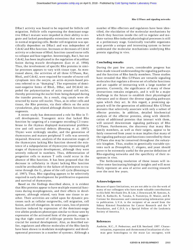
DRac1 activity was found to be required for follicle cellmigration. Follicle cells expressing the dominant-nega-tive DRac1 mutant were impeded in their ability to mi-grate and lacked pseudopodia extensions that were seenin normal migrating cells. Border cell migration was spe-cifically dependent on DRac1 and was independent ofCdc42 and Rho function. Increases or decreases of Cdc42activity or a decrease of RhoL function caused nurse cellsto collapse and fuse together. Interestingly, Rac1, but notCdc42, has been implicated in the regulation of myoblastfusion during muscle development (Luo et al. 1994).Thus, the involvement of specific Rho proteins in simi-lar processes appears to vary with tissue type. As men-tioned above, the activities of all three GTPases, Rac,RhoL, and Cdc42, were required for transfer of nurse cellcytoplasm into the oocyte, an actin–myosin-based pro-cess referred to as ‘‘dumping’’. The expression of domi-nant-negative forms of RhoL, DRac, and DCdc42 im-peded the polymerization of actin around cell nuclei,thereby preventing the nuclei from being dumped duringthe transfer process. As a result, ring canals were ob-structed by nurse cell nuclei. Thus, as in other cells andtissue, the Rho proteins, via their effects on the actincytoskeleton, play related although distinct functions inoogenesis.
A recent study has demonstrated a role for Rho in T-cell development. Transgenic mice that lacked Rhofunction by targeting of a transgene encoding C3 trans-ferase to the thymus displayed maturational prolifera-tive and cell survival defects (Henning et al. 1997).Thymi were strikingly smaller, and the generation ofthymocytes and mature peripheral T cells was severelyimpaired. However, analysis of the maturation stages ofthe thymocytes lacking Rho function showed the exis-tence of a subpopulation of thymocytes representing allstages of thymocyte development, although they wereseverely reduced in numbers. Thus, differentiation ofprogenitor cells to mature T cells could occur in theabsence of Rho function. It has been proposed that thedecrease in cellularity in thymi lacking Rho functioncould be attributable to the failure of thymocytes to tra-verse through the G1 phase of the cell cycle (Henning etal. 1997). Thus, Rho signaling appears to be selectivelyrequired in early development for proliferative expansionand survival of thymocytes.
Based on the findings described above, it is evidentthat Rho proteins appear to have multiple essential func-tions during morphogenesis, and their effects in devel-opment, although related, vary from one tissue to an-other. Their activities are called upon for dynamic pro-cesses such as cellular outgrowths, cell migration, cellfusion, and cell elongation. In some cases, loss of proteinfunction induced by expression of the dominant-nega-tive alleles produces phenotypes similar to those seen byexpression of the activated form of the protein, suggest-ing that tight control of wild-type protein function iscrucial for normal development. Also, as described ear-lier, regulators and effector molecules of the Rho familyhave been shown to modulate morphogenetic and devel-opmental processes in a number of systems. Although a
number of Rho effectors and regulators have been iden-tified, the elucidation of the molecular mechanisms bywhich they function inside the cell to regulate and me-diate various Rho-induced physiological processes is stillat a preliminary stage. Genetically tractable organismsmay provide a unique and interesting system to betterunderstand the molecular mechanisms underlying Rhoprotein signaling in vivo.
Concluding remarks
During the past few years, considerable progress hasbeen made toward understanding the signaling pathwaysand the function of Rho family members. These studieshave revealed that Rho GTPases are versatile signalingmolecules that regulate a diverse set of cellular functionsand are capable of interacting with a large number ofproteins. Currently, the significance of many of theseinteractions remains enigmatic, and it will be a majorchallenge in the future to establish their physiologicalrelevance and to dissect the diverse signaling pathwaysupon which they act. In this regard, a promising ap-proach will be the generation of additional Rho GTPasemutants that selectively associate with only a subset ofeffector proteins. In addition, detailed biochemicalanalysis of the effector proteins, along with identifi-cation of additional proteins that interact with them,will unravel downstream signaling pathways of RhoGTPases. Furthermore, the discovery that many Rhofamily members, as well as their targets, appear to befairly conserved from yeast to man implies that many ofthe signaling pathways and functions controlled by theseGTPases also may be conserved throughout the eukary-otic kingdom. Thus, studies in genetically tractable sys-tems such as Drosophila, C. elegans, and yeast shouldprove to be extremely useful for the precise definition ofRho-signaling networks and the specificity of their re-sponses in vivo.
The forthcoming resolution of these issues will in-volve some fascinating biological insights and will mostlikely represent an area of active and exciting researchover the next few years.
Acknowledgments
Because of space limitations, we are not able to cite the work ofmany of our colleagues who have made valuable contributionsto this field. We thank Drs. B. Lim, J. Downward, M. Symons, A.Hall, K. Kaibuchi, K. Tanaka, S. Narumiya, D. Manor, and R.Cerione for discussions and communicating information priorto publication. L.V.A. is the recipient of an award from theSidney Kimmel Foundation for Cancer Research and the VFoundation, and C.D.S. is a Special Fellow of the LeukemiaSociety of America.
References
Aasheim, H.C., F. Pedeutour, and E.B. Smeland. 1997. Charac-terization, expression and chromosomal localization of a hu-man gene homologous to the muse Lsc oncogene, with
Rho GTPases and signaling networks
GENES & DEVELOPMENT 2313
Cold Spring Harbor Laboratory Press on April 9, 2018 - Published by genesdev.cshlp.orgDownloaded from

strongest expression in hematopoetic tissues. Oncogene14: 1747–1752.
Abo, A., E. Pick, A. Hall, N. Totty, C.G. Teahan, and A.W.Segal. 1991. Activation of the NADPH oxidase involves thesmall GTP–binding protein p21rac1. Nature 353: 668–670.
Abo, A., A. Boyhan, I. West, A.J. Thrasher, and A.W. Segal. 1992.Reconstitution of neutrophil NADPH oxidase activity in thecell-free system by four components: p67-phox, p47-phox,p21rac1, and cytochrome b-245. J. Biol. Chem. 267: 16767–16770.
Adam, T., M. Giry, P. Boquet, and P. Sansonetti. 1996. Rho-dependent membrane folding causes Shigella entry into ep-ithelial cells. EMBO J. 15: 3315—3321.
Adamson, P., C.J. Marshall, A. Hall, and P.A. Tilbrook. 1992.Post-translational modifications of p21rho proteins. J. Biol.Chem. 267: 20033–20038.
Adler, P.N. 1992. The genetic control of tissue polarity in Dro-sophila. BioEssays 14: 735–741.
Adra, C.N., J. Ko, D. Leonard, L.J. Wirth, R.A. Cerione, and B.Lim. 1993. Identification of a novel protein with GDP dis-sociation inhibitor activity for the ras-like proteinsCDC42Hs and rac I. Genes, Chrom. Cancer 8: 253–261.
Adra, C.N., D. Manor, J.L. Ko, S. Zhu, T. Horiuchi, L. Van Aelst,R.A. Cerione, and B. Lim. 1997. Rho-GDIgamma: A GDP-dissociation inhibitor for Rho proteins with preferential ex-pression in brain and pancreas. Proc. Natl. Acad. Sci.94: 4279–4284.
Agnel, M., L. Roder, C. Vola, and S.R. Griffin. 1992. A Dro-sophila rotund transcript expressed during spermatogenesisand imaginal disc morphogenesis encodes a protein which issimilar to human Rac GTPase-activating (racGAP) proteins.Mol. Cell. Biol. 12: 5111–5122.
Allen, L.A. and A. Aderem. 1996. Mechanisms of phagocytosis.Curr. Opin. Immunol. 8: 36–40.
Amano, M., M. Ito, K. Kimura, Y. Fukata, K. Chihara, T. Na-kano, Y. Matsuura, and K. Kaibuchi. 1996a. Phosphorylationand activation of myosin by Rho-associated kinase (Rho-ki-nase). J. Biol. Chem. 271: 20246–20249.
Amano, M., H. Mukai, Y. Ono, K. Chihara, T. Matsui, Y.Hamajima, K. Okawa, A. Iwamatsu, and K. Kaibuchi. 1996b.Identification of a putative target for Rho as the serine-threo-nine kinase protein kinase N. Science 271: 648–650.
Amano, M., K. Chihara, K. Kimura, Y. Fukata, N. Nakamura, Y.Matsuura, and K. Kaibuchi. 1997. Formation of actin stressfibers and focal adhesions enhanced by Rho-kinase. Science275: 1308–1311.
Arellano, M., A. Duran, and P. Perez. 1996. Rho 1 GTPase ac-tivates the (1-3)beta-D-glucan synthase and is involved inSchizosaccharomyces pombe morphogenesis. EMBO J.15: 4584–4591.
Aspenstrom, P., U. Lindberg, and A. Hall. 1996. Two GTPases,Cdc42 and Rac, bind directly to a protein implicated in theimmunodeficiency disorder Wiskott-Aldrich syndrome.Curr. Biol. 6: 70–75.
Bagrodia, S., B. Derijard, R.J. Davis, and R.A. Cerione. 1995a.Cdc42 and PAK-mediated signaling leads to Jun kinase andp38 mitogen-activated protein kinase activation. J. Biol.Chem. 270: 27995–27998.
Bagrodia, S., S.J. Taylor, C.L. Creasy, J. Chernoff, and R.A. Ceri-one. 1995b. Identification of a mouse p21Cdc42/Rac acti-vated kinase. J. Biol. Chem. 270: 22731–22737.
Bankaitis, V.A., J.R. Aikten, A.E. Cleves, and W. Dowhan. 1990.An essential role for a phospholipid transfer protein in yeastGolgi function. Nature 347: 561–520.
Barbieri, M.A., G. Li, M.I. Colombo, and P.D. Stahl. 1994. Rab5,an early acting endosomal GTPase, supports in vitro endo-
some fusion without GTP hydrolysis. J. Biol. Chem.269: 18720–18724.
Barfod, E.T., Y. Zheng, W.J. Kuang, M.J. Hart, T. Evans, R.A.Cerione, and A. Ashkenazi. 1993. Cloning and expression ofa human CDC42 GTPase-activating protein reveals a func-tional SH3-binding domain. J. Biol. Chem.268: 26059–26062.
Bate, M. 1990. The embryonic development of larval muscles inDrosophila. Development 110: 791–804.
Bate, M. and E. Rushton. 1993. Myogenesis and muscle pattern-ing in Drosophila. C. R. Acad. Sci. III Sci. Vie 316: 1047–1061.
Bender, L., H.S. Lo, H. Lee, V. Kokojan, V. Peterson, and A.Bender. 1996. Associations among PH and SH3 domain-con-taining proteins and Rho-type GTPases in yeast. J. Cell Biol.133: 879–894.
Benian, G.M., T.L. Tinley, X. Tang, and M. Borodovsky. 1996.The Caenorhabditis elegans gene un9c-89, required formuscle M-line assembly, encodes a giant modular proteincomposed of Ig and signal transduction domains. J. Cell Biol.132: 835–848.
Beron, W., C. Alvaez-Dominguez, L. Mayorga, and P.D. Stahl.1995. Membrane trafficking along the phagocytic pathway.Trends Cell Biol. 5: 93–99.
Boguski, M.S. and F. McCormick. 1993. Proteins regulating Rasand its relatives. Nature 366: 643–654.
Bokoch, G.M. 1995. Regulation of the phagocyte respiratoryburst by small GTP-binding proteins. Trends Cell Biol.5: 109–113.
Bowtell, D., P. Fu, M. Simon, and P. Senior. 1992. Identificationof murine homologues of the Drosophila son of sevenlessgene: Potential activators of ras. Proc. Natl. Acad. Sci.89: 6511–6515.
Brill, S., S. Li, C.W. Lyman, D.M. Church, J.J. Wasmuth, L.Weissbach, A. Bernards, and A.J. Snijders. 1996. The RasGTPase-activating-protein-related human protein IQGAP2harbors a potential actin binding domain and interacts withcalmodulin and Rho family GTPases. Mol. Cell. Biol.16: 4869–4878.
Brown, J.L., L. Stowers, M. Baer, J. Trejo, S. Coughlin, and J.Chant. 1996. Human Ste20 homologue hPAK1 links GT-Pases to the JNK MAP kinase pathway. Curr. Biol. 6: 598–605.
Bucci, C., R. Parton, I.M. Mather, H. Stunnenberg, K. Simon, B.Hoflack, and M. Zerial. 1992. The small GTPase Rab5 func-tions as a regulatory factor in the early endocytic pathway.Cell 70: ??
Burbelo, P.D., D. Drechsel, and A. Hall. 1995. A conserved bind-ing motif defines numerous candidate target proteins forboth Cdc42 and Rac GTPases. J. Biol. Chem. 270: 29071–29074.
Burridge, K. and W.M. Chrzanowska. 1996. Focal adhesions,contractility, and signaling. Ann. Rev. Cell Dev. Biol.12: 463–518.
Cantor, S.B., T. Urano, and L.A. Feig. 1995. Identification andcharacterization of Ral-binding protein 1, a potential down-stream target of Ral GTPases. Mol. Cell. Biol. 8: 4578–4584.
Cao, L.G., G.G. Babcock, P.A. Rubenstein, and Y.L. Wang. 1992.Effects of profilin and profilactin on actin structure and func-tion in living cells. J. Cell Biol. 117: 1023–1029.
Castrillon, D.H. and S.A. Wasserman. 1994. Diaphanous is re-quired for cytokinesis in Drosophila and shares domains ofsimilarity with the products of the limb deformity gene. De-velopment 120: 3367–3377.
Cerione, R.A. and Y. Zheng. 1996. The Dbl family of oncogenes.Curr. Opin. Cell Biol. 8: 216–222.
Van Aelst and D’Souza-Schorey
2314 GENES & DEVELOPMENT
Cold Spring Harbor Laboratory Press on April 9, 2018 - Published by genesdev.cshlp.orgDownloaded from

Chan, A.M., E.S. McGovern, G. Catalano, T.P. Fleming, and T.Miki. 1994. Expression cDNA cloning of a novel oncogenewith sequence similarity to regulators of small GTP-bindingproteins. Oncogene 4: 1057–1063.
Chan, A.M., S. Takai, K. Yamada, and T. Miki. 1996. Isolationof a novel oncogene, NET1, from neuroepithelioma cells byexpression cDNA cloning. Oncogene 12: 1259–1266.
Chang, E.C., M. Barr, Y. Wang, V. Jung, H.P. Xu, and M.H.Wigler. 1994. Cooperative interaction of S. pombe proteinsrequired for mating and morphogenesis. Cell 79: 131–141.
Chang, J.H., S. Gill, J. Settleman, and S.J. Parsons. 1995. c-Srcregulates the simultaneous rearrangement of actin cytoskel-eton, p190RhoGAP, and p120RasGAP following epidermalgrowth factor stimulation. J. Cell Biol. 130: 355–368.
Chant, J. and L. Stowers. 1995. GTPase cascades choreographingcellular behavior: Movement, morphogenesis, and more.Cell 81: 1–4.
Chardin, P., J.H. Camonis, N.W. Gale, L. van Aelst, J. Schless-inger, M.H. Wigler, and D. Bar-Sagi. 1993. Human Sos1: Aguanine nucleotide exchange factor for Ras that binds toGRB2. Science 260: 1338–1343.
Chen, G.C., L. Zheng, and C.S. Chan. 1996a. The LIM domain-containing Dbm1 GTPase–activating protein is required fornormal cellular morphogenesis in Saccharomyces cerevisiae.Mol. Cell. Biol. 16: 1376–1390.
Chen, L.M., S. Hobbie, and J.E. Galan. 1996b. Requirement ofCDC42 for Salmonella–induced cytoskeletal and nuclear re-sponses. Science 274: 2115–2118.
Chen, W. and L. Lim. 1994. The Caenorhabditis elegans smallGTP-binding protein RhoA is enriched in the nerve ring andsensory neurons during larval development. J. Biol. Chem.269: 32394–32404.
Chen, W., J. Blanc, and L. Lim. 1994. Characterization of a pro-miscuous GTPase-activating protein that has a Bcr-relateddomain from Caenorhabditis elegans. J. Biol. Chem.269: 820–823.
Chen, W., S. Chen, S.F. Yap, and L. Lim. 1996. The Caenorhab-ditis elegans p21-activated kinase (CePAK) colocalizes withCeRac1 and CDC42Ce at hypodermal cell boundaries duringembryo elongation. J. Biol. Chem. 271: 26362–26368.
Chihara, K., M. Amano, N. Nakamura, T. Yano, M. Shibata, T.Tokui, H. Ichikawa, R. Ikebe, M. Ikebe, and K. Kaibuchi.1997. Cytoskeletal rearrangements and transcriptional acti-vation of c-fos serum response element by Rho-kinase. J.Biol. Chem. (in press).
Chong, L.D., K.A. Traynor, G.M. Bokoch, and M.A. Schwartz.1994. The small GTP-binding protein Rho regulates a phos-phatidylinositol 4-phosphate 5-kinase in mammalian cells.Cell 79: 507–513.
Chou, M.M. and J. Blenis. 1996. The 70 kDa S6 kinase com-plexes with and is activated by the Rho family G proteinsCdc42 and Rac1. Cell 85: 573–583.
Chrzanowska, W.M. and K. Burridge. 1996. Rho-stimulatedcontractility drives the formation of stress fibers and focaladhesions. J. Cell Biol. 133: 1403–1415.
Chuang, T.H., X. Xu, U.G. Knaus, M.J. Hart, and G.M. Bokoch.1993. GDP dissociation inhibitor prevents intrinsic and GT-Pase activating protein-stimulated GTP hydrolysis by theRac GTP-binding protein. J. Biol. Chem. 268: 775–778.
Chuang, T H., X. Xu, V. Kaartinen, N. Heisterkamp, J. Groffen,and G.M. Bokoch. 1995. Abr and Bcr are multifunctionalregulators of the Rho GTP-binding protein family. Proc.Natl. Acad. Sci. 92: 10282–10286.
Cicchetti, P., B.J. Mayer, G. Thiel, and D. Baltimore. 1992. Iden-tification of a protein that binds to the SH3 region of Abl andis similar to Bcr and GAP-rho. Science 257: 803–806.
Cicchetti, P., A.J. Ridley, Y. Zheng, R.A. Cerione, and D. Balti-more. 1995. 3BP-1, an SH3 domain binding protein, has GAPactivity for Rac and inhibits growth factor-induced mem-brane ruffling in fibroblasts. EMBO J. 14: 3127–3135.
Clague, M.J., A.T. Thrope, and R.T. Jones. 1995. Phosphatidyli-nositol 3-kinase regulation of fluid phase endocytosis. FEBSLett. 367: 272–276.
Collard J.G., G.G. Habets, F. Michiels, J. Stam, R.A. van derKammen, and F. van Leeuwen. 1996. Role of Tiam1 in Rac-mediated signal transduction pathways. Curr. Top. Micro-biol. Immunol. 213: 253–265.
Collins, L.R., A. Minden, M. Karin, and J.H. Brown. 1996. Ga12stimulates c-Jun NH2-terminal kinase through the small Gproteins Ras and Rac. J. Biol. Chem. 271: 17349–17353.
Cooley, L., E. Verheyen, and K. Ayers. 1992. Chikadee encodesa profilin required for intracellular cytoplasm transport dur-ing Drosophila oogenesis. Cell 69: 173–184.
Coso, O.A., M. Chiariello, J.C. Yu, H. Teramoto, P. Crespo, N.Xu, T. Miki, and J.S. Gutkind. 1995. The small GTP-bindingproteins Rac1 and Cdc42 regulate the activity of the JNK/SAPK signaling pathway. Cell 81: 1137–1146.
Coso, O.A., H. Teramoto, W.F. Simonds, and J.S. Gutkind. 1996.Signaling from G protein-coupled receptors to c-Jun kinaseinvolves beta gamma subunits of heterotrimeric G proteinsacting on a Ras and Rac1-dependent pathway. J. Biol. Chem.271: 3963–3966.
Crespo, P., K.E. Schuebel, A.A. Ostrom, J.S. Gutkind, and X.R.Bustelo. 1997. Phosphotyrosine-dependent activation ofRac-1 GDP/GTP exchange by the vav proto-oncogene prod-uct. Nature 385: 169–172.
Cvrckova, F., V.C. De, E. Manser, J.R. Pringle, and K. Nasmyth.1995. Ste20-like protein kinases are required for normal lo-calization of cell growth and for cytokinesis in buddingyeast. Genes & Dev. 9: 1817–1830.
Debant, A., C. Serra-Pages, K. Seipel, S. O’Brien, M. Tang, andS.H. Park. 1996. The multidomain protein Trio binds theLAR transmembrane tyrosine phosphatase, contains a pro-tein kinase domain, and has separate rac-specific and rho-specific guanine nucleotide exchange factor domains. Proc.Natl. Acad. Sci. 93: 5466–5471.
Derijard, B., M. Hibi, I.H. Wu, T. Barrett, B. Su, T. Deng, M.Karin, and R.J. Davis. 1994. JNK1: A protein kinase stimu-lated by UV light and Ha–Ras that binds and phosphorylatesthe c-Jun activation domain. Cell 76: 1025–1037.
Derijard, B., J. Raingeaud, T. Barrett, I.H. Wu, J. Han, R.J. Ulev-itch, and R.J. Davis. 1995. Independent human MAP-kinasesignal transduction pathways defined by MEK and MKK iso-forms. Science 267: 682–685.
Diekmann, D., S. Brill, M.D. Garrett, N. Totty, J. Hsuan, C.Monfries, C. Hall, L. Lim, and A. Hall. 1991. Bcr encodes aGTPase-activating protein for p21rac. Nature 351: 400–402.
Diekmann, D., A. Abo, C. Johnston, A.W. Segal, and A. Hall.1994. Interaction of Rac with p67phox and regulation ofphagocytic NADPH oxidase activity. Science 265: 531–533.
Dorseuil, O., G. Leca, A. Vazquez, and G. Gacon. 1995. Inhibi-tion of Rac function using antisense oligonucleotides. Meth-ods Enzymol. 256: 358–366.
Downward, J. 1996. Control of ras activation. Cancer Surv.27: 87–100.
Drechsel, D.N., A.A. Hyman, A. Hall, and M. Glotzer. 1997. Arequirement for Rho and Cdc42 during cytokinesis in Xeno-pus embryos. Curr. Biol. 7: 12–23.
Drgonova, J., T. Drgon, K. Tanaka, R. Kollar, G.C. Chen, R.A.Ford, C.S. Chan, Y. Takai, and E. Cabib. 1996. Rho1p, a yeastprotein at the interface between cell polarization and mor-phogenesis. Science 272: 277–279.
Rho GTPases and signaling networks
GENES & DEVELOPMENT 2315
Cold Spring Harbor Laboratory Press on April 9, 2018 - Published by genesdev.cshlp.orgDownloaded from

D’Souza-Schorey, C., G. Li, M.I. Colombo, and P.D. Stahl. 1995.A regulatory role for ARF6 in receptor-mediated endocyto-sis. Science 267: 1175–1178.
D’Souza-Schorey, C., R. Boshans, M. McDonough, P.D. Stahl,and L. Van Aelst. 1997. A role for POR1 a Rac1 interactingprotein in ARF6 mediated cytoskeletal rearrangements.EMBO J. (in press).
Dutartre, H., J. Davoust, J.P. Gorvel, and P. Chavrier. 1996.Cytokinesis arrest and redistribution of actin-cytoskeletonregulatory components in cells expressing the Rho GTPaseCDC42Hs. J. Cell. Sci. 109: 367–377.
Eaton, S., P. Auvinen, L. Luo, Y.N. Jan, and K. Simons. 1995.CDC42 and Rac1 control different actin-dependent pro-cesses in the Drosophila wing disc epithelium. J. Cell Biol.131: 151–164.
Eaton, S., R. Wepf, and K. Simons. 1996. Roles for Rac1 andCdc42 in planar polarization and hair outgrowth in the wingof Drosophila. J. Cell Biol. 135: 1277–1289.
Eberhard, D.A., C.L. Cooper, M.G. Low, and R.W. Holz. 1990.Evidence that the inositol phospholipids are necessary forexocytosis. Loss of inositol phospholipids and inhibition ofsecretion in permeabilized cells caused by a bacterial phos-pholipase C and removal of ATP. J. Biochem. 268: 15–25.
Edwards, S.W. 1996. The O-2 generating NADPH oxidase ofphagocytes: Structure and methods of detection. Methods9: 563–577.
Ellis, C., M. Moran, F. McCormick, and T. Pawson. 1990. Phos-phorylation of GAP and GAP-associated proteins by trans-forming and mitogenic tyrosine kinases. Nature 343: 377–381.
Emmons, S., H. Phan, J. Calley, W. Chen, B. James, and L.Manseau. 1995. Cappuccino, a Drosophila maternal effectgene required for polarity of the egg and embryo, is related tothe vertebrate limb deformity locus. Genes & Dev. 9: 2482–2494.
Erickson, J.W., C.J. Zhang, R.A. Kahn, E.T.?? and R.A. Cerione.1996. Mammalian Cdc42 ia a Brefeldin A sensitive com-pound of the Golgi apparatus. J. Biol. Chem. 271: 26850–26854.
Eva, A. and S.A. Aaronson. 1985. Isolation of a new humanoncogene from a diffuse B-cell lymphoma. Nature 316: 273–275.
Evangelista, M., K. Blundell, M.S. Longtine, C.J. Chow, N.Adames, J.R. Pringle, M. Peter, and C. Boone. 1997. Bni1p, ayeast formin linking cdc42p and the actin cytoskeleton dur-ing polarized morphogenesis. Science 276: 118–122.
Fristrom, D., P. Gotwals, S. Eaton, T.B. Kornberg, M. Sturte-vant, E. Bier, and J.W. Fristrom. 1994. Blistered: A gene re-quired for vein/intervein formation in wings of Drosophila.Development 120: 2661–2671.
Frost, J.A., S. Xu, M.R. Hutchison, S. Marcus, and M.H. Cobb.1996. Actions of Rho family small G proteins and p21-acti-vated protein kinases on mitogen-activated protein kinasefamily members. Mol. Cell. Biol. 16: 3707–3713.
Fukumoto, Y., K. Kaibuchi, Y. Hori, H. Fujioka, S. Araki, T.Ueda, A. Kikuchi, and Y. Takai. 1990. Molecular cloning andcharacterization of a novel type of regulatory protein (GDI)for the rho proteins, ras p21-like small GTP-binding pro-teins. Oncogene 5: 1321–1328.
Gabig, T.G., C.D. Crean, P.L. Mantel, and R. Rosli. 1995. Func-tion of wild-type or mutant Rac2 and Rap1a GTPases indifferentiated HL60 cell NADPH oxidase activation. Blood85: 804–811.
Galan, J., J. Pace, and M.J. Hayman. 1992. Involvement of theepidermal growth factor receptor in the invasion of culturedmammalian cells by Salmonella typhimurium. Nature
357: 588–589.Gallo, K.A., M.R. Mark, D.T. Scadden, Z. Wang, Q. Gu, and P.J.
Godowski. 1994. Identification and characterization ofSPRK, a novel src-homology 3 domain-containing proline-rich kinase with serine/threonine kinase activity. J. Biol.Chem. 269: 15092–15100.
Gerwins, P., J.L. Blank, and G.L. Johnson. 1997. Cloning of anovel mitogen-activated protein kinase kinase kinase,MEKK4, that selectively regulates the c-Jun amino terminalkinase pathway. J. Biol. Chem. 272: 8288–8295.
Gille, H., T. Strahl, and P.E. Shaw. 1995. Activation of ternarycomplex factor Elk-1 by stress-activated protein kinases.Curr. Biol. 5: 1191–1200.
Gilmore, A.P. and K. Burridge. 1996. Regulation of vinculinbinding to talin and actin by phosphatidyl-inositol-4-5-bi-sphosphate. Nature 381: 531–535.
Glaven, J.A., I.P. Whitehead, T. Nomanbhoy, R. Kay, and R.A.Cerione. 1996. Lfc and Lsc oncoproteins represent two newguanine nucleotide exchange factors for the Rho GTP-bind-ing protein. J. Biol. Chem. 271: 27374–27381.
Goldschmidt, C.P., L.M. Machesky, S.K. Doberstein, and T.D.Pollard. 1991. Mechanism of the interaction of human plate-let profilin with actin. J. Cell Biol. 113: 1081–1089.
Guillemot, J.C., B.A. Kruskal, C.N. Adra, S. Zhu, J.L. Ko, P.Burch, K. Nocka, K. Seetoo, E. Simons, and B. Lim. 1996.Targeted disruption of guanosine diphosphate-dissociationinhibitor for Rho-related proteins, GDID4: Normal hemato-poietic differentiation but subtle defect in superoxide pro-duction by macrophages derived from in vitro embryonalstem cell differentiation. Blood 88: 2722–2731.
Gulbins, E., K.M. Coggeshall, G. Baier, S. Katzav, P. Burn, andA. Altman. 1993. Tyrosine kinase–stimulated guaninenucleotide exchange activity of Vav in T cell activation. Sci-ence 260: 822–825.
Gupta, S., D. Campbell, B. Derijard, and R.J. Davis. 1995. Tran-scription factor ATF2 regulation by the JNK signal transduc-tion pathway. Science 267: 389–393.
Habets, G.G., E.H. Scholtes, D. Zuydgeest, R.A. van der Kam-men, J. Ra, C. Stam, A. Berns, and J.G. Collard. 1994. Iden-tification of an invasion-inducing gene, Tiam-1, that encodesa protein with homology to GDP-GTP exchangers for Rho-like proteins. Cell 77: 537–549.
Hall, A. 1990. ras and GAP—Who’s controlling whom? Cell61: 921–923.
Han, J., J.D. Lee, Y. Jiang, Z. Li, L. Feng, and R.J. Ulevitch. 1996.Characterization of the structure and function of a novelMAP kinase kinase (MKK6). J. Biol. Chem. 271: 2886–2891.
Han, J., B. Das, W. Wei, L. Van Aelst, R.D. Mosteller, F.R. Khos-ravi, J.K. Westwick, C.J. Der, and D. Broek. 1997. Lck regu-lates Vav activation of members of the Rho family of GT-Pases. Mol. Cell. Biol. 17: 1346–1353.
Harden, N., H.Y. Loh, W. Chia, and L. Lim. 1995. A dominantinhibitory version of the small GTP-binding protein Rac dis-rupts cytoskeletal structures and inhibits developmentalcell shape changes in Drosophila. Development 121: 903–914.
Harden, N., J. Lee, H.Y. Loh, Y.M. Ong, I. Tan, T. Leung, E.Manser, and L. Lim. 1996. A Drosophila homolog of the Rac-and Cdc42-activated serine/threonine kinase PAK is a po-tential focal adhesion and focal complex protein that colo-calizes with dynamic actin structures. Mol. Cell. Biol.16: 1896–1908.
Hariharan, I.K., K.Q. Hu, H. Asha, A. Quintanilla, R.M. Ezzell,and J. Settleman. 1995. Characterization of rho GTPase fam-ily homologues in Drosophila melanogaster: overexpressingRho1 in retinal cells causes a late developmental defect.
Van Aelst and D’Souza-Schorey
2316 GENES & DEVELOPMENT
Cold Spring Harbor Laboratory Press on April 9, 2018 - Published by genesdev.cshlp.orgDownloaded from
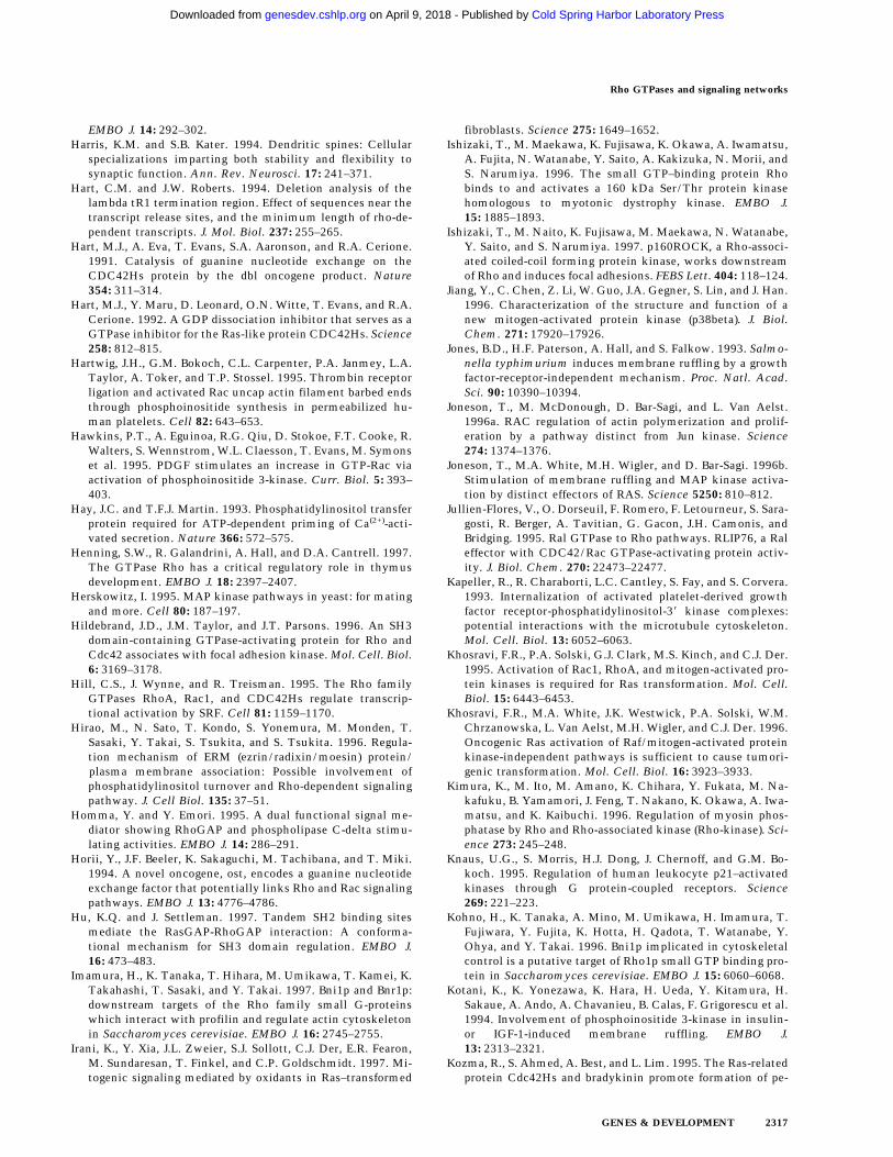
EMBO J. 14: 292–302.Harris, K.M. and S.B. Kater. 1994. Dendritic spines: Cellular
specializations imparting both stability and flexibility tosynaptic function. Ann. Rev. Neurosci. 17: 241–371.
Hart, C.M. and J.W. Roberts. 1994. Deletion analysis of thelambda tR1 termination region. Effect of sequences near thetranscript release sites, and the minimum length of rho-de-pendent transcripts. J. Mol. Biol. 237: 255–265.
Hart, M.J., A. Eva, T. Evans, S.A. Aaronson, and R.A. Cerione.1991. Catalysis of guanine nucleotide exchange on theCDC42Hs protein by the dbl oncogene product. Nature354: 311–314.
Hart, M.J., Y. Maru, D. Leonard, O.N. Witte, T. Evans, and R.A.Cerione. 1992. A GDP dissociation inhibitor that serves as aGTPase inhibitor for the Ras-like protein CDC42Hs. Science258: 812–815.
Hartwig, J.H., G.M. Bokoch, C.L. Carpenter, P.A. Janmey, L.A.Taylor, A. Toker, and T.P. Stossel. 1995. Thrombin receptorligation and activated Rac uncap actin filament barbed endsthrough phosphoinositide synthesis in permeabilized hu-man platelets. Cell 82: 643–653.
Hawkins, P.T., A. Eguinoa, R.G. Qiu, D. Stokoe, F.T. Cooke, R.Walters, S. Wennstrom, W.L. Claesson, T. Evans, M. Symonset al. 1995. PDGF stimulates an increase in GTP-Rac viaactivation of phosphoinositide 3-kinase. Curr. Biol. 5: 393–403.
Hay, J.C. and T.F.J. Martin. 1993. Phosphatidylinositol transferprotein required for ATP-dependent priming of Ca(2+)-acti-vated secretion. Nature 366: 572–575.
Henning, S.W., R. Galandrini, A. Hall, and D.A. Cantrell. 1997.The GTPase Rho has a critical regulatory role in thymusdevelopment. EMBO J. 18: 2397–2407.
Herskowitz, I. 1995. MAP kinase pathways in yeast: for matingand more. Cell 80: 187–197.
Hildebrand, J.D., J.M. Taylor, and J.T. Parsons. 1996. An SH3domain-containing GTPase-activating protein for Rho andCdc42 associates with focal adhesion kinase. Mol. Cell. Biol.6: 3169–3178.
Hill, C.S., J. Wynne, and R. Treisman. 1995. The Rho familyGTPases RhoA, Rac1, and CDC42Hs regulate transcrip-tional activation by SRF. Cell 81: 1159–1170.
Hirao, M., N. Sato, T. Kondo, S. Yonemura, M. Monden, T.Sasaki, Y. Takai, S. Tsukita, and S. Tsukita. 1996. Regula-tion mechanism of ERM (ezrin/radixin/moesin) protein/plasma membrane association: Possible involvement ofphosphatidylinositol turnover and Rho-dependent signalingpathway. J. Cell Biol. 135: 37–51.
Homma, Y. and Y. Emori. 1995. A dual functional signal me-diator showing RhoGAP and phospholipase C-delta stimu-lating activities. EMBO J. 14: 286–291.
Horii, Y., J.F. Beeler, K. Sakaguchi, M. Tachibana, and T. Miki.1994. A novel oncogene, ost, encodes a guanine nucleotideexchange factor that potentially links Rho and Rac signalingpathways. EMBO J. 13: 4776–4786.
Hu, K.Q. and J. Settleman. 1997. Tandem SH2 binding sitesmediate the RasGAP-RhoGAP interaction: A conforma-tional mechanism for SH3 domain regulation. EMBO J.16: 473–483.
Imamura, H., K. Tanaka, T. Hihara, M. Umikawa, T. Kamei, K.Takahashi, T. Sasaki, and Y. Takai. 1997. Bni1p and Bnr1p:downstream targets of the Rho family small G-proteinswhich interact with profilin and regulate actin cytoskeletonin Saccharomyces cerevisiae. EMBO J. 16: 2745–2755.
Irani, K., Y. Xia, J.L. Zweier, S.J. Sollott, C.J. Der, E.R. Fearon,M. Sundaresan, T. Finkel, and C.P. Goldschmidt. 1997. Mi-togenic signaling mediated by oxidants in Ras–transformed
fibroblasts. Science 275: 1649–1652.Ishizaki, T., M. Maekawa, K. Fujisawa, K. Okawa, A. Iwamatsu,
A. Fujita, N. Watanabe, Y. Saito, A. Kakizuka, N. Morii, andS. Narumiya. 1996. The small GTP–binding protein Rhobinds to and activates a 160 kDa Ser/Thr protein kinasehomologous to myotonic dystrophy kinase. EMBO J.15: 1885–1893.
Ishizaki, T., M. Naito, K. Fujisawa, M. Maekawa, N. Watanabe,Y. Saito, and S. Narumiya. 1997. p160ROCK, a Rho-associ-ated coiled-coil forming protein kinase, works downstreamof Rho and induces focal adhesions. FEBS Lett. 404: 118–124.
Jiang, Y., C. Chen, Z. Li, W. Guo, J.A. Gegner, S. Lin, and J. Han.1996. Characterization of the structure and function of anew mitogen-activated protein kinase (p38beta). J. Biol.Chem. 271: 17920–17926.
Jones, B.D., H.F. Paterson, A. Hall, and S. Falkow. 1993. Salmo-nella typhimurium induces membrane ruffling by a growthfactor-receptor-independent mechanism. Proc. Natl. Acad.Sci. 90: 10390–10394.
Joneson, T., M. McDonough, D. Bar-Sagi, and L. Van Aelst.1996a. RAC regulation of actin polymerization and prolif-eration by a pathway distinct from Jun kinase. Science274: 1374–1376.
Joneson, T., M.A. White, M.H. Wigler, and D. Bar-Sagi. 1996b.Stimulation of membrane ruffling and MAP kinase activa-tion by distinct effectors of RAS. Science 5250: 810–812.
Jullien-Flores, V., O. Dorseuil, F. Romero, F. Letourneur, S. Sara-gosti, R. Berger, A. Tavitian, G. Gacon, J.H. Camonis, andBridging. 1995. Ral GTPase to Rho pathways. RLIP76, a Raleffector with CDC42/Rac GTPase-activating protein activ-ity. J. Biol. Chem. 270: 22473–22477.
Kapeller, R., R. Charaborti, L.C. Cantley, S. Fay, and S. Corvera.1993. Internalization of activated platelet-derived growthfactor receptor-phosphatidylinositol-38 kinase complexes:potential interactions with the microtubule cytoskeleton.Mol. Cell. Biol. 13: 6052–6063.
Khosravi, F.R., P.A. Solski, G.J. Clark, M.S. Kinch, and C.J. Der.1995. Activation of Rac1, RhoA, and mitogen-activated pro-tein kinases is required for Ras transformation. Mol. Cell.Biol. 15: 6443–6453.
Khosravi, F.R., M.A. White, J.K. Westwick, P.A. Solski, W.M.Chrzanowska, L. Van Aelst, M.H. Wigler, and C.J. Der. 1996.Oncogenic Ras activation of Raf/mitogen-activated proteinkinase-independent pathways is sufficient to cause tumori-genic transformation. Mol. Cell. Biol. 16: 3923–3933.
Kimura, K., M. Ito, M. Amano, K. Chihara, Y. Fukata, M. Na-kafuku, B. Yamamori, J. Feng, T. Nakano, K. Okawa, A. Iwa-matsu, and K. Kaibuchi. 1996. Regulation of myosin phos-phatase by Rho and Rho-associated kinase (Rho-kinase). Sci-ence 273: 245–248.
Knaus, U.G., S. Morris, H.J. Dong, J. Chernoff, and G.M. Bo-koch. 1995. Regulation of human leukocyte p21–activatedkinases through G protein-coupled receptors. Science269: 221–223.
Kohno, H., K. Tanaka, A. Mino, M. Umikawa, H. Imamura, T.Fujiwara, Y. Fujita, K. Hotta, H. Qadota, T. Watanabe, Y.Ohya, and Y. Takai. 1996. Bni1p implicated in cytoskeletalcontrol is a putative target of Rho1p small GTP binding pro-tein in Saccharomyces cerevisiae. EMBO J. 15: 6060–6068.
Kotani, K., K. Yonezawa, K. Hara, H. Ueda, Y. Kitamura, H.Sakaue, A. Ando, A. Chavanieu, B. Calas, F. Grigorescu et al.1994. Involvement of phosphoinositide 3-kinase in insulin-or IGF-1-induced membrane ruffling. EMBO J.13: 2313–2321.
Kozma, R., S. Ahmed, A. Best, and L. Lim. 1995. The Ras-relatedprotein Cdc42Hs and bradykinin promote formation of pe-
Rho GTPases and signaling networks
GENES & DEVELOPMENT 2317
Cold Spring Harbor Laboratory Press on April 9, 2018 - Published by genesdev.cshlp.orgDownloaded from

ripheral actin microspikes and filopodia in Swiss 3T3 fibro-blasts. Mol. Cell. Biol. 15: 1942–1952.
———. 1996. The GTPase-activating protein n-chimaerin coop-erates with Rac1 and Cdc42Hs to induce the formation oflamellipodia and filopodia. Mol. Cell. Biol. 16: 5069–5080.
Kuroda, S., M. Fukata, K. Kobayashi, M. Nakafuku, N. Nomura,A. Iwamatsu, and K. Kaibuchi. 1996. Identification ofIQGAP as a putative target for the small GTPases, Cdc42and Rac1. J. Biol. Chem. 271: 23363–23367.
Kyriakis, J.M. and J. Avruch. 1996. Protein kinase cascades ac-tivated by stress and inflammatory cytokines. BioEssays18: 567–577.
Lamarche, N., N. Tapon, L. Stowers, P.D. Burbelo, P. Aspen-strom, T. Bridges, J. Chant, and A. Hall. 1996. Rac and Cdc42induce actin polymerization and G1 cell cycle progressionindependently of p65PAK and the JNK/SAPK MAP kinasecascade. Cell 87: 519–529.
Lamaze, C., T.H. Chuang, L.J. Terlecky, G.M. Bokoch, and S.L.Schmid. 1996. Regulation of receptor-mediated endocytosisby Rho and Rac. Nature 382: 177–179.
Lancaster, C.A., H.P. Taylor, A.J. Self, S. Brill, H.E. van Erp, andA. Hall. 1994. Characterization of rhoGAP. A GTPase-acti-vating protein for rho-related small GTPases. J. Biol. Chem.269: 1137–1142.
Lang, P., F. Gesbert, C.M. Delespine, R. Stancou, M. Pouchelet,and J. Bertoglio. 1996. Protein kinase A phosphorylation ofRhoA mediates the morphological and functional effects ofcyclic AMP in cytotoxic lymphocytes. EMBO J. 15: 510–519.
Lange-Carter, C., C.M. Pleiman, A.M. Gardner, K.J. Blumer, andG.L. Johnson. 1993. A divergence in the MAP kinase regu-latory network defined by MEK kinase and Raf. Science260: 315–319.
Leeuw, T., L.A. Fourest, C. Wu, J. Chenevert, K. Clark, M.Whiteway, D.Y. Thomas, and E. Leberer. 1995. Pheromoneresponse in yeast: association of Bem1p with proteins of theMAP kinase cascade and actin. Science 270: 1210–1213.
Lelias, J.M., C.N. Adra, G.M. Wulf, J.C. Guillemot, M. Khagad,D. Caput, and B. Lim. 1993. cDNA cloning of a humanmRNA preferentially expressed in hematopoietic cells andwith homology to a GDP-dissociation inhibitor for the rhoGTP-binding proteins. Proc. Natl. Acad. Sci. 90: 1479–1483.
Leonard, D., M.J. Hart, J.V. Platko, A. Eva, W. Henzel, T. Evans,and R.A. Cerione. 1992. The identification and characteriza-tion of a GDP-dissociation inhibitor (GDI) for the CDC42Hsprotein. J. Biol. Chem. 267: 22860–22868.
Leung, T., B.E. How, E. Manser, and L. Lim. 1993. Germ cellb-chimaerin, a new GTPase-activating protein for p21rac, isspecifically expressed during the acrosomal assembly stagein rat testis. J. Biol. Chem. 268: 3813–3816.
Leung, T., E. Manser, L. Tan, and L. Lim. 1995. A novel serine/threonine kinase binding the Ras-related RhoA GTPasewhich translocates the kinase to peripheral membranes. J.Biol. Chem. 270: 29051–29054.
Leung, T., X.Q. Chen, E. Manser, and L. Lim. 1996. The p160RhoA-binding kinase ROK alpha is a member of a kinasefamily and is involved in the reorganization of the cytoskel-eton. Mol. Cell. Biol. 16: 5313–5327.
Li, R. 1997. Bee1, a yeast protein with homology to Wiscott-Aldrich syndrome protein, is critical for the assembly of cor-tical actin cytoskeleton. J. Cell Biol. 136: 649–658.
Li, G., C. D’Souza-Schorey, M.A. Barbeiri, R.R. Roberts, A.Kipple, L.T. Williams, and P.D. Stahl. 1995. Evidence forphosphatidylinositol 3-kinase as a regulator of endocytosisvia activation of Rab5. Proc. Natl. Acad. Sci. 92:10207–10211.
Li, G., C. D’Souza-Schorey, M.A. Barbeiri, J.A. Cooper, and P.D.
Stahl. 1997. Uncoupling of membrane ruffling and pinocy-tosis during Ras-signal transduction. J. Biol. Chem.272: 10337–10340.
Lin, R., S. Bagrodia, R. Cerione, and D. Manor. 1997. A novelCdc42Hs mutant induces cellular transformation. Curr.Biol. (in press).
Lo, Y.Y. and T.F. Cruz. 1995. Involvement of reactive oxygenspecies in cytokine and growth factor induction of c-fos ex-pression in chondrocytes. J. Biol. Chem. 270: 11727–11730.
Ludwig, S., K. Engel, A. Hoffmeyer, G. Sithanandam, B. Neu-feld, D. Palm, M. Gaestel, and U.R. Rapp. 1996. 3pK, a novelmitogen-activated protein (MAP) kinase-activated proteinkinase, is targeted by three MAP kinase pathways. Mol. Cell.Biol. 16: 6687–6697.
Luo, L., Y.J. Liao, L.Y. Jan, and Y.N. Jan. 1994. Distinct mor-phogenetic functions of similar small GTPases: DrosophilaDrac1 is involved in axonal outgrowth and myoblast fusion.Genes & Dev. 8: 1787–17802.
Luo, L., T.K. Hensch, L. Ackerman, S. Barbel, L.Y. Jan, and Y.N.Jan. 1996. Differential effects of the Rac GTPase on Purkingecell axons and dentritic trunks and spines. Nature 379: 837–840.
Mackay, J.G., F. Esch, H. Furthmayr, and A. Hall. 1997. Rho-and Rac-dependent assembly of focal adhesion complexesand actin filaments in permeabilized fibroblasts: An essen-tial role for ERM proteins. J. Cell Biol. (in press).
Madaule, P., T. Furuyashiki, T. Reid, T. Ishizaki, G. Watanabe,N. Morii, and S. Narumiya. 1995. A novel partner forthe GTP-bound forms of rho and rac. FEBS Lett. 377: 243–248.
Manser, E., C. Chong, Z.S. Zhao, T. Leung, G. Michael, C. Hall,and L. Lim. 1995. Molecular cloning of a new member of thep21-Cdc42/Rac-activated kinase (PAK) family. J. Biol.Chem. 270: 25070–25078.
Manser, E., H.Y. Huang, T.H. Loo, X.Q. Chen, J.M. Dong, T.Leung, and L. Lim. 1997. Expression of constitutively activealpha-PAK reveals effects of the kinase on actin and focalcomplexes. Mol. Cell. Biol. 17: 1129–1143.
Marcus, S., A. Polverino, E. Chang, D. Robbins, M.H. Cobb, andM.H. Wigler. 1995. Shk1, a homolog of the Saccharomycescerevisiae Ste20 and mammalian p65PAK protein kinases, isa component of a Ras/Cdc42 signaling module in the fissionyeast Schizosaccharomyces pombe. Proc. Natl. Acad. Sci.92: 6180–6184.
Marhoul, J.F. and T.H. Adams. 1995. Identification of develop-mental regulatory genes in Aspergillus nidulans by overex-pression. Genetics 139: 537–547.
Mariot, P., A.J. O’Sullivan, A.M. Brown, and P.E. Tatham. 1996.Rho guanine nucleotide dissociation inhibitor protein (Rho-GDI) inhibits exocytosis in mast cells. EMBO J. 15: 6476–6482.
Martin, G.A., G. Bollag, F. McCormick, and A. Abo. 1995. Anovel serine kinase activated by rac1/CDC42Hs-dependentautophosphorylation is related to PAK65 and STE20. EMBOJ. 14: 1970–1978.
Martinez-Arias, A. 1993. Development and patterning of thelarval epidermis of Drosophila. In The Development of Dro-sophila. pp. 517–608. Cold Spring Harbor Laboratory Press,Cold Spring Harbor, NY.
Martys, J.L., C. Wjasow, D.M. Gangi, M.C. Kielian, T.E. Mc-Graw, and J.M. Backer. 1996. Wortmannin-sensitive traffick-ing pathways in Chinese hamster ovary cells. Differentialeffects on endocytosis and lysosomal sorting. J. Biol. Chem.271: 10953–10962.
Matsui, T., M. Amano, T. Yamamoto, K. Chihara, M. Nakaf-uku, M. Ito, T. Nakano, K. Okawa, A. Iwamatsu, and K.
Van Aelst and D’Souza-Schorey
2318 GENES & DEVELOPMENT
Cold Spring Harbor Laboratory Press on April 9, 2018 - Published by genesdev.cshlp.orgDownloaded from

Kaibuchi. 1996a. Rho-associated kinase, a novel serine/threonine kinase, as a putative target for small GTP bindingprotein Rho. EMBO J. 15: 2208–2216.
Matsui, Y., R. Matsui, R. Akada, and A. Toh-e. 1996b. Yeast srchomology region 3 domain-binding proteins involved in budformation. J. Cell Biol. 133: 865–878.
McCaffery, M., J.S. Johnson, B. Goud, A.M. Myers, J. Rossier,M.R. Popoff, P. Madaule, and P. Boquet. 1991. The smallGTP-binding protein Rho1p is localized on the Golgi appa-ratus and post Golgi vesicles in Saccharomyces cerevisiae. J.Cell Biol. 115: 309–319.
McCallum, S.J., W.J. Wu, and R.A. Cerione. 1996. Identificationof a putative effector for Cdc42Hs with high sequence simi-larity to the RasGAP-related protein IQGAP1 and a Cdc42Hsbinding partner with similarity to IQGAP2. J. Biol. Chem.271: 21732–21737.
Michiels, F., G.G. Habets, J.C. Stam, R.A. van der Kammen, andJ.G. Collard. 1995. A role for Rac in Tiam1-induced mem-brane ruffling and invasion. Nature 375: 338–340.
Michiels, F., J.C. Stam, P.L. Hordijk, R.A. van der Kammen, L.Ruuls-Van Stalle, C.A. Feltkamp, and J.G. Collard. 1997.Regulated membrane localization of Tiam1, mediated by theNH2-terminal pleckstrin homology domain, is required forRac-dependent membrane ruffling and C-Jun NH2-terminalkinase activation. J. Cell Biol. 137: 387–398.
Miki, T., C.L. Smith, J.E. Long, A. Eva, and T.P. Fleming. 1993.Oncogene ect2 is related to regulators of small GTP-binding.Nature 362: 462–465.
Minden, A., A. Lin, F.X. Claret, A. Abo, and M. Karin. 1995.Selective activation of the JNK signaling cascade and c-Juntranscriptional activity by the small GTPases Rac andCdc42Hs. Cell 81: 1147–1157.
Mitchell, H.K., J. Roach, and N.S. Petersen. 1983. The morpho-genesis of cell hairs on Drosophila wings. Dev. Biol. 95: 387–398.
Murphy, A.M. and D.J. Montell. 1996. Cell type-specific rolesfor Cdc42, Rac, and RhoL in Drosophila oogenesis. J. CellBiol. 133: 617–630.
Murphy, C., R. Saffrich, M. Grummt, H. Gournier, V. Rybin, M.Rubino, P. Auvinen, A. Lutcke, R.G. Parton, and M. Zerial.1996. Endosome dynamics regulated by a Rho protein. Na-ture 384: 427–432.
Na, S., T.H. Chuang, A. Cunningham, T.G. Turi, J.H. Hanke,G.M. Bokoch, and D.E. Danley. 1996. D4-GDI, a substrate ofCPP32, is proteolyzed during Fas-induced apoptosis. J. Biol.Chem. 271: 11209–11213.
Nakagawa, O., K. Fujisawa, T. Ishizaki, Y. Saito, K. Nakao, andS. Narumiya. 1996. ROCK-I and ROCK-II, two isoforms ofRho-associated coiled-coil forming protein serine/threoninekinase in mice. FEBS Lett. 392: 189–193.
Narumiya, S., T. Ishizaki, and N. Watanabe. 1997. Rho effectorsand reorganization of actin cytoskeleton. FEBS Lett. 410:68–72.
Nishida, E. 1985. Opposite effects of cofilin and profilin fromporcine brain on rate of exchange of actin-bound adenosine58-triphosphate. Biochemistry 24: 1160–1164.
Nishiyama, T., T. Sasaki, K. Takaishi, M. Kato, H. Yaku, K.Araki, Y. Matsuura, and Y. Takai. 1994. rac p21 is involvedin insulin-induced membrane ruffling and rho p21 is in-volved in hepatocyte growth factor- and 12-O-tetradec-anoylphorbol-13-acetate (TPA)-induced membrane rufflingin KB cells. Mol. Cell. Biol. 14: 2447–2456.
Nobes, C.D. and A. Hall. 1995. Rho, rac, and cdc42 GTPasesregulate the assembly of multimolecular focal complexesassociated with actin stress fibers, lamellipodia, and filopo-dia. Cell 81: 53–62.
Nobes, C.D., P. Hawkins, L. Stephens, and A. Hall. 1995. Acti-vation of the small GTP-binding proteins rho and rac bygrowth factor receptors. J. Cell Sci. 108: 225–233.
Nonaka, H., K. Tanaka, H. Hirano, T. Fujiwara, H. Kohno, M.Umikawa, A. Mino, and Y. Takai. 1995. A downstream tar-get of RHO1 small GTP-binding protein is PKC1, a homologof protein kinase C, which leads to activation of the MAPkinase cascade in Saccharomyces cerevisiae. EMBO J.14: 5931–5938.
Norman, J.C., L.S. Price, A.J. Ridley, and A. Koffer. 1996. Thesmall GTP-binding proteins, Rac and Rho, regulate cytoskel-etal organization and exocytosis in mast cells by parallelpathways. Mol. Biol. Cell 7: 1429–1442.
Novick, P., C. Field, and R. Scheckman. 1980. Identification of23 complementation groups required for post-translationalevents in the yeast secretory pathway. Cell 21: 205–215.
Nusslein-Volhard, C., E. Weischus, and H. Kluding. 1984. Mu-tations affecting the pattern of the larval cuticle in Dro-sophila melanogaster. I. Zygotic loci on the second chromo-some. Arch. Dev. Biol. 193: 267–282.
O’Hagan, R.C., R.G. Tozer, M. Symons, F. McCormick, and J.A.Hassell. 1996. The activity of the Ets transcription factorPEA3 is regulated by two distinct MAPK cascades. Onco-gene 13: 1323–1333.
Olson, M.F., A. Ashworth, and A. Hall. 1995. An essential rolefor Rho, Rac, and Cdc42 GTPases in cell cycle progressionthrough G1. Science 269: 1270–1272.
Olson, M.F., N.G. Pasteris, J.L. Gorski, and A. Hall. 1996. Fa-ciogenital dysplasia protein (FGD1) and Vav, two related pro-teins required for normal embryonic development, are up-stream regulators of Rho GTPases. Curr. Biol. 6: 1628–1633.
Otsu, M., I. Hiles, I. Gout, M.J. Fry, F. Ruiz-Larrea, G. Panay-otou, A. Thompson, R. Dhand, J. Hsuan, N. Totty et al. 1991.Characterization of two 85 kd proteins that associate withreceptor tyrosine kinases, middle-T/pp60c-src complexes,and P13-kinase. Cell 65: 91–104.
Ottilie, S., P.J. Miller, D.I. Johnson, C.L. Creasy, M.A. Sells, S.Bagrodia, S.L. Forsburg, and J. Chernoff. 1995. Fission yeastpak1+ encodes a protein kinase that interacts with Cdc42pand is involved in the control of cell polarity and mating.EMBO J. 14: 5908–5919.
Park, S.H. and R.A. Weinberg. 1995. A putative effector of Ralhas homology to Rho/Rac GTPase activating proteins. On-cogene 11: 2349–2355.
Pasteris, N.G., A. Cadle, L.J. Logie, M.E. Porteous, C.E.Schwartz, R.E. Stevenson, T.W. Glover, R.S. Wilroy, and J.L.Gorski. 1994. Isolation and characterization of the faciogeni-tal dysplasia (Aarskog-Scott syndrome) gene: A putativeRho/Rac guanine nucleotide exchange factor. Cell 79: 669–678.
Peifer, M.S., S. Orsulic, D. Sweeton, and E. Wieschaus. 1993. Arole for Drosophila segment polarity gene armadillo in celladhesion and cytoskeletal intergrity during oogenesis. De-velopment 118: 1191–1207.
Perona, R., S. Montaner, L. Saniger, P.I. Sanchez, R. Bravo, andJ.C. Lacal. 1997. Activation of the nuclear factor-kB by Rho,CDC42, and Rac-1 proteins. Genes & Dev. 11: 463–475.
Pesacreta, T.C., T.J. Byers, R. Dubreuil, D.P. Kiehart, and D.Branton. 1989. Drosophila spectrin: The membrane skeletonduring embryogenesis. J. Cell Biol. 108: 1697–1709.
Peter, M., A.M. Neiman, H.O. Park, M. Lohuizen, and I. Her-skowitz. 1997. Functional analysis of the interaction be-tween the small GTP binding protein Cdc42 and the Ste20protein kinase in yeast. EMBO J. 15: 7046–7059.
Peters, P.J., V.W. Hsu, C.E. Ooi, D. Finazzi, S.B. Teal, V. Oor-schot, J.G. Donaldson, and R.D. Klausner. 1995. Overexpres-
Rho GTPases and signaling networks
GENES & DEVELOPMENT 2319
Cold Spring Harbor Laboratory Press on April 9, 2018 - Published by genesdev.cshlp.orgDownloaded from
sion of wild-type and mutant ARF1 and ARF6: Distinct per-turbations of nonoverlapping membrane compartments. J.Cell Biol. 128: 1003–1017.
Platko, J.V., D.A. Leonard, C.N. Adra, R.J. Shaw, R.A. Cerione,and B. Lim. 1995. A single residue can modify target-bindingaffinity and activity of the functional domain of the Rho-subfamily GDP dissociation inhibitors. Proc. Natl. Acad.Sci. 92: 2974–2978.
Poodry, C.A. 1980. Imaginal discs: Morphology and develop-ment. In Genetics and Biology of Drosophila, Vol. 2, pp.407–441. Acedemic Press, Orlando, FL.
Proud, C.G. 1996. p70 S6 kinase: An enigma with variations.Trends Biochem. Sci. 21: 181–185.
Pulverer, B.J., J.M. Kyriakis, J. Avruch, E. Nikolakaki, and J.R.Woodgett. 1991. Phosphorylation of c-jun mediated by MAPkinases. Nature 353: 670–674.
Qadota, H., C.P. Python, S.B. Inoue, M. Arisawa, Y. Anraku, Y.Zheng, T. Watanabe, D.E. Levin, and Y. Ohya. 1996. Identi-fication of yeast Rho1p GTPase as a regulatory subunit of1,3-b-glucan synthase. Science 272: 279–281.
Qiu, R.-G. 1997. The role of Rho-like GTPases in cell prolifera-tion and Ras transformation. ‘‘Ph.D. thesis’’
Qiu, R.G., J. Chen, D. Kirn, F. McCormick, and M. Symons.1995a. An essential role for Rac in Ras transformation. Na-ture 374: 457–459.
Qiu, R.G., J. Chen, F. McCormick, and M. Symons. 1995b. Arole for Rho in Ras transformation. Proc. Natl. Acad. Sci.92: 11781–11785.
Qiu, R.G., A. Abo, F. McCormick, and M. Symons. 1997. Cdc42regulates anchorage-independent growth and is necessary forRas transformation. Mol. Cell. Biol. 17: 3449–3458.
Quilliam, L.A., Q.T. Lambert, Y.L. Mickelson, J.K. Westwick,A.B. Sparks, B.K. Kay, N.A. Jenkins, D.J. Gilbert, N.G. Cope-land, and C.J. Der. 1996. Isolation of a NCK-associated ki-nase, PRK2, an SH3-binding protein and potential effector ofRho protein signaling. J. Biol. Chem. 271: 28772–28776.
Radhakrishna, H., R.D. Klausner, and J.G. Donaldson. 1996.Aluminum fluoride stimulates surface protrusions in cellsoverexpressing the ARF6 GTPase. J. Cell Biol. 134: 935–946.
Raingeaud, J., A.J. Whitmarsh, T. Barrett, B. Derijard, and R.J.Davis. 1996. MKK3- and MKK6-regulated gene expression ismediated by the p38 mitogen-activated protein kinase signaltransduction pathway. Mol. Cell. Biol. 16: 1247–1255.
Reid, T., T. Furuyashiki, T. Ishizaki, G. Watanabe, N. Watan-abe, K. Fujisawa, N. Morii, P. Madaule, and S. Narumiya.1996. Rhotekin, a new putative target for Rho bearing ho-mology to a serine/threonine kinase, PKN, and rhophilin inthe rho-binding domain. J. Biol. Chem. 271: 13556–13560.
Reif, K., C.D. Nobes, G. Thomas, A. Hall, and D.A. Cantrell.1996. Phosphatidylinositol 3-kinase signals activate a selec-tive subset of Rac/Rho–dependent effector pathways. Curr.Biol. 6: 1445–1455.
Reinhard, J., A.A. Scheel, D. Diekmann, A. Hall, C. Ruppert,and M. Bahler. 1995. A novel type of myosin implicated insignalling by rho family GTPases. EMBO J. 14: 697–704.
Ren, X.D., G.M. Bokoch, K.A. Traynor, G.H. Jenkins, R.A.Anderson, and M.A. Schwartz. 1996. Physical association ofthe small GTPase Rho with a 68-kDa phosphatidylinositol4–phosphate 5–kinase in Swiss 3T3 cells. Mol. Biol. Cell7: 435–442.
Renshaw, M.W., C.E. Lea, and J.Y. Wang. 1996. Rac is requiredfor v-Abl tyrosine kinase to activate mitogenesis. Curr. Biol.6: 76–83.
Ridley, A.J. and A. Hall. 1992. The small GTP-binding proteinrho regulates the assembly of focal adhesions and actin stressfibers in response to growth factors. Cell 70: 389–399.
Ridley, A.J., P.M. Comoglio, and A. Hall. 1995. Regulation ofscatter factor/hepatocyte growth factor responses by Ras,Rac, and Rho in MDCK cells. Mol. Cell. Biol. 15: 1110–1122.
Ridley, A.J., H.F. Paterson, C.L. Johnston, D. Diekmann, and A.Hall. 1992. The small GTP-binding protein rac regulatesgrowth factor-induced membrane ruffling. Cell 70: 401–410.
Ridley, A.J., A.J. Self, F. Kasmi, H.F. Paterson, A. Hall, C.J. Mar-shall, and C. Ellis. 1993. Rho family GTPase activating pro-teins p190, bcr and rhoGAP show distinct specificities invitro and in vivo. EMBO J. 12: 5151–5160.
Riesgo-Escovar, J.R., M. Jenni, A. Fritz, and E. Hafen. 1996. TheDrosophila Jun-N-terminal kinase is required for cell mor-phogenesis but not for DJun-dependent cell fate specificationin the eye. Genes & Dev. 10: 2759–2768.
Rodriguez, V.P., P.H. Warne, A. Khwaja, B.M. Marte, D. Pappin,P. Das, M.D. Waterfield, A. Ridley, and J. Downward. 1997.Role of phosphoinositide 3-OH kinase in cell transformationand control of the actin cytoskeleton by Ras. Cell 89: 457–467.
Ron, D., M. Zannini, M. Lewis, R.B. Wickner, L.T. Hunt, G.Graziani, S.R. Tronick, S.A. Aaronson, and A. Eva. 1991. Aregion of proto-dbl essential for its transforming activityshows sequence similarity to a yeast cell cycle gene, CDC24,and the human breakpoint cluster gene, bcr. New Biol.3: 372–379.
Rouse, J., P. Cohen, S. Trigon, M. Morange, L.A. Alonso, D.Zamanillo, T. Hunt, and A.R. Nebreda. 1994. A novel kinasecascade triggered by stress and heat shock that stimulatesMAPKAP kinase-2 and phosphorylation of the small heatshock proteins. Cell 78: 1027–1037.
Sanchez, I., R.T. Hughes, B.J. Mayer, K. Yee, J.R. Woodgett, J.Avruch, J.M. Kyriakis, and L.I. Zon. 1994. Role of SAPK/ERK kinase-1 in the stress-activated pathway regulatingtranscription factor c-Jun. Nature 372: 794–798.
Scherle, P., T. Behrens, and L.M. Staudt. 1993. Ly-GDI, a GDP-dissociation inhibitor of the RhoA GTP-binding protein, isexpressed preferentially in lymphocytes. Proc. Natl. Acad.Sci. 90: 7568–7572.
Schmalzing, G., H.P. Richter, A. Hansen, W. Schwarz, I. Just,and K. Aktories. 1995. Involvement of the GTP binding pro-tein Rho in constitutive endocytosis in Xenopus laevis oo-cytes. J. Cell Biol. 130: 1319–1332.
Schmidt, A., J. Kunz, and M.N. Hall. 1996. TOR2 is required fororganization of the actin cytoskeleton in yeast. Proc. Natl.Acad. Sci. 93: 13780–13785.
Schmidt, A., M. Bickle, T. Beck, and M.N. Hall. 1997. The yeastphosphatidylinositol kinase homolog TOR2 activates RHO1and RHO2 via the exchange factor ROM2. Cell 88: 531–542.
Schreck, R., P. Rieber, and P.A. Baeuerle. 1991. Reactive oxygenintermediates as apparently widely used messengers in theactivation of the NF–kB transcription factor and HIV-1.EMBO J. 10: 2247–2258.
Seger, R. and E.G. Krebs. 1995. The MAPK signaling cascade.FASEB J. 9: 726–735.
Sells, M.A. and J. Chernoff. 1997. Emerging from the Pak: Thep21-activated protein kinase family. Trends Cell Biol.7: 162–167.
Sells, M., U. Knaus, S. Bagrodia, D.M. Ambrose, G.M. Bokoch,and J. Chernoff. 1997. Human p21-activated kinase (Pak1)regulates actin organization in mammalian cells. Curr. Biol.7: 202–210.
Settleman, J., V. Narasimhan, L.C. Foster, and R.A. Weinberg.1992. Molecular cloning of cDNAs encoding the GAP-asso-ciated protein p190: Implications for a signaling pathwayfrom ras to the nucleus. Cell 69: 539–549.
Shepherd, P.R., M.A. Soos, and K. Siddle. 1995. Inhibitors of
Van Aelst and D’Souza-Schorey
2320 GENES & DEVELOPMENT
Cold Spring Harbor Laboratory Press on April 9, 2018 - Published by genesdev.cshlp.orgDownloaded from
phosphoinositide 3-kinase block exocytosis but not endocy-tosis of transferrin receptors in 3T3-L1 adipocytes. Biochem.Biophys. Res. Commun. 211: 535–539.
Shou, C., C.L. Farnsworth, B.G. Neel, L.A. Feig. 1992. Molecu-lar cloning of cDNAs encoding a guanine-nucleotide-releas-ing factor for Ras p21. Nature 358: 351–354.
Sluss, H.K., Z. Han, T. Barrett, R.J. Davis, and Y.T. Ip. 1996. AJNK signal transduction pathway that mediates morphogen-esis and an immune response in Drosophila. Genes & Dev.10: 2745–2758.
Small, J.V. 1994. Lamellipodia architecture: Actin filamentturnover and the lateral flow of actin filaments during mo-tility. Semin. Cell Biol. 5: 157–163.
Sone, M., M. Hoshino, E. Suzuki, S. Kuroda, K. Kaibuchi, H.Nakagoshi, K. Saigo, Y. Nabeshima, and C. Hama. 1997. Stilllife, a protein in synaptic terminals of Drosophila homolo-gous to GDP-GTP exchangers. Science 275: 543–547.
Stevenson, B.J., B. Ferguson, V.C. De, E. Bi, J.R. Pringle, G. Am-merer, and G.J. Sprague. 1995. Mutation of RGA1, whichencodes a putative GTPase-activating protein for the polar-ity-establishment protein Cdc42p, activates the pheromone-response pathway in the yeast Saccharomyces cerevisiae.Genes & Dev. 9: 2949–2963.
Stossel, T.P. 1993. On the crawling of animal cells. Science260: 1086–1094.
Stowers, L., D. Yelon, L. Berg, and J. Chant. 1995. Regulation ofthe polarization of T-cells toward antigen presenting cells bythe Ras-related GTPase Cdc42. Proc. Natl. Acad. Sci.92: 5027–5031.
Strutt, D.I., U. Weber, and M. Mlodzik. 1997. The role of RhoAin tissue polarity and Frizzled signalling. Nature 387: 292–295.
Sulciner, D.J., K. Irani, Z.X. Yu, V.J. Ferrans, C.P. Goldschmidt,and T. Finkel. 1996. rac1 regulates a cytokine-stimulated,redox-dependent pathway necessary for NF-kB activation.Mol. Cell. Biol. 16: 7115–7121.
Swanson, J.A. and S.C. Baer. 1995. Phagocytosis by zippers andtriggers. Trends Cell Biol. 5: 89–93.
Symons, M. 1995. The Rac and Rho pathways as a source of drugtargets for Ras-mediated malignancies. Curr. Opin. Biotech-nol. 6: 668–674.
———. 1996. Rho family GTPases: The cytoskeleton and be-yond. Trends Biochem. Sci. 21: 178–181.
Symons, M., J.M. Derry, B. Karlak, S. Jiang, V. Lemahieu, F.McCormick, U. Francke, and A. Abo. 1996. Wiskott-Aldrichsyndrome protein, a novel effector for the GTPaseCDC42Hs, is implicated in actin polymerization. Cell84: 723–734.
Takahashi, K., T. Sasaki, A. Mammoto, K. Takaishi, T. Kam-eyama, Sa Tsukita, Sh. Tsukita, and Y. Takai. 1997. Directinteraction of the Rho-GDP dissociation inhibitor withezrin/radixin/moesin initiates the activation of the Rhosmall G protein. J. Biol. Chem. (in press).
Takai, Y., T. Sasaki, K. Tanaka, and H. Nakanishi. 1995. Rho asa regulator of the cytoskeleton. Trends Biochem. Sci.20: 227–231.
Tan, J.L., S. Ravid, and J.A. Spudich. 1992. Control of non-muscle myosins by phosphorylation. Annu. Rev. Biochem.61: 721–759.
Tan, E.C., T. Leung, E. Manser, and L. Lim. 1993. The humanactive breakpoint cluster region-related gene encodes a brainprotein with homology to guanine nucleotide exchange pro-teins and GTPase-activating proteins. J. Biol. Chem.268: 27291–27298.
Tarakhovsky, A., M. Turner, S. Schaal, P.J. Mee, L.P. Duddy, K.Rajewsky, and V.L. Tybulewicz. 1995. Defective antigen re-
ceptor-mediated proliferation of B and T cells in the absenceof Vav. Nature 374: 467–470.
Teramoto, H., O.A. Coso, H. Miyata, T. Igishi, T. Miki, and J.S.Gutkind. 1996a. Signaling from the small GTP-binding pro-teins Rac1 and Cdc42 to the c-Jun N-terminal kinase/stress-activated protein kinase pathway. A role for mixed lineagekinase 3/protein-tyrosine kinase 1, a novel member of themixed lineage kinase family. J. Biol. Chem.271: 27225–27228.
Teramoto, H., P. Crespo, O.A. Coso, T. Igishi, N. Xu, and J.S.Gutkind. 1996b. The small GTP-binding protein rho acti-vates c-Jun N-terminal kinases/stress-activated protein ki-nases in human kidney 293T cells. Evidence for a Pak-inde-pendent signaling pathway. J. Biol. Chem.271: 25731–25734.
Tibbles, L.A., Y.L. Ing, F. Kiefer, J. Chan, N. Iscove, J.R.Woodgett, and N.J. Lassam. 1996. MLK-3 activates theSAPK/JNK and p38/RK pathways via SEK1 and MKK3/6.EMBO J. 15: 7026–7035.
Tolias, K.F., L.C. Cantley, and C.L. Carpenter. 1995. Rho familyGTPases bind to phosphoinositide kinases. J. Biol. Chem.270: 17656–17659.
Treisman, R. 1990. The SRE: A growth factor responsive tran-scriptional regulator. Semin. Cancer Biol. 1: 47–58.
———. 1994. Ternary complex factors: Growth factor regulatedtranscriptional activators. Curr. Opin. Genet. Dev. 4: 96–101.
Tsuchiya, M., S.R. Price, S.-C. Tsai, J. Moss, and M. Vaughan.1991. Molecular identification of ADP-ribosylation factormRNAs and their expression in mammalian cells. J. Biol.Chem. 266: 2772–2777.
Tsukita, S., K. Oishi, N. Sato, J. Sagara, A. Kawai, and S. Tsu-kita. 1994. ERM family members as molecular linkers be-tween the cell surface glycoprotein CD44 and actin-basedcytoskeletons. J. Cell Biol. 126: 391–401.
Tsukita, S., S. Yonemura, and S. Tsukita. 1997. ERM proteins:Head-to-tail regulation of actin-plasma membrane interac-tion. Trends Biochem. Sci. 22: 53–58.
Ueda, T., A. Kikuchi, N. Ohga, J. Yamamoto, and Y. Takai.1990. Purification and characterization from bovine braincytosol of a novel regulatory protein inhibiting the dissocia-tion of GDP from and the subsequent binding of GTP to rhoBp20, a ras p21-like GTP-binding protein. J. Biol. Chem.265: 9373–9380.
Van Aelst, L., M.A. White, and M.H. Wigler. 1994. Ras partners.Cold Spring Harbor Symp. Quant. Biol. 59: 181–186.
Van Aelst, L., T. Joneson, and D. Bar-Sagi. 1996. Identification ofa novel Rac1-interacting protein involved in membrane ruf-fling. EMBO J. 15: 3778–3786.
van Leeuwen, F., R.A. van der Kammen, G.G. Habets, and J.G.Collard. 1995. Oncogenic activity of Tiam1 and Rac1 inNIH3T3 cells. Oncogene 11: 2215–2221.
Varner, J.A. and D.A. Cheresh. 1996. Integrins and cancer. Curr.Opin. Cell Biol. 8: 724–730.
Verma, I.M., J.K. Stevenson, E.M. Schwarz, D. Van Antwerp,and S. Miyamoto. 1995. Rel/NF-kB/IkB family: Intimatetales of association and dissociation. Genes & Dev. 9: 2723–2735.
Vincent, S. and J. Settleman. 1997. The PRK2 kinase is a poten-tial effector target of both Rho and Rac GTPases and regu-lates actin cytoskeletal organization. Mol. Cell. Biol.17: 2247–2256.
Voncken, J.W., H. van Schaik, V. Kaartinen, K. Deemer, T.Coates, B. Landing, P. Pattengale, O. Dorseuilli, G.M. Bo-koch, J. Groffen et al. 1995. Increased neutrophil respiratoryburst in bcr-null mutants. Cell 80: 719–728.
Rho GTPases and signaling networks
GENES & DEVELOPMENT 2321
Cold Spring Harbor Laboratory Press on April 9, 2018 - Published by genesdev.cshlp.orgDownloaded from

Wang, X.Z. and D. Ron. 1996. Stress-induced phosphorylationand activation of the transcription factor CHOP (GADD153)by p38 MAP kinase. Science 272: 1347–1349.
Watanabe, G., Y. Saito, P. Madaule, T. Ishizaki, K. Fujisawa, N.Morii, H. Mukai, Y. Ono, A. Kakizuka, and S. Narumiya.1996. Protein kinase N (PKN) and PKN–related proteinrhophilin as targets of small GTPase Rho. Science 271: 645–648.
Watanabe, N., P. Madaule, T. Reid, T. Ishizaki, G. Watanabe, A.Kakizuka, Y. Saito, K. Nakao, B. Jockusch, and S. Narumiya.1997. p140mDia, a mammalian homolog of Drosophila di-aphanous, is a target protein for Rho small GTPase and is aligand for profilin. EMBO J. 16: 3044–3056.
Wennstrom, S., P. Hawkins, F. Cooke, K. Hara, K. Yonezawa, M.Kasuga, T. Jackson, W.L. Claesson, and L. Stephens. 1994.Activation of phosphoinositide 3-kinase is required forPDGF-stimulated membrane ruffling. Curr. Biol.4: 385–393.
Westwick, J.K., Q.T. Lambert, G.J. Clark, M. Symons, L. VanAelst, R.G. Pestell, and C.J. Der. 1997. Rac regulation oftransformation, gene expression, and actin organization bymultiple, PAK-independent pathways. Mol. Cell. Biol.17: 1324–1335.
White, M.A., C. Nicolette, A. Minden, A. Polverino, L. VanAelst, M. Karin, and M.H. Wigler. 1995. Multiple Ras func-tions can contribute to mammalian cell transformation. Cell80: 533–541.
Whiteway, M., L. Hougan, D. Dignard, D.Y. Thomas, L. Bell,G.C. Saari, F.J. Grant, P. O’Hara, and V.L. MacKay. 1989.The STE4 and STE18 genes of yeast encode potential betaand gamma subunits of the mating factor receptor-coupled Gprotein. Cell 56: 467–477.
Whitmarsh, A.J., S.H. Yang, M.S. Su, A.D. Sharrocks, and R.J.Davis. 1997. Role of p38 and JNK mitogen-activated proteinkinases in the activation of ternary complex factors. Mol.Cell. Biol. 17: 2360–2371.
Wissmann, A., J. Ingles, J.D. McGhee, and P.E. Mains. 1997.Caenorhabditis elegans LET-502 is related to Rho-bindingkinases and human myotonic dystrophy kinase and interactsgenetically with a homolog of the regulatory subunit ofsmooth muscle myosin phosphatase to affect cell shape.Genes & Dev. 11: 409–422.
Wong, L.L. and P.N. Adler. 1993. Tissue polarity genes of Dro-sophila regulate the subcellular location for prehair initia-tion in pupal wing cells. J. Cell Biol. 123: 209–221.
Xu, S., D. Robbins, J. Frost, A. Dang, C.C. Lange, and M.H.Cobb. 1995. MEKK1 phosphorylates MEK1 and MEK2 butdoes not cause activation of mitogen-activated protein ki-nase. Proc. Natl. Acad. Sci. 92: 6808–6812.
Yamochi, W., K. Tanaka, H. Nonaka, A. Maeda, T. Musha, andY. Takai. 1994. Growth site localization of Rho1 small GTP-binding protein and its involvement in bud formation inSaccharomyces cerevisiae. J. Cell Biol. 125: 1077–1093.
Young, P.E., A.M. Richman, A.S. Ketchum, and D.P. Kiehart.1993. Morphogenesis in Drosophila requires nonmusclemyosin heavy chain function. Genes & Dev. 7: 29–41.
Zalcman, G., V. Closson, J. Camonis, N. Honore, M.M. Rous-seau, A. Tavitian, and B. Olofsson. 1996. Rho-GDI-3 is a newGDP dissociation inhibitor (GDI). Identification of a non-cytosolic GDI protein interacting with the small GTP-bind-ing proteins RhoB and RhoG. J. Biol. Chem. 271: 30366–30374.
Zervos, A.S., L. Faccio, J.P. Gatto, J.M. Kyriakis, and R. Brent.1995. Mxi2, a mitogen-activated protein kinase that recog-nizes and phosphorylates Max protein. Proc. Natl. Acad. Sci.92: 10531–10534.
Zhang, R., F.W. Alt, L. Davidson, S.H. Orkin, and W. Swat.1995a. Defective signalling through the T- and B-cell antigenreceptors in lymphoid cells lacking the vav proto-oncogene.Nature 374: 470–473.
Zhang, S., J. Han, M.A. Sells, J. Chernoff, U.G. Knaus, R.J. Ulev-itch, and G.M. Bokoch. 1995b. Rho family GTPases regulatep38 mitogen-activated protein kinase through the down-stream mediator Pak1. J. Biol. Chem. 270: 23934–23936.
Zhao, Z.S., T. Leung, E. Manser, and L. Lim. 1995. Pheromonesignalling in Saccharomyces cerevisiae requires the smallGTP-binding protein Cdc42p and its activator CDC24. Mol.Cell. Biol. 15: 5246–5257.
Zheng, Y., R. Cerione, and A. Bender. 1994. Control of the yeastbud-site assembly GTPase Cdc42. Catalysis of guaninenucleotide exchange by Cdc24 and stimulation of GTPaseactivity by Bem3. J. Biol. Chem. 269: 2369–2372.
Zheng, Y., A. Bender, and R.A. Cerione. 1995. Interactionsamong proteins involved in bud-site selection and bud-siteassembly in Saccharomyces cerevisiae. J. Biol. Chem.270: 626–630.
Zheng Y., D. Zangrilli, R.A. Cerione, and A. Eva. 1996. Thepleckstrin homology domain mediates transformation byoncogenic dbl through specific intracellular targeting. J. Biol.Chem. 271: 19017–19020.
Zigmond, S.H. 1996. Signal transduction and actin filament or-ganization. Curr. Opin. Cell Biol. 8: 66–73.
Ziman, M., D. Preuss, J. Mulholland, J. O’Brien, D. Botstein, andD.I. Johnson. 1993. Subcellular localization of Cdc42p, aSaccharomyces cerevisiae GTP-binding protein involved incontrol of cell polarity. Mol. Cell. Biol. 4: 1307–1316.
Van Aelst and D’Souza-Schorey
2322 GENES & DEVELOPMENT
Cold Spring Harbor Laboratory Press on April 9, 2018 - Published by genesdev.cshlp.orgDownloaded from

10.1101/gad.11.18.2295Access the most recent version at doi: 11:1997, Genes Dev.
Linda Van Aelst and Crislyn D'Souza-Schorey Rho GTPases and signaling networks
References
http://genesdev.cshlp.org/content/11/18/2295.full.html#ref-list-1
This article cites 286 articles, 156 of which can be accessed free at:
License
ServiceEmail Alerting
click here.right corner of the article or
Receive free email alerts when new articles cite this article - sign up in the box at the top
Cold Spring Harbor Laboratory Press
Cold Spring Harbor Laboratory Press on April 9, 2018 - Published by genesdev.cshlp.orgDownloaded from