ribosomes structure & function
TRANSCRIPT
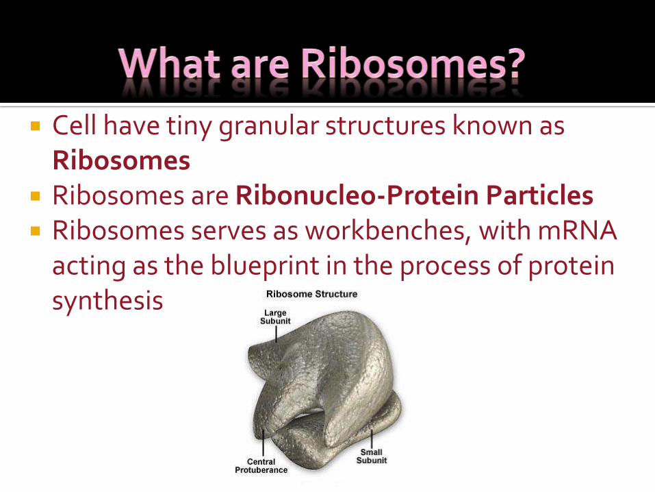
Cell have tiny granular structures known as Ribosomes
Ribosomes are Ribonucleo-Protein Particles Ribosomes serves as workbenches, with mRNA
acting as the blueprint in the process of protein synthesis
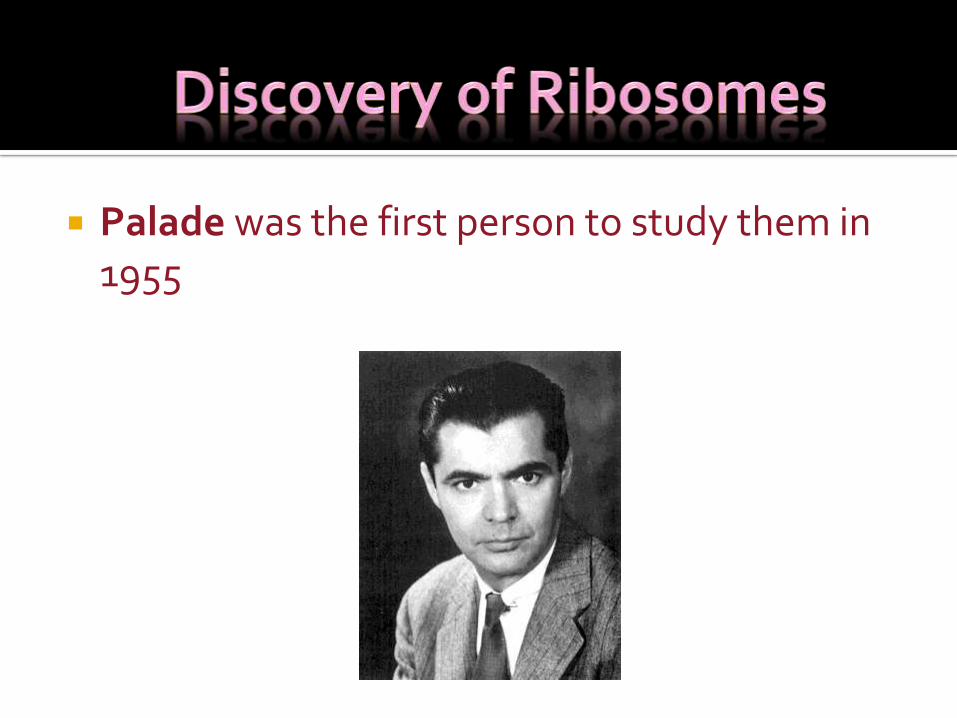
Palade was the first person to study them in 1955
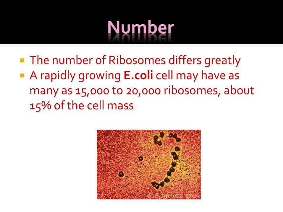
The number of Ribosomes differs greatly A rapidly growing E.coli cell may have as
many as 15,000 to 20,000 ribosomes, about 15% of the cell mass
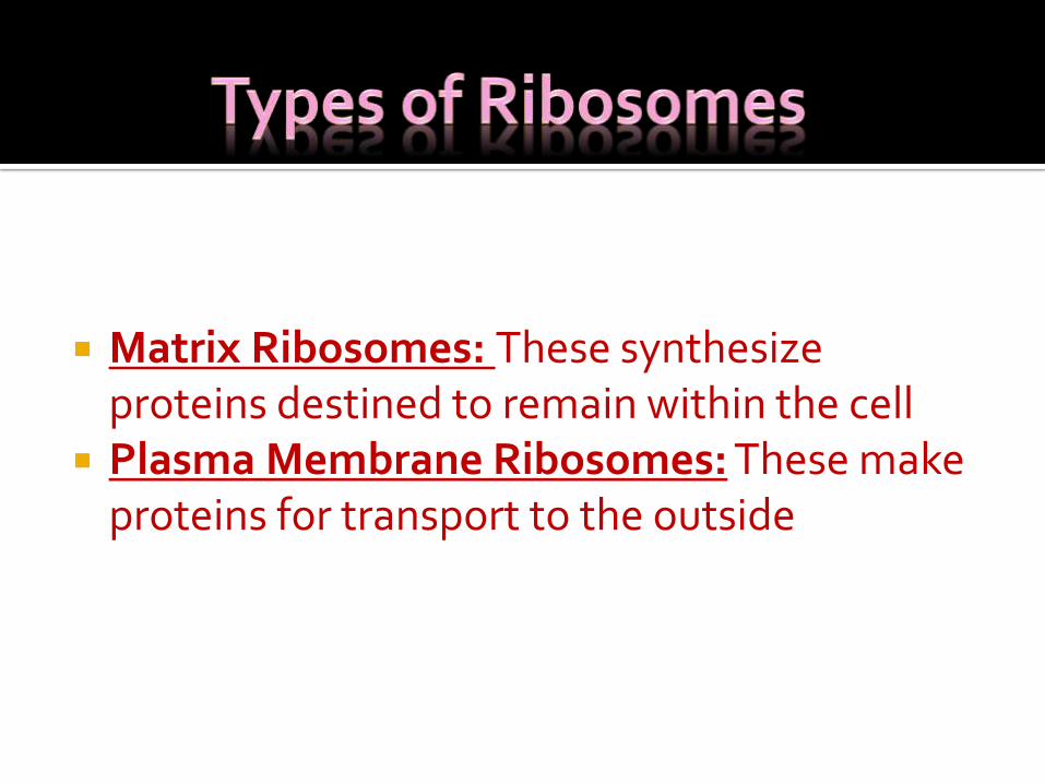
Matrix Ribosomes: These synthesize proteins destined to remain within the cell
Plasma Membrane Ribosomes: These make proteins for transport to the outside
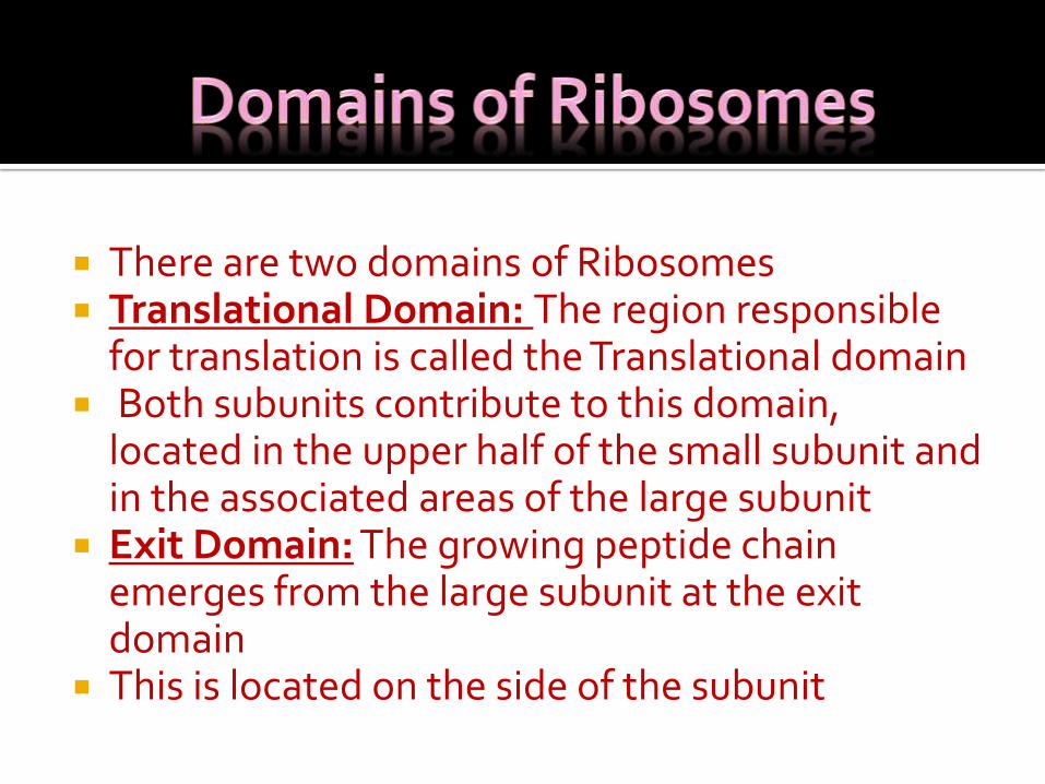
There are two domains of Ribosomes Translational Domain: The region responsible
for translation is called the Translational domain Both subunits contribute to this domain,
located in the upper half of the small subunit and in the associated areas of the large subunit
Exit Domain: The growing peptide chain emerges from the large subunit at the exit domain
This is located on the side of the subunit
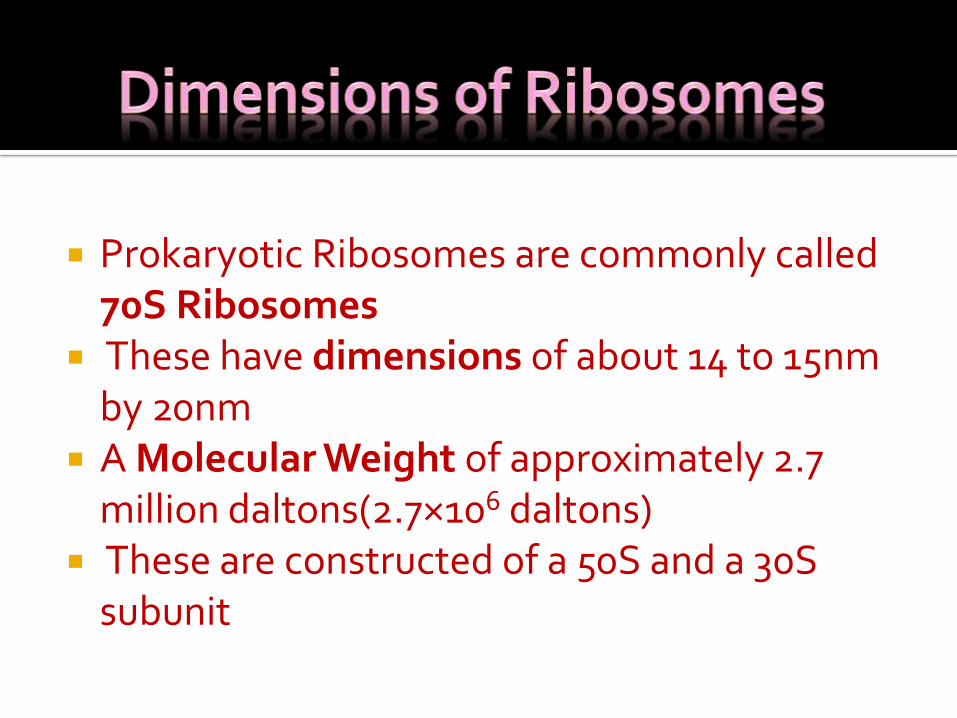
Prokaryotic Ribosomes are commonly called 70S Ribosomes
These have dimensions of about 14 to 15nm by 20nm
A Molecular Weight of approximately 2.7 million daltons(2.7×10⁶ daltons)
These are constructed of a 50S and a 30S subunit
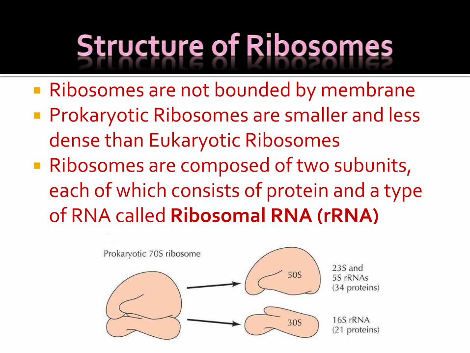
Ribosomes are not bounded by membrane Prokaryotic Ribosomes are smaller and less
dense than Eukaryotic Ribosomes Ribosomes are composed of two subunits,
each of which consists of protein and a type of RNA called Ribosomal RNA (rRNA)
Each subunit is constructed from one to two rRNA molecules and many polypeptides
30S smaller Subunit 50S larger Subunit
The S in 70S and similar values stand for Svedberg units
The faster a particle travels when centrifuged, the greater its Svedberg value or Sedimentation coefficient
The sedimentation coefficient is a function of a particles molecular weight, volume and shape
Heavier and more compact particles normally have larger Svedberg numbers or sediment faster
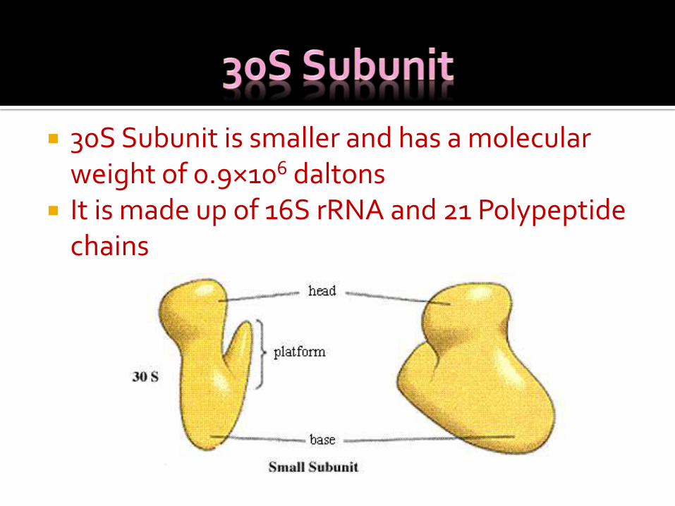
30S Subunit is smaller and has a molecular weight of 0.9×10⁶ daltons
It is made up of 16S rRNA and 21 Polypeptide chains
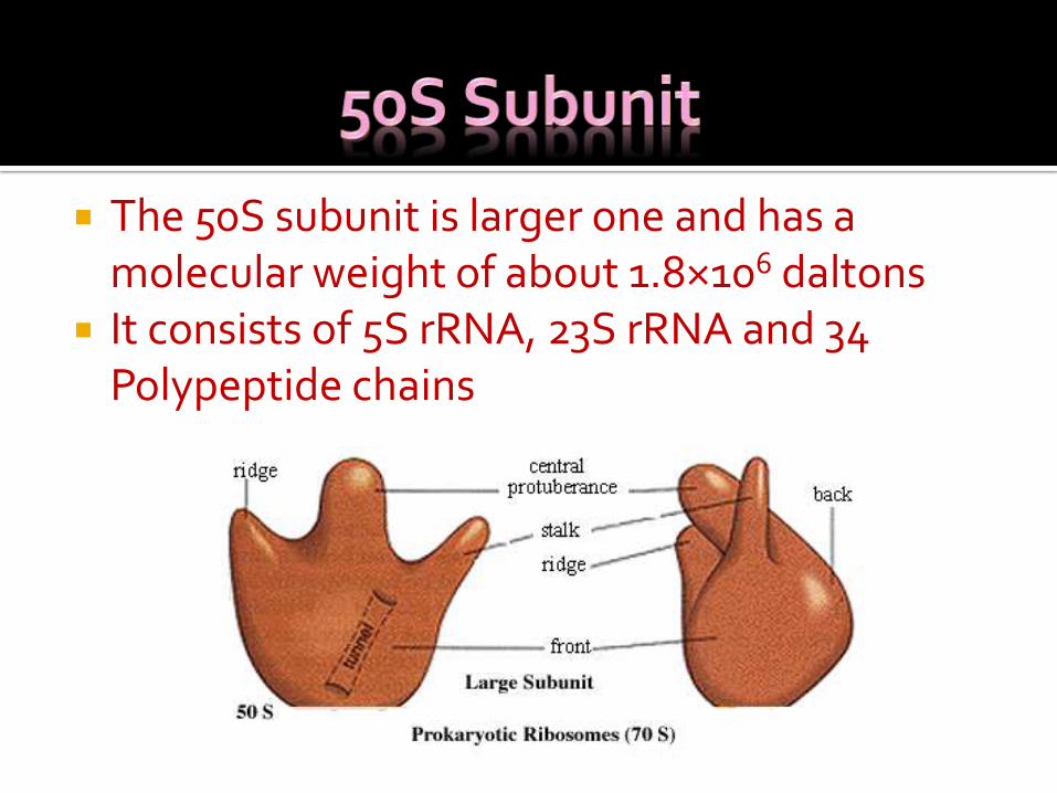
The 50S subunit is larger one and has a molecular weight of about 1.8×10⁶ daltons
It consists of 5S rRNA, 23S rRNA and 34 Polypeptide chains
rRNA is transcribed from certain portions of DNA by the same energy-requiring process used for the synthesis of mRNA and tRNA
rRNA is thought to have two rolesi. The 16S rRNA of the 30S subunit may aid in the
initiation of protein synthesis The 3` end of the 16S rRNA complexes with an
initiating signal site on the mRNA and helps position the mRNA on the ribosome
ii. 16S rRNA binds initiation factor-3 and the 3` CCA end of aminoacyl-tRNA
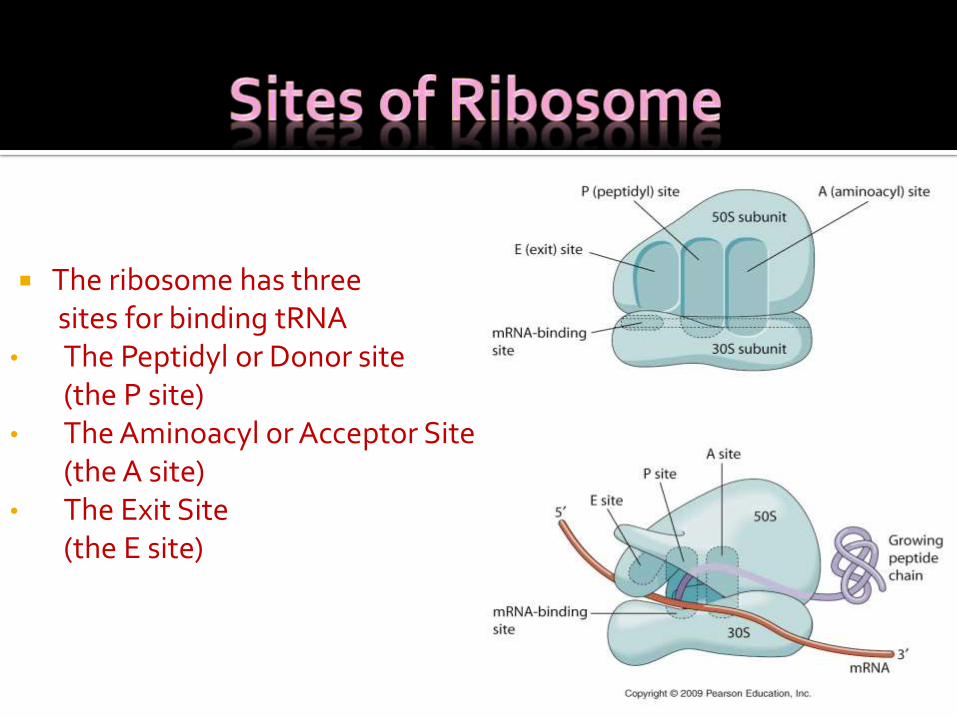
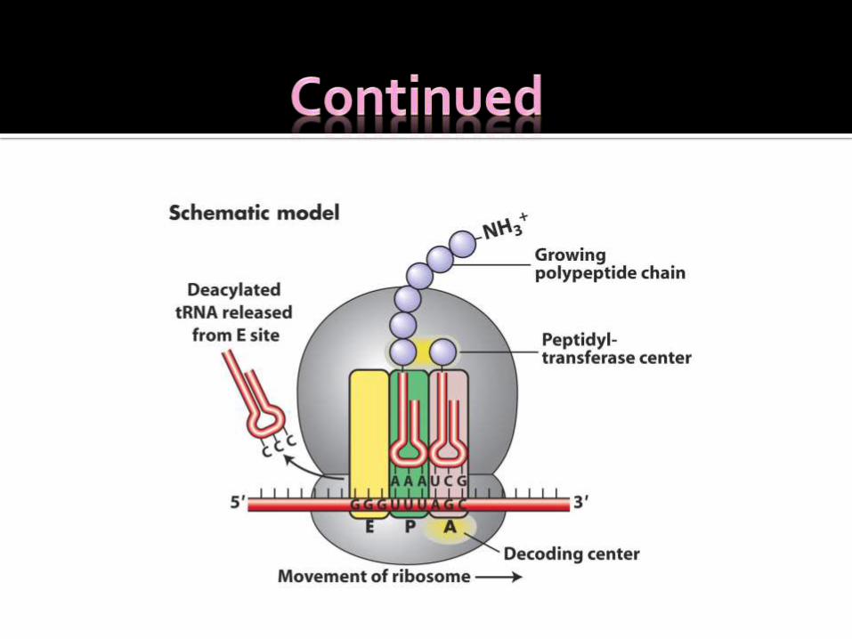
The ribosome has threesites for binding tRNA
• The Peptidyl or Donor site (the P site)
• The Aminoacyl or Acceptor Site (the A site)
• The Exit Site(the E site)
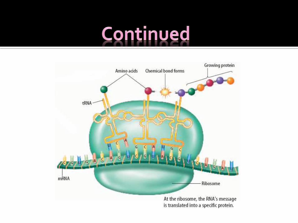
The Ribosome is involved in the process of Protein Synthesis
Protein Synthesis is divided into three stages: 1. Initiation2. Elongation3. Termination
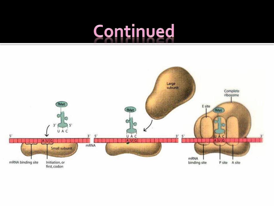
The necessary Components Assemble: i. The two ribosomal subunitsii. A tRNA with the anticodon UACiii. The mRNA molecule to be translatediv. Along with several additional protein factors In E.coli and most bacteria translation begin with
specially modified aminoacyl tRNA, N-formylmethionyl tRNA
Because the α-amino is blocked by a formyl group, this aminoacyl tRNA can be used only for initiation
This N-formylmethionyl-tRNA attaches itself to the P Site of ribosome(Peptidyl Site)
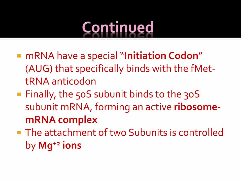
mRNA have a special “Initiation Codon” (AUG) that specifically binds with the fMet-tRNA anticodon
Finally, the 50S subunit binds to the 30S subunit mRNA, forming an active ribosome-mRNA complex
The attachment of two Subunits is controlled by Mg+2 ions
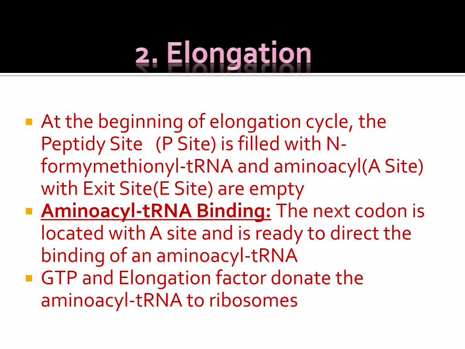
At the beginning of elongation cycle, the Peptidy Site (P Site) is filled with N-formymethionyl-tRNA and aminoacyl(A Site) with Exit Site(E Site) are empty
Aminoacyl-tRNA Binding: The next codon is located with A site and is ready to direct the binding of an aminoacyl-tRNA
GTP and Elongation factor donate the aminoacyl-tRNA to ribosomes
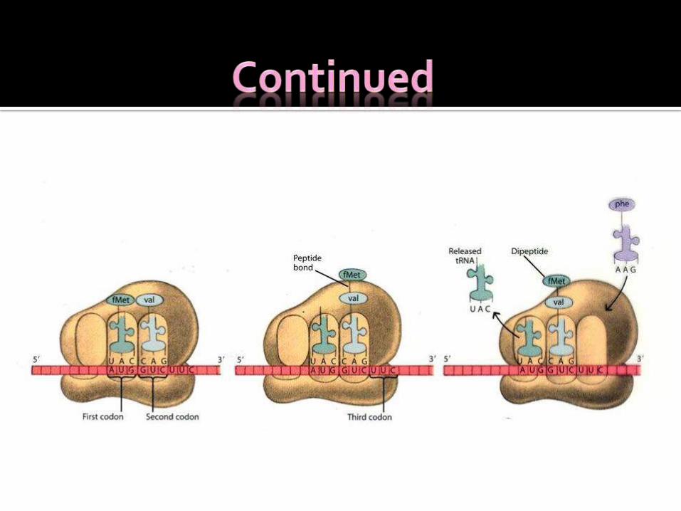
Transpeptidation Reaction: Peptidyltransferase, located on 50S Subunit catalyze the transpeptidation reaction
The α-amino group of A site amino acid attacks α-carboxyl group of C-terminal amino acid on P site tRNA in this reaction resulting in peptide bond formation
A specific adenine base seems to participate in catalyzing peptide bond formation
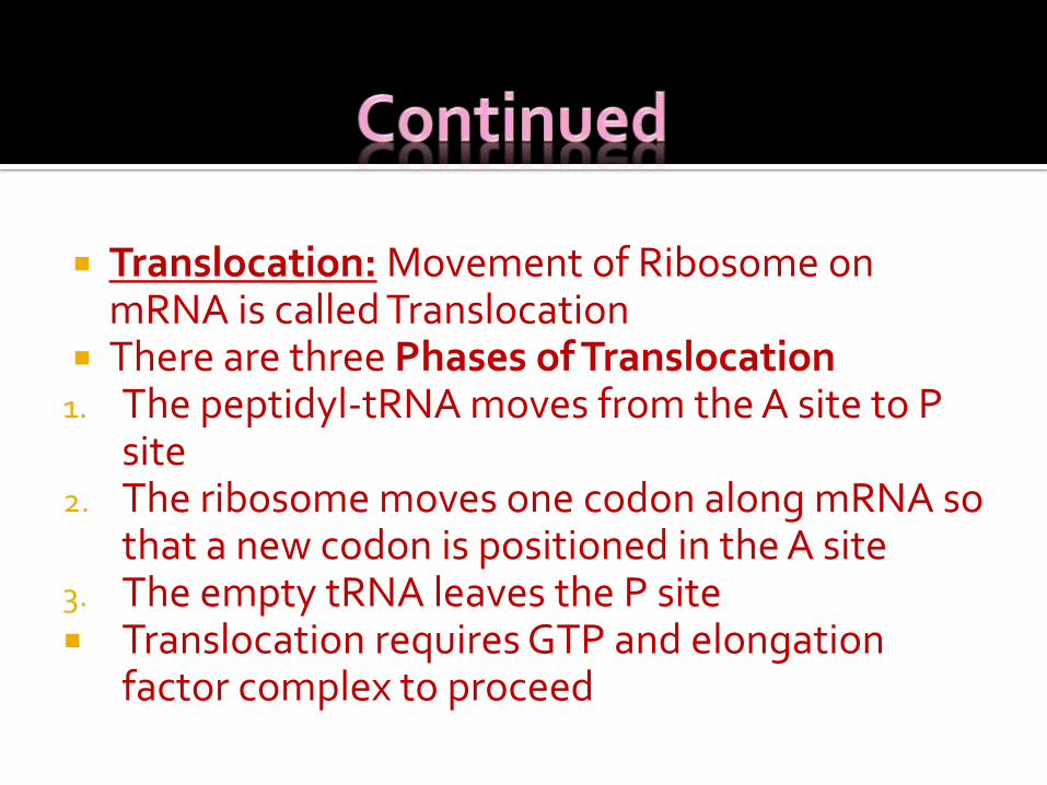
Translocation: Movement of Ribosome on mRNA is called Translocation
There are three Phases of Translocation1. The peptidyl-tRNA moves from the A site to P
site2. The ribosome moves one codon along mRNA so
that a new codon is positioned in the A site3. The empty tRNA leaves the P site Translocation requires GTP and elongation
factor complex to proceed
Protein Synthesis stops when the ribosomes reaches one of three special non-sense codons- UAA, UAG, UGA
Three release factors(RF-1, RF-2, RF-3) aid the ribosomesin recognizing these codons
After the ribosome has stopped, peptidyl transferasehydrolyzes the peptide free from its tRNA, and the empty tRNA is released
GTP hydrolyzes required for this process Next the ribosome dissociates from its mRNA and
separates into 30S and 50S subunits. IF-3 binds to 30S subunit and prevent it from re-associating with 50S subunit till next initiation starts
Several antibiotics work by inhibiting protein synthesis on prokaryotic ribosomes
Antibiotics such as Streptomycin and gentamicin attach to the 30S subunit and interfere with protein synthesis
Other Antibiotics, such as Erythromycin and Chloramphenicol, interfere with protein synthesis by attaching to the 50S subunit
Because of differences in prokaryotic and eukaryotic ribosomes, the microbial cell can be killed by the antibiotic while the eukaryotic host cell remains unaffected

Thank You