right-sided colitis' - gut.bmj.com · gut, 1963, 4, 316 right-sided colitis' e. s. r....
TRANSCRIPT

Gut, 1963, 4, 316
Right-sided colitis'E. S. R. HUGHES
From the Royal Melbourne Hospital, Melbourne, Australia
EDITORIAL SYNOPSIS The term 'right-sided colitis' has been used to describe a lesion in which theright half of the colon shows maximal inflammatory changes; although the terminal ileum is alwaysinvolved, the rectum is normal or shows minimal inflammatory changes. Some of these casesappear to be of atypical Crohn's disease, whilst the others resemble chronic ulcerative colitis.The high incidence of Jewish people in this series suggests a racial tendency towards this distributionof the inflammatory change. The right-sided nature of the lesion has led to unnecessarily prolongedmedical treatment, to right hemicolectomy, and to ileorectal anastomosis. The results of suchprocedures have been disappointing, all the more so because the rectum was almost normal andhence would seem ideally suited for ileorectal anastomosis. These patients have progressed verywell with ileostomy.
In typical ulcerative colitis the most intense inflam-matory changes are in the left colon and rectum,although the right half of the colon is often involved.In regional ileitis the disease is confined classically tothe terminal ileum but may spread into the caecum.
In some cases the right half of the colon showsquite severe inflammatory changes with involvementof the terminal ileum and minimal or no changes inthe rectum. These are either atypical examples of
ULCERATIVE COLITIS REGIONAL ILEITIS
ulcerative colitis, atypical Crohn's lesions, or areseparate disease entities. Because of this uncertaintythe term 'right-sided' colitis has been used (Fig. 1).
INCIDENCE OFRIGHT-SIDED COLITIS
In 172 patients with typical ulcerative colitis opera-tion was performed. In seven of these patients therectum was involved to a lesser degree than is usual.In seven patients the inflammatory lesion was con-fined to a segment, short or long, single or multiple,and was classified as segmental colitis. In sevenpatients the inflammation was most intense in theright colon, with involvement of the terminal ileum.This group of seven patients has been classified as'right-sided' colitis and is reviewed here. In fivepatients the lesion was typical of Crohn's regionalileitis.
SEGMENTAL COLITIS RIGHT - SIDED COLITIS
FIG. 1. Possible types of inflammatory lesions involvingthe right colon. Segmental colitis and right-sided colitis maybe variants of ulcerative colitis or regional ileitis.
316
SUMMARY OF CASE HISTORIES OFPATIENTS WITH RIGHT-SIDED COLITIS
The distribution of the inflammatory changes is shown inFigure 2.
CASE 1 C. I., a Jewish woman, began to have abdominalpain and diarrhoea at the age of 16 years. This persistedand led to repeated trials of medical treatment, whichincluded steroids. In 1957, at the age of 24 years she wasreferred for further advice. She was lethargic, pale, andtender in the right iliac fossa. Sigmoidoscopy showed verymild changes of ulcerative coltis. Barium enema revealed1 A paper read at the annual meeting of the Australian Gastroenter-ological Society 11 August 1962.
on January 2, 2020 by guest. Protected by copyright.
http://gut.bmj.com
/G
ut: first published as 10.1136/gut.4.4.316 on 1 Decem
ber 1963. Dow
nloaded from

Right-sided colitis
C.l. 2 V.S. 3 M.G. 4 M.S.
Pz< 2 <>FIG. 2. Distribution of the inflammatory changes in theseven patients with right-sided colitis.
.5 D.C. 6 M.M. 7 P.K.
persistent deformity of the ileocaecal region with colitis of At the last operation 'skip' lesions in the terminal ileumthe proximal colon. On 23 April 1957 ileostomy and were removed. The case was classified by the pathologistcolectomy was performed and on 8 October 1958 the as Crohn's disease.rectum was removed. She has been well since her firstoperation. The pathologist regarded the lesion as 'chroniculcerative colitis'.
CASE 2 V. S., a Jewish woman, first developed abdominalpain and diarrhoea at the age of 31 years. There wastenderness in the right iliac fossa. Sigmoidoscopy wasnormal; barium clysma showed deformity in the ileo-caecal region. A right hemicolectomy was performed inDecember 1955, but symptoms recurred almost at once.She failed to respond to medical treatment (which includ-ed steroid therapy), developed arthritis, and finallyrequired emergency intervention for acute, fulminatingcolitis. Even at this stage sigmoidoscopy showed minimalchanges. Ileostomy and colectomy performed on 23February 1959 was followed by an excellent recovery andas the rectum retained a healthy appearance ileo-rectalanastomosis followed on 12 October 1960. In April 1961a stricture and recto-vaginal fistula developed at the levelof the anorectal ring. The rectum between this strictureand anastomosis was only mildly inflamed. The pathologywas regarded as ulcerative colitis.
CASE 3 M. G., a Jew, began to have abdominal pain anddiarrhoea at the age of 13 years. Sigmoidoscopy wasnormal; barium enema showed colitis involving the rightcolon. In August 1959 at the age of 16 years right hemi-colectomy was performed, but pain and diarrhoea per-sisted despite steroid therapy. In 1961 sigmoidoscopy "9vshowed mild inflammation of the rectum to 12 cm. andsevere ulceration above. On 9 February 1961 colectomyand ileorectal anastomosis was performed (Fig. 3). Afteran encouraging period of two or three months diarrhoeaand abdominal pain recurred and on 12 August 1961 FIG. 3. Case 3. The segmental type of lesion classified asileostomy was performed. He has since remained well. Crohn's disease.
317
on January 2, 2020 by guest. Protected by copyright.
http://gut.bmj.com
/G
ut: first published as 10.1136/gut.4.4.316 on 1 Decem
ber 1963. Dow
nloaded from
E. S. R. Hughes
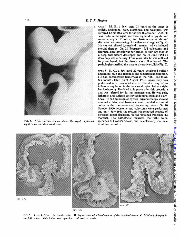
FIG. 4. M.S. Barium enema shows the rigid, deformedrig/it colon and ileocaecal zone.
CASE 4 NI. S., a Jew, aged 21 years at the onset ofcolicky abdominal pain, diarrhoea, and weakness, wasreferred 12 months later for advice (December 1957). Hewas tender in the right iliac fossa, sigmoidoscopy showedminor changes of colitis, and barium enema showeddistortion and narrowing of the ileocaecal region (Fig. 4).He was not relieved by medical treatment, which includedsteroid therapy. On 21 February 1958 colectomy andileorectal anastomosis was performed. Within two monthsa deep anal fissure developed and on 10 June 1958 anileostomy was necessary. Four years later he was well andfully employed, but the fissure was still unhealed. Thepathologist classified this case as ulcerative colitis (Fig. 5).
CASE 5 D. C., a Jew aged 22 years, developed colickyabdominal pain and diarrhoea and began to lose condition.He had considerable tenderness in the right iliac fossa.Six months later, on 9 August 1960, laparotomy wasperformed in a provincial centre. The discovery of aninflammatory lesion in the ileocaecal region led to a righthemicolectomy. He failed to improve after this procedureand was referred for further management. He was pale,lethargic, and suffered colicky abdominal pain and diarr-hoea. He had an irregular pyrexia, sigmoidoscopy showedminimal colitis, and barium enema revealed advancedcolitis in the transverse and descending colons. On 10October 1960 ileostomy and colectomy were performedand on 4 July 1961 the rectum was removed because ofpersistent rectal discharge. He has remained well since (12months). The pathologist regarded the right colonspecimen as Crohn's disease, but the colectomy specimenas ulcerative colitis.
FIG. 5B
FIG. 5. Case 4, M.S. A Whole colon B Right colon with involvement of the terminal ileum C Minimal changes inthe left colon. This lesion was regarded as ulcerative colitis.
318
on January 2, 2020 by guest. Protected by copyright.
http://gut.bmj.com
/G
ut: first published as 10.1136/gut.4.4.316 on 1 Decem
ber 1963. Dow
nloaded from

Right-sided colitis
CASE 6 M. M., a Jew, developed colicky abdominal painand diarrhoea at the age of 33 years. Four years after theonset abdominal palpation revealed a mass in the rightiliac fossa; sigmoidoscopy was normal and a bariumenema showed narrowing of the ascending colon andcaecum. On 5 September 1955 a right hemicolectomy wasperformed. He has remained well since. The pathologistregarded the lesion as non-specific inflammatory; it hassince been reclassified as possibly Crohn's disease.
CASE 7 P.K., a non-jewish woman, developed colickyabdominal pain and diarrhoea at the age of 44 years.Eighteen months later she sought surgical treatment. Atthis time her bowels were irregular, with both diarrhoeaand constipation. Her abdomen was a little distended,with excess bowel activity. Sigmoidoscopy to 20 cm.showed mild, patchy proctocolitis. A barium enemashowed persistent rigidity of the ileocaecal region. On28 June 1961 colectomy with ileorectal anastomosis wasperformed. The right half of the colon was involved intypical ulcerative colitis, which the pathologist confirmed.Her progress was uneventful and she has remained wellsince (12 months).
ANALYSIS OF CASE HISTORIES
In this series there were three females and four males.The ages at onset were 13, 16, 21, 22, 31, 37, and 44years. All but the last were Jewish patients. Therewere several notable features in the series.
ABDOMINAL PAIN Colicky pain, particularly in theright iliac fossa, was a common symptom, and in thisregard resembled Crohn's enteritis rather thanulcerative colitis.
DIARRHOEA Diarrhoea was not as severe as occursin typical left-sided ulcerative colitis. Bowel actionsnumbered three or four a day. Blood loss wasinsignificant.
WEAKNESS Persistent pain and diarrhoea and fear ofeating contributed to the lethargy, fatigue, and lossof weight produced by the colitis.
MASS OR TENDERNESS IN RIGHT ILIAC FOSSA In onecase a mass was palpable in the right iliac fossa; inthe others there was considerable tenderness.
MINIMAL SIGMOIDOSCOPIC ABNORMALITY Symptomswere out of proportion to the sigmoidoscopicchanges. Appearances were either normal or showeda mild degree of friability of the mucous membrane.Motions were soft but blood was not often seen.
ABNORMALITY OF ILEOCAECAL REGION ON BARIUMENEMA EXAMINATION In all cases there was persis-
tent narrowing and rigidity of the ileocaecal region.The colon sometimes showed changes in the trans-verse colon and upper descending colon but therectum appeared normal.
PROGRESS With medical treatment progress wasdisappointing. Temporary improvement was soonfollowed by relapse.
PATHOLOGY OF RIGHT-SIDED COLITIS
One patient in this series (case 6) had a granuloma-tous lesion confined to the right colon with someextension into the ileum. This was regarded as a caseof Crohn's disease although the pathologist was notconvinced.
In two cases (cases 3 and 5) the lesion appeared tobe 'Crohn's ileocolitis'. One had 'skip' lesions in thedistal ileum. The other had suggestive histologicalfeatures. Crohn's regional ileitis has been rare in thewriter's experience, although granulomatous lesionsof the large bowel have been more common.
In four cases in this series the macroscopic andmicroscopic appearances were indistinguishable fromulcerative colitis, except that involvement of therectal mucosa was minimal.The pathology of the cases in this group was not
comparable to the ileocolitis described by Brooke andCooke (1951) and reaffirmed by Brooke (1959a).This lesion commences in the ileum or higher andproduces steatorrhoea, which in turn causes changesin the colon. Malabsorption and liver changes arecommon and ileostomy disastrous.
TREATMENT OF RIGHT-SIDED COLITIS
Medical treatment failed in these cases. In fourpatients (cases 1, 2, 3, and 4) steroid therapy wastried. In two others the physical signs pointed sostrongly to a right-sided lesion that surgical inter-vention was advised without prolonged preliminarymedical treatment and with excellent results.Two patients (cases 2 and 5) had acute exacerba-
tions (one during the course of steroid therapy) andrequired emergency surgery. One (case 2) wastroubled by arthritis which improved after colectomybut which recurred when a stricture developed in therectum after an ileorectal anastomosis.
Right hemicolectomy was performed in fourpatients. In each the surgeon believed the lesion to belocalized to the right colon. After resection the ileumwas anastomosed to the transverse colon. One hasremained well (case 6) for seven years following theoperation. (This patient had an unusual lesion whichwas finally classified as probably Crohn's disease.)The remaining three patients were not relieved by
319
on January 2, 2020 by guest. Protected by copyright.
http://gut.bmj.com
/G
ut: first published as 10.1136/gut.4.4.316 on 1 Decem
ber 1963. Dow
nloaded from

E. S. R. Hughes
this operation and required further interventionafter two, 18, and 38 months respectively.Colectomy and ileorectal anastomosis was done in
four patients. In the last case in the series (case 7) ileo-rectal anastomosis was selected as the treatment ofchoice at the outset and operation was performedwithout prolonged medical treatment. The result hasbeen excellent so far (12 months). In the second(case 4) operation was performed after 12 months'unsuccessful medical treatment which includedsteroids. Three-and-a-half months after operation itwas necessary to establish an ileostomy because of adeep anal fissure. Four years later this patient is well,but the fissure is still unhealed. The third patient(case 3) had a right hemicolectomy; 18 months latercolectomy and ileorectal anastomosis was performed.Persistent diarrhoea and discomfort led to ileostomyafter seven months. He has been in excellent healthsince this was done (11 months), the first such periodhe has had in five years since his illness started. Thefourth patient (case 2) also had a right hemicolec-tomy; 38 months later emergency colectomy andileostomy was necessary for acute fulminating colitis.She progressed well and after 20 months an ileorectalanastomosis was performed. This has been marredby a persistent frequency of bowel action and by thegradual development of a stricture and rectovaginalfistula at the anorectal ring. The rectum between thiszone and the level of the anastomosis shows mildinflammation only.Four of the patients (cases 1, 3, 4, and 5) now have
an ileostomy. In two of these the rectum has beenremoved; in the other two excision will certainly benecessary. All four patients have progressed wellsince ileostomy. One has had the ileostomy for fiveyears, a second for four years, a third for 21 months,and the fourth for 11 months.
DISCUSSION
The seven patients in this series had characteristicclinical features. Abdominal pain, diarrhoea, andgeneral lethargy were present in all cases. In additionthere were signs referrable to the right iliac fossa:either a mass (one case) or tenderness, mild changeson sigmoidoscopic examination, and persistentnarrowing, rigidity, or distortion of the ileocaecalregion and right colon in the barium radiologicalstudies. None was controlled by medical treatment,including steroids. These features are those enumer-ated for 'right-sided colitis' by Crohn and Berg in1938.The term 'right-sided colitis' is unsatisfactory
because it is vague and does not indicate whether thelesion is related to Crohn's disease or ulcerativecolitis or whether it is some specific, as yet unrecog-
nized, condition. In all cases the terminal ileum wasaffected and in this way right-sided colitis differedfrom segmental colitis. Involvement of the ileum hasbeen regarded as indicating that Crohn's disease isresponsible for right-sided colitis. Certainly it isknown that Crohn's regional ileitis spreads into thecolon in about half the cases (Brooke, 1959b; Cornesand Stecher, 1961).Lymph node enlargement was noted in some of the
cases, but this can be a feature of either ulcerativecolitis or Crohn's disease. Care must be taken ininterpreting the pathologist's report on the nodesbecause the surgeon may keep close to the bowel inthese inflammatory conditions, leaving the proximalnodes in situ (Neuman and Dockerty, 1954; Brooke,1962).Macroscopic and microscopic appearances of the
bowel varied in this series. In three cases the lesionwas considered to be Crohn's disease and in four thebowel appeared to be typical of ulcerative colitisexcept for the distribution.Most authors have grouped segmental and right-
sided colitis together (Crohn and Berg, 1938; Crohn,Garlock, and Yarnis, 1947; Neuman, Bargen, andJudd, 1954; Watkinson, Thompson, and Goligher,1960). On the other hand, Brooke (1962) recom-mends that both terms be abandoned. He feels thatulcerative colitis is a 'left-sided' disease and that a'right-sided' lesion cannot be ulcerative colitis.Furthermore he considers that such right-sidedlesions originate in the small intestine and thatileostomy in such circumstances may be dangerous.However, in this series four cases appeared to bevariants of chronic ulcerative colitis. Further, in fourcases ileostomy has produced excellent results.The very large number of Jewish people in this
series is a feature. In the series of 172 with typicalulcerative colitis there were only three Jewishpersons. Neuman, Bargen, and Judd (1954) found thatnearly 25% of their cases with segmental colitis wereJewish (and their segmental cases included right-sided colitis). Acheson (1960) found that Jewishpeople were more prone to ulcerative colitis and toregional enteritis but he did not analyse the patho-logical features. It is possible that the right-sideddistribution of the lesion, whether it is regarded asCrohn's disease or ulcerative colitis, is a racialfeature.Apart from the pathological interest of right-sided
colitis the distribution of the inflammatory reactionon the right side led to three therapeutic procedureswhich were unsatisfactory. The minor changesobserved at sigmoidoscopy were responsible forprolonged medical treatment. Indeed persistentsymptoms prompted a diagnosis of functionalexaggeration, but the excellent results of ileostomy
320
on January 2, 2020 by guest. Protected by copyright.
http://gut.bmj.com
/G
ut: first published as 10.1136/gut.4.4.316 on 1 Decem
ber 1963. Dow
nloaded from

Right-sided colitis 321
suggested a fault in the initial medical assessment ofthe patient. Steroid therapy proved unsuccessful.
Secondly, although right hemicolectomy provedsatisfactory in one patient, in three others it failed. Aninflammatory lesion in the right colon should be verycarefully assessed before deciding to restrict surgeryto right hemicolectomy and anastomosis.
Lastly, ileorectal anastomosis was disappointing.Only one of four patients has done well, and thispatient has been followed for 12 months. These poorresults of anastomotic procedures are similar to thosereported from the Mayo Clinic and elsewhere (Neu-man and Dockerty, 1954; Manning, Warren, andAdi, 1955). On the other hand, both Watkinson et al.(1960) and Lockhart-Mummery and Morson (1960)believe that the rectum could remain free from thedisease and that therefore ileorectal anastomosis isa reasonable operation. However, they includedcases of segmental colitis in which the ileum was notinvolved and which have done well with ileorectalanastomosis in the writer's hands.
Ileostomy has given excellent results and, despitethe warning given by Brooke, such a procedure canbe advised with confidence in this group of cases.
The rectum should be left alone for some time beforea decision is made to anastomose ileum to it.
REFERENCESAcheson, E. D. (1960). The distribution of ulcerative colitis and
regional enteritis in United States veterans with particularreference to the Jewish religion. Gut, 1, 291-293.
Brooke, B. N. (1959a). Granulomatous diseases of the intestine.Lancet, 2, 745-749.
(1959b). Surgical Aspects of Ulcerative Colitis. In Diseases of theColon and Anorectum, edited by R. Turell, vol. 2, pp. 702-728.Saunders, Philadelphia and London.
(1962). Inflammatory disorders of the colon. Dis. Colon. Rect.,5, 138-144.and Cooke, W. T. (1951). Ulcerative colitis: problem andtherapeutic warning. Lancet, 2, 462-464.
Cornes, J. S., and Stecher, M. (1961). Primary Crohn's disease of thecolon and rectum. Gut, 2, 189-201.
Crohn, B. B., arid Berg, A. A. (1938). Right-sided (regional) colitis.J. Amer. med. Ass., 110, 32-38.Garlock, J. H., and Yarnis, H. (1947). Right-sided (regional)colitis. Ibid., 134, 344-338.
Lockhart-Mummery, H. E., and Morson, B. C. (1960). Crohn'sdisease (regional enteritis) of the large intestine and its dis-tinction from ulcerative colitis. Gut, 1, 87-105.
Manning, J. H., Warren, R., and Adi, A. S. (1955). Segmental colitis:results of surgery. New Engl. J. med., 252, 850-853.
Neuman, H. W., Bargen, J. A., and Judd, E. S. Jr. (1954). A clinicalstudy of 201 cases of regional (segmental) colitis. Surg. Gynec.Obstet., 99, 563-571.
, and Dockerty, M. B. (1954). The pathology of regional (seg-mental) colitis. Ibid., 99, 572-579.
W'atkinson, G., Thompson, H., and Goligher, J. C. (1960). Right-sided or segmental ulcerative colitis. Brit. J. Surg., 47, 337-351.
on January 2, 2020 by guest. Protected by copyright.
http://gut.bmj.com
/G
ut: first published as 10.1136/gut.4.4.316 on 1 Decem
ber 1963. Dow
nloaded from