ring finger protein 145 (rnf145) is a ubiquitin ligase for ... · 4048...
TRANSCRIPT

Ring finger protein 145 (RNF145) is a ubiquitin ligase forsterol-induced degradation of HMG-CoA reductaseReceived for publication, December 4, 2017, and in revised form, January 15, 2018 Published, Papers in Press, January 26, 2018, DOI 10.1074/jbc.RA117.001260
Lu-Yi Jiang1, Wei Jiang1, Na Tian, Yan-Ni Xiong, Jie Liu, Jian Wei, Kai-Yue Wu, Jie Luo, Xiong-Jie Shi2,and Bao-Liang Song3
From the Hubei Key Laboratory of Cell Homeostasis, College of Life Sciences, Institute for Advanced Studies, Wuhan University,Wuhan 430072, China
Edited by Henrik G. Dohlman
Cholesterol biosynthesis is tightly regulated in the cell. Forexample, high sterol concentrations can stimulate degradationof the rate-limiting cholesterol biosynthetic enzyme 3-hydroxy-3-methylglutaryl-coenzyme A reductase (HMG-CoA reductase,HMGCR). HMGCR is broken down by the endoplasmic reticu-lum membrane–associated protein complexes consisting ofinsulin-induced genes (Insigs) and the E3 ubiquitin ligase gp78.Here we found that HMGCR degradation is partially blunted inChinese hamster ovary (CHO) cells lacking gp78 (gp78-KO). Toidentify other ubiquitin ligase(s) that may function togetherwith gp78 in triggering HMGCR degradation, we performed asmall-scale short hairpin RNA– based screening targeting endo-plasmic reticulum–localized E3s. We found that knockdown ofboth ring finger protein 145 (Rnf145) and gp78 genes abrogatessterol-induced degradation of HMGCR in CHO cells. We alsoobserved that RNF145 interacts with Insig-1 and -2 proteins andubiquitinates HMGCR. Moreover, the tetrapeptide sequenceYLYF in the sterol-sensing domain and the Cys-537 residue inthe RING finger domain were essential for RNF145 binding toInsigs and RNF145 E3 activity, respectively. Of note, amino acidsubstitutions in the YLYF or of Cys-537 completely abolishedRNF145-mediated HMGCR degradation. In summary, ourstudy reveals that RNF145, along with gp78, promotes HMGCRdegradation in response to elevated sterol levels and identifiesresidues essential for RNF145 function.
Cholesterol is the most abundant sterol in mammalian cells.It regulates membrane function and serves as the precursor forbile acids and steroid hormones. Cholesterol can either be syn-thesized through the mevalonate pathway (1) or taken up fromdiets via Niemann–Pick C1-like 1 (NPC1L1)–mediated absorp-tion (2–5).
HMG-CoA4 reductase (HMGCR) catalyzes the rate-limitingstep in cholesterol biosynthesis in which HMG-CoA is con-verted to mevalonate. The half-life of HMGCR varies with cel-lular sterol levels: more than 12 h in sterol-depleted cells andless than 1 h in sterol-overloaded cells (6, 7). HMGCR is tightlyregulated by sterols at both transcriptional and posttransla-tional levels. High concentrations of cholesterol decrease tran-scription of the HMGCR gene by inhibiting the activation ofsterol regulatory element– binding protein 2 (SREBP-2) (8, 9).In addition, excess levels of 24,25-dihydrolanosterol, an inter-mediate in the mevalonate pathway, promote ubiquitinationand degradation of the HMGCR protein (10 –12). Oxysterolscan inhibit HMGCR transcription and stimulate HMGCRdegradation (13, 14). Besides sterols, geranylgeraniol, a non-steroid product downstream of mevalonate, acts on the post-ubiquitination step to accelerate sterol-induced HMGCRdegradation (7).
The sterol-induced degradation of HMGCR initiates whenthe endoplasmic reticulum (ER)-localized Insig-1 and -2 pro-teins bind to HMGCR (15) and recruit the ubiquitin ligase (E3)gp78 to catalyze ubiquitination (16). The HMGCR protein iseventually degraded in the proteasome. Ufd1 enhances the E3activity of gp78 and accelerates the degradation of HMGCR(17). Ablation of gp78 in mouse liver increases the stability ofHMGCR, Insig-1, and Insig-2 (18, 19). Elevated levels of Insigsinhibit the SREBP pathway and decrease cholesterol synthesis(18). These data suggest that gp78 is a major E3 essential forHMGCR degradation in the hepatocytes.
Besides gp78, TRC8 and MARCH6 are two other ER-local-ized E3s involved in HMGCR degradation (20, 21). TRC8 inter-acts with Insig-1 and -2 and ubiquitinates HMGCR for protea-somal degradation. In addition to sterol-regulated degradation,the basal turnover of HMGCR is mediated by Hrd1, an ER-an-chored E3 homologous to gp78 (22, 23). Interestingly, sterol-induced HMGCR degradation has been found to persist ingp78-deficient primary mouse embryonic fibroblasts (24),This work was supported by grants from the Ministry of Science and Technol-
ogy of China (2016YFA0500100), the National Natural Science Foundationof China (31600651, 31430044, 31690102, 31771568, and 31701030), theChina Postdoctoral Science Foundation (2016M592380), the 111 Project ofthe Ministry of Education of China (B16036), and the Natural Science Foun-dation of Hubei Province (2016CFA012 and 2017CFB617). The authorsdeclare that they have no conflicts of interest with the contents of thisarticle.
This article contains Figs. S1 and S2 and Table S1.1 Both authors contributed equally to this work.2 To whom correspondence may be addressed: E-mail: [email protected] To whom correspondence may be addressed: E-mail: [email protected].
4 The abbreviations used are: HMG-CoA, 3-hydroxy-3-methylglutaryl-CoA;HMGCR, 3-hydroxy-3-methylglutaryl-CoA reductase; RNF145, ring fingerprotein 145; ER, endoplasmic reticulum; Insig, insulin-induced gene;SREBP, sterol regulatory element– binding protein; SCAP, SREBP cleavage–activating protein; CHO, Chinese hamster ovary; KO, knockout; 25-HC,25-hydroxycholesterol; shRNA, short hairpin RNA; aa, amino acids; SSD,sterol-sensing domain; LXR, liver X receptor; HA, hemagglutinin; TBS, Tris-buffered saline; IP, immunoprecipitation.
croARTICLE
J. Biol. Chem. (2018) 293(11) 4047–4055 4047© 2018 by The American Society for Biochemistry and Molecular Biology, Inc. Published in the U.S.A.
by guest on April 30, 2020
http://ww
w.jbc.org/
Dow
nloaded from

which leads us to speculate that there might be other E3(s)compensating for the function of gp78 in cultured cells.
In this study, we identified that an ER-anchored E3 namedRNF145 catalyzed sterol-induced ubiquitination of HMGCR.Knockout of gp78 or Rnf145 alone had partial or little effect onHMGCR degradation in Chinese hamster ovary (CHO) cells.However, knockout of both genes dramatically blunted sterol-induced degradation of HMGCR. The E3 activity– deficientRNF145 (C537A) failed to promote sterol-induced ubiquitina-tion and degradation of HMGCR. Moreover, we found thatInsigs were required for RNF145-catalyzed HMGCR degrada-tion and that RNF145 interacted with Insigs constitutivelythrough its transmembrane domains. We therefore concludethat RNF145 is a new E3 promoting sterol-induced degradationof HMGCR.
Results
Identification of Rnf145 involved in HMGCR degradation
To determine whether gp78 is exclusively responsible forHMGCR degradation, we treated WT CHO and gp78 knockout(gp78-KO) cells with increasing concentrations of 25-hydroxy-cholesterol (25-HC). Although the degradation of HMGCR waspartially impaired when cells were treated with low concentra-tions of 25-HC (0.03 and 0.1 �g/ml), high concentrations of25-HC (0.3 and 1 �g/ml) almost or completely diminishedHMGCR in gp78-KO cells (Fig. 1, A and B), suggesting thatgp78 is not the only E3 mediating HMGCR degradation.Because previous studies identified a total of 24 ER membrane–spanning E3s (25), we next transfected plasmids expressingshRNAs targeting each E3 together with those expressingHMGCR-T7 and Insig-1-Myc into gp78-KO cells and exam-ined sterol-induced degradation of HMGCR (Fig. S1). In con-trast to control shRNA transfected cells where HMGCRdegraded rapidly in response to sterols (Fig. 1C, compare lanes1 and 2), no HMGCR degradation was detected upon Rnf145deficiency in gp78-KO cells (Fig. 1C, lanes 3– 6). These resultssuggest that RNF145 is involved in HMGCR degradation. Wefurther generated Rnf145-KO and gp78 plus Rnf145 knockout(double KO) CHO cells using the CRISPR/Cas9 technique (26).Knockout of gp78 slightly affected HMGCR degradation rela-tive to WT cells (Fig. 1D, lanes 4 – 6), and knockout of Rnf145alone had little influence on HMGCR degradation (Fig. 1D,lanes 7–9). However, HMGCR degradation was largely bluntedin the double KO cells (Fig. 1D, lanes 10 –12). Next we mea-sured sterol-induced ubiquitination of HMGCR in cells lackingeither E3 or both. Knockout of gp78, Rnf145, or both genesdecreased sterol-induced ubiquitination of HMGCR (Fig. 1E).These results suggest that RNF145 plays a critical role in sterol-induced degradation of HMGCR.
RNF145 is an ER-localized ubiquitin ligase mediating HMGCRdegradation
RNF145 is a putative ER transmembrane protein (Fig. 2A)(25). To confirm the subcellular location of RNF145, weperformed immunofluorescence experiments by co-stainingtransfected RNF145 together with the endogenous ER markerCalnexin. RNF145-FLAG was largely colocalized with Cal-nexin, indicating that RNF145 is indeed an ER-localized protein
(Fig. 2B). Next, we purified the recombinant cytosolic domain(aa 511– 663) of RNF145 and performed an in vitro ubiquitina-tion assay. The recombinant cytosolic domain of gp78 (309 –643) was used as a positive control. We found that RNF145(511– 663) could efficiently catalyze the formation of polyubiq-uitin chains in the presence of E1, E2, FLAG-ubiquitin, andATP (Fig. 2C). Replacement of the conserved Cys-537 residuewith alanine (C537A) in the RING finger domain of RNF145(27), however, completely abolished the E3 ligase activity ofRNF145 (Fig. 2D). These results indicate that RNF145 is anER-localized ubiquitin ligase and that the Cys-537 residue isrequired for its E3 activity.
We then sought to determine whether Cys-537 is requiredfor sterol-induced HMGCR ubiquitination. As shown in Fig.3A, lanes 1 and 2, sterols substantially increased the ubiquiti-nation of HMGCR, as evidenced by the high-molecular-weightsmears of the immunoprecipitates. Addition of RNF145(C537A) competitively blocked the ubiquitination of HMGCRinduced by sterols (Fig. 3A, lane 3). Consistent with the ubi-quitination results, the WT form of RNF145 acceleratedHMGCR degradation (Fig. 3B, lanes 1– 4), whereas the C537Amutant completely abrogated HMGCR degradation (Fig. 3B,lanes 5–10). These results suggest that RNF145 acts as a ubiq-uitin ligase promoting HMGCR degradation.
Insigs are required for RNF145-mediated HMGCR degradation
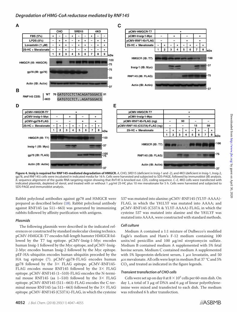
Insigs are indispensable for sterol-induced degradation ofHMGCR mediated by gp78 (7, 10, 16). Indeed, SRD15 cells, acell line lacking both Insig-1 and Insig-2 (28), failed to displaysterol-regulated degradation of HMGCR (Fig. 4A, lanes 4 – 6).To test whether RNF145-mediated degradation of HMGCRrequires Insigs, we generated an SRD15 cell line deficient inRnf145 and gp78 (4KO) (Fig. 4B) that was also insensitive tosterol treatment (Fig. 4A, lanes 7–9). Re-expression of Insig-1or RNF145 alone in 4KO cells did not restore sterol-induceddegradation of HMGCR (Fig. 4C, lanes 3– 6). Interestingly, co-expression of Insig-1 and RNF145 triggered HMGCR degrada-tion in 4KO cells exposed to sterols (Fig. 4C, lanes 7 and 8). Itwas noteworthy that sterol-regulated HMGCR degradationwas also detected in 4KO cells co-expressing Insig-1 and gp78(Fig. 4D, lanes 7 and 8), suggesting that gp78 and RNF145 mayfunction redundantly in mediating HMGCR degradation. Incontrast to these findings, RNF145 (C537A) did not induce thedegradation of HMGCR in the reconstitution system, even inthe presence of Insig-1 (Fig. 4E, lanes 5– 8).
We then tested whether RNF145 could interact with Insigs.Fig. 5A shows that RNF145 co-immunoprecipitated with bothInsig-1 and Insig-2 regardless of sterol levels. Specifically, it wasthe transmembrane domain (aa 1–510) but not the cytosolicdomain (aa 511– 663) of RNF145 that bound to Insig-1 (Fig.5B). Further, overexpression of the transmembrane domain(aa 1–510) of RNF145 effectively blunted sterol-regulatedHMGCR degradation (Fig. 5C).
The sterol-sensing domain of RNF145 is crucial for HMGCRdegradation
It is known that SREBP cleavage–activating protein (SCAP)and HMGCR harbor the sterol-sensing domain (SSD) for Insig
Degradation of HMG-CoA reductase mediated by RNF145
4048 J. Biol. Chem. (2018) 293(11) 4047–4055
by guest on April 30, 2020
http://ww
w.jbc.org/
Dow
nloaded from

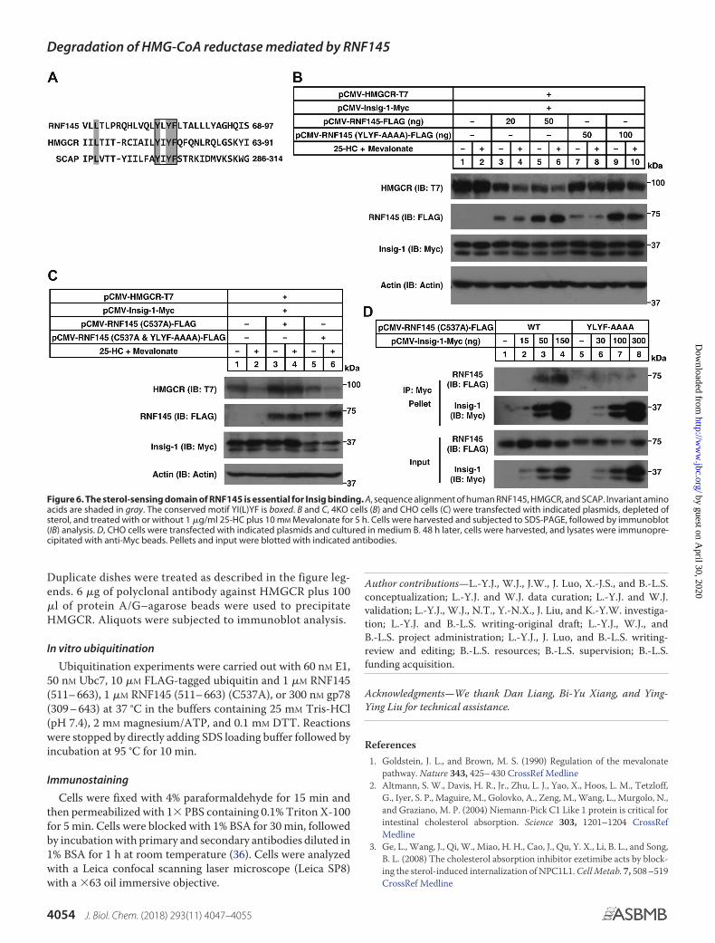
binding and that the tetrapeptide YIYF is highly conservedwithin the SSD (7, 15). There is a putative SSD in RNF145 (Fig.2A). Sequence alignment revealed a YLYF tetrapeptide in
RNF145 corresponding to the YIYF found in HMGCR andSCAP (Fig. 6A). To test whether this motif was essential forRNF145-mediated degradation of HMGCR, we mutated
Figure 1. RNF145 is involved in sterol-regulated HMGCR degradation. A, WT CHO and gp78-KO CHO cells were depleted of sterol in medium C for16 h. Cells were then treated with medium C supplemented with indicated concentrations of 25-HC plus 10 mM mevalonate for 5 h. Cells were harvestedand subjected to SDS-PAGE, followed by immunoblot (IB) analysis. B, quantification of the HMGCR protein in A. C, the gp78-KO CHO cells weretransfected with plasmids encoding HMGCR-T7, Insig-1-Myc, and the shRNA targeting RNF145. After 48 h, cells were depleted of sterol and then treatedwith or without 0.3 �g/ml 25-HC plus 10 mM mevalonate for 5 h as described in A. Cells were harvested and subjected to SDS-PAGE, followed byimmunoblot analysis. Results shown are representative of three independent experiments. D, WT, gp78-KO, Rnf145-KO, and double KO CHO cellswere depleted of sterol and then treated with indicated concentrations of 25-HC plus 10 mM mevalonate for 5 h as described in A. Cells were harvestedand subjected to SDS-PAGE, followed by immunoblot analysis. Asterisks indicate nonspecific bands. Results shown are representative of two inde-pendent experiments. E, WT, gp78-KO, Rnf145-KO, and double KO CHO cells were depleted of sterol and then treated with 10 �M MG132 in the presenceor absence of 1 �g/ml 25-HC and 10 mM mevalonate for 2 h. Cells were harvested, and lysates were immunoprecipitated with protein A/Gbeads coupled with the anti-HMGCR antibody. Pellet fractions were immunoblotted with anti-ubiquitin (P4D1) and polyclonal anti-HMGCRantibodies.
Degradation of HMG-CoA reductase mediated by RNF145
J. Biol. Chem. (2018) 293(11) 4047–4055 4049
by guest on April 30, 2020
http://ww
w.jbc.org/
Dow
nloaded from

YLYF to AAAA and evaluated its function in 4KO cells.Unlike WT RNF145, the RNF145 (YLYF-AAAA) mutantcould not elicit the degradation of HMGCR following steroltreatment (Fig. 6B). We next combined the YLYF-AAAAmutation with E3 activity deficiency together. As shown inFig. 6C, the RNF145 (C537A and YLYF-AAAA) mutant nolonger blocked sterol-regulated degradation of HMGCR.Moreover, the RNF145 (YLYF-AAAA) mutant could notbe co-immunoprecipitated with Insig-1 (Fig. 6D). Collec-tively, these results suggest that the YLYF motif is requiredfor Insig binding as well as RNF145-mediated HMGCRdegradation.
Discussion
In this study, we identified RNF145 as another E3 mediat-ing the degradation of HMGCR through unbiased shRNAscreening. RNF145 resides in the ER with 14 putative trans-membrane segments, one to five of which constitute an SSD
that is also present in SCAP, HMGCR, NPC1, NPC1L1, andTRC8 (29 –31). The RING finger domain localized in thecytosolic C terminus confers RNF145 E3 activity. RNF145and TRC8 display similar structures and sequences.
In CHO cells, knockout of gp78 partially delayed the turnoverof HMGCR in response to low concentrations of sterols, andablation of Rnf145 alone also had little effect (Fig. 1). Notably,knockout of both genes largely abolished sterol-induced degra-dation of HMGCR (Fig. 1D). Similar to gp78 and TRC8, Insigsare required by RNF145 for HMGCR ubiquitination. The bind-ing between Insigs and HMGCR is regulated by sterol levels,whereas the Insig and E3 (gp78, TRC8, and RNF145) interac-tion is constitutive. The inactivated mutation (C537A) ofRNF145 had a dominant-negative effect on HMGCR degrada-tion. A similar C356G mutation was found in gp78 (16). Inter-estingly, HMGCR, SCAP, and RNF145 all bind Insigs throughthe SSD and require the conserved YI(L)YF tetrapeptide forbinding.
Figure 2. RNF145 is an ER-localized ubiquitin ligase. A, predicted topology of RNF145. YLYF, the amino acids from 81 to 84 of RNF145. B, subcellularlocalization of RNF145. HeLa cells were transfected with a plasmid encoding RNF145-FLAG and stained with anti-FLAG and anti-calnexin antibodies. Scale bar �10 �m. C, in vitro ubiquitination assay showing that RNF145 (511– 663) possesses E3 activity. Recombinant proteins, including E1, E2 (Ubc7), FLAG-ubiquitin(FLAG-Ub), RNF145 (511– 663), and gp78 (309 – 643), were added to the reaction system as indicated. After incubation at 37 °C for 15 min, samples weresubjected to SDS-PAGE, followed by immunoblot (IB) analysis. D, in vitro ubiquitination assay comparing RNF145 (511– 663) and RNF145 (511– 663) (C537A).Experiments were carried out as described in C.
Degradation of HMG-CoA reductase mediated by RNF145
4050 J. Biol. Chem. (2018) 293(11) 4047–4055
by guest on April 30, 2020
http://ww
w.jbc.org/
Dow
nloaded from

The question why multiple E3s are involved in sterol-in-duced degradation of HMGCR is intriguing. One possibility isthat different cells express different levels of E3s in response todistinct signals. In fact, recent studies have identified Rnf145 asa Liver X receptor (LXR) target gene (32, 33). We hypothesizethat activation of LXR might elevate the RNF145 level andsubsequently down-regulate cholesterol biosynthesis throughdegrading HMGCR. Another possibility is that the existence ofmultiple E3s for HMGCR degradation prevents saturation ofspecific E3(s) and ensures that ER-associated degradation func-tions properly when HMGCR is degraded.
The protein machineries involved in HMGCR degradationmay also participate in other cholesterol-regulating processes.gp78 is the first characterized E3 catalyzing HMGCR ubiquiti-nation (16). It is highly expressed in the liver. Knockout of gp78in the hepatocytes largely blunted the degradation of HMGCR(18). However, gp78 deficiency also stabilizes Insigs (especiallyInsig-2), resulting in suppressed processing of SREBP and sub-sequently decreased expression of Hmgcr and other genes inthe mevalonate pathway (18). As the protein levels of Insigswere dramatically increased in gp78-deficient cells (18) (Fig.S2), the effects of other E3s such as TRC8 and RNF145 might beboosted, which may explain why HMGCR is still degraded ingp78-KO cells (24). gp78 can also catalyze ubiquitination ofacyl-CoA:cholesterol acyltransferase (ACAT)-2 on a cysteineresidue (34). In addition to degrading HMGCR, RNF145 trig-gers the ubiquitination of SCAP and interferes with SCAP bind-ing to coat protein complex II (COPII), thus inhibiting SREBP-2maturation (33).
In summary, we here identify that RNF145 is an E3 governingsterol-regulated degradation of HMGCR. Together with theprevious findings that Rnf145 is an LXR-regulated gene andthat RNF145 inhibits the SREBP pathway through ubiquitinat-ing SCAP, RNF145 serves as an important negative regulator ofcholesterol biosynthesis. Activation of RNF145 may be effectivefor treating hypercholesterolemia through inhibiting endoge-nous cholesterol synthesis.
Experimental procedures
Reagents
We obtained lovastatin, mevalonate, and 25-hydroxycholes-terol from Sigma, MG132 from Calbiochem, FLAG-ubiquitinfrom Boston Biochem, and linear polyethyleneimine from Poly-sciences; lipoprotein-deficient serum was prepared from new-born calf serum as described before (35).
Antibodies
Primary antibodies used were as follows. Mouse monoclonalantibody against the T7 tag (Novagen), mouse monoclonalantibody P4D1 against ubiquitin and goat polyclonal antibodyagainst Calnexin (Santa Cruz Biotechnology), mouse monoclo-nal antibody (clone 16B12) against the HA tag (Biolegend),mouse monoclonal antibody (clone AC-15) against �-actin andmonoclonal antibody (clone M2) against the FLAG tag (Sigma),mouse monoclonal antibody (clone 10E2) against the His tag(Abmart), and mouse monoclonal antibody (clone 9E10)against the Myc tag and monoclonal antibody (clone A9)against HMGCR were prepared from hybridomas (ATCC).
Figure 3. The E3 activity– deficient RNF145 blocks sterol-induced ubiquitination and degradation of HMGCR. A, CHO cells were transfected withindicated plasmids, depleted of sterol, and treated with 10 �M MG132 in the presence or absence of 1 �g/ml 25-HC and 10 mM mevalonate for 3 h. Cells wereharvested, and lysates were immunoprecipitated with anti-FLAG beads. Input and pellet fractions were immunoblotted (IB) with anti-HA, anti-FLAG, andpolyclonal anti-RNF145 antibodies. Results shown are representative of two independent experiments. B, CHO cells were transfected with indicated plasmids,depleted of sterol, and treated with or without 1 �g/ml 25-HC plus 10 mM mevalonate for 5 h. Cells were harvested and subjected to SDS-PAGE andimmunoblot analysis. Results shown are representative of two independent experiments.
Degradation of HMG-CoA reductase mediated by RNF145
J. Biol. Chem. (2018) 293(11) 4047–4055 4051
by guest on April 30, 2020
http://ww
w.jbc.org/
Dow
nloaded from
Rabbit polyclonal antibodies against gp78 and HMGCR wereprepared as described before (18). Rabbit polyclonal antibodyagainst RNF145 (aa 511– 663) was generated by immunizingrabbits followed by affinity purification with antigens.
Plasmids
The following plasmids were described in the indicated ref-erences or constructed by standard molecular cloning technics.pCMV-HMGCR-T7 encodes full-length hamster HMGCR fol-lowed by the T7 tag epitope. pCMV-Insig-1-Myc encodeshuman Insig-1 followed by the Myc epitope, and pCMV-Insig-2-Myc encodes human Insig-2 followed by the Myc epitope.pEF-HA-ubiquitin encodes human ubiquitin preceded by theHA tag epitope (7). pCMV-gp78-FLAG encodes humangp78 followed by the 3� FLAG epitope. pCMV-RNF145-FLAG encodes mouse RNF145 followed by the 3� FLAGepitope. pCMV-RNF145 (1–510)-FLAG encodes the N-termi-nal mouse RNF145 (aa 1–510) followed by the 3� FLAGepitope. pCMV-RNF145 (511– 663)-FLAG encodes the C-ter-minal mouse RNF145 (aa 511– 663) followed by the 3� FLAGepitope. pCMV-RNF145 (C537A)-FLAG, in which the cysteine
537 was mutated into alanine; pCMV-RNF145 (YLYF-AAAA)-FLAG, in which the Y81LYF was mutated into AAAA; andpCMV-RNF145 (C537A & YLYF-AAAA)-FLAG, in which thecysteine 537 was mutated into alanine and the Y81LYF wasmutated into AAAA, were constructed with standard methods.
Cell culture
Medium A contained a 1:1 mixture of Dulbecco’s modifiedEagle’s medium and Ham’s F-12 medium containing 100units/ml penicillin and 100 �g/ml streptomycin sulfate.Medium B contained medium A supplemented with 5% fetalbovine serum. Medium C contained medium A supplementedwith 5% lipoprotein-deficient serum, 1 �M lovastatin, and 50�M mevalonate. All cells were kept in medium B at 37 °C and 5%CO2 and treated as indicated in the figure legends.
Transient transfection of CHO cells
Cells were set up on day 0 at 8 � 105 cells per 60-mm dish. Onday 1, a total of 3 �g of DNA and 6 �g of linear polyethylene-imine were mixed and transfected to each dish. The mediumwas refreshed 6 h after transfection.
Figure 4. Insig is required for RNF145-mediated degradation of HMGCR. A, CHO, SRD15 (deficient in Insig-1 and -2), and 4KO (deficient in Insig-1, Insig-2,gp78, and RNF145) cells were incubated in indicated media for 16 h. Cells were harvested and subjected to SDS-PAGE, followed by immunoblot (IB) analysis.B, sequence alignment of the guide RNA targeting region showing that Rnf145 is knocked out. CDS, coding sequence. C–E, 4KO cells were transfected withindicated plasmids, depleted of sterol, and treated with or without 1 �g/ml 25-HC plus 10 mM mevalonate for 5 h. Cells were harvested and subjected toSDS-PAGE and immunoblot analysis.
Degradation of HMG-CoA reductase mediated by RNF145
4052 J. Biol. Chem. (2018) 293(11) 4047–4055
by guest on April 30, 2020
http://ww
w.jbc.org/
Dow
nloaded from

Generating of knockout cells
CHO cells deficient in gp78 were generated by transcrip-tion activator-like effector nuclease (TALEN) technology asdescribed before (34). CHO cells deficient in Rnf145 andRnf145 plus gp78 were generated by CRISPR/Cas9 (26). Theguide RNA sequences were as follows: Cricetulus griseus gp78,CTTATCCAGTGTATTGTGTT; C. griseus Rnf145, GCTGA-CGTCCCATCTGTAG.
Immunoblot analysis
Cells were lysed with 200 �l of radioimmune precipitationassay buffer and mixed with the loading buffer (23.4 mM Tris-HCl, 5.625% (w/v) SDS, 1 M urea, 3.75% (v/v) glycerol, and 37.5mM DTT in final concentrations) and incubated at 37 °C for 30min. The protein concentration of each lysate was quantified byPierce BCA protein assay, and equal amounts of total proteinswere loaded for SDS-PAGE gels and transferred to polyvi-nylidene difluoride membranes. Membranes were blocked byTBS–Tween (1‰) supplemented with 5% skim milk for 1 h atroom temperature and then incubated with the indicated pri-mary antibodies overnight at 4 °C. Membranes were washedthree times with TBS–Tween and incubated with secondaryantibodies (1:5000) diluted in TBS–Tween supplemented with5% skim milk for 1 h at room temperature, followed by at leastthree washes with TBS–Tween. Quantification of the immuno-blot was performed with ImageJ.
Immunoprecipitation
Cells were harvested and lysed in 1 ml of immunoprecipita-tion (IP) buffer (1 � PBS, 0.5% (v/v) Nonidet P-40, 5 mM EDTA,5 mM EGTA, and protease inhibitors), followed by centrifuga-tion at 12,000 � g for 10 min at 4 °C. Supernatants were immu-noprecipitated with anti-Myc beads for 4 – 6 h at 4 °C. Beadswere washed three times with IP buffer at 4 °C and boiled at95 °C for 10 min. Aliquots were subjected to immunoblotanalysis.
Ubiquitination of HMGCR
CHO cells were transfected and treated as described in thefigure legends. Cells were lysed in HMG-IP buffer (1 � PBScontaining 1% (v/v) Nonidet P-40, 1% (w/v) deoxycholate, 5 mM
EDTA, 5 mM EGTA, 0.1 mM leupeptin, protease inhibitors, 10�M MG132, and 10 mM N-ethylmaleimide). Lysates werefirst immunoprecipitated at 4 °C with 3 �g of polyclonalantibody against GFP plus 40 �l of protein A/G–agarosebeads for 1 h. Beads were removed by centrifugation, andsupernatants were then immunoprecipitated with 40 �l ofanti-FLAG–agarose beads at 4 °C for 5 h. After incubation,beads were washed three times with HMG-IP buffer at 4 °Cand boiled at 95 °C for 10 min. Aliquots were subjected toimmunoblot analysis.
For detecting ubiquitination of endogenous HMGCR,CHO cells were set up on day 0 at 2.5 � 106 per 100-mm dish.
Figure 5. RNF145 interacts with Insigs through its transmembrane domain. A, CHO cells were transfected with indicated plasmids, depleted of sterol, andtreated with or without 1 �g/ml 25-HC plus 10 mM mevalonate for 2 h. The cell lysates were immunoprecipitated with anti-Myc beads. Pellets and input wereimmunoblotted (IB) with indicated antibodies. Results shown are representative of two independent experiments. B, CHO cells were transfected with indicatedplasmids and cultured in medium B. 48 h later, cells were harvested, and lysates were immunoprecipitated with anti-Myc beads. Pellets and input were blotted withindicated antibodies. C, CHO cells were transfected with indicated plasmids, depleted of sterol, and treated with or without 1 �g/ml 25-HC plus 10 mM mevalonate for5 h. Cells were harvested and subjected to SDS-PAGE, followed by immunoblot analysis. Results shown are representative of two independent experiments.
Degradation of HMG-CoA reductase mediated by RNF145
J. Biol. Chem. (2018) 293(11) 4047–4055 4053
by guest on April 30, 2020
http://ww
w.jbc.org/
Dow
nloaded from
Duplicate dishes were treated as described in the figure leg-ends. 6 �g of polyclonal antibody against HMGCR plus 100�l of protein A/G–agarose beads were used to precipitateHMGCR. Aliquots were subjected to immunoblot analysis.
In vitro ubiquitination
Ubiquitination experiments were carried out with 60 nM E1,50 nM Ubc7, 10 �M FLAG-tagged ubiquitin and 1 �M RNF145(511– 663), 1 �M RNF145 (511– 663) (C537A), or 300 nM gp78(309 – 643) at 37 °C in the buffers containing 25 mM Tris-HCl(pH 7.4), 2 mM magnesium/ATP, and 0.1 mM DTT. Reactionswere stopped by directly adding SDS loading buffer followed byincubation at 95 °C for 10 min.
Immunostaining
Cells were fixed with 4% paraformaldehyde for 15 min andthen permeabilized with 1� PBS containing 0.1% Triton X-100for 5 min. Cells were blocked with 1% BSA for 30 min, followedby incubation with primary and secondary antibodies diluted in1% BSA for 1 h at room temperature (36). Cells were analyzedwith a Leica confocal scanning laser microscope (Leica SP8)with a �63 oil immersive objective.
Author contributions—L.-Y.J., W.J., J.W., J. Luo, X.-J.S., and B.-L.S.conceptualization; L.-Y.J. and W.J. data curation; L.-Y.J. and W.J.validation; L.-Y.J., W.J., N.T., Y.-N.X., J. Liu, and K.-Y.W. investiga-tion; L.-Y.J. and B.-L.S. writing-original draft; L.-Y.J., W.J., andB.-L.S. project administration; L.-Y.J., J. Luo, and B.-L.S. writing-review and editing; B.-L.S. resources; B.-L.S. supervision; B.-L.S.funding acquisition.
Acknowledgments—We thank Dan Liang, Bi-Yu Xiang, and Ying-Ying Liu for technical assistance.
References1. Goldstein, J. L., and Brown, M. S. (1990) Regulation of the mevalonate
pathway. Nature 343, 425– 430 CrossRef Medline2. Altmann, S. W., Davis, H. R., Jr., Zhu, L. J., Yao, X., Hoos, L. M., Tetzloff,
G., Iyer, S. P., Maguire, M., Golovko, A., Zeng, M., Wang, L., Murgolo, N.,and Graziano, M. P. (2004) Niemann-Pick C1 Like 1 protein is critical forintestinal cholesterol absorption. Science 303, 1201–1204 CrossRefMedline
3. Ge, L., Wang, J., Qi, W., Miao, H. H., Cao, J., Qu, Y. X., Li, B. L., and Song,B. L. (2008) The cholesterol absorption inhibitor ezetimibe acts by block-ing the sterol-induced internalization of NPC1L1. Cell Metab. 7, 508 –519CrossRef Medline
Figure 6. The sterol-sensing domain of RNF145 is essential for Insig binding. A, sequence alignment of human RNF145, HMGCR, and SCAP. Invariant aminoacids are shaded in gray. The conserved motif YI(L)YF is boxed. B and C, 4KO cells (B) and CHO cells (C) were transfected with indicated plasmids, depleted ofsterol, and treated with or without 1 �g/ml 25-HC plus 10 mM Mevalonate for 5 h. Cells were harvested and subjected to SDS-PAGE, followed by immunoblot(IB) analysis. D, CHO cells were transfected with indicated plasmids and cultured in medium B. 48 h later, cells were harvested, and lysates were immunopre-cipitated with anti-Myc beads. Pellets and input were blotted with indicated antibodies.
Degradation of HMG-CoA reductase mediated by RNF145
4054 J. Biol. Chem. (2018) 293(11) 4047–4055
by guest on April 30, 2020
http://ww
w.jbc.org/
Dow
nloaded from
4. Ge, L., Qi, W., Wang, L. J., Miao, H. H., Qu, Y. X., Li, B. L., and Song, B. L.(2011) Flotillins play an essential role in Niemann-Pick C1-like 1-medi-ated cholesterol uptake. Proc. Natl. Acad. Sci. U.S.A. 108, 551–556CrossRef Medline
5. Li, P. S., Fu, Z. Y., Zhang, Y. Y., Zhang, J. H., Xu, C. Q., Ma, Y. T., Li, B. L.,and Song, B. L. (2014) The clathrin adaptor Numb regulates intestinalcholesterol absorption through dynamic interaction with NPC1L1. Nat.Med. 20, 80 – 86 CrossRef Medline
6. Goldstein, J. L., DeBose-Boyd, R. A., and Brown, M. S. (2006) Proteinsensors for membrane sterols. Cell 124, 35– 46 CrossRef Medline
7. Sever, N., Song, B. L., Yabe, D., Goldstein, J. L., Brown, M. S., and DeBose-Boyd, R. A. (2003) Insig-dependent ubiquitination and degradation ofmammalian 3-hydroxy-3-methylglutaryl-CoA reductase stimulated bysterols and geranylgeraniol. J. Biol. Chem. 278, 52479 –52490 CrossRefMedline
8. Horton, J. D., Goldstein, J. L., and Brown, M. S. (2002) SREBPs: activatorsof the complete program of cholesterol and fatty acid synthesis in the liver.J. Clin. Invest. 109, 1125–1131 CrossRef Medline
9. Brown, A. J., Sun, L., Feramisco, J. D., Brown, M. S., and Goldstein, J. L.(2002) Cholesterol addition to ER membranes alters conformation ofSCAP, the SREBP escort protein that regulates cholesterol metabolism.Mol. Cell 10, 237–245 CrossRef Medline
10. Song, B. L., and DeBose-Boyd, R. A. (2004) Ubiquitination of 3-hydroxy-3-methylglutaryl-CoA reductase in permeabilized cells mediated by cyto-solic E1 and a putative membrane-bound ubiquitin ligase. J. Biol. Chem.279, 28798 –28806 CrossRef Medline
11. Song, B. L., Javitt, N. B., and DeBose-Boyd, R. A. (2005) Insig-mediateddegradation of HMG CoA reductase stimulated by lanosterol, an interme-diate in the synthesis of cholesterol. Cell Metab. 1, 179 –189 CrossRefMedline
12. Lange, Y., Ory, D. S., Ye, J., Lanier, M. H., Hsu, F. F., and Steck, T. L. (2008)Effectors of rapid homeostatic responses of endoplasmic reticulum cho-lesterol and 3-hydroxy-3-methylglutaryl-CoA reductase. J. Biol. Chem.283, 1445–1455 CrossRef Medline
13. Radhakrishnan, A., Ikeda, Y., Kwon, H. J., Brown, M. S., and Goldstein, J. L.(2007) Sterol-regulated transport of SREBPs from endoplasmic reticulumto Golgi: oxysterols block transport by binding to Insig. Proc. Natl. Acad.Sci. U.S.A. 104, 6511– 6518 CrossRef Medline
14. Faust, J. R., Luskey, K. L., Chin, D. J., Goldstein, J. L., and Brown, M. S.(1982) Regulation of synthesis and degradation of 3-hydroxy-3-methyl-glutaryl-coenzyme A reductase by low density lipoprotein and 25-hy-droxycholesterol in UT-1 cells. Proc. Natl. Acad. Sci. U.S.A. 79, 5205–5209CrossRef Medline
15. Sever, N., Yang, T., Brown, M. S., Goldstein, J. L., and DeBose-Boyd, R. A.(2003) Accelerated degradation of HMG CoA reductase mediated bybinding of insig-1 to its sterol-sensing domain. Mol. Cell 11, 25–33CrossRef Medline
16. Song, B. L., Sever, N., and DeBose-Boyd, R. A. (2005) Gp78, a membrane-anchored ubiquitin ligase, associates with Insig-1 and couples sterol-reg-ulated ubiquitination to degradation of HMG CoA reductase. Mol. Cell19, 829 – 840 CrossRef Medline
17. Cao, J., Wang, J., Qi, W., Miao, H. H., Wang, J., Ge, L., DeBose-Boyd, R. A.,Tang, J. J., Li, B. L., and Song, B. L. (2007) Ufd1 is a cofactor of gp78 andplays a key role in cholesterol metabolism by regulating the stability ofHMG-CoA reductase. Cell Metab. 6, 115–128 CrossRef Medline
18. Liu, T. F., Tang, J. J., Li, P. S., Shen, Y., Li, J. G., Miao, H. H., Li, B. L., andSong, B. L. (2012) Ablation of gp78 in liver improves hyperlipidemia andinsulin resistance by inhibiting SREBP to decrease lipid biosynthesis. CellMetab. 16, 213–225 CrossRef Medline
19. Lee, J. N., Song, B., DeBose-Boyd, R. A., and Ye, J. (2006) Sterol-regulateddegradation of Insig-1 mediated by the membrane-bound ubiquitin ligasegp78. J. Biol. Chem. 281, 39308 –39315 CrossRef Medline
20. Jo, Y., Lee, P. C., Sguigna, P. V., and DeBose-Boyd, R. A. (2011) Sterol-induced degradation of HMG CoA reductase depends on interplay of twoInsigs and two ubiquitin ligases, gp78 and Trc8. Proc. Natl. Acad. Sci.U.S.A. 108, 20503–20508 CrossRef Medline
21. Zelcer, N., Sharpe, L. J., Loregger, A., Kristiana, I., Cook, E. C., Phan, L.,Stevenson, J., and Brown, A. J. (2014) The E3 ubiquitin ligase MARCH6degrades squalene monooxygenase and affects 3-hydroxy-3-methyl-glu-taryl coenzyme A reductase and the cholesterol synthesis pathway. Mol.Cell. Biol. 34, 1262–1270 CrossRef Medline
22. Jiang, W., and Song, B. L. (2014) Ubiquitin ligases in cholesterol metabo-lism. Diabetes Metab. J. 38, 171–180 CrossRef Medline
23. Kikkert, M., Doolman, R., Dai, M., Avner, R., Hassink, G., van Voorden, S.,Thanedar, S., Roitelman, J., Chau, V., and Wiertz, E. (2004) Human HRD1is an E3 ubiquitin ligase involved in degradation of proteins from theendoplasmic reticulum. J. Biol. Chem. 279, 3525–3534 CrossRef Medline
24. Tsai, Y. C., Leichner, G. S., Pearce, M. M., Wilson, G. L., Wojcikiewicz,R. J., Roitelman, J., and Weissman, A. M. (2012) Differential regulation ofHMG-CoA reductase and Insig-1 by enzymes of the ubiquitin-protea-some system. Mol. Biol. Cell 23, 4484 – 4494 CrossRef Medline
25. Neutzner, A., Neutzner, M., Benischke, A. S., Ryu, S. W., Frank, S., Youle,R. J., and Karbowski, M. (2011) A systematic search for endoplasmic re-ticulum (ER) membrane-associated RING finger proteins identifiesNixin/ZNRF4 as a regulator of calnexin stability and ER homeostasis.J. Biol. Chem. 286, 8633– 8643 CrossRef Medline
26. Ran, F. A., Hsu, P. D., Wright, J., Agarwala, V., Scott, D. A., and Zhang, F.(2013) Genome engineering using the CRISPR-Cas9 system. Nat. Protoc.8, 2281–2308 CrossRef Medline
27. Lipkowitz, S., and Weissman, A. M. (2011) RINGs of good and evil: RINGfinger ubiquitin ligases at the crossroads of tumour suppression and on-cogenesis. Nat. Rev. Cancer 11, 629 – 643 CrossRef Medline
28. Lee, P. C., Sever, N., and Debose-Boyd, R. A. (2005) Isolation of sterol-resistant Chinese hamster ovary cells with genetic deficiencies in bothInsig-1 and Insig-2. J. Biol. Chem. 280, 25242–25249 CrossRef Medline
29. Kuwabara, P. E., and Labouesse, M. (2002) The sterol-sensing domain:multiple families, a unique role? Trends Genet. 18, 193–201 CrossRefMedline
30. Davies, J. P., Levy, B., and Ioannou, Y. A. (2000) Evidence for a Niemann-pick C (NPC) gene family: identification and characterization of NPC1L1.Genomics 65, 137–145 CrossRef Medline
31. Gemmill, R. M., West, J. D., Boldog, F., Tanaka, N., Robinson, L. J., Smith,D. I., Li, F., and Drabkin, H. A. (1998) The hereditary renal cell carcinoma3;8 translocation fuses FHIT to a patched-related gene, TRC8. Proc. Natl.Acad. Sci. U.S.A. 95, 9572–9577 CrossRef Medline
32. Cook, E. C., Nelson, J. K., Sorrentino, V., Koenis, D., Moeton, M., Scheij, S.,Ottenhoff, R., Bleijlevens, B., Loregger, A., and Zelcer, N. (2017) Identifi-cation of the ER-resident E3 ubiquitin ligase RNF145 as a novel LXR-regulated gene. PLoS ONE 12, e0172721 CrossRef Medline
33. Zhang, L., Rajbhandari, P., Priest, C., Sandhu, J., Wu, X., Temel, R., Cas-trillo, A., de Aguiar Vallim, T. Q., Sallam, T., and Tontonoz, P. (2017)Inhibition of cholesterol biosynthesis through RNF145-dependent ubiq-uitination of SCAP. eLife 6,
34. Wang, Y. J., Bian, Y., Luo, J., Lu, M., Xiong, Y., Guo, S. Y., Yin, H. Y., Lin, X.,Li, Q., Chang, C. C. Y., Chang, T. Y., Li, B. L., and Song, B. L. (2017)Cholesterol and fatty acids regulate cysteine ubiquitylation of ACAT2through competitive oxidation. Nat. Cell Biol. 19, 808 – 819 CrossRefMedline
35. Goldstein, J. L., Basu, S. K., and Brown, M. S. (1983) Receptor-mediatedendocytosis of low-density lipoprotein in cultured cells. Methods Enzy-mol. 98, 241–260 CrossRef Medline
36. Chu, B. B., Liao, Y. C., Qi, W., Xie, C., Du, X., Wang, J., Yang, H., Miao,H. H., Li, B. L., and Song, B. L. (2015) Cholesterol transport throughlysosome-peroxisome membrane contacts. Cell 161, 291–306 CrossRefMedline
Degradation of HMG-CoA reductase mediated by RNF145
J. Biol. Chem. (2018) 293(11) 4047–4055 4055
by guest on April 30, 2020
http://ww
w.jbc.org/
Dow
nloaded from

Luo, Xiong-Jie Shi and Bao-Liang SongLu-Yi Jiang, Wei Jiang, Na Tian, Yan-Ni Xiong, Jie Liu, Jian Wei, Kai-Yue Wu, Jie
degradation of HMG-CoA reductaseRing finger protein 145 (RNF145) is a ubiquitin ligase for sterol-induced
doi: 10.1074/jbc.RA117.001260 originally published online January 26, 20182018, 293:4047-4055.J. Biol. Chem.
10.1074/jbc.RA117.001260Access the most updated version of this article at doi:
Alerts:
When a correction for this article is posted•
When this article is cited•
to choose from all of JBC's e-mail alertsClick here
http://www.jbc.org/content/293/11/4047.full.html#ref-list-1
This article cites 35 references, 15 of which can be accessed free at
by guest on April 30, 2020
http://ww
w.jbc.org/
Dow
nloaded from