risk factors for developing tb disease. - sihtasutus tartu … · 2017-09-21 · role of tnf in...
TRANSCRIPT

Progression to active TB disease
In most individuals with LTBI, the combination of macrophages, dendritic cells and T cells is sufficient to maintain a controlled, asymptomatic infection.
in a subset of hosts, for reasons that are not completely clear, the infection can progress to clinical disease,in as early as weeks or as long as decades.
• HIV TNF - tumour necrosis factor, neutralizing antibodies and inborn errors in immunity. The
role of TNF in containing M. tuberculosis infection was experimentally demonstrated in mice in the early 1990s and confirmed in observational studies that showed an increased risk of active TB disease in patients receiving anti-TNF treatments. However, further investigation has shown that TNF mechanisms are complex.
Inborn errors in immunity
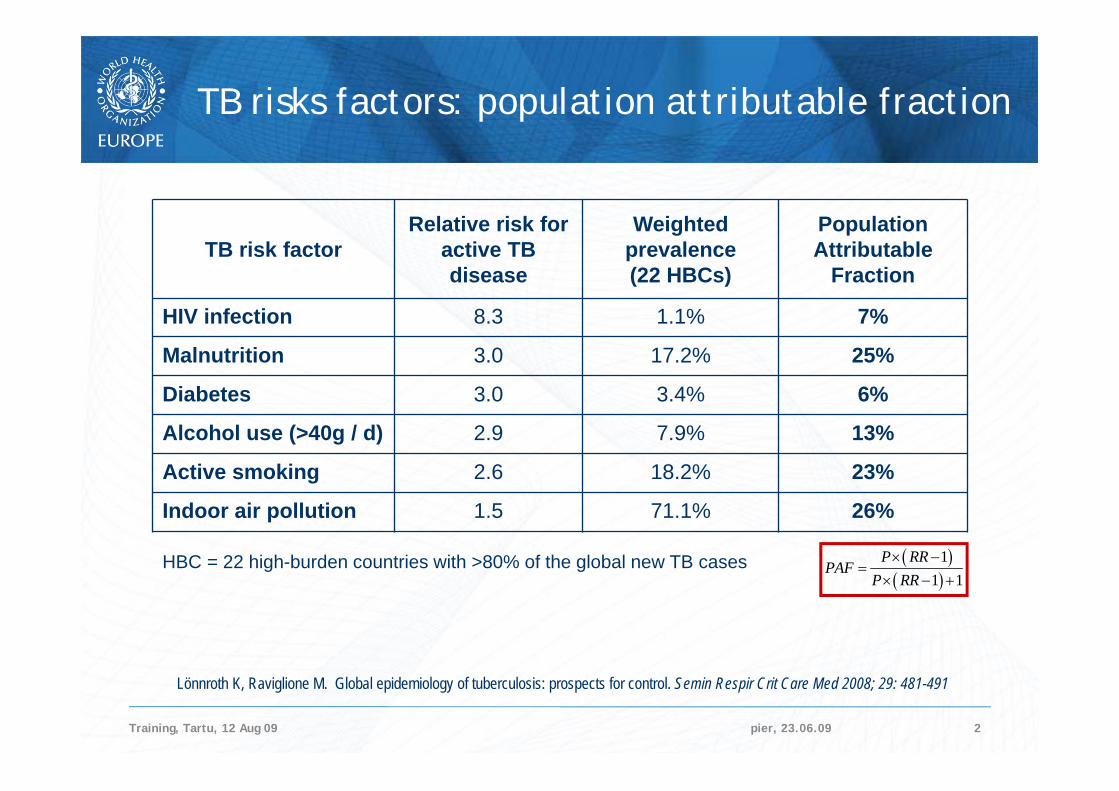
Risk factors for developing TB disease.
pier, 23.06.09Training, Tartu, 12 Aug 09 2
TB risk factorRelative risk for
active TB disease
Weighted prevalence (22 HBCs)
Population Attributable
Fraction
HIV infection 8.3 1.1% 7%
Malnutrition 3.0 17.2% 25%
Diabetes 3.0 3.4% 6%
Alcohol use (>40g / d) 2.9 7.9% 13%
Active smoking 2.6 18.2% 23%
Indoor air pollution 1.5 71.1% 26%
11 1
P RRPAF
P RR
Lönnroth K, Raviglione M. Global epidemiology of tuberculosis: prospects for control. Semin Respir Crit Care Med 2008; 29: 481-491
TB risks factors: population attributable fraction
HBC = 22 high-burden countries with >80% of the global new TB cases

Diagnosis of TB Disease
1. The medical history. A medical history is the part of a patient's life history that is important for diagnosing and treating the patient's medical condition. It includes social, family, medical, and occupational information about the
patient. To obtain a medical history, the clinician should ask whether the patient
has: a. Been exposed to a person who has infectious TB b. Symptoms of TB disease c. Had TB infection or TB disease before d. Risk factors for developing TB disease
Anyone with symptoms of TB or a positive skin testreaction should be evaluated for TB disease

Diagnosis of TB DiseaseDiagnosis of TB Disease
Previous TB infection or TB disease. During the medical history, the clinician should ask the patientwhether he or she has ever been diagnosed with ortreated for TB infection or disease.
Patients known to have a positive skin test reactionprobably have TB infection.
If they were infected within the past 2 years, they are at high risk for TB disease.
Patients who have had TB disease before should be asked when they had the disease and how the disease was treated.
If the regimen prescribed was inadequate or if the patient did not follow the recommended treatment, TB may recur, and it may be resistant to one or more of the drugs used

People with TB disease may or may not have symptoms.
Most patients with TB disease have one or more symptoms that led them to seek medical care. Occasionally, TB is discovered during a medical examination for an unrelated condition (for
example, when a patient is given a chest x-ray before undergoing surgery). Usually, when patients do have symptoms, the symptoms have developed gradually, and they
have been present for weeks or even months.
Pulmonary TB disease usually causes one or more of the following symptoms:--- Coughing--- Pain in the chest when breathing or coughing--- Coughing up sputum (phlegm from deep in the lungs) or blood
Occasionally patients may present massive haemoptysis due to erosion of a bronchial artery .
The majority of patients has only minimal constitutional manifestations (Baydur, 1977, Weir, 1985)The general symptoms of TB disease (pulmonary or extrapulmonary) include
--- Weight loss--- Fatigue--- Malaise--- Fever
--- Night sweats
The symptoms of extrapulmonary TB disease depend on site of disease. For example, TB of the spine may cause pain in the back; TB of the kidney may cause blood in the urine. All of these symptoms may be caused by other diseases, but they should prompt the clinician to suspect TB disease.
Symptoms of TB disease

Radiology. Primary Tuberculosis (1)
Chest radiography remains the mainstay of diagnosis; however, normal radiographic findings may be seen in up to 15% of patients with proved tuberculosis.
At radiology, primary tuberculosis manifests as four main entities: 1) parenchymal disease, 2) lymphadenopathy, 3) miliary disease, 4) pleural effusion
1) Parenchymal Disease Typically, homogeneous parenchymal consolidation in any lobe; Predominance in the lower and middle lobes is suggestive of the disease In approximately two-thirds of cases, the parenchymal focus resolves without sequelae
at conventional radiography; however, this resolution can take up to 2 years. In the remaining cases, a radiologic scar persists that can calcify in up to 15% of cases,
an entity that is known as a Ghon focus. Cavitation is uncommon in primary TB, seen only in 10-30% of cases Tuberculomas can cavitate and undergo calcification.
http://pubs.rsna.org/doi/full/10.1148/rg.275065176

Radiology. Primary Tuberculosis. (2)
2) Lymphadenopathy. Lymphadenopathy is seen in up to 96% of children and 43% of adults. Lymphadenopathy is typically unilateral and right sided, involving the hilum and right
paratracheal region, it is bilateral in about one-third of cases. It is usually seen in the elderly, infants, and immunocompromised persons,
manifesting within 6 months of initial exposure
3) Miliary tuberculosis It is uncommon but carries a poor prognosis. It represents haematogenous dissemination of an uncontrolled tuberculous infection. It is seen both in primary and post-primary tuberculosis. Although implants are seen throughout the body, the lungs are the easiest location to the
image. The classic radiographic findings of evenly distributed diffuse small 2–3-mm nodules,
with a slight lower lobe predominance, are seen in 85% of cases. CT is more sensitive than conventional radiography,with nodules seen in a random
distribution
Radiology. Primary Tuberculosis. (2)
Pleural Effusion. A pleural effusion is seen in approximately one-fourth of patients with proved primary
tuberculosis. The effusion is often the sole manifestation of TB and usually manifests 3–7 months after
initial exposure. Pleural effusions are more frequent in adults, seen in 30-40% of cases,
and a very uncommon finding in infants. The effusion is usually unilateral, and complications ) are rare (eg, empyema formation,
fistulization, bone erosion). Residual pleural thickening and calcification can result. Ultrasonography (US) often
demonstrates a complex septated effusion.

Postprimary tuberculosis remains primarily a disease of adolescence and adulthood
The term postprimary tuberculosis is generally used to refer to both reinfection with and reactivation of tuberculosis.
Primary tuberculosis is usually self-limiting, whereas postprimary tuberculosis is progressive, with cavitations, hematogenous dissemination of the disease as well as disease spreadthroughout the lungs.
Healing usually occurs with fibrosis and calcification. The earliest finding in parenchymal disease is poorly defined
consolidation, in the apical and posterior segments of the upper lobes Cavities are usually multiple and occur within areas of consolidation. Resolution can result in emphysematous change or scarring. A minority of cavities demonstrate air-fluid levels; however, these
findings can indicate the presence of superinfection. If there is airway disease and, in particular, endobronchial spread of
infection, tree-in-bud opacities may develop.
Radiology. Post primary Tuberculosis.

Consolidations, cavities typicallyin the apical and posterior segments of
the upper lobes.
Rarely in the lower and middle lobes


Primary TB infectionAcute Haematological dissemination,
Miliary tuberculosis


Tuberculoma
Tuberculomas are persistent nodules or mass-like lesions which can be seen in both primary TB and PPT.
Pulmonary tuberculomas can range in size from being subcentimetric to 5 cm in diameter, and may be solitary or multiple.
They are most often the result of healed primary TB and are usually smooth-walled and sharply defined.
The majority of these lesions remain stable in size and may calcify. Nodular or diffuse calcification can be seen in 20-30% of tuberculomas.
Cavitation is seen in 10-50% of cases. In 80% of cases, small round opacities (satellite lesions) may be observed
in the immediate vicinity of the main lesion

Post primary TB. Rigid fibrous cavern, unfavorable course of TB
Develops slowly , 2 yearsUsually smear and culture posit, epidemiologically dangerous
Male, 62 y

Pleural Effusion

Tuberculous Spondylitis
Wikimedia Commons Yale Rosen from USA. Uploaded by CFCF

Tuberculous SpondylitisMale, 23 a., MDR-TB

Tuberculous Spondylitis
Otis Historical Archives Nat'l Museum of Health & MedicineThoracic spine showing collapsed vertebrae from infection with tuberculosis. Photo by Brian Spatola. Wikimedia Commons

Osteoarticular TB
Characteristically a monoarthritis affecting large weight-bearing joints.
The imaging findings are similar to those of other infectious and inflammatory arthritides and are, therefore, nonspecific.
These findings include osteopenia, synovitis and other soft-tissue swellings, marginal erosions, and varying degrees of cartilage destruction.
Joint space narrowing occurs with highly variable rapidity but is usually delayed.
As with any chronic infection, synovial involvement in the young results in hyperemia and epiphyseal overgrowth, most commonly in the knee.
With progression of infection, bone sequestration and sinus formation can develop. The end result is usually fibrous ankylosis of the joint.