risk factors for pressure ulcers including...
TRANSCRIPT

Risk Factors for Pressure Ulcers IncludingSuspected Deep Tissue Injury in NursingHome Facility Residents: Analysis ofNational Minimum Data Set 3.0
C M E1 AMA PRACategory 1 CreditTM
ANCC3.0 Contact Hours
Hyochol Ahn, PhD, ARNP, ANP-BC & Assistant Professor & University of Florida College of Nursing & Gainesville
Linda Cowan, PhD, ARNP, FNP-BC, CWS & Research Health Scientist & North Florida/South Georgia Veterans HealthSystem & Gainesville
Cynthia Garvan, PhD & Research Associate Professor & University of Florida College of Nursing & Gainesville
Debra Lyon, PhD, RN, FAAN & Professor and Executive Associate Dean & University of Florida College of Nursing &Gainesville
Joyce Stechmiller, PhD, ACNP-BC, FAAN & Department Chair and Associate Professor & Department of BiobehavioralNursing Science, University of Florida College of Nursing & Gainesville
All authors, staff, and planners, including spouses/partners (if any), in any position to control the content of this CME activity have disclosed that they have no financial relationships with, orfinancial interests in, any commercial companies pertaining to this educational activity.
To earn CME credit, you must read the CME article and complete the quiz and evaluation on the enclosed answer form, answering at least 13 of the 18 questions correctly.
This continuing educational activity will expire for physicians on April 30, 2017, and for nurses on April 30, 2018.
All tests are now online only; take the test at: http://cme.lww.com for physicians and www.nursingcenter.com for nurses. Complete CE/CME information is on the last page of this article.
Acknowledgment: The authors disclose that this study was funded by the University of Florida College of Nursing, Gainesville, Florida. The sponsor had no role in the design, methods, datacollection, analysis, or preparation of this manuscript. The authors thank Debra McDonald for her work on the project.
PURPOSE:
To provide information on risk factors associated with pressure ulcers (PrUs), including suspected deep tissue
injury (sDTI), in nursing home residents in the United States.
TARGET AUDIENCE:
This continuing education activity is intended for physicians and nurses with an interest in skin and wound care.
APRIL 2016
C L I N I C A L M A N A G E M E N T
extra
ADVANCES IN SKIN & WOUND CARE & VOL. 29 NO. 4 178 WWW.WOUNDCAREJOURNAL.COM
Copyright © 2016 Wolters Kluwer Health, Inc. All rights reserved.

OBJECTIVES:
After participating in this educational activity, the participant should be better able to:
1. Examine the literature related to risk factors for the development of PrUs.
2. Compare risk factors associated with the prevalence of PrUs and sDTI from the revised Minimum Data Set 3.0
2012 using a modified Defloor_s conceptual model of PrUs as a theoretical framework.
ABSTRACT
OBJECTIVE: This study aims to characterize and compare riskfactors associated with pressure ulcers (PrUs), includingsuspected deep tissue injury (sDTI), in nursing home (NH) residentsin the United States.DESIGN: Secondary analysis of the 2012 Minimum Data Set(MDS 3.0).SETTING: Medicare- or Medicaid-certified NHs in the United States.PARTICIPANTS: Nursing home residents (n = 2,936,146) 18 yearsor older with complete PrU data, who received comprehensiveassessments from January to December 2012.MEASUREMENTS: Pressure ulcer by stage was the outcomevariable. Explanatory variables (age, gender, race and ethnicity,body mass index, skin integrity, system failure, disease, infection,mobility, and cognition) from the MDS 3.0 were aligned with the4 elements of Defloor_s conceptual model: compressive forces,shearing forces, tissue tolerance for pressure, and tissue tolerancefor oxygen.RESULTS: Of 2,936,146 NH residents who had complete data for PrU,89.9% had no PrU; 8.4% had a Stage 2, 3, or 4 or unstagable PrU;and 1.7% had an sDTI. The MDS variables corresponding to the4 elements of Defloor_s model were significantly predictive of bothPrU and sDTI. Black residents had the highest risk of any-stagePrU, and Hispanic residents had the highest risk of sDTI. Skinintegrity, system failure, infection, and disease risk factors hadlarger effect sizes for sDTI than for other PrU stages.CONCLUSIONS: The MDS data support Defloor_s model andinform clinicians, educators, researchers, and policymakers on riskfactors associated with PrUs and sDTI in NH residents in theUnited States participating in Medicare and Medicaid.KEYWORDS: pressure ulcer, deep tissue injury, nursing home,Minimum Data Set, Defloor model
ADV SKIN WOUND CARE 2016;29:178-90.
INTRODUCTIONEach year in the United States, more than 2.5 million people
experience pressure ulcers (PrUs), which occur across all
healthcare settings: 0.4% to 38% in acute care, 0% to 17% in
home care, and 2% to 24% in long-term-care facilities.1,2 The
prevalence of PrUs is a major threat to public health and the
US healthcare system.3 Because of the increasing occurrence
and severity of PrUs, in 2007 the National Pressure Ulcer
Advisory Panel revised the classifications of the 4 stages of
PrU (I, II, III, and IV) and added 2 more classifications:
Bunstageable[ and Bsuspected deep tissue injury[ (sDTI). The
first type is termed Bunstageable[ because eschar or slough
obscures the depth of the pressure-related injury, which is
characterized by full-thickness tissue loss where the full depth
(Stage III vs IV) is unable to be determined.4,5 The second
type, sDTI, is due to a pressure-related injury under intact
skin.4,5 This internal injury originates in the muscular tissue
that overlies bony prominences as a result of soft tissue
(skeletal muscle and fat tissues) deformations and progresses
outward6 until it appears on the skin surface as a purple or
maroon localized area of discolored intact skin or blood-filled
blister due to damage of underlying soft tissue from pressure
and/or shear.5 The area may be preceded by tissue that is
painful, firm, mushy, boggy, or warmer or cooler as compared
with adjacent tissue. Suspected DTI may be difficult to detect
in individuals with dark skin tones. Evolution may include a
thin blister over a dark wound bed. These serious wounds may
further evolve and be covered by thin eschar. Progression may
be rapid, exposing additional layers of tissue even with
optimal treatment.4,5,7 Suspected DTI may precede the
development of an open Stage III-IV PrU,5 which commonly
occurs in points of high pressure, such as the sacrum,
buttocks, and heels.1,4,5,8–10
Considerable research is ongoing in understanding the
mechanisms underlying the onset and progression of sDTI.6,11
Prior research has focused on ischemic/hypoxic damage,
ischemia-reperfusion injury, structural damage to cells, and
interference with lymphatic and interstitial fluid drainage.6
However, recent studies by Slomka et al6 have shown that
the involvement of sustained deformation-related events at the
cellular level is directly responsible for the distortion of cells,
the deformation of plasma membranes leading to increased
membrane permeability, impairment of cellular control
mechanisms, and eventual cell death caused by loss of cell
homeostasis leading to sDTI.
Skin Changes at Life_s End (SCALE) is a term introduced in
2009 in reference to unusual wounds that occur at the end of
life.10,12 The wound types include PrUs, DTI, unavoidable
pressure injury, and mottling that appear in individuals who
are in the process of dying.12,13 Edsberg et al13 identify some of
ADVANCES IN SKIN & WOUND CARE & APRIL 2016179WWW.WOUNDCAREJOURNAL.COM
Copyright © 2016 Wolters Kluwer Health, Inc. All rights reserved.

the SCALE wounds as examples of unavoidable pressure
injuries. In persons who are actively dying, SCALE pressure
injuries may occur because of nonmodifiable intrinsic and
extrinsic factors that make the individual at risk for pressure,
shear, friction, deformation, hemodynamic instability, ische-
mia, and or reperfusion injury.
Pressure ulcer risk factors have been mentioned in the
scientific literature for hundreds of years.1–45 Lyder9 reported
more than 100 PrU risk factors identified from the scientific
literature in 2003. Berlowitz et al36 validated a risk adjustment
model for the development of PrUs in nursing home (NH)
residents using the Minimum Data Set (MDS) from 1998.
Results of their logistic regression model related NH resident
characteristics and the development of PrUs. The highest risk
factors identified by Berlowitz et al19 include age, male gender,
nonwhite ethnicity, nonroutine assessment, bed mobility self-
performance, transfer self-performance, bedfast, bladder incon-
tinence, deterioration in cognitive status, diabetes, peripheral
vascular disease, hip fracture within 180 days, body mass index
(BMI), end-stage disease, Stage I PrU, history of resolved PrU,
and edema. In comparison and more recently, Fogerty et al34
identified the highest 45 risk factors associated with PrU
prevalence among a sample of 6,610,787 patients hospitalized
in acute care settings. Common risk factors include advanced
age, immobility, friction, shear, poor nutrition, excessive
moisture and incontinence, altered level of consciousness, poor
perfusion, certain skin infections, and comorbid conditions.
Furthermore, Fogerty et al34 found that persons who were
identified as black were more than twice as likely (odds ratio
[OR], 2.3) to develop PrUs than those identified as being white,
and age was an interacting variable, so that as black persons
increase in age, their risk of PrUs increased more than a white
person_s risk of PrUs as they aged.
Defloor38 created a theoretical model of PrU that contained
4 main elements: (1) compressive forces, (2) shearing forces,
(3) tissue tolerance for pressure, and (4) tissue tolerance for
oxygen. The first element, compressive forces, relates to when
mechanical loads exert pressure on a patient_s tissue that is
compressed between a surface, such as a chair, and the bony
prominences of the human body, such as the sacrum or
buttocks. These mechanical loads increase the risk of PrUs,
especially when they exist for an extended period on patients
who are immobile and in sitting positions. The second element
of Defloor_s model, shearing forces, relates to sliding forces that
occur when a human body is moved along a surface. In the
clinical setting, shearing forces are best exemplifiedwhen a nurse
slides a patient up in bed or transfers a patient from the bed to the
chairVactions that entail the patients_ continual contact with a
surface throughout the motion and result in separation of skin
layers. Defloor38 included friction along with shearing forces,
although friction is related to more superficial abrasive or
rubbing forces and may not result in separation between skin
layers. The third element, tissue tolerance for pressure, relates to
the fact that external factors, such as mechanical loads and/or
moisture, interact with internal factors for the fourth element,
tissue tolerance for oxygen, such as low arteriolar pressure or
poor oxygenation, leading to an increased risk of PrUs.38 For
example, if tissue tolerance is low, a shorter duration of pressure
can damage tissues, and likewise, if tissue tolerance is high,
tissue damage may occur only with longer duration of sustained
pressures.38Althoughnodata exist that have precisely timedPrU
development, Slomka et al6 and Gefen11 have shown that PrUs
may develop in high-risk individuals in less than 1 hour of
sustained pressure to vulnerable body tissue.
In Defloor_s38 conceptual model for PrU, several factors
affect tissue tolerance for pressure, and these are age, stress,
cognition, sensory awareness to pain and discomfort (acuity),
dehydration, and tissue mass, as well as protein and vitamin C
deficiency. Similarly, factors that affect tissue tolerance for
oxygen are temperature, medication, protein deficiency, smok-
ing, blood pressure, and presence of certain diseases (ie, those
that affect oxygen supply, reactive hyperemia, and vascular
occlusion). Studies suggest that the development of PrUs may
result from major alterations in the normal functioning of these
human mechanisms. Pressure ulcers may also develop from the
cumulative effects of minor changes in several of these
mechanisms/factors, particularly in combination with sustained
external pressure (forces perpendicular to the skin), and/or the
presence of external moisture (fecal or urinary incontinence,
excess perspiration), and/or friction and shear forces (slipping,
sliding, or rubbing forces parallel to the skin).11,38
In general, studies suggest PrUs will develop because of
major or continual minor alterations in the normal functioning
of human mechanisms in combination with sustained external
pressure, external moisture, and/or friction/shear forces.11,38
With respect to frequency, an analysis of the International
Pressure Ulcer Prevalence survey 2006–2009 showed that the
overall and nosocomial PrU prevalence decreased by approxi-
mately 1% in 2009 after remaining constant in earlier years41;
however, the proportion of ulcers identified as sDTI had
increased 3 times, to 9%, in 2009 and was more prevalent than
either a Stage III or Stage IV PrU. Patients with sDTIs tended to
be older than patients with Stage III or IV ulcers, and unstageable
ulcers tended to occur in people with a slightly lower BMI than
the surveyed population. In attempting an explanation of these
data, the researchers suggested that sDTIs were being identified
more frequently because of staff education of staging definitions
and that actual prevalence had not increased.41
ADVANCES IN SKIN & WOUND CARE & VOL. 29 NO. 4 180 WWW.WOUNDCAREJOURNAL.COM
Copyright © 2016 Wolters Kluwer Health, Inc. All rights reserved.

The MDS, a federally mandated tool, guides the regular
assessment of all residents in NHs certified to participate in
US Medicare and Medicaid programs. The revised Section M:
Skin Conditions of MDS 3.0 now includes at-risk status and
ulcer characteristics, including stage, size, history, tissue type,
number of ulcers, and ulcer identification. For the first time,
the revised Nursing Home MDS 3.0 added a section related
to the presence/absence and number of sDTIs. It is anticipated
that the revisedMDS 3.0 will change PrU tracking and recording
in long-term care and impact the generation of quality indicators
for skin conditions, including PrUs. More important, the MDS
data may provide further understanding of whether PrUs are
actually a reflection of quality care in long-term-care facilities in
the United States.
Many variables aligned with the elements of the Defloor
model were also available in the MDS 3.0. Thus, the objectives
of this study were to characterize and compare risk factors
associated with the prevalence of PrUs and sDTI in NH
residents in a secondary analysis of the revised MDS 3.0 2012
national data set using a modified Defloor_s38 conceptual
model (Figure) of PrUs as a theoretical framework.
METHODS
Data SourceData were drawn from the national MDS 3.0 assessment data
from January to December 2012. The MDS data are mandatory
in all NHs certified to participate in Medicaid and Medicare.
Details about the methods of the MDS assessment data have
been published previously.42,43 This study used the admission
assessment (66.34%) or first comprehensive assessment
(27.35%) or significant change in status assessment (6.31%)
for residents in each NH. The exclusion criteria were age
younger than 18 years, missing age data, missing data on PrU
status, and having a Stage I PrU. Stage I PrUs were excluded
because this stage of pressure-related injury is considered to
be reversible, and literature suggests Stage I PrUs are frequently
misidentified. The MDS 3.0 national data set included 2,936,146
residents whomet the study criteria. Approval for this study was
obtained from the University of Florida Health Science Center
Institutional Review Board prior to commencement.
VariablesOutcome Variables
The outcome variables of interest were (1) the presence of
sDTI; (2) a Stage II, III, or IV or unstageable PrU (excluding
sDTI); and (3) a Stage II, III, or IV or unstageable PrU or the
presence of sDTI (all PrUs, including sDTI). In the validation
study of the MDS 3.0, the average for interrater agreement in
identifying sDTI was 0.94.44
Risk Factors (Explanatory Variables)
The risk factors used in the MDS analysis were classified as
resident characteristics (age, gender, race, ethnicity, and BMI)
and factors that influence skin integrity, including nutritional
factors (anemia, malnutrition, dehydration, urinary incontinence,
and bowel incontinence), system failure (comatose, heart failure,
respiratory failure, end-stage renal disease, cirrhosis), disease
burden (diabetes, multiple sclerosis, coronary artery disease
[CAD], chronic obstructive pulmonary disease, peripheral
vascular disease [venous and arterial]), infection (septicemia,
pneumonia, urinary tract infection [UTI], multidrug resistant
organism), mobility (hemiplegia, paraplegia, quadriplegia, hip
fracture, activities of daily living [ADLs] impairment), sensory
alterations, and cognition. These risk factors were selected a
priori based on the hypothesized association with PrU and
sDTI supported by extant literature and Defloor_s model.
Table 1 shows ORs found in previous literature and shows how
selected MDS variables correspond with Defloor_s model.
AnalysisTo examine group differences on study variables, the groups
Bno PrU[ and Bany PrU[ (including Stages II-IV, unstageable,
and sDTI)were comparedusing descriptive statistics. Descriptive
statistics for the BsDTI only group[ were also calculated.
Descriptive statistics were presented as mean or percentages.
Logistic regression was used to compute the unadjusted OR and
Figure.
MODIFIED DEFLOOR’S CONCEPTUAL MODEL OF
PRESSURE ULCER DEVELOPMENT INCLUDING
SUSPECTED DEEP TISSUE INJURY
ADVANCES IN SKIN & WOUND CARE & APRIL 2016181WWW.WOUNDCAREJOURNAL.COM
Copyright © 2016 Wolters Kluwer Health, Inc. All rights reserved.

Table 1.
RISK FACTORS FOR PRUS WITH CORRESPONDENCE TO DEFLOOR_S MODEL AND ODDS RATIOS FOUND IN
PREVIOUS LITERATURE32–36
Variables Odds Ratios Found in Previous Literature32–34
Corresponds to Defloor_s Model
Age Age >75 y = OR, 12.6332
Tissue tolerance for pressure: Bthe pressure distribution
capacity of tissue correlates negatively with age[36
(aging leads to decrease in muscle and fat mass, altered
cell senescence, altered nutrient transport,
and oxygenation)10,11,33-36
Age (per additional 10 y)33 = OR, 1.134
Gender Female gender = OR, 0.8432
Male gender = OR, 1.434Tissue changes relating to age and relationship to
compressive and shearing forces
Race and
ethnicity
Black = OR, 2.3; Asian = OR, 1.01;
Hispanic = OR, 1.37; other = OR, 1.1132;
nonwhite ethnicity = OR, 1.334
This is an additional risk factor not specifically listed in the
Defloor model, but impacting early identification of tissue
changes relating to both compressive and shearing forces
BMI OR, 0.9534
Compressive forces: Intensity affected by body weight/
build; Bwith cachectic persons, higher peak pressures are
measured at the skin surface than those of normal weight.
For obese persons, greater areas of increased pressure
but lower peak pressures are recorded.[36 Tissue
tolerance for pressure affected by body build (amount of
subcutaneous tissue mass for Bpadding[)11,33,35,36
Malnutrition OR, 9.18 for poor nutrition32
Tissue tolerance for pressure and oxygen (malnutrition alters
cellular senescence, nutrient and waste transport, and
cellular repair).11,33 BLong-term protein deficiency causes an
edema as a result of hypoalbuminemia. This edema
decreases oxygen supply to tissue[34,36
Dehydration No OR listed in Fogerty et al32 article Shearing forces and tissue tolerance for pressure and
oxygen: dehydration shrinks cells and alters cellular
senescence, nutrient and waste transport, cellular repair,
and tolerance to tissue deformation and shearing
forces.11,33 BDehydration decreases skin elasticity and
increases the capacity for deformation of the tissue,
increasing the risk of damage[36
Anemia OR, 2.62 for anemia32
Tissue tolerance for pressure and oxygen: anemia reduces
oxygen levels in the blood and oxygen tissue transport,
which may result in cellular hypoxia and alter cellular
senescence, nutrient andwaste transport, cellular repair, and
tolerance to tissue deformation11,33,36
Urinary or fecal
incontinence
Bladder incontinence = OR, 1.434
Shearing forcesVintensity: maceration and friction
(incontinence alters skin integrity, making tissue more
susceptible to damage by pressure and friction or shearing
forces)36
Neuro (coma) Deterioration in cognitive
status = OR, 1.334
Coma impacts duration of compressive and shearing forces
by impacting sensory-motor activity and mobility11,36
BA comatose state ablates the ability of the individual to
move independently, making them vulnerable to PrUS
unless they are repositioned[11
(continues)
ADVANCES IN SKIN & WOUND CARE & VOL. 29 NO. 4 182 WWW.WOUNDCAREJOURNAL.COM
Copyright © 2016 Wolters Kluwer Health, Inc. All rights reserved.

Table 1.
RISK FACTORS FOR PRUS WITH CORRESPONDENCE TO DEFLOOR_S MODEL AND ODDS RATIOS FOUND IN
PREVIOUS LITERATURE,32–36 CONTINUED
Variables Odds Ratios Found in Previous Literature32-34
Corresponds to Defloor_s Model
Heart failure/CHF
respiratory failure
Renal failure
Cirrhosis
Edema34
CHF = OR, 2.6332
Respiratory failure = OR, 4.32
Acute renal failure = OR, 4.16 32,34
OR, 1.334
All of these biological system failures impact tissue oxygenation
and tissue tolerance to pressure and shear forces, as well as
contribute to skin failure.10 Cardiac failure and respiratory failure
play a part in reduced oxygen supply, delayed reactive
hyperemia, and accelerated vascular occlusion.36 Renal and
liver failure results in alterations in cellular nutrient and waste
transport. Both result in loss of tissue protection during pressure
and shearing forces and greater risk of tissue ischemia36
Diabetes
Coronary artery disease
Vascular disease
Chronic obstructive
pulmonary disease
Diabetes with complication = OR,
2.6332; OR, 1.334
Other circulatory disease = OR, 1.6832
Peripheral atherosclerosis = OR, 2.0932;
OR, 1.034
COPD = OR, 1.6232
All of these biological system failures impact tissue oxygenation
and tissue tolerance to pressure and shear forces as well as
contribute to skin failure.10 Persons with diabetes display a
delayed reactive hyperemia, reduced sympathetic nervous
system function, increased blood viscosity, and thickening of
basement membrane in capillaries. CAD, vascular disease, and
COPD directly impact tissue oxygenation by reduced oxygen
supply, delayed reactive hyperemia, and accelerated vascular
occlusion36
Pneumonia
UTI
Septicemia
Multidrug-resistant
organism
Pneumonia = OR, 3.4732
UTI = OR, 7.1732
Septicemia = OR, 9.7832
Sepsis = OR, 11.311
Tissue tolerance to pressure and oxygenation, and tolerance
to friction and shear forces10
BInfection is the invasion and multiplication of microorganisms
within the body, causing cellular injury due to competitive
metabolism, toxins, intracellular replication, or antigen-antibody
response[11
MS Decreased mobility results in increases in duration and intensity
of compressive, as well as shearing forces and friction.35 MS
results in decreased mobility, as well as Bintermittent or
sustained involuntary muscle activation that may be painful and
deforming. Involuntary spasticmovements can create shear and
contribute to the development of PrU[11
Hemiplegia
Paraplegia
Quadriplegia
Paralysis = OR, 10.3032
Paralysis results in increased compressive forces (both intensity
and duration) and shearing forces (and friction) due to sensory
motor deficits, as well as decreased tissue tolerance to pressure
due to tissue changes that occur below the level of paralysis11,36
Hip fracture Hip fracture within 180 d34
Hip fracture results in decreased mobility, which results in
increased compressive forces (both intensity and duration)
and shearing forces (and friction) due to sensory-motor
changes, as well as pain34,36
ADL impairment Activity was strongly associated with risk
of PrU in previous literature, such that as
activity increases, PrU risk decreases32,34
Decreased mobility/activity results in increased compressive
forces (both intensity and duration) and shearing forces (and
friction)36
Bed mobility self-performance = OR, 1.334
Bedridden = OR, 1.434
Cognition Senility = OR, 4.8432
Senility is associated with decreased mobility (or increased
repetitive motions)
Abbreviations: BMI, body mass index; CAD, coronary artery disease; CHF, congestive heart failure; COPD, chronic obstructive pulmonary disorder; OR, odds ratio; PrU, pressure ulcer;
UTI, urinary tract infection.
ADVANCES IN SKIN & WOUND CARE & APRIL 2016183WWW.WOUNDCAREJOURNAL.COM
Copyright © 2016 Wolters Kluwer Health, Inc. All rights reserved.

associated 95% confidence interval (CI) of risk of any PrU
(comparison is no PrU) due to age, sex, race and ethnicity, BMI,
skin integrity, biological system failure, disease burden, infection,
mobility, and cognition. Odds ratios and associated 95% CIs
were then calculated for the risk of PrU excluding sDTI
(comparison is no PrU). Odds ratios and associated 95% CIs
were also calculated for the risk of sDTI only (comparison is no
PrU). The SAS version 9.3 software (SAS Institute Inc, Cary,
North Carolina) was used for all analyses. To examine whether
resident characteristics were differentially associated with PrU
excluding sDTI or sDTI only, those residents with any PrUs were
analyzed. In the subset of NH residents with any PrU, 2 groups
were compared: PrU excluding sDTI and sDTI only. #2 analyses
were used to compare these 2 groups on resident characteristics.
Multiple logistic stepwise regression was used to determine the
risk factors most associated with the prevalence of PrUS.
RESULTSOf the 2,936,146 NH residents included in the analysis, 89.9%
did not have a PrU, whereas 10.1% had a PrU (4.9% had a
Stage II PrU, 1.0% had a Stage III PrU, 1.8% had an
unstageable PrU, 0.7% had a Stage IV PrU, and 1.7% had
sDTI). Table 2 describes the overall sample, the subgroup who
had no PrU, the subgroup who had a PrU including sDTI, and
the subgroup that had only sDTIs. Table 2 also shows the
percentage of missing data for each variable. The majority of
residents were older than 75 years (65%), female (65%), and
white (81%) and had normal, overweight, or obese BMI (94%).
Among factors influencing the skin integrity variables, urinary
and bowel incontinence (42% and 36%, respectively) and
anemia (29%) were conditions with the highest prevalence in
the sample population. Among the biological system failure
variables, heart failure (20%) and end-stage renal disease (12%)
were the most prevalent, whereas among the disease variables,
diabetes (33%), CAD (23%), and chronic obstructive pulmonary
disease (22%) were the most prevalent. Almost 13% of residents
had a UTI, and 13% had severe mobility limitations due to
paralysis or hip fracture. Sensory alterations included deficits of
hearing (8%), speech (13%), and vision (10%) and inability to
understand others (14%). Approximately 30% of residents were
reported to have severe cognitive impairment, and 21% were
reported to have moderate impairment. Forty-one percent of
residents were on insulin, and 26% were on an anticoagulant.
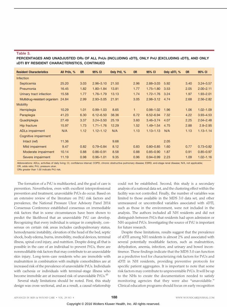
Table 3 presents the magnitude of risk of the prevalence of
all PrUs (including sDTI), only PrU (excluding sDTI), and only
sDTIs. The youngest group of NH residents (aged 18–39 years)
had the highest risk of PrU (13%). Males had a higher risk of
both PrU and sDTI. Black residents and those who were very
severely underweight had the highest risk of any-stage PrU.
Among the skin integrity variables, ORs for both PrU and
sDTI were greater than 2 for residents with malnutrition and
dehydration. Among the biological system failure variables,
ORs for both PrUs and sDTIs were greater than 2 for comatose
residents and residents with respiratory failure. Among disease
factors, the ORs for all PrUs were greater than 2.0 for multiple
sclerosis. The ORs for any PrUs were close to or greater than 2.0
for all of the measured infection variables. Among the mobility
factors, the OR for any PrUs for paraplegia was 6.3, and the OR
for quadriplegia was 3.4. Mean ADL impairment was higher in
both the PrU and sDTI groups.
In comparing the magnitude of risk of only PrU (excluding
sDTI) and only sDTI, the risk of sDTI (OR, 1.17) was larger for
NH residents older than 75 years than for all other PrU stages
(OR, 0.70). The sDTI risk effect size was larger for Asian (OR,
1.11) and Hispanic (OR, 1.53) residents than it was for white
residents. The sDTI ORs were larger than the ORs for all other
stages of PrUs and for all the skin integrity variables. Odds
ratios were larger for all of the system failure variables in the
sDTI group compared with all other stages of PrU. Among the
disease variables, ORs were larger for CAD and vascular dis-
ease in the sDTI group compared with all other stages of PrUs.
Among the infection variables, ORs were larger for septice-
mia, pneumonia, and UTI in the sDTI group compared with all
other PrUs. Among other conditions, OR was higher for sDTI
than all other PrUs if hip fracture was present (OR for sDTI
2.88 vs 1.73 for all other PrUs).
In a multiple logistic stepwise regression to model presence/
absence of PrU, the following risk factors were retained in all
3 models (ie, any PrU, any PrU except sDTI, sDTI only): race,
sex, age, BMI, anemia, malnutrition, dehydration, heart failure,
respiratory failure, end-stage renal disease, diabetes, multiple
sclerosis, peripheral vascular disease, cirrhosis, septicemia, pneu-
monia, multidrug resistant organism, UTI, bowel continence, hip
fracture, mobility (ADL impairment), and impaired cognition. In
addition, the Bany PrU[ and BsDTI only[ models retained the
multiple sclerosis and CAD variables. The nature of association of
risk was the same in the adjusted and unadjusted models.
DISCUSSIONThis prevalence study is the first to provide a detailed
summary of the relationship between NH resident character-
istics and PrUs including sDTI using the national MDS 3.0.
Results demonstrated that just over 10% of NH residents had
a Stage II–IV PrU, unstageable PrU, or sDTI. This number is
slightly lower than that of other studies and may reflect clinical
improvements in prevention and treatment strategies.45 In
reviewing the prevalence of PrUs in long-term care using the
2004 National Nursing Home Survey, approximately 11% of
ADVANCES IN SKIN & WOUND CARE & VOL. 29 NO. 4 184 WWW.WOUNDCAREJOURNAL.COM
Copyright © 2016 Wolters Kluwer Health, Inc. All rights reserved.

Table 2.
RESIDENT CHARACTERISTICS, OVERALL, AND BY PRU STATUS
Resident Characteristics Missing Overall No PrU Any PrU Only sDTI
Age, y 0.00
18–39 1.00 0.97 1.24 0.87
40–58 8.03 7.97 8.49 6.36
59–75 26.47 26.32 27.80 25.23
>75 64.50 64.74 62.47 67.54
Gender 0.02
Male 35.35 34.59 42.00 42.21
Female 64.65 65.41 58.00 57.79
Race 2.39
Asian 1.61 1.62 1.54 1.68
Black 12.23 11.81 15.93 14.91
Hispanic 4.71 4.62 5.43 6.61
Other 0.55 0.54 0.62 0.50
White 80.90 81.41 76.47 76.30
Body mass index (BMI), kg/m2
4.94
Very severely underweight (BMI < 15) 0.39 0.32 0.98 0.87
Severely underweight (15 e BMI < 16.0) 0.75 0.66 1.54 1.41
Underweight (16.0 e BMI < 18.5) 5.15 4.81 8.29 8.08
Normal (18.5 e BMI < 25.0) 37.63 37.21 41.37 42.96
Overweight (25.0 e BMI < 30.0) 28.16 28.57 24.48 25.37
Obese class I (30.0 e BMI < 35.0) 15.15 15.47 12.28 11.72
Obese class II (35.0 e BMI < 40.0) 7.10 7.23 5.90 5.39
Obese class III (BMI > 40.0) 5.67 5.73 5.16 4.20
Skin integrity
Anemia 0.02 29.02 28.17 36.51 37.85
Malnutrition 0.01 2.97 2.60 6.25 6.56
Dehydration 0.02 0.31 0.28 0.60 0.71
Urinary incontinence 7.57 42.44 41.05 57.34 60.56
Bowel incontinence 1.94 35.81 33.45 57.14 60.56
System failure
Comatose 0.02 0.21 0.17 0.62 0.70
Heart failure 0.01 19.56 19.05 24.05 24.57
Respiratory failure 0.01 2.37 2.03 5.31 5.53
ESRD 0.01 12.44 11.73 18.71 20.28
Disease
Diabetes 0.01 32.77 31.89 40.51 40.70
Multiple sclerosis 0.01 0.95 0.86 1.72 1.19
Coronary artery disease 0.01 22.58 22.24 25.57 27.95
COPD 0.02 21.89 21.79 22.76 22.12
Vascular disease 0.01 8.24 7.78 12.27 13.23
Cirrhosis 0.01 0.83 0.80 1.07 0.89(continues)
ADVANCES IN SKIN & WOUND CARE & APRIL 2016185WWW.WOUNDCAREJOURNAL.COM
Copyright © 2016 Wolters Kluwer Health, Inc. All rights reserved.

NH residents had a PrU of any stage; 50% were Stage II, and
the other 50% were Stages I, III, or IV. These PrUs were more
likely to present in those 64 years or younger (14%) versus
those older than 64 years. Men (10%) were less likely to have
PrUs than women (13%). Persons who were residents of NHs
for less than 1 year duration were less likely to have PrUs than
those who were residents for more than 1 year. Other risk
variables identified in the 2004 National Nursing Home Survey,
included weight loss, high immobility, and polypharmacy.2
Notably, this study found that risk factors of Stage II–IV and
unstageable PrUs identified in this analysis were also risk
factors of sDTI. In addition, several risk factors were strongly
associated with all stages of PrUs, including anemia, malnutri-
tion, dehydration, infection, urinary incontinence, and bowel
incontinence. Many of these risk factors are potentially modi-
fiable and may facilitate the development of strategies to help
prevent PrUs in this population in the future.
This study also had 2 findings that represent promising
areas for future research. For example, data revealed that the
youngest group of NH residents had a higher risk of PrUs.
This finding may indicate greater impairment or medical
complexity in this age group and/or the fact that preventive
interventions are focused on older residents. In addition, the
MDS data demonstrated significant racial and ethnic dispar-
ities in the prevalence of PrUs in NH residents. Data for this
study showed that black residents had the highest risk of any-
stage PrU, and Hispanic residents had the highest risk of sDTI.
Considering these findings, it is clear that more research is
needed that examines young, racial/ethnic minority NH
residents, who are at high risk of PrUs, in order to reduce PrU
prevalence in these populations.
Consistent with Defloor_s38 conceptual model, this study
found that a very high risk of PrU and sDTI was evidenced in the
mobility-impaired groups,which includedpatientswithmultiple
sclerosis and other neurodegenerative diseases. This study also
determined that NH residents older than 75 years had the
greatest risk of sDTI. Finally, the data revealed potential rela-
tionships between PrU risk and level of paralysis (paraplegia vs
quadriplegia), as well as PrU risk and specific neurodegenerative
disorders (multiple sclerosis), both of which warrant further
investigation. Other contributing factors related to younger
patients and those with spinal cord injury or paraplegia (such as
biobehavioral and psychological aspects of care and compliance)
also warrant further investigation.13
Table 2.
RESIDENT CHARACTERISTICS, OVERALL, AND BY PRU STATUS, CONTINUED
Resident Characteristics Missing Overall No PrU Any PrU Only sDTI
Infection
Septicemia 0.01 1.31 1.09 3.24 3.62
Pneumonia 0.01 6.54 6.08 10.54 11.73
Urinary tract infection 0.03 12.70 11.94 19.39 21.05
Multidrug-resistant organism 0.01 1.63 1.37 3.98 3.59
Mobility
Hemiplegia 0.01 6.31 6.31 6.36 6.64
Paraplegia 0.01 0.62 0.40 2.49 1.68
Quadriplegia 0.01 0.47 0.38 1.27 0.85
Hip fracture 0.01 5.38 5.03 8.41 13.22
ADLs impairment, mean (SD) 0.01 17.26 (6.49) 16.83 (6.46) 21.04 (5.51) 21.60 (5.18)
Cognitive impairment 2.17
Intact 1.91 1.88 2.13 2.10
Mild impairment 47.84 48.20 44.61 41.56
Moderate impairment 20.65 20.65 20.63 20.87
Severe impairment 29.61 29.27 32.63 35.46
Abbreviations: ADLs, activities of daily living; COPD, chronic obstructive pulmonary disease; ESRD, end-stage renal disease; MDS, Minimum Data Set; PrU, pressure ulcer;
sDTI, suspected deep tissue injury.
Note: Values are presented in percentages. Overall n = 2,936,146 no PrU (n = 2,639,104 89.9%), and any PrU, including sDTI (n = 297,042; 10.1%) and only sDTI (n = 50,073; 1.7%). The level
of activities of daily living impairment was measured using the MDS ADL-Long form.36 Cognition was measured using the MDS 3.0 Brief Interview for Mental Status for residents who could
be interviewed and the MDS-Cognitive Performance Scale for those who could not be interviewed.
ADVANCES IN SKIN & WOUND CARE & VOL. 29 NO. 4 186 WWW.WOUNDCAREJOURNAL.COM
Copyright © 2016 Wolters Kluwer Health, Inc. All rights reserved.

Table 3.
PERCENTAGES AND UNADJUSTED ORS OF ALL PRUS (INCLUDING SDTI), ONLY PRU (EXCLUDING SDTI), AND ONLY
SDTI BY RESIDENT CHARACTERISTICS, CONTINUED
Resident Characteristics All PrUs, % OR 95% CI Only PrU, % OR 95% CI Only sDTI, % OR 95% CI
Age
18–39 (ref) 12.62 11.31 1.66
40–58 10.80 0.84 0.81–0.87 9.58 0.83 0.80–0.86 1.49 0.90 0.81–0.99
59–75 10.72 0.83 0.80–0.86 9.25 0.80 0.77–0.83 1.79 1.08 0.98–1.18
>75 9.89 0.76 0.73–0.79 8.25 0.70 0.68–0.73 1.94 1.17 1.06–1.29
Gender
Male 12.13 1.37 1.36–1.38 10.30 1.37 1.36–1.38 2.26 1.38 1.36–1.41
Female (ref) 9.16 7.75 1.65
Race
Asian 9.76 1.01 0.98–1.05 8.13 1.00 0.96–1.03 1.92 1.11 1.03–1.19
Black 13.28 1.44 1.42–1.45 11.44 1.45 1.44–1.47 2.33 1.35 1.31–1.38
Hispanic 11.76 1.25 1.23–1.27 9.60 1.20 1.17–1.22 2.63 1.53 1.47–1.58
Other 11.52 1.22 1.16–1.28 10.13 1.27 1.20–1.34 1.71 0.98 0.87–1.12
White (ref) 9.63 8.16 1.74
Body mass index
Very severely underweight 25.33 2.76 2.64–2.88 22.37 2.84 2.71–2.97 4.86 2.37 2.14–2.62
Severely underweight 20.45 2.09 2.02–2.16 17.86 2.14 2.06–2.22 3.81 1.84 1.70–1.99
Underweight 16.05 1.55 1.53–1.58 13.78 1.57 1.55–1.60 3.04 1.46 1.41–1.51
Normal (ref) 10.97 9.23 2.11
Overweight 8.67 0.77 0.76–0.78 7.27 0.77 0.76–0.78 1.63 0.77 0.75–0.79
Obese class I 8.09 0.71 0.71–0.72 6.88 0.73 0.72–0.74 1.40 0.66 0.64–0.68
Obese class II 8.29 0.73 0.72–0.75 7.11 0.75 0.74–0.77 1.37 0.65 0.62–0.67
Obese class III 9.07 0.81 0.80–0.82 7.93 0.85 0.83–0.86 1.35 0.63 0.61–0.66
Skin integrity
Anemia 12.84 1.47 1.46–1.48 10.86 1.45 1.44–1.46 2.49 1.55 1.53–1.58
Malnutrition 21.47 2.50 2.46–2.54 18.40 2.47 2.43–2.52 4.57 2.63 2.54–2.73
Dehydration 19.66 2.16 2.05–2.27 16.42 2.08 1.97–2.20 4.59 2.54 2.29–2.83
Urinary continence 13.33 1.95 1.93–1.97 9.69 1.88 1.86–1.90 2.22 2.21 2.16–2.25
Bowel continence 18.11 2.59 2.57–2.61 13.46 2.58 2.56–2.6 3.27 3.05 3.00–3.11
System failure
Comatose 29.73 3.74 3.54–3.95 25.55 3.65 3.44–3.87 7.38 4.23 3.79–4.71
Heart failure 12.55 1.35 1.33–1.36 10.64 1.34 1.33–1.35 2.39 1.38 1.36–1.41
Respiratory failure 22.92 2.71 2.66–2.76 19.72 2.69 2.63–2.74 4.91 2.82 2.71–2.93
ESRD 15.36 1.73 1.72–1.75 12.94 1.70 1.68–1.72 3.18 1.92 1.87–1.96
Disease
Diabetes 12.62 1.46 1.44–1.47 10.73 1.45 1.44–1.47 2.36 1.47 1.44–1.49
Multiple sclerosis 18.48 2.01 1.95–2.07 16.71 2.14 2.07–2.21 2.55 1.39 1.28–1.50
Coronary artery disease 11.56 1.20 1.19–1.21 9.65 1.17 1.16–1.18 2.33 1.36 1.33–1.38
COPD 10.61 1.06 1.05–1.07 9.05 1.07 1.06–1.08 1.89 1.02 1.00–1.04
Vascular disease 15.20 1.66 1.64–1.68 12.81 1.63 1.61–1.65 3.12 1.81 1.76–1.85
Cirrhosis 13.20 1.34 1.29–1.39 11.57 1.39 1.33–1.44 2.07 1.12 1.02–1.23
(continues)
ADVANCES IN SKIN & WOUND CARE & APRIL 2016187WWW.WOUNDCAREJOURNAL.COM
Copyright © 2016 Wolters Kluwer Health, Inc. All rights reserved.
The formation of a PrU is multifaceted, and the goal of care is
prevention. Nevertheless, even with excellent interprofessional
prevention and treatment, unavoidable PrUs do occur. Based on
an extensive review of the literature on PrU risk factors and
prevalence, the National Pressure Ulcer Advisory Panel 2014
Consensus Conference established consensus on irremediable
risk factors that in some circumstances have been shown to
predict the likelihood that an unavoidable PrU can develop.
Recognizing that every individual is unique in complexity, con-
sensus on certain risk areas includes cardiopulmonary status,
hemodynamic instability, elevation of the head of the bed, septic
shock, body edema, burns, immobility,medical devices, terminal
illness, spinal cord injury, and nutrition. Despite doing all that is
possible in the care of an individual to prevent PrUs, there are
nonmodifiable risk factors thatmay contribute to an unavoidable
skin injury. Long-term-care residents who are immobile with
malnutrition in combination with multiple comorbidities are at
increased risk of the prevalence of unavoidable PrUs. Individuals
with cachexia or individuals with terminal-stage illness who
become immobile are at increased risk of unavoidable PrUs.45
Several study limitations should be noted. First, this study
design was cross-sectional, and as a result, a causal relationship
could not be established. Second, this study is a secondary
analysis of a national data set, and the clustering effect within the
facility was not controlled. Finally, the number of variables was
limited to those available in the MDS 3.0 data set, and other
unmeasured or uncontrolled variables associated with sDTI,
such as those in the environment, were not included in the
analysis. The authors included all NH residents and did not
distinguish between PrUs that residents had upon admission or
NH-acquired PrUs. Investigating the source of PrU is important
for future research.
Despite these limitations, results suggest that the prevalence
of sDTI among NH residents is almost 2% and associated with
several potentially modifiable factors, such as malnutrition,
dehydration, anemia, infection, and urinary and bowel incon-
tinence. These findings indicate that the MDS 3.0 can function
as a predictive tool for characterizing risk factors for PrUs and
sDTI in NH residents, providing preventive protocols for
specific patient aggregates. It is important to note that some
risk factors may contribute to unpreventable PrUs. It will be up
to the NHs to create the documentation needed to satisfy
monitoring agencies that they were also Bunavoidable.[
Clinical education programs should focus on early recognition
Table 3.
PERCENTAGES AND UNADJUSTED ORS OF ALL PRUS (INCLUDING SDTI), ONLY PRU (EXCLUDING SDTI), AND ONLY
SDTI BY RESIDENT CHARACTERISTICS, CONTINUED
Resident Characteristics All PrUs, % OR 95% CI Only PrU, % OR 95% CI Only sDTI, % OR 95% CI
Infection
Septicemia 25.20 3.03 2.96–3.10 21.50 2.96 2.88–3.03 5.92 3.40 3.24–3.57
Pneumonia 16.45 1.82 1.80–1.84 13.81 1.77 1.75–1.80 3.53 2.05 2.00–2.11
Urinary tract infection 15.58 1.77 1.76–1.79 13.13 1.74 1.72–1.76 3.24 1.97 1.93–2.01
Multidrug-resistant organism 24.84 2.99 2.93–3.05 21.91 3.05 2.98–3.12 4.74 2.68 2.56–2.82
Mobility
Hemiplegia 10.29 1.01 0.99–1.03 8.65 1 0.98–1.02 1.96 1.06 1.02–1.09
Paraplegia 41.23 6.30 6.12–6.50 38.36 6.72 6.52–6.94 7.32 4.22 3.93–4.53
Quadriplegia 27.49 3.37 3.24–3.50 25.19 3.60 3.46–3.74 4.07 2.25 2.04–2.48
Hip fracture 15.97 1.73 1.71–1.76 12.29 1.52 1.49–1.54 4.75 2.88 2.8–2.95
ADLs impairment N/A 1.12 1.12–1.12 N/A 1.13 1.13–1.13 N/A 1.13 1.13–1.14
Cognitive impairment
Intact (ref) 11.36 9.68 2.05
Mild impairment 9.47 0.82 0.79–0.84 8.12 0.83 0.80–0.85 1.60 0.77 0.73–0.82
Moderate impairment 10.14 0.88 0.86–0.91 8.58 0.88 0.85–0.90 1.87 0.91 0.85–0.97
Severe impairment 11.19 0.98 0.96–1.01 9.35 0.96 0.94–0.99 2.23 1.09 1.02–1.16
Abbreviations: ADLs, activities of daily living; CI, confidence interval; COPD, chronic obstructive pulmonary disease; ESRD, end-stage renal disease; N/A, not applicable;
OR, odds ratio; PrU, pressure ulcer.
ORs greater than 1.00 indicate PrU risk.
ADVANCES IN SKIN & WOUND CARE & VOL. 29 NO. 4 188 WWW.WOUNDCAREJOURNAL.COM
Copyright © 2016 Wolters Kluwer Health, Inc. All rights reserved.
of NH residents at risk of the development of PrUs and sDTI, as
well as specific risk characteristics and preventive strategies.
Pressure ulcer Bprevention protocols[ in most facilities do not
include interventions that alter some of the important potentially
modifiable risk factors noted here. It remains an area of deep
concern that these protocols continue to focus on turning and
moisture rather than on interventions that impact the other factors
clinicians identify (eg, anemia, infection, other variables that affect
tissue oxygen levels). Similarly, clinical translational researchers
should test preventive strategies in future studies and develop
tools to determine sDTI risk in order to reduce sDTI prevalence in
high-risk populations.
CONCLUSIONSIn summary, this study characterized the risk factors associa-
ted with PrUs and sDTI. The strongest risk factors associated
with all stages of PrU prevalence, including sDTI, were anemia,
malnutrition, dehydration, infection, urinary incontinence, and
bowel incontinence. These risk factors aremodifiable andmay aid
in the development of strategies to prevent PrUs in NH residents
in the future. The magnitude of risk of sDTI is higher for factors
that include age, race, skin integrity, system failure, disease, and
infection variables. Individuals with these factors should be
targeted with frequent skin assessments and implementation of
off-loading pressure strategies. Modifiable risk factors for sDTI
in NHs that should be targeted by interventions include malnutri-
tion, dehydration, anemia, UTI, urinary and bowel incontinence,
reduction of friction and shearing, and effective pressure off-
loading/redistribution in mobility-impaired individuals.
PRACTICE PEARLS
REFERENCES1. Agency for Healthcare Research and Quality. Preventing pressure ulcers in hospitals.
http://www.ahrq.gov/professionals/systems/long-term-care/resources/pressure-ulcers/
pressureulcertoolkit/putoolkit.pdf. Last accessed January 27, 2016.
2. National Pressure Ulcer Advisory Panel (NPUAP) Monograph. In: Pieper B, ed. Pressure
Ulcers: Prevalence, Incidence, and Implications for the Future. Washington, DC: NPUAP;
2012.
3. Sen CK, Gordillo GM, Roy S, et al. Human skin wounds: a major and snowballing threat
of public health and the economy. Wound Repair Regen 2009;17:763-71.
4. National Pressure Ulcer Advisory Panel (NPUAP). NPUAP pressure ulcer stages/categories.
http://www.npuap.org/resources/educational-and-clinical-resources/npuap-pressure-ulcer-
stagescategories/. Last accessed January 27, 2016.
5. Black J, Baharestani MM, Cuddigan J, et al. National Pressure Ulcer Advisory Panel_s
updated pressure ulcer staging system. Adv Skin Wound Care 2007;20:269-74.
6. Slomka N, Or-Tzadikario S, Gefen A. Cellular Deformations under Compression in Cells
Involved in Deep Tissue Injury. In: Gefen A, ed. Bioengineering Research of Chronic Wounds:
A Multidisciplinary Study Approach. Berlin Heidelberg, Germany: Springer-Verlag Berlin
Heidelberg; 2009.
7. Niezgoda JA, Mendez-Eastman S. The effective management of pressure ulcers. Adv
Skin Wound Care 2006;19(Suppl 1):3-15.
8. Perneger TV, Heliot C, Rae AC, Borst F, Gaspoz JM. Hospital-acquired pressure ulcers:
risk factors and use of preventive devices. Arch Intern Med 1998;158:1940-5.
9. Lyder CH. Pressure ulcer prevention and management. JAMA 2003;289:223-6.
10. Vangilder C, Macfarlane GD, Meyer S. Results of nine international pressure ulcer prevalence
surveys: 1989 to 2005. Ostomy Wound Manage 2008;54(2):40-54.
11. Gefen A. How much time does it take to get a pressure ulcer? Integrated evidence from
human, animal, and in vitro studies. Ostomy Wound Manage 2008;54(10):26-8.
12. Sibbald RG, Krasner DL, Lutz J. SCALE: Skin Changes at Life_s End: final consensus
statement. October 1, 2009. Adv Skin Wound Care 2010;23:225-38.
13. Edsberg LE, Langemo D, Baharestani MM, Posthauer ME, Goldberg M. Unavoidable
pressure injury: state of the science and consensus outcomes. J Wound Ostomy Continence
Nurs 2014;41:313-34.
14. Allman RM, Goode PS, Patrick MM, Burst N, Bartolucci AA. Pressure ulcer risk factors
among hospitalized patients with activity limitation. JAMA 1995;273:865-70.
15. Baldwin KM, Ziegler SM. Pressure ulcer risk following critical traumatic injury. Adv Wound
Care 1998;11:168-73.
16. Bergquist S, Frantz R. Pressure ulcers in community-based older adults receiving home
health care. Prevalence, incidence, and associated risk factors. Adv Wound Care 1999;
12:339-51.
17. Bergstrom N, Braden B. A prospective study of pressure sore risk among institutionalized
elderly. J Am Geriatr Soc 1992;40:747-58.
18. Bergstrom N, Braden B, Kemp M, Champagne M, Ruby E. Multi-site study of incidence
of pressure ulcers and the relationship between risk level, demographic characteristics,
diagnoses, and prescription of preventive interventions. J Am Geriatr Soc 1996;44(1):22-30.
19. Berlowitz DR, Wilking SV. Risk factors for pressure sores. A comparison of cross-sectional
and cohort-derived data. J Am Geriatr Soc 1989;37:1043-50.
20. Brandeis GH, Ooi WL, Hossain M, Morris JN, Lipsitz LA. A longitudinal study of risk factors
associated with the formation of pressure ulcers in nursing homes. J Am Geriatr Soc
1994;42:388-93.
21. Chan WS, Pang SM, Kwong EW. Assessing predictive validity of the modified Braden
scale for prediction of pressure ulcer risk of orthopaedic patients in an acute care setting.
J Clin Nurs 2009;18:1565-73.
22. Fife C, Otto G, Capsuto EG, et al. Incidence of pressure ulcers in a neurologic intensive
care unit. Crit Care Med;29:283-90.
23. Goodridge DM, Sloan JA, LeDoyen YM, McKenzie JA, Knight WE, Gayari M. Risk-assessment
scores, prevention strategies, and the incidence of pressure ulcers among the elderly in four
Canadian health-care facilities. Can J Nurs Res 1998;30(2):23-44.
24. Hatanaka N, Yamamoto Y, Ichihara K, et al. A new predictive indicator for development
of pressure ulcers in bedridden patients based on common laboratory tests results. J Clin
Pathol 2008;61:514-8.
25. Okuwa M, Sanada H, Sugama J, et al. A prospective cohort study of lower-extremity
pressure ulcer risk among bedfast older adults. Adv Skin Wound Care 2006;19:391-7.
26. Schultz A, Bien M, Dumond K, Brown K, Myers A. Etiology and incidence of pressure
ulcers in surgical patients. AORN J 1999;70:434, 437-440, 443-39.
& The revised Nursing Home MDS 3.0 added a section
related to the presence/absence and number of sDTI.
& Suspected DTI may precede the development of an open
Stage III-IV PrU, which commonly occurs in points of high
pressure, such as the sacrum, buttocks, and heels.
& Study results suggest that the prevalence of sDTI among NH
residents is almost 2% and associated with several potentially
modifiable factors, such as malnutrition, dehydration, anemia,
infection, and urinary and bowel incontinence.
& Studies suggest PrUs will develop because of major or
continual minor alterations in the normal functioning of
human mechanisms in combination with sustained external
pressure, external moisture, and/or friction/shear forces.
&Defloor_s model of PrUs contained 4 main elements: (1)
compressive forces, (2) shearing forces, (3) tissue tolerance
for pressure, and (4) tissue tolerance for oxygen.
ADVANCES IN SKIN & WOUND CARE & APRIL 2016189WWW.WOUNDCAREJOURNAL.COM
Copyright © 2016 Wolters Kluwer Health, Inc. All rights reserved.

27. Stordeur S, Laurent S, D_Hoore W. The importance of repeated risk assessment for
pressure sores in cardiovascular surgery. J Cardiovasc Surg (Torino) 1998;39:343-9.
28. Tescher AN, Branda ME, Byrne TJ, Naessens JM. All at-risk patients are not created
equal: analysis of Braden pressure ulcer risk scores to identify specific risks. J Wound
Ostomy Continence Nurs 2012;39:282-91.
29. Baumgarten M, Margolis D, van Doorn C, et al. Black/White differences in pressure ulcer
incidence in nursing home residents. J Am Geriatr Soc 2004;52:1293-8.
30. Levine JM. Historical perspective: impact of plaster of paris splints on pressure ulcer
occurrence in World War II. Adv Skin Wound Care 2008;21:526-8.
31. Levine JM. Historical notes on pressure ulcers: the decubitus ominosus of Jean-Martin
Charcot. J Am Geriatr Soc 2005;53:1248-51.
32. Levine JM. Historical notes on pressure ulcers: the cure of Ambrose Pare. Decubitus
1992;5(2):23-4, 26.
33. Levine JM. Historical perspective: the neurotrophic theory of skin ulceration. J Am Geriatr
Soc 1992;40:1281-3.
34. Fogerty MD, Abumrad NN, Nanney L, Arbogast PG, Poulose B, Barbul A. Risk factors for
pressure ulcers in acute care hospitals. Wound Repair Regen 2008;16:11-8.
35. Stechmiller JK, Cowan L, Whitney JD, et al. Guidelines for the prevention of pressure
ulcers. Wound Repair Regen 2008;16:151-68.
36. Berlowitz D, Brandeis G, Anderson J, et al. Evaluation of a risk-adjustment model for
pressure ulcer development using the Minimum Data Set. J Am Geriatr Soc 2001;49:872-6.
37. Cowan LJ, Stechmiller JK, Rowe M, Kairalla JA. Enhancing Braden pressure ulcer
risk assessment in acutely ill adult veterans. Wound Repair Regen 2012;20:137-48.
38. Defloor T. The risk of pressure sores: a conceptual scheme. J Clin Nurs 1999;8:
206-16.
39. Braden B, Bergstrom N. A conceptual schema for the study of the etiology of pressure
sores. Rehabil Nurs 1987;12:8-12.
40. Baranoski S. Pressure ulcers: a renewed awareness. Nursing 2006;36(8):36-41.
41. VanGilder C, MacFarlane GD, Harrison P, Lachenbruch C, Meyer S. The demographics
of suspected deep tissue injury in the United States: an analysis of the International
Pressure Ulcer Prevalence Survey 2006-2009. Adv Skin Wound Care 2010;23:254-61.
42. Ahn H, Horgas A. The relationship between pain and disruptive behaviors in nursing
home resident with dementia. BMC Geriatr 2013;13:14.
43. Ahn H, Stechmiller J, Horgas A. Pressure ulcer-related pain in nursing home residents
with cognitive impairment. Adv Skin Wound Care 2013;26:375-80.
44. Saliba D, Buchanan J. Development and validation of a revised nursing home assessment
tool: MDS 3.0. 2008. http://www.cms.gov/Medicare/Quality-Initiatives-Patient-Assessment-
instruments/NursingHomeQualityInits/downloads/MDS30FinalReport.pdf. Last accessed
February 3, 2016.
45. National Pressure Ulcer Advisory Panel, European Pressure Ulcer Advisory Panel and
Pan Pacific Pressure Injury Alliance. Prevention and Treatment of Pressure Ulcers: Clinical
Practice Guideline. Haesler E, ed. Perth, Australia: Cambridge Media; 2014.
For more than 137 additional continuing education articles related to Skin and Wound Care topics,go to NursingCenter.com/CE.
CONTINUING MEDICAL EDUCATION INFORMATION FOR PHYSICIANSLippincott Continuing Medical Education Institute, Inc. is accredited by the Accreditation
Council for Continuing Medical Education to provide continuing medical education
for physicians.
LippincottContinuingMedical Education Institute, Inc. designates this journal-basedCMEactivity
for a maximum of 1 AMA PRA Category 1 CreditTM. Physicians should only claim credit
commensurate with the extent of their participation in the activity.
PROVIDER ACCREDITATION INFORMATION FOR NURSES
Lippincott Williams & Wilkins, publisher of the Advances in Skin & Wound Care journal, will
award 3.0 contact hours for this continuing nursing education activity.
LWW is accredited as a provider of continuing nursing education by the American Nurses
Credentialing Center’s Commission on Accreditation.
This activity is also provider approved by the California Board of Registered Nursing, Provider
Number CEP 11749 for 3.0 contact hours. LWW is also an approved provider by the District of
Columbia, Georgia, and Florida CE Broker #50-1223. Your certificate is valid in all states.
OTHER HEALTH PROFESSIONALS
This activity provides ANCC credit for nurses and AMA PRA Category 1 CreditTM for MDs and
DOs only. All other healthcare professionals participating in this activity will receive a certificate
of participation that may be useful to your individual profession’s CE requirements.
CONTINUING EDUCATION INSTRUCTIONS
&Read the article beginning on page 178. For nurses who wish to take the test for CE contact
hours, visit www.nursingcenter.com. For physicians, who wish to take the test for CME credit,
visit http://cme.lww.com.
&You will need to register your personal CE Planner account before taking online tests. Your planner
will keep track of all your Lippincott Williams & Wilkins online CE activities for you.
& There is only one correct answer for each question. A passing score for this test is 13 correct
answers. If you pass, you can print your certificate of earned contact hours or credit and access
the answer key. Nurses who fail have the option of taking the test again at no additional cost. Only
the first entry sent by physicians will be accepted for credit.
Registration Deadline: April 30, 2018 (nurses); April 30, 2017 (physicians).
PAYMENT AND DISCOUNTS
& The registration fee for this test is $27.95 for nurses; $22 for physicians.
ADVANCES IN SKIN & WOUND CARE & VOL. 29 NO. 4 190 WWW.WOUNDCAREJOURNAL.COM
Copyright © 2016 Wolters Kluwer Health, Inc. All rights reserved.