rna families the rnase p family
TRANSCRIPT
[RNA Biology 6:4, 362-369; September/October 2009]; ©2009 Landes Bioscience
362 RNA Biology 2009; Vol. 6 Issue 4
Ribonuclease P (RNase P) is a ribonucleoprotein comprised of a catalytic RNA subunit and one or several protein subunits. RNase P is best known for its role in 5'-processing of tRNA precursors. RNase P enzymes from almost all forms of life, including protein-synthesizing organelles, contain an RNase P with a conserved, homologous RNA. Five distinct structure classes of RNase P RNAs have been identified in bacteria and archaea; eukaryotic RNase P RNAs are not yet sufficiently well surveyed for structure classes to be defined. Here we will examine the structure variations in RNase P RNAs in bacteria, archaea, eukaryotes, plastids and mitochondria with special emphasis on the functional roles these unique secondary structures perform.
Introduction
Ribonuclease P (RNase P) is an ancient ribonuclease, arising before the last common ancestor and thought to be a relic of the RNA world. The RNase P holoenzyme is a ribonucleoprotein complex comprised of an RNA subunit and one or more protein subunits. The RNA, not the associated protein(s), is the catalytic subunit; RNase P is a truly catalytic RNA. RNase P enzymes from all three domains of life (bacteria, archaea and eukarya) have a homologous RNA subunit, and with a single known exception (that of the human mitochondrion, see below) this RNA is found in all organisms and is an essential component of life.
RNase P is best known for its role in the maturation of tRNA. RNase P recognizes the mature region of the precursor-tRNA (pre-tRNA) substrate and cleaves it at the location of the 5' end, generating the 5'-mature tRNA and a leader fragment. RNase P is also responsible for the 5' maturation of other RNAs, e.g., 2S, 4.5S, tmRNAs and snoRNAs.1-6 It has also been shown to cleave riboswitchs in E. coli and Bacillus subtilis.7-9
Five distinct structural classes of RNase P RNAs (types A, type B, type C, type M and type T) have been defined, and many less well-defined types and subtypes have been observed in nature. Here we will examine the structure variations in RNase P RNAs
in bacteria, archaea, eukaryotes, plastids and mitochondria with special emphasis on the functional roles these unique secondary structures perform.
Bacterial RNase P RNA
In bacteria, the RNase P holoenzyme is composed of a single RNA (ca. 400 nts) and a single small protein subunit (ca.14 kDa). The secondary structure of RNase P RNA in bacteria has been well-established primarily on the basis of comparative analysis of several hundred sequences.10-13 Two distinct secondary structure types predominate in bacteria: Type A and Type B. Within each type, the RNAs share a common conserved “body plan”, often decorated by volatile insertions and deletions. Each of the types can be further separated into subtypes that vary in conserved specific elements.
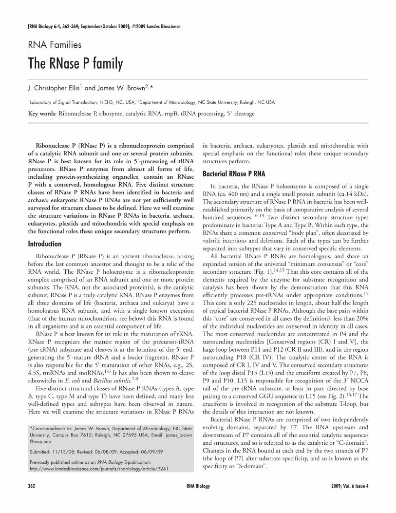
All bacterial RNase P RNAs are homologous, and share an expanded version of the universal “minimum consensus” or “core” secondary structure (Fig. 1).14,15 That this core contains all of the elements required by the enzyme for substrate recognition and catalysis has been shown by the demonstration that this RNA efficiently processes pre-tRNAs under appropriate conditions.15 This core is only 225 nucleotides in length, about half the length of typical bacterial RNase P RNAs. Although the base pairs within this “core” are conserved in all cases (by definition), less than 20% of the individual nucleotides are conserved in identity in all cases. The most conserved nucleotides are concentrated in P4 and the surrounding nucleotides [Conserved regions (CR) I and V], the large loop between P11 and P12 (CR II and III), and in the region surrounding P18 (CR IV). The catalytic center of the RNA is composed of CR I, IV and V. The conserved secondary structures of the loop distal P15 (L15) and the cruciform created by P7, P8, P9 and P10. L15 is responsible for recognition of the 3' NCCA tail of the pre-tRNA substrate, at least in part directed by base pairing to a conserved GGU sequence in L15 (see Fig. 2).16,17 The cruciform is involved in recognition of the substrate T-loop, but the details of this interaction are not known.
Bacterial RNase P RNAs are comprised of two independently evolving domains, separated by P7. The RNA upstream and downstream of P7 contains all of the essential catalytic sequences and structures, and so is referred to as the catalytic or “C-domain”. Changes in the RNA bound at each end by the two strands of P7 (the loop of P7) alter substrate specificity, and so is known as the specificity or “S-domain”.
*Correspondence to: James W. Brown; Department of Microbiology; NC State University; Campus Box 7615; Raleigh, NC 27695 USA; Email: james_brown @ncsu.edu
Submitted: 11/13/08; Revised: 06/08/09; Accepted: 06/09/09
Previously published online as an RNA Biology E-publication: http://www.landesbioscience.com/journals/rnabiology/article/9241
RNA Families
The RNase P familyJ. Christopher Ellis1 and James W. Brown2,*
1Laboratory of Signal Transduction; NIEHS; NC, USA; 2Department of Microbiology; NC State University; Raleigh, NC USA
Key words: Ribonuclease P, ribozyme, catalytic RNA, rnpB, tRNA processing, 5´ cleavage
www.landesbioscience.com RNA Biology 363
In all bacterial RNase P enzymes examined, the RNA subunit is associated with a single, small conservative protein. This protein, encoded by the rnpA gene, has an unusual left-handed βαβ crossover connection and a large central cleft.18 This secondary structure is shared by ribosomal protein S5 and ribosomal trans-locase elongation factor (EF-G), suggesting that these proteins evolved from a common ancestry component of the ancient trans-lational machinery.18
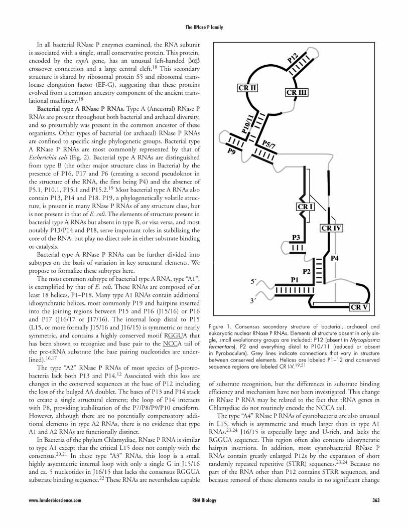
Bacterial type A RNase P RNAs. Type A (Ancestral) RNase P RNAs are present throughout both bacterial and archaeal diversity, and so presumably was present in the common ancestor of these organisms. Other types of bacterial (or archaeal) RNase P RNAs are confined to specific single phylogenetic groups. Bacterial type A RNase P RNAs are most commonly represented by that of Escherichia coli (Fig. 2). Bacterial type A RNAs are distinguished from type B (the other major structure class in Bacteria) by the presence of P16, P17 and P6 (creating a second pseudoknot in the structure of the RNA, the first being P4) and the absence of P5.1, P10.1, P15.1 and P15.2.19 Most bacterial type A RNAs also contain P13, P14 and P18. P19, a phylogenetically volatile struc-ture, is present in many RNase P RNAs of any structure class, but is not present in that of E. coli. The elements of structure present in bacterial type A RNAs but absent in type B, or visa versa, and most notably P13/P14 and P18, serve important roles in stabilizing the core of the RNA, but play no direct role in either substrate binding or catalysis.
Bacterial type A RNase P RNAs can be further divided into subtypes on the basis of variation in key structural elements. We propose to formalize these subtypes here.
The most common subtype of bacterial type A RNA, type “A1”, is exemplified by that of E. coli. These RNAs are composed of at least 18 helices, P1–P18. Many type A1 RNAs contain additional idiosynchratic helices, most commonly P19 and hairpins inserted into the joining regions between P15 and P16 (J15/16) or P16 and P17 (J16/17 or J17/16). The internal loop distal to P15 (L15, or more formally J15/16 and J16/15) is symmetric or nearly symmetric, and contains a highly conserved motif RGGUA that has been shown to recognize and base pair to the NCCA tail of the pre-tRNA substrate (the base pairing nucleotides are under-lined).16,17
The type “A2” RNase P RNAs of most species of β-proteo- bacteria lack both P13 and P14.12 Associated with this loss are changes in the conserved sequences at the base of P12 including the loss of the bulged AA doublet. The bases of P13 and P14 stack to create a single structural element; the loop of P14 interacts with P8, providing stabilization of the P7/P8/P9/P10 cruciform. However, although there are no potentially compensatory addi-tional elements in type A2 RNAs, there is no evidence that type A1 and A2 RNAs are functionally distinct.
In Bacteria of the phylum Chlamydiae, RNase P RNA is similar to type A1 except that the critical L15 does not comply with the consensus.20,21 In these type “A3” RNAs, this loop is a small highly asymmetric internal loop with only a single G in J15/16 and ca. 5 nucleotides in J16/15 that lacks the consensus RGGUA substrate binding sequence.22 These RNAs are nevertheless capable
of substrate recognition, but the differences in substrate binding efficiency and mechanism have not been investigated. This change in RNase P RNA may be related to the fact that tRNA genes in Chlamydiae do not routinely encode the NCCA tail.
The type “A4” RNase P RNAs of cyanobacteria are also unusual in L15, which is asymmetric and much larger than in type A1 RNAs.23,24 J16/15 is especially large and U-rich, and lacks the RGGUA sequence. This region often also contains idiosyncratic hairpin insertions. In addition, most cyanobacterial RNase P RNAs contain greatly enlarged P12s by the expansion of short tandemly repeated repetitive (STRR) sequences.23,24 Because no part of the RNA other than P12 contains STRR sequences, and because removal of these elements results in no significant change
The RNase P family
Figure 1. Consensus secondary structure of bacterial, archaeal and eukaryotic nuclear RNase P RNAs. Elements of structure absent in only sin-gle, small evolutionary groups are included: P12 (absent in Mycoplasma fermentans), P2 and everything distal to P10/11 (reduced or absent in Pyrobaculum). Grey lines indicate connections that vary in structure between conserved elements. Helices are labeled P1–12 and conserved sequence regions are labeled CR I-V.19,51
The RNase P family
364 RNA Biology 2009; Vol. 6 Issue 4
length, and its loop (L18) interacts with P8.25 This interaction, like that of P14 with P8, stabilizes this critical helix.25 However, also like type A2 RNase P RNAs, the loss of this stabilizing element has no significant affect on substrate affinity, although in the case of type A5 RNAs this is presumably because of compensatory increases in the length of P18 (resulting in additional stabilizing contacts elsewhere) and perhaps P9.
in catalytic activity, it seems that these extra elements are tolerated rather than inferring any evolutionary distinction.23,24
The type “A5” RNase P RNAs of the bacterial phyla Chlorobi (the green sulfur bacteria) and Aquificae (except for Aquifex aeolicus, which may not contain a traditional RNase P) lack P18, and there is a concomitant increase in the length of P8.25 P18 in other bacterial RNase P RNAs is rooted in CR IV, is conserved in
Figure 2. Representative bacterial type A RNase P RNAs. Type A1, exemplified by that of E. coli, is common throughout bacterial phylogeny. Type A2 (lacking P13 and P14) is found in most β-proteobacteria. Type A3 (with an altered L15 internal loop, in which the substrate 3'-NCCA tail is recognized) in found in Chlamydiae. Type A4 (also with an altered L15) is present in cyanobacteria. Type A5 (lacking P18) is present in Chlorobi and Aquificae. Structures are modified from the RNase P Database.20
www.landesbioscience.com RNA Biology 365
The RNase P family
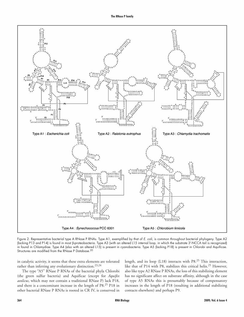
An extreme case of modification of the type B RNA is the type “B3” RNase P RNA of Mycoplasma fermentans, which lacks the otherwise universally present (except in type T RNAs, see below) P12 as well as P10.1.30 Unlike the type B2 RNA, P3 and P15.2 are not enlarged, and P9 is reduced to a minimum.30 The M. fermentans RNA is the smallest known naturally-occurring bacterial RNase P RNA.
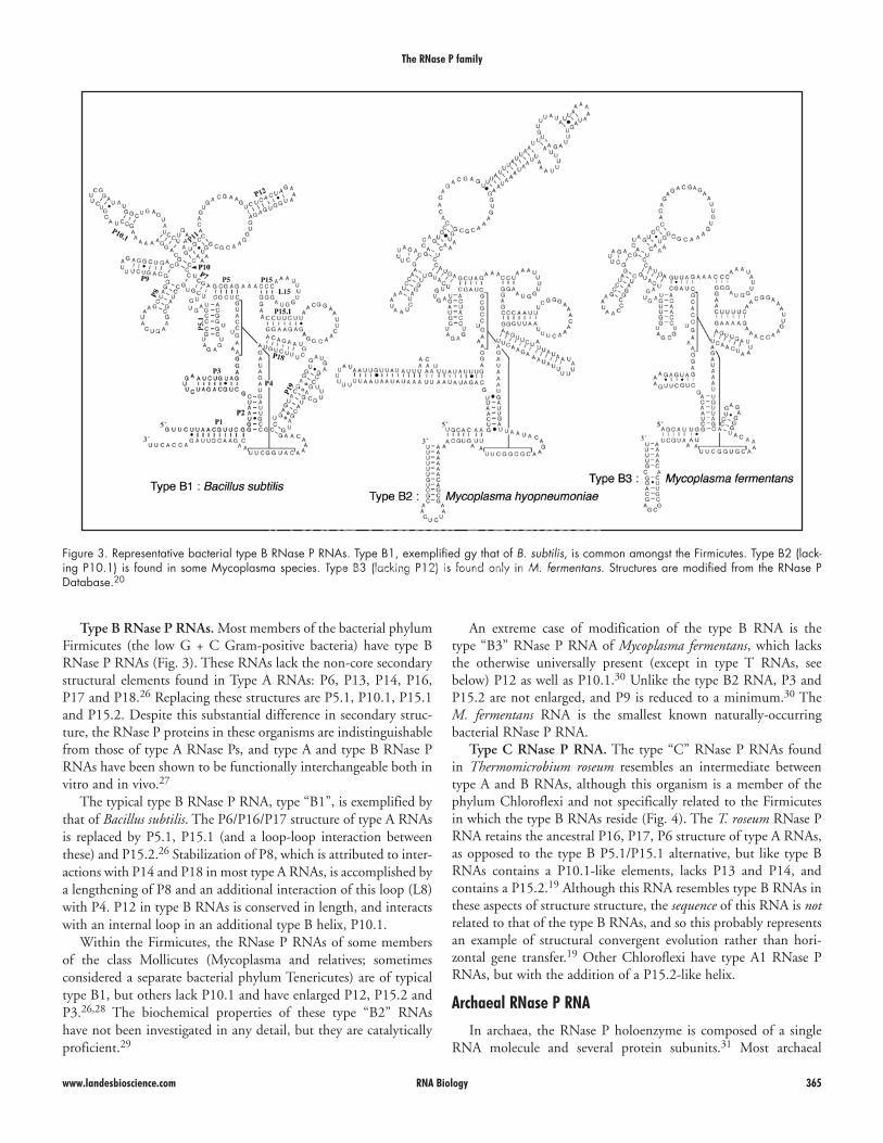
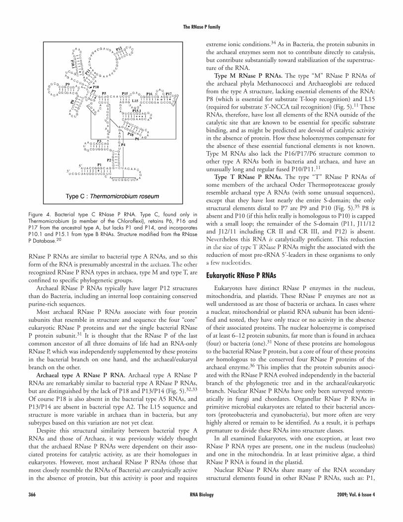
Type C RNase P RNA. The type “C” RNase P RNAs found in Thermomicrobium roseum resembles an intermediate between type A and B RNAs, although this organism is a member of the phylum Chloroflexi and not specifically related to the Firmicutes in which the type B RNAs reside (Fig. 4). The T. roseum RNase P RNA retains the ancestral P16, P17, P6 structure of type A RNAs, as opposed to the type B P5.1/P15.1 alternative, but like type B RNAs contains a P10.1-like elements, lacks P13 and P14, and contains a P15.2.19 Although this RNA resembles type B RNAs in these aspects of structure structure, the sequence of this RNA is not related to that of the type B RNAs, and so this probably represents an example of structural convergent evolution rather than hori-zontal gene transfer.19 Other Chloroflexi have type A1 RNase P RNAs, but with the addition of a P15.2-like helix.
Archaeal RNase P RNA
In archaea, the RNase P holoenzyme is composed of a single RNA molecule and several protein subunits.31 Most archaeal
Type B RNase P RNAs. Most members of the bacterial phylum Firmicutes (the low G + C Gram-positive bacteria) have type B RNase P RNAs (Fig. 3). These RNAs lack the non-core secondary structural elements found in Type A RNAs: P6, P13, P14, P16, P17 and P18.26 Replacing these structures are P5.1, P10.1, P15.1 and P15.2. Despite this substantial difference in secondary struc-ture, the RNase P proteins in these organisms are indistinguishable from those of type A RNase Ps, and type A and type B RNase P RNAs have been shown to be functionally interchangeable both in vitro and in vivo.27
The typical type B RNase P RNA, type “B1”, is exemplified by that of Bacillus subtilis. The P6/P16/P17 structure of type A RNAs is replaced by P5.1, P15.1 (and a loop-loop interaction between these) and P15.2.26 Stabilization of P8, which is attributed to inter-actions with P14 and P18 in most type A RNAs, is accomplished by a lengthening of P8 and an additional interaction of this loop (L8) with P4. P12 in type B RNAs is conserved in length, and interacts with an internal loop in an additional type B helix, P10.1.
Within the Firmicutes, the RNase P RNAs of some members of the class Mollicutes (Mycoplasma and relatives; sometimes considered a separate bacterial phylum Tenericutes) are of typical type B1, but others lack P10.1 and have enlarged P12, P15.2 and P3.26,28 The biochemical properties of these type “B2” RNAs have not been investigated in any detail, but they are catalytically proficient.29
Figure 3. Representative bacterial type B RNase P RNAs. Type B1, exemplified gy that of B. subtilis, is common amongst the Firmicutes. Type B2 (lack-ing P10.1) is found in some Mycoplasma species. Type B3 (lacking P12) is found only in M. fermentans. Structures are modified from the RNase P Database.20
The RNase P family
366 RNA Biology 2009; Vol. 6 Issue 4
extreme ionic conditions.34 As in Bacteria, the protein subunits in the archaeal enzymes seem not to contribute directly to catalysis, but contribute substantially toward stabilization of the superstruc-ture of the RNA.
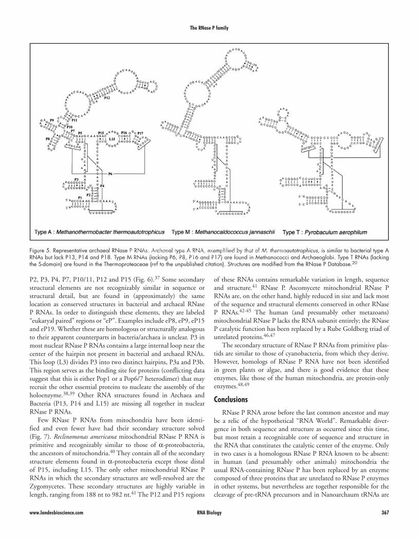
Type M RNase P RNAs. The type “M” RNase P RNAs of the archaeal phyla Methanococci and Archaeoglobi are reduced from the type A structure, lacking essential elements of the RNA: P8 (which is essential for substrate T-loop recognition) and L15 (required for substrate 3'-NCCA tail recognition) (Fig. 5).11 These RNAs, therefore, have lost all elements of the RNA outside of the catalytic site that are known to be essential for specific substrate binding, and as might be predicted are devoid of catalytic activity in the absence of protein. How these holoenzymes compensate for the absence of these essential functional elements is not known. Type M RNAs also lack the P16/P17/P6 structure common to other type A RNAs both in bacteria and archaea, and have an unusually long and regular fused P10/P11.11
Type T RNase P RNAs. The type “T” RNase P RNAs of some members of the archaeal Order Thermoproteaceae grossly resemble archaeal type A RNAs (with some unusual sequences), except that they have lost nearly the entire S-domain; the only structural elements distal to P7 are P9 and P10 (Fig. 5).35 P8 is absent and P10 (if this helix really is homologous to P10) is capped with a small loop; the remainder of the S-domain (P11, J11/12 and J12/11 including CR II and CR III, and P12) is absent. Nevertheless this RNA is catalytically proficient. This reduction in the size of type T RNase P RNAs might the associated with the reduction of most pre-tRNA 5'-leaders in these organisms to only a few nucleotides.
Eukaryotic RNase P RNAs
Eukaryotes have distinct RNase P enzymes in the nucleus, mitochondria, and plastids. These RNase P enzymes are not as well understood as are those of bacteria or archaea. In cases where a nuclear, mitochondrial or plastid RNA subunit has been identi-fied and tested, they have only trace or no activity in the absence of their associated proteins. The nuclear holoenzyme is comprised of at least 6–12 protein subunits, far more than is found in archaea (four) or bacteria (one).31 None of these proteins are homologous to the bacterial RNase P protein, but a core of four of these proteins are homologous to the conserved four RNase P proteins of the archaeal enzyme.36 This implies that the protein subunits associ-ated with the RNase P RNA evolved independently in the bacterial branch of the phylogenetic tree and in the archaeal/eukaryotic branch. Nuclear RNase P RNAs have only been surveyed system-atically in fungi and chordates. Organellar RNase P RNAs in primitive microbial eukaryotes are related to their bacterial ances-tors (proteobacteria and cyanobacteria), but more often are very highly altered or remain to be identified. As a result, it is perhaps premature to divide these RNAs into structure classes.
In all examined Eukaryotes, with one exception, at least two RNase P RNA types are present, one in the nucleus (nucleolus) and one in the mitochondria. In at least primitive algae, a third RNase P RNA is found in the plastid.
Nuclear RNase P RNAs share many of the RNA secondary structural elements found in other RNase P RNAs, such as: P1,
RNase P RNAs are similar to bacterial type A RNAs, and so this form of the RNA is presumably ancestral in the archaea. The other recognized RNase P RNA types in archaea, type M and type T, are confined to specific phylogenetic groups.
Archaeal RNase P RNAs typically have larger P12 structures than do Bacteria, including an internal loop containing conserved purine-rich sequences.
Most archaeal RNase P RNAs associate with four protein subunits that resemble in structure and sequence the four “core” eukaryotic RNase P proteins and not the single bacterial RNase P protein subunit.31 It is thought that the RNase P of the last common ancestor of all three domains of life had an RNA-only RNase P, which was independently supplemented by these proteins in the bacterial branch on one hand, and the archaeal/eukaryal branch on the other.
Archaeal type A RNase P RNA. Archaeal type A RNase P RNAs are remarkably similar to bacterial type A RNase P RNAs, but are distinguished by the lack of P18 and P13/P14 (Fig. 5).32,33 Of course P18 is also absent in the bacterial type A5 RNAs, and P13/P14 are absent in bacterial type A2. The L15 sequence and structure is more variable in archaea than in bacteria, but any subtypes based on this variation are not yet clear.
Despite this structural similarity between bacterial type A RNAs and those of Archaea, it was previously widely thought that the archaeal RNase P RNAs were dependent on their asso-ciated proteins for catalytic activity, as are their homologues in eukaryotes. However, most archaeal RNase P RNAs (those that most closely resemble the RNAs of Bacteria) are catalytically active in the absence of protein, but this activity is poor and requires
Figure 4. Bacterial type C RNase P RNA. Type C, found only in Thermomicrobium (a member of the Chloroflexi), retains P6, P16 and P17 from the ancestral type A, but lacks P1 and P14, and incorporates P10.1 and P15.1 from type B RNAs. Structure modified from the RNase P Database.20
www.landesbioscience.com RNA Biology 367
The RNase P family
of these RNAs contains remarkable variation in length, sequence and structure.41 RNase P. Ascomycete mitochondrial RNase P RNAs are, on the other hand, highly reduced in size and lack most of the sequence and structural elements conserved in other RNase P RNAs.42-45 The human (and presumably other metazoans) mitochondrial RNase P lacks the RNA subunit entirely; the RNase P catalytic function has been replaced by a Rube Goldberg triad of unrelated proteins.46,47
The secondary structure of RNase P RNAs from primitive plas-tids are similar to those of cyanobacteria, from which they derive. However, homologs of RNase P RNA have not been identified in green plants or algae, and there is good evidence that these enzymes, like those of the human mitochondria, are protein-only enzymes.48,49
Conclusions
RNase P RNA arose before the last common ancestor and may be a relic of the hypothetical “RNA World”. Remarkable diver-gence in both sequence and structure as occurred since this time, but most retain a recognizable core of sequence and structure in the RNA that constitutes the catalytic center of the enzyme. Only in two cases is a homologous RNase P RNA known to be absent: in human (and presumably other animals) mitochondria the usual RNA-containing RNase P has been replaced by an enzyme composed of three proteins that are unrelated to RNase P enzymes in other systems, but nevertheless are together responsible for the cleavage of pre-tRNA precursors and in Nanoarchaum tRNAs are
P2, P3, P4, P7, P10/11, P12 and P15 (Fig. 6).37 Some secondary structural elements are not recognizably similar in sequence or structural detail, but are found in (approximately) the same location as conserved structures in bacterial and archaeal RNase P RNAs. In order to distinguish these elements, they are labeled “eukaryal paired” regions or “eP”. Examples include eP8, eP9, eP15 and eP19. Whether these are homologous or structurally analogous to their apparent counterparts in bacteria/archaea is unclear. P3 in most nuclear RNase P RNAs contains a large internal loop near the center of the hairpin not present in bacterial and archaeal RNAs. This loop (L3) divides P3 into two distinct hairpins, P3a and P3b. This region serves as the binding site for proteins (conflicting data suggest that this is either Pop1 or a Pop6/7 heterodimer) that may recruit the other essential proteins to nucleate the assembly of the holoenzyme.38,39 Other RNA structures found in Archaea and Bacteria (P13, P14 and L15) are missing all together in nuclear RNase P RNAs.
Few RNase P RNAs from mitochondria have been identi-fied and even fewer have had their secondary structure solved (Fig. 7). Reclinomonas americana mitochondrial RNase P RNA is primitive and recognizably similar to those of α-proteobacteria, the ancestors of mitochondria.40 They contain all of the secondary structure elements found in α-proteobacteria except those distal of P15, including L15. The only other mitochondrial RNase P RNAs in which the secondary structures are well-resolved are the Zygomycetes. These secondary structures are highly variable in length, ranging from 188 nt to 982 nt.41 The P12 and P15 regions
Figure 5. Representative archaeal RNase P RNAs. Archaeal type A RNA, exemplified by that of M. thermoautotrophicus, is similar to bacterial type A RNAs but lack P13, P14 and P18. Type M RNAs (lacking P6, P8, P16 and P17) are found in Methanococci and Archaeoglobi. Type T RNAs (lacking the S-domain) are found in the Thermoproteaceae (ref to the unpublished citation). Structures are modified from the RNase P Database.20
The RNase P family
368 RNA Biology 2009; Vol. 6 Issue 4
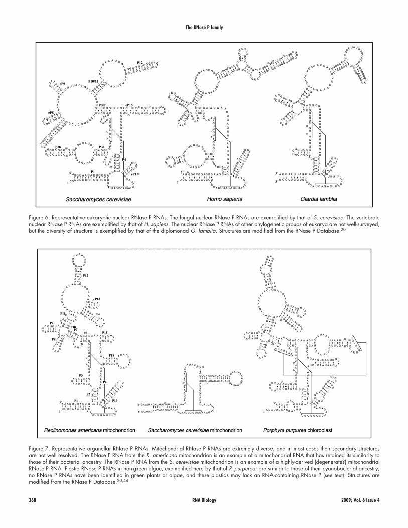
Figure 6. Representative eukaryotic nuclear RNase P RNAs. The fungal nuclear RNase P RNAs are exemplified by that of S. cerevisiae. The vertebrate nuclear RNase P RNAs are exemplified by that of H. sapiens. The nuclear RNase P RNAs of other phylogenetic groups of eukarya are not well-surveyed, but the diversity of structure is exemplified by that of the diplomonad G. lamblia. Structures are modified from the RNase P Database.20
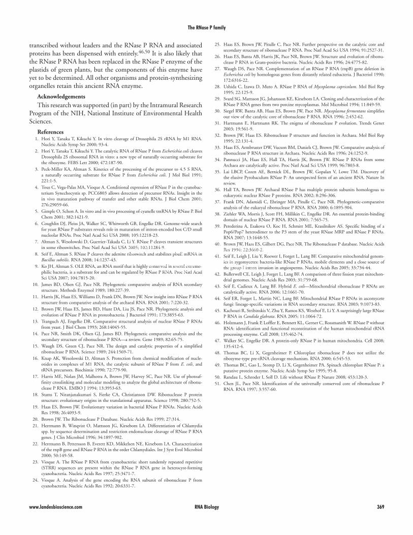
Figure 7. Representative organellar RNase P RNAs. Mitochondrial RNase P RNAs are extremely diverse, and in most cases their secondary structures are not well resolved. The RNase P RNA from the R. americana mitochondrion is an example of a mitochondrial RNA that has retained its similarity to those of their bacterial ancestry. The RNase P RNA from the S. cerevisiae mitochondrion is an example of a highly-derived (degenerate?) mitochondrial RNase P RNA. Plastid RNase P RNAs in non-green algae, exemplified here by that of P. purpurea, are similar to those of their cyanobacterial ancestry; no RNase P RNAs have been identified in green plants or algae, and these plastids may lack an RNA-containing RNase P (see text). Structures are modified from the RNase P Database.20,44
www.landesbioscience.com RNA Biology 369
The RNase P family
25. Haas ES, Brown JW, Pitulle C, Pace NR. Further perspective on the catalytic core and secondary structure of ribonuclease P RNA. Proc Natl Acad Sci USA 1994; 91:2527-31.
26. Haas ES, Banta AB, Harris JK, Pace NR, Brown JW. Structure and evolution of ribonu-clease P RNA in Gram-positive bacteria. Nucleic Acids Res 1996; 24:4775-82.
27. Waugh DS, Pace NR. Complementation of an RNase P RNA (rnpB) gene deletion in Escherichia coli by homologous genes from distantly related eubacteria. J Bacteriol 1990; 172:6316-22.
28. Ushida C, Izawa D, Muto A. RNase P RNA of Mycoplasma capricolum. Mol Biol Rep 1995; 22:125-9.
29. Svard SG, Mattsson JG, Johansson KE, Kirsebom LA. Cloning and characterization of the RNase P RNA genes from two porcine mycoplasmas. Mol Microbiol 1994; 11:849-59.
30. Siegel RW, Banta AB, Haas ES, Brown JW, Pace NR. Mycoplasma fermentans simplifies our view of the catalytic core of ribonuclease P RNA. RNA 1996; 2:452-62.
31. Hartmann E, Hartmann RK. The enigma of ribonuclease P evolution. Trends Genet 2003; 19:561-9.
32. Brown JW, Haas ES. Ribonuclease P structure and function in Archaea. Mol Biol Rep 1995; 22:131-4.
33. Haas ES, Armbruster DW, Vucson BM, Daniels CJ, Brown JW. Comparative analysis of ribonuclease P RNA structure in Archaea. Nucleic Acids Res 1996; 24:1252-9.
34. Pannucci JA, Haas ES, Hall TA, Harris JK, Brown JW. RNase P RNAs from some Archaea are catalytically active. Proc Natl Acad Sci USA 1999; 96:7803-8.
35. Lai LBCP, Cozen AE, Bernick DL, Brown JW, Gopalan V, Lowe TM. Discovery of the elusive Pyrobaculum RNase P: An unexpected form of an ancient RNA. Nature In review.
36. Hall TA, Brown JW. Archaeal RNase P has multiple protein subunits homologous to eukaryotic nuclear RNase P proteins. RNA 2002; 8:296-306.
37. Frank DN, Adamidi C, Ehringer MA, Pitulle C, Pace NR. Phylogenetic-comparative analysis of the eukaryal ribonuclease P RNA. RNA 2000; 6:1895-904.
38. Ziehler WA, Morris J, Scott FH, Millikin C, Engelke DR. An essential protein-binding domain of nuclear RNase P RNA. RNA 2001; 7:565-75.
39. Perederina A, Esakova O, Koc H, Schmitt ME, Krasilnikov AS. Specific binding of a Pop6/Pop7 heterodimer to the P3 stem of the yeast RNase MRP and RNase P RNAs. RNA 2007; 13:1648-55.
40. Brown JW, Haas ES, Gilbert DG, Pace NR. The Ribonuclease P database. Nucleic Acids Res 1994; 22:3660-2.
41. Seif E, Leigh J, Liu Y, Roewer I, Forget L, Lang BF. Comparative mitochondrial genom-ics in zygomycetes: bacteria-like RNase P RNAs, mobile elements and a close source of the group I intron invasion in angiosperms. Nucleic Acids Res 2005; 33:734-44.
42. Bullerwell CE, Leigh J, Forget L, Lang BF. A comparison of three fission yeast mitochon-drial genomes. Nucleic Acids Res 2003; 31:759-68.
43. Seif E, Cadieux A, Lang BF. Hybrid E. coli—Mitochondrial ribonuclease P RNAs are catalytically active. RNA 2006; 12:1661-70.
44. Seif ER, Forget L, Martin NC, Lang BF. Mitochondrial RNase P RNAs in ascomycete fungi: lineage-specific variations in RNA secondary structure. RNA 2003; 9:1073-83.
45. Kachouri R, Stribinskis V, Zhu Y, Ramos KS, Westhof E, Li Y. A surprisingly large RNase P RNA in Candida glabrata. RNA 2005; 11:1064-72.
46. Holzmann J, Frank P, Loffler E, Bennett KL, Gerner C, Rossmanith W. RNase P without RNA: identification and functional reconstitution of the human mitochondrial tRNA processing enzyme. Cell 2008; 135:462-74.
47. Walker SC, Engelke DR. A protein-only RNase P in human mitochondria. Cell 2008; 135:412-4.
48. Thomas BC, Li X, Gegenheimer P. Chloroplast ribonuclease P does not utilize the ribozyme-type pre-tRNA cleavage mechanism. RNA 2000; 6:545-53.
49. Thomas BC, Gao L, Stomp D, Li X, Gegenheimer PA. Spinach chloroplast RNase P: a putative protein enzyme. Nucleic Acids Symp Ser 1995; 95-8.
50. Randau L, Schroder I, Soll D. Life without RNase P. Nature 2008; 453:120-3. 51. Chen JL, Pace NR. Identification of the universally conserved core of ribonuclease P
RNA. RNA 1997; 3:557-60.
transcribed without leaders and the RNase P RNA and associated proteins has been dispensed with entirely.46,50 It is also likely that the RNase P RNA has been replaced in the RNase P enzyme of the plastids of green plants, but the components of this enzyme have yet to be determined. All other organisms and protein-synthesizing organelles retain this ancient RNA enzyme.
Acknowledgements
This research was supported (in part) by the Intramural Research Program of the NIH, National Institute of Environmental Health Sciences.
References 1. Hori Y, Tanaka T, Kikuchi Y. In vitro cleavage of Drosophila 2S rRNA by M1 RNA.
Nucleic Acids Symp Ser 2000; 93-4. 2. Hori Y, Tanaka T, Kikuchi Y. The catalytic RNA of RNase P from Escherichia coli cleaves
Drosophila 2S ribosomal RNA in vitro: a new type of naturally occurring substrate for the ribozyme. FEBS Lett 2000; 472:187-90.
3. Peck-Miller KA, Altman S. Kinetics of the processing of the precursor to 4.5 S RNA, a naturally occurring substrate for RNase P from Escherichia coli. J Mol Biol 1991; 221:1-5.
4. Tous C, Vega-Palas MA, Vioque A. Conditional expression of RNase P in the cyanobac-terium Synechocystis sp. PCC6803 allows detection of precursor RNAs. Insight in the in vivo maturation pathway of transfer and other stable RNAs. J Biol Chem 2001; 276:29059-66.
5. Gimple O, Schon A. In vitro and in vivo processing of cyanelle tmRNA by RNase P. Biol Chem 2001; 382:1421-9.
6. Coughlin DJ, Pleiss JA, Walker SC, Whitworth GB, Engelke DR. Genome-wide search for yeast RNase P substrates reveals role in maturation of intron-encoded box C/D small nucleolar RNAs. Proc Natl Acad Sci USA 2008; 105:12218-23.
7. Altman S, Wesolowski D, Guerrier-Takada C, Li Y. RNase P cleaves transient structures in some riboswitches. Proc Natl Acad Sci USA 2005; 102:11284-9.
8. Seif E, Altman S. RNase P cleaves the adenine riboswitch and stabilizes pbuE mRNA in Bacillus subtilis. RNA 2008; 14:1237-43.
9. Ko JH, Altman S. OLE RNA, an RNA motif that is highly conserved in several extremo-philic bacteria, is a substrate for and can be regulated by RNase P RNA. Proc Natl Acad Sci USA 2007; 104:7815-20.
10. James BD, Olsen GJ, Pace NR. Phylogenetic comparative analysis of RNA secondary structure. Methods Enzymol 1989; 180:227-39.
11. Harris JK, Haas ES, Williams D, Frank DN, Brown JW. New insight into RNase P RNA structure from comparative analysis of the archaeal RNA. RNA 2001; 7:220-32.
12. Brown JW, Haas ES, James BD, Hunt DA, Liu JS, Pace NR. Phylogenetic analysis and evolution of RNase P RNA in proteobacteria. J Bacteriol 1991; 173:3855-63.
13. Tranguch AJ, Engelke DR. Comparative structural analysis of nuclear RNase P RNAs from yeast. J Biol Chem 1993; 268:14045-55.
14. Pace NR, Smith DK, Olsen GJ, James BD. Phylogenetic comparative analysis and the secondary structure of ribonuclease P RNA—a review. Gene 1989; 82:65-75.
15. Waugh DS, Green CJ, Pace NR. The design and catalytic properties of a simplified ribonuclease P RNA. Science 1989; 244:1569-71.
16. Knap AK, Wesolowski D, Altman S. Protection from chemical modification of nucle-otides in complexes of M1 RNA, the catalytic subunit of RNase P from E. coli, and tRNA precursors. Biochimie 1990; 72:779-90.
17. Harris ME, Nolan JM, Malhotra A, Brown JW, Harvey SC, Pace NR. Use of photoaf-finity crosslinking and molecular modeling to analyze the global architecture of ribonu-clease P RNA. EMBO J 1994; 13:3953-63.
18. Stams T, Niranjanakumari S, Fierke CA, Christianson DW. Ribonuclease P protein structure: evolutionary origins in the translational apparatus. Science 1998; 280:752-5.
19. Haas ES, Brown JW. Evolutionary variation in bacterial RNase P RNAs. Nucleic Acids Res 1998; 26:4093-9.
20. Brown JW. The Ribonuclease P Database. Nucleic Acids Res 1999; 27:314. 21. Herrmann B, Winqvist O, Mattsson JG, Kirsebom LA. Differentiation of Chlamydia
spp. by sequence determination and restriction endonuclease cleavage of RNase P RNA genes. J Clin Microbiol 1996; 34:1897-902.
22. Herrmann B, Pettersson B, Everett KD, Mikkelsen NE, Kirsebom LA. Characterization of the rnpB gene and RNase P RNA in the order Chlamydiales. Int J Syst Evol Microbiol 2000; 50:149-58.
23. Vioque A. The RNase P RNA from cyanobacteria: short tandemly repeated repetitive (STRR) sequences are present within the RNase P RNA gene in heterocyst-forming cyanobacteria. Nucleic Acids Res 1997; 25:3471-7.
24. Vioque A. Analysis of the gene encoding the RNA subunit of ribonuclease P from cyanobacteria. Nucleic Acids Res 1992; 20:6331-7.