rna-guided endonuclease provides a therapeutic strategy … · rna-guided endonuclease provides a...
TRANSCRIPT
RNA-guided endonuclease provides a therapeuticstrategy to cure latent herpesviridae infectionJianbin Wanga and Stephen R. Quakea,b,c,1
aDepartment of Bioengineering, bDepartment of Applied Physics, and cHoward Hughes Medical Institute, Stanford University, Stanford, CA 94305
Contributed by Stephen R. Quake, June 10, 2014 (sent for review May 28, 2014)
Latent viral infection is a persistent cause of human disease.Although standard antiviral therapies can suppress active viralreplication, no existing treatment can effectively eradicate latentinfection and therefore a cure is lacking for many prevalent viraldiseases. The prokaryotic immune system clustered regularly inter-spaced short palindromic repeat (CRISPR)/Cas evolved as a naturalresponse to phage infections, and we demonstrate here that theCRISPR/Cas9 system can be adapted for antiviral treatment inhuman cells by specifically targeting the genomes of latent viralinfections. Patient-derived cells from a Burkitt’s lymphoma with la-tent Epstein–Barr virus infection showed dramatic proliferation ar-rest and a concomitant decrease in viral load after exposure toa CRISPR/Cas9 vector targeted to the viral genome.
genome editing | latency | herpes virus
The herpesviridae virus family consists of some of the mostwidespread human pathogens in the world. More than 90%
of adults have been infected with at least one of the eight sub-types of herpes viruses, and latent infection persists in mostpeople (1). These herpes virus subtypes infect a wide range ofcells, including epithelium, neuron, monocyte, and lymphocyte,and the consequences can be either mild (herpes simplexby HSV-1) or severe [cancer by Epstein–Barr virus (EBV) andKaposi’s sarcoma-associated herpes virus]. HSV infection is alsoa known risk factor for HIV (2). In its latent state, the viralgenome persists within the host cells and it has not been possibleto find therapeutic approaches that completely eradicate suchinfections.Since its discovery 50 y ago, EBV has been a closely studied
member of the herpesviridae. As one of the most common humanviruses, EBV causes infectious mononucleosis and is associatedwith certain forms of lymphoma. To date, however, no EBVvaccine or treatment exists. EBV is highly efficient at transformingquiescent human B lymphocytes; the resulting lymphoblastoid celllines are now commonly used for human genetic studies, and it ispossible to use patient-derived cells that propagate directly inculture because of the viral infection and require no other ma-nipulation. The EBV genome encodes about 85 genes, several ofwhich are essential for lytic or latent infection. During latency, theEBV genome circularizes and resides in the cell nucleus as anepisome. EBV latency usually progresses through three programs,with protein production decreasing from full sets of EBV nuclearantigens (EBNAs) and latent membrane proteins to just EBNA1.EBNA1 binds to the EBV origin of replication (oriP) to maintainviral episomes; it also regulates expression of other viral genes.Most current antiviral drug development programs are focused
on protein targets and are only effective in preventing active viralreplication. It has been recognized that it would be useful to targetlatent infections with viral genome-specific nucleases (3–5), butthe challenges of engineering sequence-specific nucleases havehampered progress. Clustered regularly interspaced short palin-dromic repeat (CRISPR)/Cas9 is a naturally occurring bacterialimmune system that uses a novel nuclease system to protectbacteria from phage infection (6–9), and it has recently beenharnessed for a variety of genome-engineering applications (10–17). DNA sequence recognition requires only a single 20-nt guide
RNA and a protospacer adjacent motif, which enables one torapidly engineer and test a large number of DNA cleavage sites(17–20). Here we demonstrate a therapeutic strategy for herpesvirus by targeting the CRISPR/Cas9 system directly to essentialviral genome sequences.
ResultsA Natural Model of Latent EBV Infection. As the first discoveredhuman tumor virus, EBV was initially isolated from culturedBurkitt’s lymphoma samples (21). The Raji cell line was the firstestablished long-term culture from Burkitt’s lymphoma patients(22, 23), and is one of the most extensively studied EBV models.The close relationship between the Raji EBV genome and theEBV reference (24) provides a straightforward blueprint forgenome engineering. We therefore used Raji cells for most ofthe work that follows.
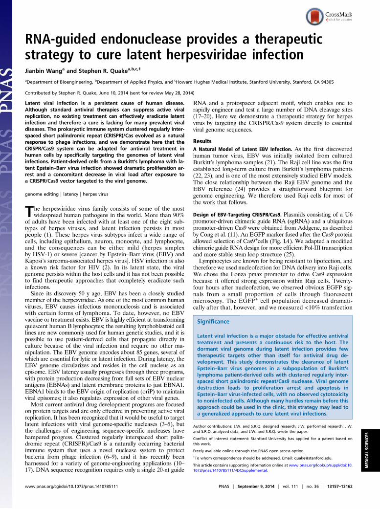
Design of EBV-Targeting CRISPR/Cas9. Plasmids consisting of a U6promoter-driven chimeric guide RNA (sgRNA) and a ubiquitouspromoter-driven Cas9 were obtained from Addgene, as describedby Cong et al. (11). An EGFP marker fused after the Cas9 proteinallowed selection of Cas9+cells (Fig. 1A). We adapted a modifiedchimeric guide RNA design for more efficient Pol-III transcriptionand more stable stem-loop structure (25).Lymphocytes are known for being resistant to lipofection, and
therefore we used nucleofection for DNA delivery into Raji cells.We chose the Lonza pmax promoter to drive Cas9 expressionbecause it offered strong expression within Raji cells. Twenty-four hours after nucleofection, we observed obvious EGFP sig-nals from a small proportion of cells through fluorescentmicroscopy. The EGFP+ cell population decreased dramati-cally after that, however, and we measured <10% transfection
Significance
Latent viral infection is a major obstacle for effective antiviraltreatment and presents a continuous risk to the host. Thedormant viral genome during latent infection provides fewtherapeutic targets other than itself for antiviral drug de-velopment. This study demonstrates the clearance of latentEpstein–Barr virus genomes in a subpopulation of Burkitt’slymphoma patient-derived cells with clustered regularly inter-spaced short palindromic repeat/Cas9 nuclease. Viral genomedestruction leads to proliferation arrest and apoptosis inEpstein–Barr virus-infected cells, with no observed cytotoxicityto noninfected cells. Although many hurdles remain before thisapproach could be used in the clinic, this strategy may lead toa generalized approach to cure latent viral infections.
Author contributions: J.W. and S.R.Q. designed research; J.W. performed research; J.W.and S.R.Q. analyzed data; and J.W. and S.R.Q. wrote the paper.
Conflict of interest statement: Stanford University has applied for a patent based onthis work.
Freely available online through the PNAS open access option.1To whom correspondence should be addressed. Email: [email protected].
This article contains supporting information online at www.pnas.org/lookup/suppl/doi:10.1073/pnas.1410785111/-/DCSupplemental.
www.pnas.org/cgi/doi/10.1073/pnas.1410785111 PNAS | September 9, 2014 | vol. 111 | no. 36 | 13157–13162
MED
ICALSC
IENCE
S
efficiency 48 h after nucleofection (Fig. 1B). We attributedthis transfection efficiency decrease to the plasmid dilutionwith cell division. To actively maintain the plasmid levelwithin the host cells, we redesigned the CRISPR plasmid toinclude the EBV origin of replication sequence, oriP. Withactive plasmid replication inside the cells, the transfectionefficiency rose to >60% (Fig. 1B).To design guide RNA targeting the EBV genome, we relied on
the EBV reference genome from strain B95-8. We targeted sixregions with seven guide RNA designs for different genome-editing purposes (Fig. 1C and Table S1). EBNA1 is crucial formany EBV functions, including gene regulation and latent ge-nome replication. We targeted guide RNA sgEBV4 and sgEBV5to both ends of the EBNA1 coding region to excise this wholeregion of the genome. Guide RNAs sgEBV1, -2, and -6 fall inrepeat regions, so that the success rate of at least one CRISPRcomplex is multiplied. These “structural” targets enable sys-tematic digestion of the EBV genome into smaller pieces.EBNA3C and latent membrane protein-1 are essential for host
cell transformation, and we designed guide RNAs sgEBV3 andsgEBV7 to target the 5′ exons of these two proteins, respectively.
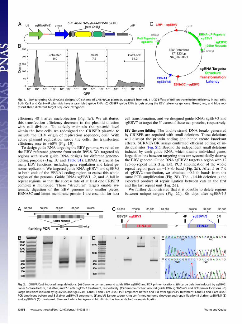
EBV Genome Editing. The double-strand DNA breaks generatedby CRISPR are repaired with small deletions. These deletionswill disrupt the protein coding and hence create knockouteffects. SURVEYOR assays confirmed efficient editing of in-dividual sites (Fig. S1). Beyond the independent small deletionsinduced by each guide RNA, which disable individual genes,large deletions between targeting sites can systematically destroythe EBV genome. Guide RNA sgEBV2 targets a region with 12125-bp repeat units (Fig. 2A). PCR amplification of the wholerepeat region gave an ∼1.8-kb band (Fig. 2B). After 5 or 7 dof sgEBV2 transfection, we obtained ∼0.4-kb bands from thesame PCR amplification (Fig. 2B). The ∼1.4-kb deletion is theexpected product of repair ligation between cuts in the firstand the last repeat unit (Fig. 2A).We further demonstrated that it is possible to delete regions
between unique targets (Fig. 2C). Six days after sgEBV4-5
A C
B
Fig. 1. EBV-targeting CRISPR/Cas9 designs. (A) Scheme of CRISPR/Cas plasmids, adapted from ref. 11. (B) Effect of oriP on transfection efficiency in Raji cells.Both Cas9 and Cas9-oriP plasmids have a scrambled guide RNA. (C) CRISPR guide RNA targets along the EBV reference genome. Green, red, and blue rep-resent three different target sequence categories.
A C
B D E
F
Fig. 2. CRISPR/Cas9 induced large deletions. (A) Genome context around guide RNA sgEBV2 and PCR primer locations. (B) Large deletion induced by sgEBV2.Lanes 1–3 are before, 5 d after, and 7 d after sgEBV2 treatment, respectively. (C) Genome context around guide RNA sgEBV3/4/5 and PCR primer locations. (D)Large deletions induced by sgEBV3/5 and sgEBV4/5. Lanes 1 and 2 are 3F/5R PCR amplicons before and 8 d after sgEBV3/5 treatment. Lanes 3 and 4 are 4F/5RPCR amplicons before and 8 d after sgEBV4/5 treatment. (E and F) Sanger sequencing confirmed genome cleavage and repair ligation 8 d after sgEBV3/5 (E)and sgEBV4/5 (F) treatment. Blue and white background highlights the two ends before repair ligation.
13158 | www.pnas.org/cgi/doi/10.1073/pnas.1410785111 Wang and Quake
transfection, PCR amplification of the whole flanking region(with primers EBV4F and -5R) returned a shorter amplicon,together with a much fainter band of the expected 2 kb (Fig. 2D).Sanger sequencing of amplicon clones confirmed the directconnection of the two expected cutting sites (Fig. 2F). A similarexperiment with sgEBV3-5 also returned an even larger deletion,from EBNA3C to EBNA1 (Fig. 2 D and E).
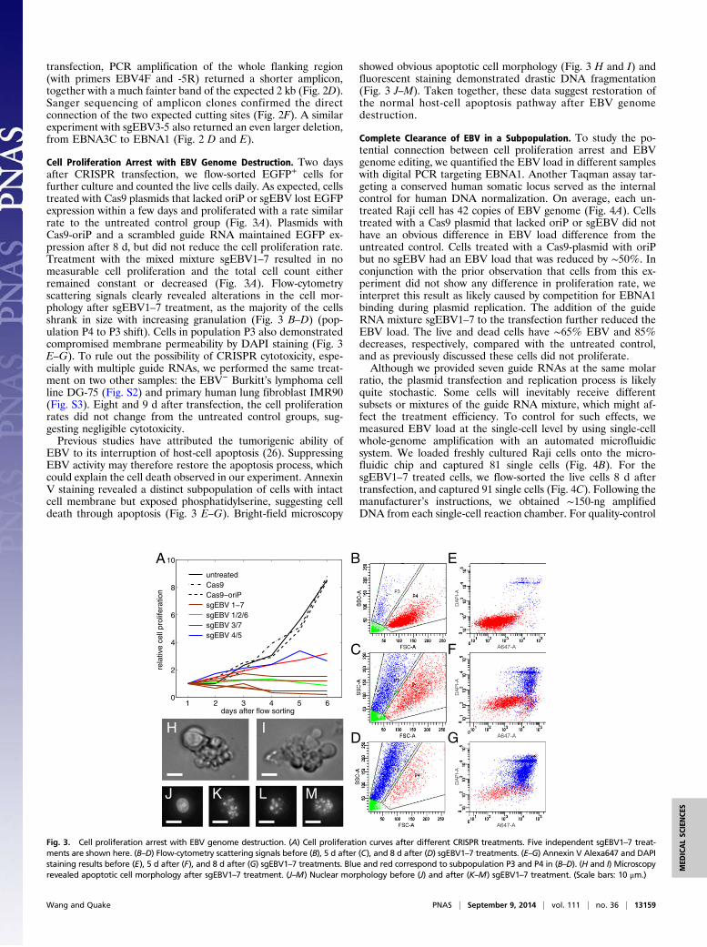
Cell Proliferation Arrest with EBV Genome Destruction. Two daysafter CRISPR transfection, we flow-sorted EGFP+ cells forfurther culture and counted the live cells daily. As expected, cellstreated with Cas9 plasmids that lacked oriP or sgEBV lost EGFPexpression within a few days and proliferated with a rate similarrate to the untreated control group (Fig. 3A). Plasmids withCas9-oriP and a scrambled guide RNA maintained EGFP ex-pression after 8 d, but did not reduce the cell proliferation rate.Treatment with the mixed mixture sgEBV1–7 resulted in nomeasurable cell proliferation and the total cell count eitherremained constant or decreased (Fig. 3A). Flow-cytometryscattering signals clearly revealed alterations in the cell mor-phology after sgEBV1–7 treatment, as the majority of the cellsshrank in size with increasing granulation (Fig. 3 B–D) (pop-ulation P4 to P3 shift). Cells in population P3 also demonstratedcompromised membrane permeability by DAPI staining (Fig. 3E–G). To rule out the possibility of CRISPR cytotoxicity, espe-cially with multiple guide RNAs, we performed the same treat-ment on two other samples: the EBV− Burkitt’s lymphoma cellline DG-75 (Fig. S2) and primary human lung fibroblast IMR90(Fig. S3). Eight and 9 d after transfection, the cell proliferationrates did not change from the untreated control groups, sug-gesting negligible cytotoxicity.Previous studies have attributed the tumorigenic ability of
EBV to its interruption of host-cell apoptosis (26). SuppressingEBV activity may therefore restore the apoptosis process, whichcould explain the cell death observed in our experiment. AnnexinV staining revealed a distinct subpopulation of cells with intactcell membrane but exposed phosphatidylserine, suggesting celldeath through apoptosis (Fig. 3 E–G). Bright-field microscopy
showed obvious apoptotic cell morphology (Fig. 3 H and I) andfluorescent staining demonstrated drastic DNA fragmentation(Fig. 3 J–M). Taken together, these data suggest restoration ofthe normal host-cell apoptosis pathway after EBV genomedestruction.
Complete Clearance of EBV in a Subpopulation. To study the po-tential connection between cell proliferation arrest and EBVgenome editing, we quantified the EBV load in different sampleswith digital PCR targeting EBNA1. Another Taqman assay tar-geting a conserved human somatic locus served as the internalcontrol for human DNA normalization. On average, each un-treated Raji cell has 42 copies of EBV genome (Fig. 4A). Cellstreated with a Cas9 plasmid that lacked oriP or sgEBV did nothave an obvious difference in EBV load difference from theuntreated control. Cells treated with a Cas9-plasmid with oriPbut no sgEBV had an EBV load that was reduced by ∼50%. Inconjunction with the prior observation that cells from this ex-periment did not show any difference in proliferation rate, weinterpret this result as likely caused by competition for EBNA1binding during plasmid replication. The addition of the guideRNA mixture sgEBV1–7 to the transfection further reduced theEBV load. The live and dead cells have ∼65% EBV and 85%decreases, respectively, compared with the untreated control,and as previously discussed these cells did not proliferate.Although we provided seven guide RNAs at the same molar
ratio, the plasmid transfection and replication process is likelyquite stochastic. Some cells will inevitably receive differentsubsets or mixtures of the guide RNA mixture, which might af-fect the treatment efficiency. To control for such effects, wemeasured EBV load at the single-cell level by using single-cellwhole-genome amplification with an automated microfluidicsystem. We loaded freshly cultured Raji cells onto the micro-fluidic chip and captured 81 single cells (Fig. 4B). For thesgEBV1–7 treated cells, we flow-sorted the live cells 8 d aftertransfection, and captured 91 single cells (Fig. 4C). Following themanufacturer’s instructions, we obtained ∼150-ng amplifiedDNA from each single-cell reaction chamber. For quality-control
rela
tive
cell
prol
ifera
tion
A B
C
D
E
F
G
A647-A
A647-A
A647-A
H I
J LK M
DA
PI-
AD
AP
I-A
DA
PI-
A
1 2 3 4 5 60
2
4
6
8
10
days after flow sorting
untreated Cas9 Cas9−oriP sgEBV 1−7 sgEBV 1/2/6 sgEBV 3/7 sgEBV 4/5
Fig. 3. Cell proliferation arrest with EBV genome destruction. (A) Cell proliferation curves after different CRISPR treatments. Five independent sgEBV1–7 treat-ments are shown here. (B–D) Flow-cytometry scattering signals before (B), 5 d after (C), and 8 d after (D) sgEBV1–7 treatments. (E–G) Annexin V Alexa647 and DAPIstaining results before (E), 5 d after (F), and 8 d after (G) sgEBV1–7 treatments. Blue and red correspond to subpopulation P3 and P4 in (B–D). (H and I) Microscopyrevealed apoptotic cell morphology after sgEBV1–7 treatment. (J–M) Nuclear morphology before (J) and after (K–M) sgEBV1–7 treatment. (Scale bars: 10 μm.)
Wang and Quake PNAS | September 9, 2014 | vol. 111 | no. 36 | 13159
MED
ICALSC
IENCE
S
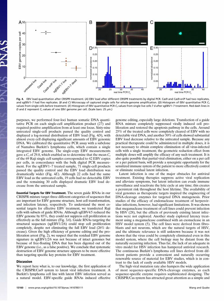
purposes, we performed four-loci human somatic DNA quanti-tative PCR on each single-cell amplification product (27) andrequired positive amplification from at least one locus. Sixty-nineuntreated single-cell products passed the quality control anddisplayed a log-normal distribution of EBV load (Fig. 4D), withalmost every cell displaying significant amounts of EBV genomicDNA. We calibrated the quantitative PCR assay with a subcloneof Namalwa Burkitt’s lymphoma cells, which contain a singleintegrated EBV genome. The single-copy EBV measurementsgave a Ct of 29.8, which enabled us to determine that the mean Ctof the 69 Raji single cell samples corresponded to 42 EBV copiesper cells, in concordance with the bulk digital PCR measure-ment. For the sgEBV1–7 treated sample, 71 single-cell productspassed the quality control and the EBV load distribution wasdramatically wider (Fig. 4E). Although 22 cells had the sameEBV load as the untreated cells, 19 cells had no detectable EBVand the remaining 30 cells displayed dramatic EBV load de-crease from the untreated sample.
Essential Targets for EBV Treatment. The seven guide RNAs in ourCRISPRmixture target three different categories of sequences thatare important for EBV genome structure, host cell transformation,and infection latency, respectively. To understand the most es-sential targets for effective EBV treatment, we transfected Rajicells with subsets of guide RNAs. Although sgEBV4/5 reduced theEBV genome by 85%, they could not suppress cell proliferation aseffectively as the full mixture (Fig. 3A). Guide RNAs targeting thestructural sequences (sgEBV1/2/6) could stop cell proliferationcompletely, despite not eliminating the full EBV load (26% de-crease). Given the high efficiency of genome editing and the pro-liferation arrest (Fig. 2), we suspect that the residual EBV genomesignature in sgEBV1/2/6 was not because of intact genomes butbecause of free-floating DNA that has been digested out of theEBV genome (i.e., as a false positive). We conclude that systematicdestruction of EBV genome structure appears to be more effectivethan targeting specific key proteins for EBV treatment.
DiscussionWe report here what is, to our knowledge, the first application ofthe CRISPR/Cas9 system to latent viral infection treatment. ABurkitt’s lymphoma cell line with latent EBV infection served asa natural model. EBV-specific guide RNAs induced effective
genome editing, especially large deletions. Transfection of a guideRNA mixture completely suppressed virally induced cell pro-liferation and restored the apoptosis pathway in the cells. Around25% of the treated cells were completely cleared of EBV with nodetectable viral DNA, and another 50% of cells showed substantialEBV load decrease relative to the untreated sample. Because anypractical therapeutic could be administered in multiple doses, it isnot necessary to obtain complete elimination of all virus-infectedcells with a single treatment; the geometric reduction effect frommultiple doses will amplify the efficacy of any such treatment. It isalso quite possible that partial viral elimination, either on a per cellor a per patient basis, will provide a synergistic opportunity for thesensitized immune system of the patient to more effectively controlor eliminate residual latent infection.Latent infection is one of the major obstacles for antiviral
treatment. Existing therapies suppress active viral replicationand alleviate symptoms, but latent infection can evade immunesurveillance and reactivate the lytic cycle at any time; this createsa persistent risk throughout the host lifetime. The availability ofviral genomes as therapeutic targets inspired the engineering ofDNA-cleavage enzymes for targeted DNA mutagenesis. Priorstudies of the efficacy of endonuclease treatment of herpesvir-idae infections, however, had significant limitations. It was shownthat meganuclease treatment of cell lines could prevent infectionby HSV (28), but the effects of previously existing latent infec-tions were not explored. Another study explored latency treat-ment using a meganuclease combined with an exonuclease in anHSV model system (5). The cell lines were derived from fibro-blasts and not neurons, which are the natural targets of HSV,and the ultimate relevance is still unknown because it was notshown that the virus could be completely eliminated even in thismodel system, where the cell biology may be distant from thenaturally occurring infection. Thus far, the lack of an adequate invitro model for HSV infection has hampered antiviral research.The continuous Burkitt’s lymphoma cell lines derived from dif-ferent patients provide a convenient and naturally occurringrenewable source of material for EBV studies, which is in con-trast to the lack of easily available HSV models.The high viral mutation rate also hampers the wide application
of most sequence-specific DNA-cleavage enzymes, as eachsequence-specific enzyme requires sophisticated designing. TheCRISPR/Cas system has attracted great attention as a simple and
20 25 30 35 400
0.05
0.10
0.15
0.20
0.25
0.30
real time PCR Ct
norm
aliz
ed a
bund
ance
20 25 30 35 400
0.05
0.10
0.15
0.20
0.25
0.30
real time PCR Ct
norm
aliz
ed a
bund
ance
0
10
20
30
40
50E
BV
genom
e c
opie
s per
cell
A B C
D E
Fig. 4. EBV load quantitation after CRISPR treatment. (A) EBV load after different CRISPR treatments by digital PCR. Cas9 and Cas9-oriP had two replicates,and sgEBV1–7 had five replicates. (B and C) Microscopy of captured single cells for whole-genome amplification. (D) Histogram of EBV quantitative PCR Ct
values from single cells before treatment. (E) Histogram of EBV quantitative PCR Ct values from single live cells 7 d after sgEBV1–7 treatment. Red dash lines inD and E represent Ct values of one EBV genome per cell. (Scale bars: 25 μm.)
13160 | www.pnas.org/cgi/doi/10.1073/pnas.1410785111 Wang and Quake
efficient genome-engineering technique whose sequence speci-ficity can be easily programmed by an RNA molecule. Althoughmost applications of CRISPR have focused mainly on gene-functionstudies, its original role as a prokaryotic immune system couldinspire a new approach for next-generation antiviral therapy.With the appropriate guide RNA, the Cas9 enzyme can rec-ognize and cleave virtually any viral genome (29). However,off-target cleavage is a major concern for any nuclease ther-apy. Single-molecule microscopy (30) and immunoprecipitationassays (31, 32) have measured the off-target sites bindingbehavior of the Cas9 endonuclease. ChiP-seq further allowedunbiased detection of genome-wide Cas9 binding (31, 32). To-gether with recent improvements on CRISPR target speci-ficity with dual-guide RNA designs (33–35), these studies showthat off-target effects are not a substantial impediment to hu-man genome-engineering applications and certainly do notappear to be an obstacle for applications that target non-human genomes (36).When used for gene-function studies, CRISPR usually targets
key coding regions, aiming to disrupt protein production. Wedesigned guide RNAs for key EBV protein disruption, includingthe double incision of the master regulator EBNA1. Despite effi-cient DNA cleavage, we were surprised to see that the subsequentlyrepaired EBV genomes could still drive host-cell proliferation. Thisfinding could be because of functional redundancy from otherproteins or perhaps noncoding RNA in the massive EBV genome.We obtained the best results by systematically destroying the entireEBV genome using guide RNAs targeting several repeat regions,thus maximizing the chance of DNA cleavage. Even with thepresence of DNA repair processes, we estimated that the possibilityof reconstructing the full genome from many smaller pieces is ex-tremely low. Indeed, cleaving these repeat regions suppressed host-cell proliferation as efficiently as using the full guide-RNA mixture.Further development of the strategy reported here into a prac-
tical therapeutic will require more sophisticated approaches todelivery. Although that is beyond the scope of this study, the nextsteps in developing a therapy will take advantage of the vastexisting literature on gene therapy, as well as the numerous de-livery technologies that have been developed for that purpose.Adenovirus and the related adeno-associeated virus are promising
potential approaches because they do not integrate into the host’sgenome. One could also imagine using a delivery approach thattook advantage of the fact that many herpesvirus infections arehighly localized and would not necessarily require systemic treat-ment. For example, HSV-1 latent infections are found only ina few sensory neurons in the trigeminal ganglia, HSV-2 latentinfections are found only in genital dorsal root ganglia, and EBVlatent infections are found only in B cells. One could try to en-gineer delivery viruses that mimic the cell-type tropism of thetarget virus, design targeted delivery vehicles that are cell typespecific, or one could apply the delivery material directly to theaffected tissues.
Materials and MethodsCell Culture. Burkitt’s lymphoma cell lines Raji, Namalwa, and DG-75 wereobtained from ATCC and cultured in RPMI 1640 supplemented with10% (vol/vol) FBS and 1× PSA, following ATCC recommendations. Humanprimary lung fibroblast IMR-90 was obtained from Coriell and cultured inAdvanced DMEM/F-12 supplemented with 10% (vol/vol) FBS and 1× PSA.
CRISPR/Cas9 Treatment. EBV-specific CRISPR/Cas9 plasmids were modifiedfrom pX458 (11), to include the pmax promoter from Lonza, sgRNA(F+E) fromref. 25, and EBV replication origin oriP from strain B95-8. We performedDNA transfection with the Lonza Nucleofector II.
Flow Cytometry Analysis and Cell Sorting. We conducted flow cytometryexperiments on the BD FACSAria II machines in Stanford Stem Cell FACS Core.Annexin V Alex647 (Life Technologies) was used following the manufacturer’srecommendations.
DNA Analysis. Genomic DNA from flow-sorted cells was used for downstreamDNAanalysis. Digital PCRwas performedon the FluidigmBioMark system. Single-cell whole-genome amplification was performed on the Fluidigm C1 system.
A complete description of the materials and methods is provided in SIMaterials and Methods. See Table S2 for PCR primers.
ACKNOWLEDGMENTS. We thank Jill Chinen and Dalong Qian for laboratorymanagement; Lolita Penland and Christopher Emig for cloning suggestions;Patricia Lovelace, Jennifer Ho, and Evan Chen for flow cytometry guidance;Brian Yu for assistance with single-cell whole-genome amplification; andCharles Gawad, Michael Rothenberg, and Shang Cai for helpful discus-sions. This project was supported by National Institutes of Health GrantsU54CA151459, U01HL099995, P01CA139490, and U01HL099999.
1. Hjalgrim H, Friborg J, Melbye M (2007) Human Herpesviruses: Biology, Therapy, andImmunoprophylaxis, eds Arvin A, et al. (Cambridge Univ Press, Cambridge), pp929–959.
2. Wald A, Link K (2002) Risk of human immunodeficiency virus infection in herpessimplex virus type 2-seropositive persons: A meta-analysis. J Infect Dis 185(1):45–52.
3. Wayengera M (2011) Identity of zinc finger nucleases with specificity to herpessimplex virus type II genomic DNA: Novel HSV-2 vaccine/therapy precursors.Theor Biol Med Model 8:23.
4. Qu X, et al. (2013) Zinc-finger-nucleases mediate specific and efficient excision ofHIV-1 proviral DNA from infected and latently infected human T cells. Nucleic AcidsRes 41(16):7771–7782.
5. Aubert M, et al. (2014) In vitro inactivation of latent HSV by targeted mutagenesisusing an HSV-specific homing endonuclease. Mol Ther Nucleic Acids 3:e146.
6. Horvath P, Barrangou R (2010) CRISPR/Cas, the immune system of bacteria and ar-chaea. Science 327(5962):167–170.
7. Terns MP, Terns RM (2011) CRISPR-based adaptive immune systems. Curr Opin Mi-crobiol 14(3):321–327.
8. Bhaya D, Davison M, Barrangou R (2011) CRISPR-Cas systems in bacteria and archaea:Versatile small RNAs for adaptive defense and regulation. Annu Rev Genet 45:273–297.
9. Wiedenheft B, Sternberg SH, Doudna JA (2012) RNA-guided genetic silencing systemsin bacteria and archaea. Nature 482(7385):331–338.
10. Jinek M, et al. (2012) A programmable dual-RNA-guided DNA endonuclease inadaptive bacterial immunity. Science 337(6096):816–821.
11. Cong L, et al. (2013) Multiplex genome engineering using CRISPR/Cas systems. Science339(6121):819–823.
12. Jinek M, et al. (2013) RNA-programmed genome editing in human cells. Elife (Cam-bridge) 2:e00471.
13. Mali P, et al. (2013) RNA-guided human genome engineering via Cas9. Science339(6121):823–826.
14. Qi LS, et al. (2013) Repurposing CRISPR as an RNA-guided platform for sequence-specific control of gene expression. Cell 152(5):1173–1183.
15. Gilbert LA, et al. (2013) CRISPR-mediated modular RNA-guided regulation of tran-scription in eukaryotes. Cell 154(2):442–451.
16. Yang H, et al. (2013) One-step generation of mice carrying reporter and conditionalalleles by CRISPR/Cas-mediated genome engineering. Cell 154(6):1370–1379.
17. Wang H, et al. (2013) One-step generation of mice carrying mutations in multiplegenes by CRISPR/Cas-mediated genome engineering. Cell 153(4):910–918.
18. Wang T, Wei JJ, Sabatini DM, Lander ES (2014) Genetic screens in human cells usingthe CRISPR-Cas9 system. Science 343(6166):80–84.
19. Shalem O, et al. (2014) Genome-scale CRISPR-Cas9 knockout screening in human cells.Science 343(6166):84–87.
20. Zhou Y, et al. (2014) High-throughput screening of a CRISPR/Cas9 library for func-tional genomics in human cells. Nature 509(7501):487–491.
21. Epstein MA, Achong BG, Barr YM (1964) Virus particles in cultured lymphoblasts fromBurkitt’s lymphoma. Lancet 1(7335):702–703.
22. Pulvertaft JV (1964) Cytology of Burkitt’s tumour (African lymphoma). Lancet 1(7327):238–240.
23. Epstein MA, et al. (1966) Morphological and virological investigations on culturedBurkitt tumor lymphoblasts (strain Raji). J Natl Cancer Inst 37(4):547–559.
24. Hatfull G, Bankier AT, Barrell BG, Farrell PJ (1988) Sequence analysis of Raji Epstein–Barr virus DNA. Virology 164(2):334–340.
25. Chen B, et al. (2013) Dynamic imaging of genomic loci in living human cells by anoptimized CRISPR/Cas system. Cell 155(7):1479–1491.
26. Ruf IK, et al. (1999) Epstein-barr virus regulates c-MYC, apoptosis, and tumorigenicityin Burkitt lymphoma. Mol Cell Biol 19(3):1651–1660.
27. Wang J, Fan HC, Behr B, Quake SR (2012) Genome-wide single-cell analysis of re-combination activity and de novo mutation rates in human sperm. Cell 150(2):402–412.
28. Grosse S, et al. (2011) Meganuclease-mediated inhibition of HSV1 Infection in cul-tured cells. Mol Ther 19(4):694–702.
29. Ebina H, Misawa N, Kanemura Y, Koyanagi Y (2013) Harnessing the CRISPR/Cas9system to disrupt latent HIV-1 provirus. Sci Rep 3:2510.
30. Sternberg SH, Redding S, Jinek M, Greene EC, Doudna JA (2014) DNA interrogation bythe CRISPR RNA-guided endonuclease Cas9. Nature 507(7490):62–67.
Wang and Quake PNAS | September 9, 2014 | vol. 111 | no. 36 | 13161
MED
ICALSC
IENCE
S

31. Wu X, et al. (2014) Genome-wide binding of the CRISPR endonuclease Cas9 inmammalian cells. Nat Biotechnol 32(7):670–676.
32. Kuscu C, Arslan S, Singh R, Thorpe J, Adli M (2014) Genome-wide analysis reveals charac-teristics of off-target sites bound by the Cas9 endonuclease. Nat Biotechnol 32(7):677–683.
33. Ran FA, et al. (2013) Double nicking by RNA-guided CRISPR Cas9 for enhanced ge-nome editing specificity. Cell 154(6):1380–1389.
34. Fu Y, Sander JD, Reyon D, Cascio VM, Joung JK (2014) Improving CRISPR-Cas nucleasespecificity using truncated guide RNAs. Nat Biotechnol 32(3):279–284.
35. Tsai SQ, et al. (2014) Dimeric CRISPR RNA-guided FokI nucleases for highly specificgenome editing. Nat Biotechnol 32(6):569–576.
36. Hsu PD, Lander ES, Zhang F (2014) Development and applications of CRISPR-Cas9 forgenome engineering. Cell 157(6):1262–1278.
13162 | www.pnas.org/cgi/doi/10.1073/pnas.1410785111 Wang and Quake