robert m. kosanke - university of...
TRANSCRIPT

ROBERT M. KOSANKE
PRINTED BY AUTHORITY OF THE STATE OF ILLINOIS
URBANA, ILLINOIS
1 9 5 5

STATE O F ILLINOIS
WILLIAM G. STRATTON, Governor
D E P A R T M E N T O F REGISTRATION A N D EDUCATION
VERA M. BINICS, Director
DIVISION O F T H E
S T A T E G E O L O G I C A L S U R V E Y JOHN C. F R Y E , Chief
URBANA
REPORT OF INVESTIGATIONS 180
MAZOSTACHYS - A NEW CALAMITE P;RUCTIFICATION
BY
ROBERT M. KOSANKE
PRINTED BY AUTHORITY OF T H E STATE OF ILLINOIS
URBANA, ILLINOIS
1 9 5 5

ORGANIZATION
STATE OF ILLINOIS HON. WILLIAM G. STRATTON, Governor
DEPARTMENT OF REGISTRATION AND EDUCATION HON. VERA M. BINKS, Director
BOARD OF NATURAL RESOURCES AND CONSERVATION HON. VERA M. BINKS, Chairman
W. H. NEWIIOUSE, PH.D., Geology
ROGER ADAMS, PH.D., D.Sc., Chemistry
ROBERT H. ANDERSON, B .S., Engineering
A. E . EMERSON, PH.D., Biology
LEWIS H. TIFFANY, PH.D., PD.D., Forestry
W. L. EVERITT, E.E., PH.D. Representing the President of the Uniz~ersity of Illinois
DELYTE W. MORRIS, Pw.D. President of Southern Illinois University
GEOLOGICAL SURVEY DIVISION JOHN C. FRYE, Pw.D., D.Sc., Chief

STATE GEOLOGICAL SURVEY DIVISION Natural Resources Building, Urbane
JOHN C. FRYE, PH.D., D.Sc., Chief M. M. LEIGHTON, PH.D., D.Sc., Chief, Emeritus
ENID TOWNLEY, MS. , Geologist and Assistant to the Chief VELDA A. MILLARD, Junior Assistant to the Chief
HELEN E. MCMORRIS, Secretary to the Chief
R E S E A R C H (not including part-time personnel)
GEOLOGICAL RESOURCES ARTHUR BEVAN, PH.D., D.Sc., Principal Geologist FRANCES H . ALSTERLUND, A.B., Research Assistant
Coal JACK A. SIMON M.S Geologist and Head G. H . CADY P ~ D 'Senior Geologist and Head, Emeritus ROBERT M.'KOSA&E, PH.D., Geologist RAYMOND SIEVER PH.D Geologist JOHN A. H A R R I S ~ N M.S' Associate Geologist PAUL EDWIN POTTAR P~:D. , Associate Geologist HAROLD B. STONEHO~SE, PH.D., Associate Geologist MARGARET A. PARKER, M.S., Assistant Geologist
(on leave) M. E. HOPKINS, M.S., Assistant Geologist KENNETH E. CLEGG, M.S., Assistant Geologist
Oil and Gas A. H. BELL, PH.D., Geologist and Head LESTER L. WHITING, B.A., Associate Geologist VIRGINIA KLINE, PH.D., Associate Geologist WAYNE F. MEENTS, Assistant Geologist MARGARET 0. OROS, B.A., Assistant Geologist KENNETH R. LARSON, A.B., Research Assistant JACOB VAN DEN BERG, B.S., Research Assistant
Petroleum Engineering PAUL A. WITHERSPOON, MS. , Petroleum Engineer and
Head FREDERICK SQUIRES, A.B., B.S., D.Sc., Petroleum Engi-
neer, Emeritus
Industrial Minerals J. E. LAMAR, B.S., Geologist and Head DONALD L. GRAF, PH.D., Geologist JAMES C. BRAD BURY, A.M ., Assistant Geologist MEREDITH E. OSTROM, M.S., Assistant Geologist DONALD L. BIGGS, M.A., Assistant Geologist
Clay Resources and Clay Mineral Technology RALPH E. GRIM, PH.D., Consulting Clay Mineralogist W . ARTHUR WHITE, M.S., Associate Geologist HERBERT D. GLASS, PH.D ., Associate Geologist CHARLES W. SPENCER, M.S., Research Assistant
Groundwater Geology and Geophysical Exploration ARTHUR BEVAN, PH.D., D.Sc., Acting Head MERLYN B. BUHLE, M.S., Associate Geologist ROBERT E. BERGSTROM, PH.D., Assistant Geologist JOHN W . FOSTER, MS. , Assistant Geologist JAMES E. HACKETT, M.S., Assistant Geologist MARGARET J. CASTLE, Assistant Geologic Draftsman
(on leavei WAYNE A. PRYOR, M.S., Assistant Geologist LIDIA SELKREGG, D.N.S., Assistant Geologist ROBERT C. PARKS, Technical Assistant
Engineering Geology and Topographic Mapping GEORGE E. EKBLAW, PH.D., Geologist and Head WILLIAM C. SMITH, M.A., Assistant Geologist
Stratigraphy and Areal Geology H. B. WILLMAN, PH.D., Geologist and Head DAVID H. SWANN, PH.D., Geologist ELWOOD ATHERTON, PII.D., Geologist CHARLES W. COLLINSON, PH.D., Associate Geologist DONALD B. SAXBY, M.S., Assistant Geologist T. C. BUSCHBACH, M.S., Assistant Geologist HOWARD R. SCHWALB, B.S., Research Asszstant FRANK B. TITUS, JR., B.S., Research Assistant CHARLES C . ENGEL, Technical Assistant JOSEPH F. HOWARD, Assistant
Physics R. J. PIERSOL, PH.D., Physicist, Emeritus
Topographic Mapping in Cooperation with the United States Geological Survey.
GEOCHEMISTRY FRANK H. REED, PH.D., Chief Chemist GRACE C. JOHNSON, B.S., Research Assistant
Coal Chemistry G. R. YOHE, PH.D., Chemist and Head E.~RLE C. SMITH, B.S., Research Assistant GUEY H. LEE, M.S., Research Assistant
Physical Chemistry J. S. MACHIN, PH.D., Chemist and Head JUANITA WITTERS, MS. , Assistant Physicist TIN BOO YEE, PH.D., Assistant Chemist DANIEL L. DEADMORE, B.S., Research Assistant
Fluorine Chemistry G. C. FINGER, PH.D., Chemist and Head ROBERT E. OESTERLING, B.A., Assistant Chemist CARL W . KRUSE, MS. , Special Research Assistant RAYMOND H. WHITE, B.S., Special Research Assistant RICHARD H . SHILEY, B.S., Special Research Assistant
Chemical Engineering H. W. JACKMAN, M.S.E., Chemical Engineer and Head R. J. HELFINSTINE, M.S., Mechanical Engineer and
Supervisor of Physical Plant B. J. GREENWOOD, B.S., Mechanical Engineer JAMES C. MCCULLOUGH, Research Associate (on leave) ROBERT L. EISSLER, B.S., Assistant Chemical Engineer WALTER E. COOPER, Technical Assistant EDWARD A. SCHAEDE, Technical Assistant CORNEL MARTA, Technical Assistant
X- Ray W. F. BRADLEY, PH.D., Chemist and Head
Analytical Chemistry 0. W. REES, PH.D., Chemist and Head L. D. MCVICKER, B.S., Chemist EMILE D. PIERRON, M.S., Associate Chemist DONALD R. DICKERSON, B.S.. Assistant Chemist FRANCIS A. COOLICAN, B.S., Assistant Chemist CHARLES T. ALLBRIGHT, B.S., Research Assistant WILLIAM J. ARMON, B.S., Research Assistant JOSEPH M. HARRIS, B.A., Research Assistant TOANNE E. KUNDE. B.A.. Research Assistant JOAN M. CEDZRSTRAND, Research Assistant EUGENE LANGE, Technical Assistant GEORGE R . JAMES, Technical Assistant FRANCES L. SCHEIDT, Technical Assistant
MINERAL ECONOMICS W. H. VOSKUIL, PH.D., Mineral Economist W. L. BUSCH, A.B., Assistant Mineral Economist ETHEL M. KING, Research Assistant
EDUCATIONAL EXTENSION GEORGE M. WILSON, MS. , Geologist and Head DOROTHY E . ROSE, B.S., Assistant Geologist
RESEARCH AFFILIATES IN GEOLOGY J HARLEN BRETZ, PH.D., Univarsity of Chicago JOHN A. BROPHY, M.S., Research Assistant, State Geolog-
ical Survey STANLEY E. HARRIS, JR., PH.D., Southern Illinois Uni -
versity C. LELAND HORBERG, PH.D., University of Chicago M. M. LEIGHTON, PH.D., D.Sc., Research Professional
Scientist, State Geological Survey HEINZ A. LOWENSTAM. PH.D., California Institute o f
Technology WILLIAM E. POWERS, PH.D., Northwestern University PAUL R . SHAFFER, PH.D., University of Illinois HAROLD R . WANLESS, PH.D., University of Illinois J. MARVIN WELLER, PH.D., University of Chicago
CONSULTANTS Geology, GEORGE W. WHITE, PH.D., University of Illinois
RALPH E. GRIM, PH.D., University of Illinois L. E. WORKMAN. M.S.. Former Head. Subsurface
Division Ceramics, RALPH K. HURSH, B.S., University of Illinois Mechanical Engineering, SEICHI K o ~ z o , M.S., University of
Illinois March 16, 1955

GENERAL ADMINISTRATION (not including part-time personnel)
LIBRARY ANNE E. KOVANDA, B.S., B.L.S., Librarian RUBY D. FREON. Technical Assistant
MINERAL RESOURCE RECORDS VIVIAN GORDON, Head MARGARET B. BROPHY, B.A., Research Assistant SUE J . CUNNINGHAM, Technical Assistant BETTY CLARK, B.S., Technical Assistant JE.~NINE CLIMER, Technical Assistant MARILYN W . THIES, B.S., Technical Assistant HANNAH FISHER, Technical Assistant LAROY PETERSON, Technical Assistant CARY JEAN STEVENSON, Technical Assistant PATRICIA L. LUEDTKE, B.A., Technical Assistant KATHRYN BROWN, Technical Assistant
PUBLICATIONS BARBARA ZEIDERS, B.S., Assistant Technical Editor MEREDITH M. CALKINS, Geologic Draftsman MARLENE PONSHOCK, Assistant Geologic Draftsman
TECHNICAL RECORDS BERENICE REED, Supervisory Technical Assistant MARILYN DELAND, B.S., Technical Assistant MARY LOUISE LOCKLIN, B.A., Technical Assistant
OTHER TECHNICAL SERVICES WM. DALE FARRIS, Research Associate BEULAH M. UNFER, Technical Assistant A. W . GOTSTEIN, Research Associate GLENN G. POOR, Research Associate* GILBERT L. TINBERG, Technical Assistant WAYNE W. NOFFTZ, Supervisory Technical Assistant DONOVAN M. WATKINS, Technical Assistant
FINANCIAL RECORDS VELDA A. MILLARD, In Charge LEONA K. ERICKSON, Clerk-Typist I I I VIRGINIA C. SANDERSON, B.S., Clerk-Typist I I IRMA E. SAMSON, Clerk-Typist I
CLERICAL SERVICES MARY CECIL, Clerk-Stenographer I I I MARY M. SULLIVAN, Clerk-Stenographer I I I LYLA NOFFTZ, Clerk-Stenographer I I LILLIAN WEAKLEY, Clerk-Stenographer I I SHARON ELLIS, Clerk-Stenographer I BARBARA BARHAM, Clerk-Stenographer I MARY ALICE JACOBS, Clerk-Stenographer I LORRAINE CUNNINGHAM, Clerk-Stenographer I IRENE BENSON, Clerk-Typist I MARY J. DE HAAN, Messenger-Clerk I
AUTOMOTIVE SERVICE GENERAL SCIENTIFIC INFORMATION GLENN G. POOR, I n Charge*
ROBERT 0. ELLIS, Automotive Mechanic ANN P. OSTROM, B.A., Technical Assistant EVERETTE EDWARDS, Automotive Mechanic JILL B. CAHILL, Technical Assistant DAVID B. COOLEY, Automotive Mechanic's Helper
March 16, 1955 *Divided time

CONTENTS
. . . . . . . . . . . . . . . . . . . . . . . . . . . Introduction 7 . . . . . . . . . . . . . . . . . . . . . . . . Acknowledgments 8
Mazostachys pendulata gen . et sp . nov . . . . . . . . . . . . . . . . . . . . . . . . . General description 9
. . . . . . . . . . . . . . . . . . . . . . . . . . Shoot axis 10 . . . . . . . . . . . . . . . . . . . . . . . . . . . Peduncles 10
. . . . . . . . . . . . . . . . . . . . . . . . . . Cone axis 10 . . . . . . . . . . . . . . . . . . . . . . . . . . . . Bracts 11
. . . . . . . . . . . . . . . . . . . . . . . . . Sporangiophore 14 . . . . . . . . . . . . . . . . . . . . . . . . . . . Sporangia 14
. . . . . . . . . . . . . . . . . . . . . . . . . . . . Spores 15 . . . . . . . . . . . . . . . . . . . . . . . . . . . Diagnosis 17
. . . . . . . . . . . . . . . . . . . . . . . . . . Discussion 17 . . . . . . . . . . . . . . . . . Pre-Calamostachys type of fructification 17
. . . . . . . . . . . . . . . . . . Calamostachys type of fructification 17
. . . . . . . . . . . . . . . . . . Palaeostachya type of fructification 18 . . . . . . . . . . . . . . . . . . . Cingularia type of fructification 19
. . . . . . . . . . . . . . . . . Metacalamostach~s type of fructification 19 . . . . . . . . . . . . . . . . . . Phylogeny of the cones of the Calamitaceae 20
. . . . . . . . . . . . . . . . . . . . . . . . . . . . Summary 22
. . . . . . . . . . . . . . . . . . . . . . . . . . . . References 23
ILLUSTRATIONS
FIGURE . . . . . . . . . . . . . . 1 . Diagrammatic drawing of holotype specimen B-1304A 8 . . . . . . . . . . . . . . 2 . Diagrammatic drawing of holotype specimen B-1304B 9
. . . . . . . . . . . . . . . . . . 3 . Reconstruction of Mazostach-vs pendulata 12 . . . . . . . . . . . . . . . . . . 4 . Transverse section of Mazostacliys pendulata 13
. . . . . . . . . . . . . . 5 . Proposed phyletic sequence for cones of Calamitaceae 22
PLATES
PLATE . . . . . . . . . . . . . . . . . . . . 1 . Habit and longitudinal sections 27
. . . . . . . . . . . . . . . . . . . . . . . . 2 . Transverse sections 28
. . . . . . . . . . . . . . . . . . . . . . . . 3 . Transverse sections 31 . . . . . . . . . . . . . . . . . . . . . 4 . Habit and sections of tissues 32
. . . . . . . . . . . . . . . . 5 . Transverse section of cone axis and sporangia 35 . . . . . . . . . . . . . 6 . Longitudinal section, reconstructions. tissues. and spore 36


MAZOSTACHYS - A NEW CALAMITE FRUCTIFICATION
BY
ROBERT M. KOSANKE
A B S T R A C T
T h e specimen described is preserved in an ironstone concretion collected from spoil piles of the No. 2 coal bed of Pennsylvanian age in Wi l l County, Ill. I t is a calamite shoot bearing fifteen cones. Each cone axis, with nodes and internodes, bears appendages of two types. A whorl of twelve bracts occurs on each node and high on each internode are six whorled sporangiophores. Whorls of sporangiophores and bracts alternate along the vertical extent of the cone axis. T w o sporangia a re attached to each sporangiophore in a pendulant manner. T h e striking features of this type of calamite fructification a re : (1) the position of attachment of the sporangiophore, and (2) !he presence of two sporangia per sporangiophore, a new feature of calamite fruct~ficatlons. T h e second feature demonstrates that the phylogeny has progressed with reduction in number of sporangia per sporangiophore from four in AsterocaLa- mites Pothocites, ProtocalamosiachysJ Calamostachys, Palaeostachya, and Cingularia typica, to two in this specimen (described as Maaostachys), to only one in MeLacaCa- mostachys. Mazostachys is homosporous. T h e foliage of Annularia sphenophylloides occurs in the ironstone concretion in association with Maaostachys but not in organic connection with the fertile shoot.
I N T R O D U C T I O N
T h e following report is a contribution to fundamental studies of fossil plants of Pennsylvanian age of Illinois. I t is a con- tinuation of the Survey's efforts to explore past plant life seeking knowledge of coal- forming plants of potential value in the correlation of coal beds and in related stud- ies in the field of coal petrography. Studies of fossil cones are also useful to paleobot- anists in associating isolated spores in coal with their parent plants.
Th i s report describes a new type of cala- mite fructification and gives a re-evalua- tion of previous ideas on the phylogeny of these fructifications in light of new evi- dence. T h e specimen, with preserved cellu- lar structure, was found in an ironstone concretion on the spoil piles of the strip- ping operations of the Northern Illinois Coal Corp., near the Mazon Creek area, Wi l l County, Ill.
Ironstone concretions with preserved plant remains occur in the Francis Creek shale immediately above the NO. 2 coal bed in the area. I t is assumed that the iron- stone concretion bearing the specimen of
Mazostacizys was removed from the shale during stripping operations.
T h e Francis Creek shale lies in the lower part of the Carbondale group of Pennsyl- vanian age in Illinois. According to IVoore et al. (1944), an equivalent stratigraphic ~os i t ion in the mid-continent region would be in the upper Cherokee group of the Des Moines series and in the Allegheny series of the Appalachian region of eastern United States. For additional information about the Francis Creek shale, see Cady (1919) , Savage ( l 9 2 7 ) , W anless ( l 9 2 9 ) , and Willman and Payne (1942).
T h e external appearance of the concre- tion containing the specimen of M a a o - stachys is typical of others found in the area, but the mode of preservation is not en- tirely of the compression type. T h e main axis and branches of the specimen are prin- cipally carbonaceous material, but each cone is encased in, or surrounded by, calcium carbonate. Therefore, preservation is some- what similar to a coal ball in which histo- logical details can be observed. Peels and thin sections were used in studying the pre- served cellular structure of the fructifica- tions. Individual sporangia from several

8 ILLINOIS STATE: GEOLOGICAL SURVEY
cones were dissected and macerated so that above the Murphysboro coal bed in south- the spores could be studied individually in western Illinois. Condit and Miller (1951) the isolated state. Specimens with pre- reported plant-bearing ironstone concre- served cellular structure from the Mazon tions from Iowa, Creek area are rare, but the author has a specimen of Calarnostachys which is not as ACKNOWLEDGMENTS well preserved as the specimen described. Darrah ( 1936) described Macrostachya thonzpsonii in an ironstone concretion from the Mazon Creek area that is preserved in a similar manner.
Ironstone concretions containing plant compressions occur at a number of strati- graphic levels and in various areas of the United States. I have observed these con- cretions from a shale overlying the Pome- roy coal bed in Ohio, above the Minshall coal bed in War r en County, Ind., and
I t is a pleasure to acknowledge the as- sistance of D r . Wilson N. Stewart, under whose supervision this report was submit- ted in 1952 t o the Graduate College, Uni- versity of Illinois, as part one of a doctoral thesis in botany. D r . Theodore Just, Chi- cago Natural History Museum, assisted in the selection of the new names proposed. D r . James R. Kamp and M r . and Mrs. John A4. McLuckie, Coal City, Ill., as- sisted in obtaining the described specimen.
FIG. 1.-A diagrammatic drawing of the holytype specimen of Mazostachys pendulata gen. et sp. nov. (B- 1304A). The cones were assigned numbers for ease of discussion in text. Cones 3 and 9 did not occur on
this half of the specimen, so their position is shown by dashed lines. About natural size.

G E N E R A L DESCRIPTION 9
M A Z O S T A C H Y S P E N D U L A T A GEN. ET SP. NOV.
GENERAL DESCRIPTION
T h e specimen is in the Illinois State Geo- logical Survey, paleobotanical collection, number B-1304. I t was split in half and, accordingly, will be referred to as B-1304A and B-1304B (figs. 1 and 2 ; pl. 1, fig. 1 ; and pl. 4, fig. 1 ) . I t is a fertile calamite shoot with 15 attached cones. Foliage is not attached, but six whorls of leaves iden- tified as Annularia sphenophylloides are preserved as a very thin carbonaceous film in the same plane as the specimen.
T h e shoot axis has two nodes and one complete internode. There is also a small portion of another internode. T h e complete internode is 4.2 cm. long and slightly less than 1 cm. wide. T w o branches depart from the shoot axis opposite each other; thus the mode of branching places this speci-
men in the subgenus Ezrcalamites, typified by Calamites carinatus as illustrated by Hirmer (1927, figs. 537 to 543). Each branch redivides (figs. 1 and 2 ) , one bear- ing seven cones and the other eight. T h e divisions of the two branches are somewhat irregular and not entirely typical of Cala- mites carinatus. T h e internode of .each branch is 1 cm. long and less than 2 mm. wide. T h e branch that gives rise to cones 1 through 7 (figs. 1 and 2) bifurcates; one dichotomy gives rise to cones 1 through 3 and the other dichotomy bears cones 4 through 7. T h e branch giving rise to cones 8 through 15 divides in true eucalamite style at the first node. Cones 12 and 13 are formed in the same manner. T h e first node of the other division gives rise to cone 11 and a sterile node leading to the culmina- tion of cones 8 through 10 (as in the for- mation of cones 4 through 7 ) . T h e second node of the branch bearing cones 8 through
FIG. 2.-A diagrammatic drawing of the holotype specimen of Mazostachys pendulnta gen. et sp. nov. (B- 1304B). All fifteen cones occur on this half of the specimen. About natural size.

10 ILLINOIS STATE GEOLOGICAL SURVEY
15 appears t o bifurcate, with one of the re- sulting branches bearing cones 14 and 15 and the other perhaps bearing foliage. There is no organic connection between the cone-bearing and leaf-bearing shoots, but their proximity suggests that they may be- long to the same plant.
Each cone is attached to a short peduncle usually less than 2 mm. in length; the pe- duncle for cone 7 is somewhat longer.
T h e cones vary in length, depending upon the number of whorls per cone. Cone 1 has nine whorls; cones 8 and 15, eight; cones 4 and 7, seven; cone 6, six; cones 10 and 14, five ; cones 11, 12, and 13, three; and cones 2, 3, and 9, one. T h e largest cone is number 1, which is 2.6 cm. long and just under 4 mm. wide.
SHOOT AXIS
T h e shoot axis is preserved as a true com- pression. Three peels of B-1304A con- tained carbonaceous matter, whereas only the first peel of B-1304B contained any ap- preciable carbonaceous matter. Because of degradation and poor structural preserva- tion, superficial ribbing and furrowing is apparent only from the external surface of the shoot. Examination of peels shows only a few scattered tracheids with scalariform and multiseriate scalariform pitting, the latter similar to that described by Delevor- pas and Morgan (1952). Similar pitting was termed scalariform or multiseriate pit- ting by H$eg (1942). Patches of paren- chyma cells were observed, but these could not be correlated with any specific tissues of the shoot axis. T h e branche,~ arising from the shoot axis are also poorly pre- served, and only a few tracheids with sca- lariform pitting were observed. Parenchy- ma cells are present, and their position sug- gests a central pith.
PEDUNCLES
T h e peduncles attached to the cones are exceedingly thin, delicate structures, and their preservation is very poor. Only one or two peels in which peduncles were in- cluded contained structural details. These
consisted of patches of preserved parenchy- ma and a few tracheids with scalariform and multiseriate scalariform pitting. The re is no indication of a pith cavity.
CONE AXIS
T h e preservation of the cone axis is usu- ally good except for the phloem and cortex. Trac ing the vascularization in the longitu- dinal section is difficult, owing to its slen- der nature and to the fact that these axes are preserved with minor curvatures or twists. Plates 2 and 3 show the axis of cone 14 in transverse section from the base of the sporangia of the second whorl from the proximal end of the cone to the base of the sporangia of the next higher whorl (pl. 1, fig. 2 ) . Thirty-eight peels were made between these two points, a distance of ap- proximately l9GO microns.
I t is apparent from an examination of plates 2 and 3 that no internodal pith cav- ity is present in the transverse sections of the cone axis. T h e absence of a pith cav- ity in longitudinal section may be seen in figure 3, plate 1, and figure 1, plate 6. I n this respect, Mazostachys pendulata sp. nov. is similar to Calamostachys binneyann.
T h e pith is composed of parenchyma cells which are largest in diameter a t the center of the cone axis. A t the middle of the inter- nodes, these cells are about seven times longer than their maximum width. Be- tween the vascular bundles they are about one-half this size.
Carinal canals are located between the extensions of the pith of the internodal re- gion, a t the level of departure of the spo- rangiophore traces (pl. 5, fig. 1 ). Six canals are still evident at the level of the bract traces, but they fuse or pair a short dis- tance above this level, where they lie adja- cent to three regions of secondary wood similar t o Calamostachys binneyana as illus- trated by Williamson and Scott (1894). I n a transverse section of cone 14 (B- l304A) , peel 25, four carinal canals are paired close together, whereas two are still somewhat separated. All ultimately pair and fuse at a slightly higher level on the axis, so a somewhat triangular stele re-

BRACTS 11
sults at a point just above the middle of the nodal region. T h e pairing of carinal ca- nals is believed to be the result of rearrange- ment of cells in the nodal region. A n at- tempt was made to determine whether or not there is an alternation of vascular bun- dles between successive nodes, a possibility strongly suggested by the pairing of the carinal canals. Peels 1 through 38 of cone 14 were examined and no evidence was found for an alternation of the bundles, al- though there is a shift in the position of the carinal canals. This shift could be due in part to preservational factors.
T h e protoxylem of the cone axis is largely disintegrated, leaving the carinal canals. T h e remains of annular protoxylem elements line the canal walls. Plate 4, fig- ure 3, shows the protoxylem remains in the canal. Portions of three tracheids lie side by side, filling the canal at a point near the top of the figure. I t is believed that the protoxylem mass, prior to the formation of the canals, was composed of three, perhaps four, vertical rows of tracheids. A t the periphery of the protoxylem is the metaxy- lem; its tracheids have scalariform pitting (pl. 4, fig. 3 ) . T h e remains of an annular protoxylem element may be seen in plate 5, figure 1.
N o evidence was found that the secon- dary wood forms a complete ring in the nodal region. I n all sections prepared, por- tions of the pith extend between the vascu- lar bundles to the periphery of the pre- served portion of the cone axis, although in several sections the peripheral width of the parenchyma was only one or two cells. T h e phloem, cortex, and epidermis are not pre- served, so it is possible that a portion of the secondary wood is also missing. T h e vascu- lar tissue in the enlarged nodal region dis- plays scalariform (pl. 4, fig. 6 ) , multiseri- ate scalariform (pl. 6, fig. 4 ) , and reticu- late pitting of the metaxylem and secondary wood. Node enlargement in the cone axis results from formation of secondary wood and departure of vascular traces, as in Pa - laeostachya Vera and Calamostachys binne- yana.
T h e cone axis of Mazostachys pendulata sp. nov. is a dictyostele consisting of sep- arate vascular bundles which form a net- work. T h e primary xylem develops cen- trifugally and is endarch in arrangement. Only a few annular thickenings of the pro- toxylem remain in the carinal canals. Abun- dant metaxylem lies to the outside of the canals of the internodal region. Secondary wood is developed a t the nodes and beyond the metaxylem. Tissues from the cambium to the epidermis are not preserved. Meas- urements of a number of cones in transverse and longitudinal section show that the max- imum preserved nodal diameter is 750 microns a t the base of the cone. I n cone 1 this nodal diameter decreases to 450 mi- crons a t the cone apex. From base t o apex of the cones, the internodal diameter de- creases from about 475 microns to about 325 microns.
BRACTS
There are twelve bracts per whorl, lo- cated at the nodes of the cone axis. From an external view and in longitudinal sec- tion, the bracts appear to depart from the axis simultaneously. A double whorl of six bracts given off at each node is suggested, perhaps as the result of slightly oblique sec- tions. T h e bracts are connate at the sessile attachment with the cone axis. T h e degree of fusion of the bracts is much less than in Calamostachys binneyana, in which the en- tire basal portion of the bracts is connate for a considerable distance from its inser- tion. T h e bracts are located at right angles to the axis of the cone (fig. 3 ; pl. 1, figs. 2 and 3 ; pl. 6, figs. 1 and 2 ) . They arch around the base of the sporangia and then turn upward parallel to the cone axis. T h e apex of the bract slightly overlaps the base of the sporangium of the next whorl above (pl. 1, figs. 2 and 3 ; pl. 6, figs. 1 and 2) . T h e vertical extent of the bracts where they parallel the axis is from 3 to 3.5 mm. T h e bracts vary in width depending upon the point of measurement. A t the level of the base of the sporangia the bracts are about 1.25 mm. wide; a t the top of thz sporangia they are 500 microns. From this

ILLINOIS S T A T E GEOLOGICAL SURVEY
FIG. 3.-A diagrammatic reconstruction of Mazostachys pendulata; the lower half (below the dashed line) shows the position and attachment of the sporangiophores and sporangia. The position of the bracts is shown below. Aerenchyma tissue above the sporangia has been removed for clarity. The upper half of the drawing shows two nodes and one internode in longitudinal section. The vascular supply is shown by solid black lines. Note that the sporangia originate below the bracts of the node above and that they are pendulant. The bracts and sporangia are superposed. Abbreviations: AX, cone axis; BR, bract; PBR,
position of bract; SM, sporangium; and SP, sporangiophore.

BRACTS 13
point upward their width decreases rap- figure 3 and in transverse section in plates idly and they form a sharp apex. Where 2 and 3. T h e twelve bracts are in both op- they turn upward and parallel the cone posite and alternate positions relative to axis the bracts are about 200 microns thick; the six sporangiophores. T h e bracts are to they thin to about 100 microns near theapex the outside of, and alternate with, the of the bracts. T h e relative position of the twelve sporangia (fig. 4 ) . I n the early de- bracts to the sporangiophores is shown in velopmental stages of the cones, the bracts
FIG. 4.-Diagrammatic transverse section of Mazostachys pendulata just below a node; for clarity, the aeren- chyma tissue is not shown. There are 12 sporangia attached to 6 sporangiophores and 12 bracts opposite and alternate to the sporangiophore. This reconstruction is based on peels from whorl 2 of cone 14. Ab-
breviations: AX, cone axis; BR, bract; SM, sporangium; and SPY sporangiophore.

14 I L L I N O I S S T A T E G E O L O G I C A L SURVEY
were closely pressed against the sporangia. Evid.ence for this can be seen in transverse plane, where the bracts have assumed some- thing of the shape of the available space between the sporangia. Seen in transverse plane, the sporangia also arch at the ex- terior surface, which was the exposed spo- rangial portion between adjacent bracts.
T h e single vascular trace supplying each bract is median and adaxial, and it main- tains this position throughout the entire bract. T h e tracheids of the bract trace have spiral thickenings (pl. 4, fig. 4 ) , and the diameter of the trace is about the same or perhaps slightly smaller than that of the sporangiophore trace.
SPORANGIOPHORE
T h e most striking feature of Mazo- sfnchys is the position of the sporangio- phore. I t departs at right angles from the cone axis high on the internode and only slightly below the bract of the next whorl (fig. 3 ; pl. 1, figs. 2 and 3 ; pl. 6, figs. 1 and 2 ) . T h e sporangiophore extends out over and between the two pendulate spo- rangia which it bears (fig. 3 ) ; then it curves down around the upper peripheral portion of the sporangia and parallels the axis for a short distance. Hirmer (1927) proposed the name Metacalamostachys for two species formerly assigned to Calamo- stachgs. His reasons for creating the genus were based on the fact that the sporangio- phore originated below the bract of the next whorl above and the fact that there was only one sporangium per sporangiophore. Metncnlnmostachys is known only from the compression state of preservation. This in- vestigation is the first to report anatomical details of calamite fructifications with pen- dulate sporangiophores.
As stated above, the preservation of the bracts and sporangiophores leaves much to he desired, but it has been possible to locate the sporangiophore trace a t its point of de- parture from the cone axis and also throughout the sporangiophore (fig. 3 ; ~ 1 . 3, fig. 5 ) .
As the sporangiophore trace departs from the vascular bundle of the cone axis, it pro-
ceeds up and out and then bends slightly downward into the sporangiophore. The re are four or five rows of tracheids with spiral thickenings (pl. 4, fig. 5 ) forming the trace. No gaps have been observed in the vascular bundle of the axis supplying the vascular tissue for the sporangiophores.
T h e departure of the sporangiophore first appears below the mid-point of the internodal region of the cone axis, but it does not become free from the axis until much higher on the internode. T h e mode of departure was first observed in peel 7 of cone 14, which, as nearly as can be cal- culated, is about 700 microns above the subtending bract. T h e sporangiophore continues to enlarge (pl. 2, figs. 2 and 3 ) . Probably a t one stage in their development, the sporangia were pressed against the spo- rangiophores; this is suggested by the con- tour of the underside of the sporangio- phore, which parallels that of the sporangia until contact between the two is estab- lished.
T h e sporangiophore first appears low on the internodal region only a short distance above the subtending bract; it becomes free of the axis and receives its vascular supply at a point seven-eighths of the way up the internode.
SPORANGIA
There are twelve sporangia per whorl in mature cones, borne on six sporangiophores ( two sporangia per sporangiophore) . T h e sporangiophore forms an arm from which the sporangia hang in a pendulant manner (pl. 1, figs. 2 and 3 ; pl. 6, figs. 1 and 2 ) . Th i s mode of attachment is not peltate as in Cnla?nostaclzys and Palaeostachya ; instead, the sporangia hang downward as in Cingu- laria and Metacalamostuchys. T h e sporan- gia average slightly more than 1.5 mm. in length.
T h e sporangial wall is usually only one cell layer thick, and this is similar to that described by Hickling (1907) for Palaeo- stachya and by Scott (1920) for Calamo- staclzys. Seen in transverse plane, the indi- vidual cells are rectangular, about 100 mi- crons long by 35 microns wide. They are

SPORES 15
characterized by thickenings of the cell wall, which project into the lumen of the cell. These projections have been called buttresses by Hickling ( 1907) and stiffened projecting ridges by Scott (1920). An ex- amination of plate 4, figure 2, reveals that a few of these may be traced the full width of the cell lumen. Such configurations are produced by thickenings that originate on the lateral wall of the cells and extend across the lumen to the opposite lateral wall. I n some cells the thickenings also ex- tend from the bottom t o the top of the cell. This is probably why they do not all ex- tend the ful l width of the lumen when seen in tangential view. T h e thickenings are somewhat similar to the partition in sep- tate fibers; they are not t rue cross walls because the middle lamella does not extend into the partition. T h e projections of spo- rangial cell wells are unlike those of sep- tate fibers in that they are thicker or a t least more opaque.
As previously stated, the sporangia are only one cell thick (pl. 5, fig. 2 ) . The re is a possibility, however, that the sporangia wall gives rise to the tissue lying on top of the sporangia and extending t o the sterile bracts (pl. 6, figs. 1 and 2 ) . As seen in plate 6, figure 5, this- tissue is composed of parenchyma cells between which are very large lacunae. Th i s is similar in over-all appearance t o the lacunar cortex in the free roots of Psaronius. A microscopic ex- amination of the two types of tissue shows the similarity to be one of cell size plus the presence of lacunae. T h e cells walls of in- dividual cells are vastly different, however ; those of Psaronius are comparatively thin- walled, whereas those of Mazostachys are thicker and much more irregular. I n fact, the cell-wall structure of the lacunar tissue of Mazostachys is similar to that of its spo- rangial wall and is suggestive of an epi- dermal proliferation of an aerenchymatous- type tissue. T h e extent to which the aeren- chyma tissue originally covered the spo- rangia is unknown. Only fragmental por- tions of it are preserved ( ~ 1 . 1, fig. 3 ; pl. 6, fig. 1 ) . If the function of this aeren- chymatous tissue was to protect the sporan-
gia, it probably completely covered the sporangia. However, not a single cell of this tissue has been observed below the up- per third of the sporangia. There is an or- ganic residue a t the contact between the ironstone concretion and the calcium car- bonate (pls. 2 and 3 ) , more marked in some places than others, that may represent the degradated aerenchymatous tissue.
T h e method of dehiscence is unknown. Hoskins and Cross (1943) found no evi- dence to indicate that the irregular thick- enings of the s~orangia l wall in Bouwnn- ites are similar to the thickened walls of an annulus as had been suggested by Zeil- ler ( 1893) and Williamson and Scott ( 18%). I n Mazostachys the tangential walls of the sporangial cells are thinner than the radially arranged fibrous thick- ening. Th i s would suggest an annulus, but unlike the sporangial wall of ferns and some brpophytes, these thickenings are present uniformly throughout all cells of the wall layer.
SPORES
Mazostachys pendulata is homosporous, and the spores from these fructifications are densely packed in the sporangia (pl. 1, figs. 2 and 3 ; pl. 2 ; pl. 3 ; ~ 1 . 5, fig. 2 ; ~ l . 6, fig. 1 ) . T h e spores are of great value when as- sociating the isolated calamite spores from the Illinois No. 2 coal bed with the parent fructifications, because the possibility of ob- taining similar fructifications from coal balls is remote. Coal balls have been col- lected from the No. 2 coal bed, but they are rare and contain very poorly preserved plant remains owing to a large amount of pyrite and a low concentration of calcite. Accordingly, if affinities are to be estab- lished between isolated spores of the No. 2 coal bed and the parent fructifications, the only known source of material is from iron- stone concretions with cellular structure.
T h e spores were examined from peels, thin sections, and in isolation. T o prepare material in an isolated state, several spo- rangia were dissected from the rock matrix and placed in a 10 percent solution of hy- drochloric acid. within several minutes,
16 ILL1,NOIS STATE G E O L O G I C A L SURVEY
water was added to the samples. After six changes of water a concentrated aqueous solution of safranine Y was added. Three hours later the aqueous solution of safra- nine was removed by pipette. This was fol- lowed by changes of alcohol, and ultimately the spores were mounted in diaphane.
T h e spores are radial, trilete, round in outline, and invariably folded. T w o hun- dred spores were measured at their largest diameter and also a t right angles to the first measurement. T h e average diameter was 59.5 x 56.1 microns; the smallest speci- men was 51.8 x 51.8 microns, the largest, '70.9 x 65.5. Most spores measured be- tween 54.6 and 69.6 microns a t their larg- est diameter. T h e trilete rays are short, usually less than 10 microns. T h e lips are thin but distinct, and the commissure is usu- ally thin but also distinct. T h e area con- tagionis is very sharply defined (pl. 5, fig. 2 ; pl. 6, fig. 6 ) . Hartung's (1933) illustrations of Macrostachya infundibuli- formis spores show the area contagionis clearly but not nearly so sharply defined as in most isolated specimens from Mazo- staclzys pendulata. More than 500 speci- mens were examined and all possessed the area contagionis, although it is interest- ing to note that this feature was less prominent in the larger specimens. By chance, several specimens had been broken and mounted in such a way that the thick- ness of the ama contagionis could be meas- ured. I had always thought that this area was greatly thickened, but surprisingly it was found to be no more than 3 microns thick, whereas the spore coat was slightly Iess than 2 microns. T h e darker color of these areas in this species is evidently not due to thickness alone, for these areas are considerably darker than those in which the spore coat may be badly folded and, there- fore, thicker than the area contagionis. T h e spore coat is levigate and not more than 2 microns thick. I n thin sections or peels, the spore coat appears to be slightly granu- lar rather than levigate (pl. 5, fig. 2 ) . I n some sections i t a lmos t appeared as if there were an additional thin layer surrounding the spores.
Kosanke (1950) described the spores from the No. 2 and a number of other Illi- nois coal beds. Spores with calamite fruc- tifications were assigned to Ca7amospora following Schopf, Wilson, and Bentall (1941). I found Calanzospora t o have a stratigraphic range throughout almost every coal bed examined in the Caseyville, Trade- water, Carbondale, and McLeansboro groups in Illinois and to be abundant in the last three. CaZamospora was found in larg- est quantity in the lower McLeansboro group and was present in moderate abun- dance in the No. 2 coal bed immediately below the Francis Creek shale, the unit from which Mazostachys pendulata was col- lected. Three species of Calavzospora were identified from the No. 2 coal bed; they are C. hartungiana, C. flexilis, and C. brev- iradiata. T h e spores of Mazostachys pen- dulata certainly are not comparable with those of the first two, and some doubt exists that they are conspecific with C. breviradi- ata. They are similar to this last species in all but one respect-the area contagionis is present in C. breviradiata but is not as clear- ly defined. Th i s comparison is based not on examination of a single specimen but of many specimens, and there seems to be suf- ficient evidence to distinguish between them as separate species. Perhaps this dis- tinction would not exist if the spores of Mazostachys pendulata were fully mature and shed from their cones. This has not been proved, however, so they must be considered distinct from Calamospora brev- iradiata.
Mazostachys pendulata was deposited less than 10 feet from the top of the coal bed, so it might be cause for concern that spores of A4azostachys pendulata are not known to occur in the No. 2 coal bed. T h e flora of the Mazon Creek ironstone concre- tions is well known from the works of Lesquereux ( 1866, l87O), Noe ( l 9 2 5 ) , Dar rah (1936) , Schopf (1938), Janssen ( 1940), Stewart (1950) , and many others, but this type of fructification has not pre- viously been recorded. Also the spores, as preserved in the cones, have not been ob- served in any coals in Illinois. Th i s sug-

DISCUSSION 17
gests that M . pendulata was not an abun- dant member of the Pennsylvanian flora of Illinois, o r that the spores have undergone significant morphological changes during a late stage of development.
T h e cones of Mazostachys pendulata gen. eL sp. nov. are approximately 2.6 cm. long and about 4 mm. wide. They are at- tached by a peduncle, usually less than 2 mm. long, to a shoot with a branching pat- tern like that of the eucalamite group. T h e cone axis is a dictyostele, and the primary xylem develops centrifugally. There are six vascular bundles in the internode; they pair to form three in the region of secondary wood in the nodes. Twelve bracts arise a t each node; they are in part connate at the sessile attachment with the cone axis. There are six sporangiophores, each originating just below the sterile bract of the node above. There are twelve sporangia, two attached to each sporangiophore in a pen- dulant manner. T h e sporangia are about 1.5 mm. long, and the sporangial wall is one cell thick with radial projections. A tissue composed of parenchyma with nu- merous lacunae covers the sporangiophores and the upper portion of the sporangia. M . pendulata is homosporous, the spores aver- aging about 60 microns in diameter, and there is a distinct area contagionis.
Locality .-Northern Illinois Coal Com- pany, recreation area, secs. 29-32, T. 33 N., K. 9 E., Wi l l County, 111.
Horizon .-Francis Creek shale, lower portion of Carbondale group, Pennsplva- nian system, Illinois.
Type specimen. - B-1304A and B- 1304B, paleobotanical collection, Illinois State Geological Survey, Urbana, Ill.
A brief review of the genera of calamite fructifications is essential to understand the similarities and differences that have an im- portant bearing on the phyletic placement of Mazostachys. There are six basic types of calamite fructifications. Included in the
first, or pre-Calamostachys, type are a num- ber of related fructifications such as Potho- cites, Archaeocala~nites radiatzrs, Astero- calamites scrobiculatus, Protocalamosta- chys, and others. These are known only from compressions with the exception of Protocalamostachys, which is a petrifaction (very well described by Walton, 1919). T w o other distinct types, Calamostaclzys and Palaeos~aclzya, are known both from compressions and petrifactions. Macro- stachya is of the Calamostachys type and is known from compressions, although some anatomical data is available as a result of Darrah's work (1936). Hutionia is known only from compressions; according to Hi r - mer (1927), it is related to Palaeostachya. T w o more distinct types, Cingularia and AIetacalanzostachys, are known only from compressions. Mazostachys, described in this report, is the sixth type of calamite fructification.
PRE-Calamostachys T Y P E O F FRUCTIFZ-
CATION
Calamite fructifications of this type dif- fer from the remaining five types in that they are essentially bractless. They occur in strata assigned to the lower portion of the Lower Carboniferous, so are geolog- ically the oldest known fructifications of the Equisetales. The i r geologic age and bractless condition seem to warrant the use of this type of fructification as a starting point in developing a ~ h ~ l e t i c series.
T h e sporangiophores are ~ e l t a t e or, as Wal ton (1949) has indicated for Proto- calamostachys, the sporangiophore divides into four pedicels, each bearing a single sporangium. I n any case, however, each sporangiophore bears four sporangia di- rected toward the cone axis, as in Calamo- stachys.
Calamostachys TYPE OF FRUCTIFICATION
This type of calamite fructification is characterized by the placement of sporan- giophore whorls midway between the whorls of sterile bracts. T h e sporangio- phores have been termed peltate, but Lacey ( 1913) has found that in one species, C. binneyana, the sporangiophore head is not

18 ILLINOIS STATE GEOLOGICAL SURVEY
peltate but cruciate (divided into four arms). Six to sixteen sporangiophores per whorl are known, depending upon the spe- cies, and variations in number occur within some species. There are four sporangia on each sporangiophore, so the sporangia face the cone axis. T h e bracts of successive whorls alternate with each other, whereas the sporangiophores are superposed. Both homosporaus and heterosporous species are recorded in the literature.
According to Darrah (1936) , the sporan- giophores in Macrostachya thompsonii are borne as in Cnlamostachys. Th i s important discovery poses the problem as to whether &facrostachya and Calamostachys represent distinct genera, because the only apparent distinction is that of size.
From an anatomical standpoint, the cone axis of C. binneyana is best known. There is a distinct pith in this species. Surround- ing the pith are three or four vascular bun- dles, dep,ending upon the stele form. If there are six vascular bundles, the central axis is triangular with paired bundles; if there are eight vascular bundles, the axis is quadrangular. T h e vascular bundles sup- plying the bracts alternate in the cone axis through successive whorls. O n the inside of each bundle is the carinal canal with re- mains of spirally thickened protoxylem. Phloem has been recognized outside the wood, so the vascular bundles are collat- eral. Secoadary woad is found in the axis.
A comparison between Mazosiachys and Calarnostachys reveals certain similarities in the anatomy of the cone axis: in the cen- tral pith, in carinal canals and their ar- rangement in the axis, in collateral bun- dles, in number of sporangiophores to some extent, and in spore type. T h e dissimilari- ties are: position of attachment of sporan- giophore to cone axis, number of sporangia per sporangiophore, heterospory in certain species of Calamostachys, and alternation of successive bract whorls of Calamo- stachys. Calarnostachys occurs in older and younger strata than Mazostachys, ranging from Lower to Upper Carboniferous. T h e antiquity of the genus is based on the work of Bureau (1914).
P f f h e o ~ t ~ c h y ~ TYPE O F FRUCTIFICATION
This type of calamite fructification is characterized by the insertion of sparangio- phores in the bract axils. T h e paired vascu- lar bundles of P. vera depart at the nodes. According to Hickling (1907) , one enters the bract and the other continues up through the axis t o a point midway between the nodes, where it turns downward to the axil of the bract and out into the sporangio- phore. Although the position of the spo- rangiophores of Palaeostachya is strikingly different from that in Calamostachys, the rest of the ~po rang io~ho re structure is sim- ilar. T h e number of sporangiophores per whorl in Palaeostachya varies with the spe- cies, There are ten sporangiophores and twenty bracts per whorl in P. gracilis, and sixteen t o twenty sporangiophores in P. vera with an equal number of bracts. There are four sporangia attached to each peltate sporangiophore. T h e bracts of successive whorls in Palaeostachya are not known to alternate with one another as in Calamo- stachys. They are usually homosporous, but one species, thought to be a Palaeostachya, is reported to be heterosporous.
From an anatomical standpoint, the cone axis of Palaeostachya veral is best known. I t was first described by Williamson (1 871 ) , and he discussed it again later in greater detail (Williamson, 1888). H e originally named the fructification Cala- mites pedunculatus, but Seward ( 1 898) re- named it Palcrsostachya vera. Hickling ( 1907) gives a very complete description.
Hickling (1907) reported that the pe- duncle of P. vera could not be distinguished from a young vegetative shoot, and that the cone axis differs only in the slight pairing of the bundles and the lack of nodal discs or sclerized cortex. T h e axis has a distinct pith cavity surrounded by a thin layer of parenchyma cells up to ten cells thick with sixteen to twenty carinal canals. Scott ( 1920) reported spiral tracheids occasion- zlly present in the canals. T h e canals are
lBaxter recently submitted a paper on a new species of Palaeastachya to the American Journal of Botany, and Delevoryas is submitting a paper to the same journal de- scribing still another species of this same genus. These two papers will greatly extend our knowledge of the genus.

DISCUSSION 19
paired, so the vascular bundles are in pairs and arranged alternately with the vallecu- lar canals of the cortex. Metaxylem, sec- ondary wood, and some phloem tissue were recognized. T h e cortex occupies about one- fourth of the radius of the axis and is char- acterized by sclerenchyma strands which Williamson ( 1 87 1 ) called nodal discs.
Comparison of Mazostachys and Pa'laeo- stachya reveals a number of differences. One major distinction is the position of the sporangiophore on the internode. T h e spo- rangiophore in Mazostachys is high on the internode; low on the internode in Palaeo- stachya. This suggests that they possibly represent separate phyletic lines, o r that one is a plant of much later origin. Smith and White's (1905) specimen from the Devonian of Maine, which they question- ably assigned to Palaeostachya, has been examined by D r . S. H. Mamay (personal communication), and he reports that the specimen does not show enough detail to even assign it t o Calanzites. Accordingly, the lower limit of the geologic range of Palaeo- siachya is Upper Carboniferous and the upper limit is Autunian. Thus , in geolog- ical terms, both genera first appeared about the same time.
Cingularia TYPE O F FRUCTIFICATION
Cingularia is known only from compres- sions, and largely through the efforts of Weiss (1876) and Kidston (1917). T w o species have been described, C . typica Weiss and C . cantrilli Kidston. I n the former, the strap-like ~~orangiophores are found inserted at the nodes immediately below the bracts. T h e sporangiophores bear four large pendulant sporangia which face down. T h e apex of the sporangiophores is divided, and two sporangia are borne on each mem- ber of this bifurcation, one behind the other. T h e sporangia average 5 mm. in di- ameter or five times that of Calamostachys binneyana. There are twice as many bracts as sporangiophores; they are connate for half their length and form sharply pointed teeth. Fischer (1893) found the bracts and sporangiophores adnate o r partly joined to one another, a position analogous to that
in the Sphenophyllales (Scott, 1920). T h e successive whorls of bracts are superposed, and thus do not alternate as in Calamo- stachys.
Cingularia cantrilli differs from C . typica in the lack of bracts; both insertions at the nodes bear four sporangia each.
Basically C . typica and Mazostachys p ~ n d u l a t a are similar, not only as to the general position of the sporangiophores ( just beneath the sterile bracts), but also with regard to the sporangia (pendulant and facing down) . They differ in number of sporangia per sporangiophore, in shape and degree of bract fusion, and in the fact that the sporangiophore is inserted slightly lower on the internode in Mazostachys.
Both species of Cingularia occur in the Middle-Upper Carboniferous of Europe, which would be approximately the same stratigraphic position as Mazostaclzys, and hence both genera are of approximately the same geologic age.
Metacalamostachys TYPE OF FRUCTIFICA-
TION
T h e genus Metacalam os tac hys, known only from compressions, was proposed by Hirmer ( 1927). Volkrnannia pseudo-ses- silis and Calamostaclzys calathifera were placed in Metacalamostachys by Hirmer. According to Browne ( 1927 ) , M . calathif- era is most likely a Calamostachys. Grand'- Eury (1877) believed that M. pseudo-ses- silis represented the cone of Calamites pa- laeaceus. Additional excellent descriptions by Jongmans (1911) and Kidston (191 1 ) serve to clarify the morphology of this type of fructification.
T h e cone is attached to a shoot with foli- age of Asterophyllites palaeaceus. Cones measure up to 2.8 cm. long and 4 mm. broad. T h e sporangiophores originate im- mediately below the bracts as illustrated by Grand'Eurp (1877) or are partly fused to the bracts as illustrated by Kidston (191 1 ) . T h e sporangiophores are pendulant as in Mazostachys, and they bear one sporan- gium about 1 mm. long. T h e exact number of bracts and sporangiophores is unknown, but they are equal.

2 0 I L L I N O I S S T A T E G E O L O G I C A L SURVEY
There can be little doubt that this form is closely allied with M. pendulata with re- lation t o size and general organization. I t differs from Pd. pendulata in that the spo- rangiophore is either at the node and fused with the bracts or immediately below the bract, and each sporangiophore bears a sin- gle sporangium. Also, it would appear that the sporangia are attached at a single point to the sporangiophores, in contrast to Mazostachys, in which the sporangia are attached to the sporangiophore arm for some distance. According to Jongmans (personal communication, 1952) , there is definitely just one sporangium per sporan- glophore.
Weiss' ( 1876) species originally de- scribed by him as Stachannularia calathif- era, and subsequently transferred t o Cala- ~nostachys, would appear to be a problemat- ical form. Browne (1927) believes it to be a species of Calamostachys, and Hirmer (1927) has assigned it to Metacalamo- stachys. Th i s confusion probably arose be- cause the species is known only from com- pressions. T h e original illustration (fig. 11) by Weiss has been subject t o interpre- tation. However, illustrations of other spe- cies of Stachannularia by Weiss definitely show sporangiophores with a single at- tached sporangium like those covered by Hirmer's genus Metaca.'amostachys.
T h e works of Schenk ( 1 576) and Ster- zel (1882) are interesting because the fructifications of Annularia sphenop hyl- loides shown are now known to be of the Metacalamostachys type. Th i s is of par- ticular interest because of the fact that the foliage of Annularia sphenophylloides occurs in the same plane, although it is not organ- ically connected, as the specimen herein de- scribed as Mazostachys. I have copied Schenk's ( 1576) reconstruction of the fruc- tification of Annularia sphenophylloldes (pl. 6, fig. 3 ) . If this reconstruction is ac- curate, it is strikingly similar to Mazo- stachys pendulata in that the sporangio- phore departs from the cone axis high on the internode just below the node and bract. However, it differs from Mazostachys in that the apical portion of the sporangio-
phore does not bifurcate and bear two spo- sangia but terminates, bearing one sporan- gium. Also, the sporangia are attached only a t the end of the sporangiophore and not to the sporangiophore arm as in Mazostachys. 'The exact number of sporangiophores and bracts is unknown but they are probably equal. The re are two bracts to each spo- rangiophore in Mazostachys.
A critical re-examination of all the older material would seem to be appropriate, de- spite the fact that perhaps little new in- formation could be obtained because of lim- itations imposed by the type of preserva- tion. I t is hoped that this paper may stimu- late further investigation a t European insti- tutions that have type specimens.
Metacalamostachys and Mazostachys are of about the same geological age; the for- mer is from the Middle-Upper Carbonifer- ous.
PHYLOGENY OF THE C O N E S OF THE CALAMITACEAE
A sound basis for a starting point in a series is the first essential in developing any phpletic relationship. T h e geological evi- dence in the case of the Calamitaceae is very helpful. As pointed out by Browne ( 192:'), the so-called bractless fructifica- tions, which I have called the pre-Calamo- stachys type, are geologically the oldest type known, and occur in the very lowest portion of the Lower Carboniferous. Also, some of these fructifications have been found attached to morphologically primi- tive calamites. They are sometimes called bractless ; they may have infrequent whorls of bracts scattered over the axis at consid- erable intervals. Walton's ( 1949) Proto- calamostachys was collected from the Cal- ciferous sandstone on the Island of Arran. I ts geologic age and anatomy place it with the pre-Calamostachys type of fructifica- tions.
Browne ( 1927) reported the infrequent bract whorls associated with some of the bractless forms consist of almost unreduced leaves. She has postulated that the phylet- ically primitive forms were this type and gave rise to the bracteate types, such as
P H Y L O G E N Y OF C O N E S 025' CALAMITACE.dE
Calamostachys and Palaeostachya. I n the bracteate types, the leaves became inter- calated between the whorls of sporangio- phores and functioned largely t o protect the sporangia rather than as photosynthetic leaves.
T h e reports of Nathorst ( 1914), Krau- sel and Weyland (1926), and others on the Middle Devonian Hyeniales have greatly clarified previous views concern- ing morphological interpretations of the primitive cones of the Calamitaceae. One can readily see a series from the Psilophy- tales to Hyenia and Calamophyton and then t o the pre-Calamostachys type of fruc- tification. T h e postulation that the bracts were originally parts of the cones and there- fore primitive is immediately confronted with an obstacle-most of the pre-Calamo- stachys types were definitely bractless. Where bracts are present, they resemble leaf-like organs. Jeffrey's ( 1899) view that the bracts became lost in Asterocafmnites and Equisetum is confronted with a prob- lem, a t least for Asterocalamites; Hyenia and Calamophyton may have preceded Asterocalamites phyletically as neither had leaves or bracts on the fertile shoots.
Wal ton (1949) calls attention to the nsnpeltate character of the sporangio- phores of Pro~ocala~nostarhys. T h e sporan- gia are borne on short pediczls and compare with the cruciate markings in Pothocites. H e feels that the sporangiophore may have been a forked organ comparable to the forked leaf of the Protoarticulatineae and that the peltate sporangiophore associated with the bracteate fructifications is a later modification.
Other views on the morphology and phy- letic significance of bracts may be found in Bower ( l 908 ) , Scott ( l 9 2 3 ) , and else- where. A n excellent review may be found in Browne (1927).
O n the assumption that the bractless forms are primitive, we must decide which genus of the bracteate forms was first de- rived from the bractless forms. I attach considerable significance t o the position of insertion of the sporangiophore on the cone axis with respect to the bracts and the
number of sporangia per sporangiophore. Accordingly, the Calamostachys type of in- sertion and the number of sporangia per sporangiophore suggest that the Calamo- stachys type of fructification was derived from the Pothocites and Protocalamo- stachys type. Geological evidence favors Calamostachys rather than Palaeostachya if Bureau's ( 1914) Lower Carboniferous Calamostachys occidentalis is valid. Wa l - ton's (1949) statement that the peltate sporangiophore is derived by a later modifi- cation of the pedicellate sporangiophore of Protocalamostachys and possibly Potho- cites, and Lacey's (1943) report that the sporangiophore of Calamostachys binneyana is not peltate but cruciate, may be consid- ered evidence that certain primitive char- acters of the bractless genus Protocalamo- stachys are present in the well-known genus Calnmostachys. I believe that Calamo- stachys was derived from the bractless forms during Lower Carboniferous.
I am well aware that any proposed phy- letic arrangement, particularly with frag- mentary fossil plants, is subject to indi- vidual evaluation of the characteristics used in developing phyletic lines. Agreement is frequently lacking, but by attempting to re- construct phyletic development, ideas and a new perspective of the problem or problems are more clearly evident. I n the case at hand, evidence can be presented suggesting a more axillary origin for the sporangio- phore trace in certain species of Calamo- stachys. Hickling (1910) reports such a situation for C. binneyana and Browne ( 1927) reviews such evidence for C. Zeil- leri and C. magnae-crucis. Such data cer- tainly suggest a variance of the path of the sporangiophore trace and could be used to support the theory that Palaeostachya was an early derivation from Calamostachys, or that the first genus actually preceded. As it is clear, however, that Protocalamo- stachys preceded both Calamostachys and Palaeostachya phyletically, and that it is more closely related to the former than the latter, I believe that the axillary origin of the sporangiophore trace in certain mem- bers of the Calamitaceae was derived.

22 I L L I N O I S S T A T E G E O L O G I C A L SURVEY
I agree with Hickling (1907) that Pal- aeostachya is derived from the Calamo- stachys type of cone but am not convinced that the sporangiophore was a ventral lobe of a sporophyll and the bract a dorsal lobe. A downward slide of the sporangiophore to an axillary position did occur, but the vascularization, at least in Palaeostachys Vera, is not far removed from that in Cal- amostachys.
T h e work of Darrah (1936) has raised the question as to whether size alone is sufficient to retain Macrostachya distinct from Calamostachys. I have retained the distinction here, as shown in figure 5, but considered Macrostachya with the Calamo- stachys line of development. Therefore, two lines of calamite cones (fig. 5 ) have arisen from Ca!anzostachys: the Palaeo- stachya line, terminating with Palaeo- stachya but also including Huttonia, and a second line, including Mazostachya, with Cingulal-ia questionably assigned as a side branch, and terminating with Metacala- ~nostachys. T h e Palaeostachya line is char- acterized by the axillary position of the peltate sporangiophore, four sporangia per sporangiophore, and the presence of a pith cavity. T h e second line is characterized by a nonpeltate sporangiophore borne high on the internode or at the next node above, and the sporangia are attached in a pen- dulant manner. O n this main line of devel-
Metacalamostachys
Maeostachvs ?
1 1 Huttonia
Calamostachys 4
2 Protocalamostachys
FIG. 5.-A proposed phyletic sequence for the fructifications of the Calamitaceae.
opment there are two sporangia per sporan- giophore in Mazostachys and only one in Metaca!anzostachys, and the pith cavity is lacking in Mazostachys.
T h e side branch of Cingularia poses an interesting problem. Cingularia is placed in this line of development because the spo- rangia hang down in a pendulant manner, and the ~~orangiophores occur either very high on the internode or at the node above. If we exclude Cingularia, there appears to be a reduction in the number of sporangia per sporangiophore. There are four spo- rangia per sporangiophore in Cingularia typica, but in C. cnntrilli there appears to be a double set of sporangiophores, each sporangiophore bearing four sporangia.
S U M M A R Y
1. A new type of calamite fructification from the paleobotanically famous Mazon Creek area of Wi i l County, Ill., from the lower part of the Carbondale group of Pennsylvanian age (Francis Creek shale above northern Illinois No. 2 coal bed) is described as Mazostachys gendulata gen. et sp. nov.
2. A4. pendulata is known from a com- bination petrification-and-compression type of preservation.
3. M. pendulatn is characterized ana- tomically by a central pith and a pairing of vascular bundles with annular protoxylem, spirally thickened sporangiophore, and bract traces. Metaxylem and secondary wood have tracheids with scalariform, mul- tiseriate scalariform, and reticulate pit- ting.
I . Morphologically M. pendulata is characterized by six sporangiophores, in- serted just below the node of the next whorl above ; each sporangiophore bears two pendulant sporangia. There are sterile bracts, six opposite and six alternating with the six sporangiophores.
5. Phyletically M. pendulata is placed above Calamastaclzys and below Metacala- mostachys. Ci7zgularia is a side branch on the main line of phpletic derivation in which Mazostachys and P ~ l a e o s t a c h ~ a are the basis for a forked phyletic tree.

REFERENCES
REFERENCES
BOWER, F. O., 1908, T h e origin of a land flora: London, MacMillan, 727 p.
BROWNE, I. M. P., 1927, A new theory of the morphology of the Calamarian cone: Ann. Bot., v. 41, p. 301-320.
BUREAU, E., 1914, Bassin Houiller de la Basse- Loire: Fasc. 11. Descriptions des Flores fos- siles: ~ t u d e des gites min i raur de l a France, p. 209-248.
CADY, G . H., 1919, Geology and mineral re- sources of the Hennepin and LaSalle quad- rangles: Illinois Geol. Survey Bull. 37.
CONDIT, CARLTON, and MILLER, A. K., 1951, Concretions from Iowa like those from Mazon Creek, Illinois: Jour. Geol., v. 59, p. 395-396.
DARRAH, W . G., 1936, A new M,acrostachya from the Carboniferous of Illinois: Bot. MUS. Leafl. H a r v a r d Univ., v. 4, p. 53-63.
DELEVORYAS, THEODORE, and MORGAN, JEANNE, 1952, Tubicazrlis multiscalariformis: A new American Coenopterid: Am. Jour. Bot., V. 39, p. 160-166.
FISCHER, E., 1893, Einige Bemerkungen iiber die Calamarieen-Gattung Cingularia: Mitt. d. Naturf. Ges. in Bern, p. 1-7.
GRAND~EURY, I?. C., 1877, MCmoire sur la Flore Carbonifer; du DCpartement de la Loiret et du Centre de l a France: MCm. Acad. Sci. Inst. Nat. France, v. XXIV, no. 1, p. 1-616.
HARTUNG, W., 1933, Die Sporenverhaltnisse der Calamariaceen: Inst. Palaobot. u. Petrog. der Brennsteine Arb., 3, p. 95-149.
HICKLING, GEORGE, 1907, T h e anatomy of Palae- ostachya Vera: Ann. Bot , v. 21, p. 369-386.
, 1910, T h e anatomy of Calamosfachys binneyana, Schpr.: Mem. Proc. Manchester Lit. Phil. Soc., v. 54, pt. 3, no. 17, p. 1-16.
HIRMER, MAX, 1927, Handbuch der Palaobot- anik: Miinchen and Berlin, R. Oldenbourg, 708 p.
H ~ E G , 0. A., 1942, T h e Downtonian and De- vonian flora of Spitsbergen: Norges Svalbard- og Ishava-Undersokelser, Skrifter Nr., v. 83, p. 1-228.
HOSKINS, J. H., and CROSS, A. T . , 1943, Mono- graph of the Paleozoic cone genus Bowman- ites (Sphenophyllales) : Am. Mid. Nat., v. 30, p. 113-163.
JANSSEN, R. E., 1940, Some fossil plant types of Illinois: Illinois State Museum Sci. Papers, v. 1.
JEFFREY, E. C., 1899, T h e development, struc- ture, and affinities of the genus Equisetum: Mem. Boston Soc. Nat. Hist., v . 5, p. 155-190.
JONGMANS, W. J., 1911, Anleitung zur Bestim- mung der Karbonpflanzen West-Europas : Mededeelingen v a n de Rijksoporing v a n Delf- stoffen, v. 1, p. 1-482.
KIDSTON, R., 1911, Les VCgCtaux houillers re- cueillis dans le Hainault belge et se t rouvant dans les collections du MusCe royal de Bel- gique: tom. IV, p. 93-129.
, in Kidston, R., Cantrill, T. C., and Dixon, E. E., 1917, T h e Forest of W y r e and the Titterstone Clee Hill Coalfield: T r a n s . Royal Soc. Edinburgh, v. 51, p. 139-147.
KOSANKE, R. M., 1950, Pennsylvanian spores of Illinois and their use in correlation: Illinois Geol. Survey Bull. 74.
KRAUSEL, R., and WEYLAND, H., 1926, Beitrage zur Kenntnis der Devon-flora, 11: Abhandl. der Senekenbergischen Naturforschenden Ge- sellschaft, Band 40, v. 2, p. 115-155.
LESQUEREUX, LEO, 1866, A n enumeration of the fossil plants found in the Coal Measures of Illinois: Illinois Geol. Survey Paleont., v. 2, p. 427-470.
, 1870, Report on the fossil plants of Illinois: v. 4, p. 375-508.
LACEY, W. S., 1943, T h e sporangiophore of Cala- mostachys: New Phytologist, v. 42, p. 98-102.
MOORE, R. C., WANLESS, H. R., WELLER, J. M., et al., 1944, Correlation of Pennsylvanian for- mations of North America: Geol. Soc. Am. Bull., v. 55, p. 657-706.
NATHORST, A. G., 1914, Nachtrage zur palao- zoischen Flora Spitzbergens: Fossilen Flora Polarlander, v. 1, no. 4, p. 1-110.
NOE, A. C., 1925, Pennsylvanian flora of north- ern Illinois: Illinois Geol. Survey Bull. 52.
SAVAGE, T. E., 1927, Significant breaks and over- laps in the Pennsylvanian rocks of Illinois: Am. Jour. Sci., v. 14, p. 309.
SCHOPF, J. M., 1938, T w o new Lycopod seeds from the Illinois Pennsylvanian: Trans . Illi- nois Acad. Sci., v. 30, p. 139-146.
SCHOPF, J, M., WILSON, L. R., and BENTALL, RAY, 1944, A n annotated synopsis of Paleo- zoic fossil spores and the definition of generic groups: Illinois Geol. Survey Rept. Inv. 91.
SCOTT, D. H., 1920, Studies in fossil botany: 3rd ed., v. I, London, Black. 434 p.
, 1923, Studies in fossil botany: 3rd ed., v. 11, London, Black. 416 p.
SCHENK, A., 1876, Ueber die Fructstande fos siler Equisetin Botanische Zeitung: v. 34, p. 528-540, 625-634.
SMITH, G. O., and WHITE, DAVID, 1905, Geology of the Per ry Basin in southeastern Maine : U. S. Geol. Survey Prof. Paper 35, p. 64-65.
STERZEL, J. T . , 1882, Ueber die Fruchtahren von Annularia splzenophylloides Zenker Sp. : Zeitschr. de r Deut. Geol. Gesell., v. 39, p. 685- 691.
STEWART, W . N., 1950, Report on the C a r r and Daniels collections of fossil plants from M a - zon Creek: Trans . Illinois Acad. Sci., v. 43, p. 41-45.
WALTON, JOHN, 1949, On some Lower Carbonif- erous Equisetineae from the Clyde area. I . Protocalamostachys arranensis gen. et sp. nov. -a hitherto undescribed type of Strobilus: Trans . Royal Soc. Edinburgh, v. LXI, pt. 111, no. 26, p. 729-732.
WANLESS, H. R., 1929, Geology and mineral re- sources of the Alexis quadrangle: Illinois Geol. Survey Bull. 57, p. 49 and 89.
, Geology and mineral resources of the Beardstown, Glassford, Havana , and Ver- mont quadrangles (unpublished manuscript, Illinois Geol. Survey).
WEISS, C. E., 1876, Steinkohlen-Calamarien: Abhandlugen zur geologischen Specialkarte von Preussen und den Thuringischen Staaten, Band 11, Hef t 1, 149 p. and atlas.
WILIJAMSON, W . C., 1871, On a new form of Calamitean Strobilus from the Lancashire Coal Measures: Mem. Lit. and Philos. Soc. Manchester, v. 4, p. 248-265.
24 ILLIATOIS STATE GEOLOGICAL SURVEY
, 1888, Organization of the fossil plants of the Coal Measures. XIV. T h e true fructifications of Calamites: Philos. Trans . Royal Soc. London, 179 B, p. 47-57.
, and SCOTT, D. H., 1894, Further ob- servations on the organization of fossil plants from the Coal Measures, I. Calamites, Cala- mostachys, and Sphenophyllum : Philos. Trans . Royal Soc. London, 185 B, p. 863-959.
\VILLMAN, H. B., and PAYNE, J. N., 1942, Geol- ogy and mineral resources of the Marseilles, Ottawa, and Streator quadrangles : Illinois Geol. Survey Bull. 66, p. 100.
ZEILLER, R., 1893, e tude sur la constitution de I'appareil fructicateur des Sphenophyllum :
Mem. Soc. Geol. France, Paleont. Mem. 11, p. 3-39.


EXPLANATION O F PLATE 1
FIG. 1-Mazostachys pendulata gen. et sp. nov., specimen B-1304A, shown natural size. The cones are attached by means of a short peduncle to a shoot of Calamites with a primary branching pattern of the Eucalamite group. The scattered foliar whorls are those of Annularia spheno- pliyiylloides which are not connected to Mazostacliys pendulata.
%-A photomicrograph of B-1304A, cone 14, peel 3, of a fracture-surface peel made prior to grjnding and peeling, in transverse plane. Whorl 2 of this cone covers a distance of 1900 microns. There were 38 peels taken; the peel numbers, shown in their relative position at the right, are illustrated in plates 2 and 3.
3-Essentially a longitudinal section of cone 6 from B-1304B, peel 3. This is a fracture-surface peel and shows the cone axis, nodes, bracts, sporangiophores, and apical portion of the cone (X9).


EXPLANATION OF PLATE 2
FIG. I-Transverse section of cone 14 (B-1304A), peel 1, from the base of whorl 2 as shown in plate 1, figure 2. The rough surface of the sporangia at the bottom represents the fracture line be- tween the two halves of the specimen. The preserved portion of the cone axis is shown in the center surrounded by twelve sporangia and the bracts (X30).
%-Transverse section of cone 14 (B-1304A), peel 9, from whorl 2 as shown in plate 1, figure 2. The sporangiophores were first observed departing from the cone axis in peel 7. Six spo- rangiophore arms are seen departing from the cone axis in this peel. The position of the bracts is clearly shown a t the top as is the arching of the sporangia next to the bracts. The arching is thought to be the result of the cone's compactness (X30).
3-Transverse section of cone 14 (B-1304A), peel 17, as shown in plate 1, figure 2. All six sporangiophore arms are well developed, and the aerenchyma tissue is plainly shown to the right between the two sporangiophore arms. The absence of all twelve sporangia indicates a slightly oblique section or curvature of the cone (X30).

EXPLANATION O F PLATE 3
FIG. 1-Transverse section of cone 14 (B-1304A), peel 19, as shown in plate 1, figure 2. Almost all traces of three of the sporangiophore arms have disappeared from this section, which sug- gests that they do not depart from the axis a t precisely the same time. Further evidence of this is that the bottom arm is still present, although it should have completely disap- peared first, owing to the slightly oblique section. The two sporangiophore arms in the upper half of the photomicrograph are shown in almost complete development. The arm to the left is forked at its apex and overlaps adjacent sporangia. Thus each sporangiophore arm bore two sporangia. The forking is accentuated because the arm follows the curvature of the sporangia and lies immediately above and between the two sporangia which i t bore. The aerenchyma tissue with large lacunae is well shown to the right (X30).
5'-Transverse section of cone 14 (B-1304A), peel 35, from the base of whorl 3 as shown in plate 1, figure 2. Seven sporangia are visible in the lower half of the section, and the bracts have begun to ascend. Note that the bracts of whorl 3 appear to be alternate with the remaining bracts from the whorl below. Actually it can be demonstrated that they do not alternate (X3O).
3-Transverse section of cone 14 (B-1304A), peel 38, as shown in plate 1, figure 2. There are twelve sporangia, and this peel approximately duplicates that of peel 1 from whorl 2 one node below. The bracts appear to alternate with those from the node below, but they do not be- cause there has been apical shifting of the bracts from whorl 2 (X30).


EXPLANATION OF PLATE 4
FIG. I-Mazostachys pendzdatn gen. et sp. nov., specimen B-1304B, shown natural size. The opposite half may be seen in plate 1, figure 1.
2-Sporangial wall tissue of a sporangium from whorl 7, cone 1, peel 3, B-1304A. The thickenings of the lateral walls are shown. Most of these do not entirely cross from one lateral wall to another, but some of them do (X17.5).
3-Longitudinal section from cone 13 (B-1304A), peel 4, through a portion of the cone axis show- ing the carinal canal with annular thickenings, all that remains of the protoxylem. Scalari- form tracheids of the metaxylem are adjacent to the carinal canal (X350).
4-Longitudinal section from cone 15 (B-1304A), peel 6, through a portion of a bract showing a mass of spiral tracheids constituting the vascular trace of the bract (X350).
5-Sporangiophore vascular trace from cone 1 (B-1304A), peel 3, departing from the axis of the cone. These spiral tracheids are similar to those of the bracts (X350).
6-Longitudinal section from cone 6 (B-1304B), peel 3, through a small portion of the cone axis showing scalariform pitted tracheids (X350).

EXPLANATION OF PLATE 5
FIG. I-Transverse section of cone axis of cone 14 (B-1304A), peel 10. The six sporangiophore arms may be seen at the periphery of the cone axis. Six carinal canals are present, and there are remains of annular thickenings in two of the canals. Note the absence of a pith cavity (X115).
$-Two sporangia of Mazostachyiys pendulata from cone 15 (B-1304A), peel 4, showing sporangial walls and spores. The sporangia are typically completely filled with spores; the sporangial wall is one cell thick. Note the areae contagionis on two of the spores (X398).

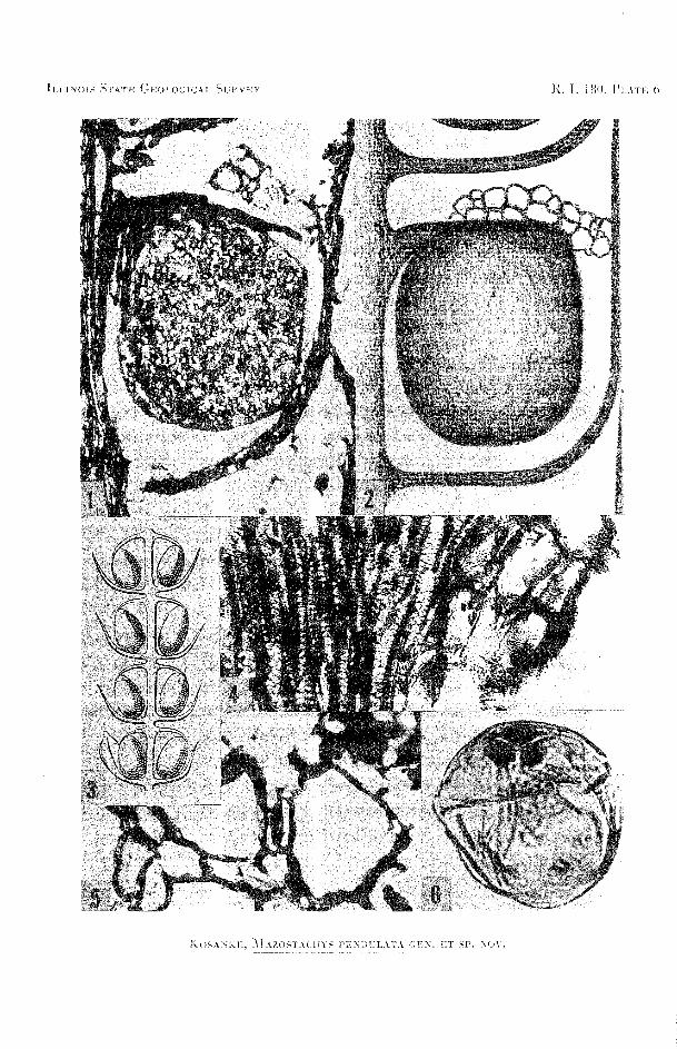

EXPLANATION O F PLATE 6
FIG. I-Longitudinal section of a part of whorl 4, cone 1 (B-1304A), peel 3. At the base of the photo- micrograph is a portion of the sporangiophore arm of whorl 6. Above this level is the node and departing bract enclosing a sporangium. Much higher is the sporangiophore and above this the aerenchyma tissue (X43).
g-A reconstruction of two nodes and one internode showing bracts, a sporangium, sporangio- phore, and above this the aerenchyma tissue. The section shown in figure 1 served as the basis for this reconstruction (X36).
3-Reconstruction of a longitudinal section of a portion of a cone associated by Schenk with Annularia splienophy2ylloides. This fructification is similar to Mazostachys in size and position of pendulate sporangiophores, but each sporangiophore had only one sporangium, sporangia are attached to the end of the sporangiophore, and bracts are shorter. (By Schenk from Jongmans.)
4-Oblique section through a small portion of a node from cone 13 (B-1304A), peel 6. This type of tracheid pitting was called scalariform or multiseriate pitting by I--Iqbeg (1942) and multi- seriate scalariform by Delevoryas and Morgan (1952) (X357).
5-Aerenchyma tissue showing large lacunae and a network of parenchyma cells. This sec- tion is from cone 14 (B-1304A), peel 19, from the second whorl above the base of the cone (Xl75).
6-One spore of Marostachys pendulata, obtained in the isolated state by mechanically dissecting a sporangium from a cone and macerating the sporangium in dilute HC1. The spore meas- ures 60 microns in its largest diameter and 58.6 microns at right angles to that diameter. Note the area contagionis between the rays of the triradiate suture.

ILLINOIS STATE GEOLOGICAL SURVEY, REPORT OF INVESTIGATIONS 180
37 p., 6 pls., 5 figs., 1955