robotic navigation to sub-cortical neural tissue for...
TRANSCRIPT
1
Robotic navigation to sub-cortical neural tissue for intracellular 1
electrophysiology in vivo 2
3
WA Stoy1, I Kolb1, GL Holst2, YJ Liew1, A Pala1, B Yang2, ES Boyden3,4, GB Stanley1, 4
CR Forest1,2 5
6
1 Wallace H. Coulter Department of Biomedical Engineering, Georgia Institute of 7
Technology, Atlanta GA, 30332 USA 8
2 George W. Woodruff School of Mechanical Engineering, Georgia Institute of 9
Technology, Atlanta GA, 30332 USA 10
3 Media Lab, Massachusetts Institute of Technology, Cambridge, MA 11
4 McGovern Institute for Brain Research, Massachusetts Institute of Technology, 12
Cambridge, MA 13
14
Articles in PresS. J Neurophysiol (June 7, 2017). doi:10.1152/jn.00117.2017
Copyright © 2017 by the American Physiological Society.
2
ABSTRACT 15
In vivo studies of neurophysiology using the whole-cell patch clamp technique enable 16
exquisite access to both intracellular dynamics and cytosol of cells in the living brain but 17
are underrepresented in deep subcortical nuclei due to fouling of the sensitive electrode 18
tip. We have developed an autonomous method to navigate electrodes around 19
obstacles such as blood vessels, after identifying them as a source of contamination 20
during regional pipette localization (RPL) in vivo. In mice, robotic navigation prevented 21
fouling of the electrode tip, increasing RPL success probability 3 mm below the pial 22
surface to 82% (n=72/88) over traditional, linear localization (25%, n=24/95) and 23
resulted in high quality thalamic whole-cell recordings with average access resistance 24
(32.0 MΩ), and resting membrane potential (-62.9 mV) similar to cortical recordings in 25
isoflurane-anesthetized mice. Whole-cell yield improved from 1% (n=1/95) to 10% 26
(n=9/88) when robotic navigation was used during RPL. This method opens the door to 27
whole-cell studies in deep subcortical nuclei, including multimodal cell typing and 28
studies of long range circuits. 29
30
NEW AND NOTEWORTHY 31
This work represents an automated method for accessing subcortical neural tissue for 32
intracellular electrophysiology in vivo. We have implemented a novel algorithm to detect 33
obstructions during regional pipette localization and move around them while minimizing 34
lateral displacement within brain tissue. This approach leverages computer control of 35
pressure, manipulator position, and impedance measurements to create a closed-loop 36
3
platform for pipette navigation in vivo. This technique enables whole-cell patching 37
studies to be performed throughout the living brain. 38
39
INTRODUCTION 40
In vivo patch clamp recording is one of the most important and versatile techniques in 41
neuroscience. Whole-cell recordings have enabled stable investigation of subthreshold 42
activity to identify cell types and circuits in the intact brain. This technique is also 43
uniquely positioned to enable concurrent measurements of intrinsic and sensory evoked 44
electrophysiology in either voltage- or current-clamp modes (Harvey, Collman, 45
Dombeck, & Tank, 2009) (Constantinople & Bruno, 2013), morphology (Margrie, Brecht, 46
& Sakmann, 2002), and the genetic profile of single neurons (Cadwell et al., 2016), as 47
well as the ability to introduce foreign genetic material into the cell ((Rancz et al., 2011), 48
(Vélez-Fort et al., 2014)). There is growing interest in multimodal cell type classification 49
(electrophysiological, morphological, and/or genetic, etc.) throughout the brain, a major 50
goal of the BRAIN Initiative (Bargmann, Newsome, & Anderson, 2014). Though recently 51
developed genetic voltage indicators have been used with some success to measure 52
activity both from individual neurons and populations (Lin & Schnitzer, 2016), the 53
heterogeneous tissue of the brain acts as a scattering medium, limiting recordings to 54
superficial cortical layers in the mammalian brain. Sharp intracellular recording 55
techniques similarly have sub-threshold resolution, but suffer from short recording times 56
(Fee, 2000) and are unsuitable for voltage-clamp recordings due to high electrode 57
impedance. 58
59

4
While whole-cell patch clamping is the gold standard for in vivo electrophysiology, it 60
requires skill to perform and has thus been more extensively used for in vitro 61
experiments. Recent efforts to automate it have been productive, yet sub-cortical 62
recording with whole-cell patch clamping remains a low yield endeavor. Published 63
papers are scant ((Margrie et al., 2002), (Brecht & Sakmann, 2002), (Groh et al., 2014)), 64
but whole-cell recordings in deep subcortical nuclei are difficult to obtain and are known 65
to suffer from increased access resistance (Margrie et al., 2002). In a literature review of 66
the 60 most cited papers that reported low access resistance, blind, whole-cell 67
recordings in vivo, only 5% of all 2350 recorded cells were recorded at depths 68
exceeding 3 mm from the pial surface in rodents. Several other studies have 69
demonstrated recordings in the cat thalamus in vivo (several centimeters below the pial 70
surface), but report high access resistance of the electrodes (X. Wang, Vaingankar, 71
Sanchez, Sommer, & Hirsch, 2011; X. Wang et al., 2007). 72
To investigate these subcortical nuclei in vivo, researchers have used extracellular, 73
juxtasomal, cell-attached, or sharp intracellular recordings ((Q. Wang, Webber, & 74
Stanley, 2010a), (Yu, Xiong, Chan, & He, 2004b),(Petersen et al., 2008),(Higley & 75
Contreras, 2007),(Friedberg, Lee, & Ebner, 2004),(Polack & Charpier, 2006),(Yu, Xiong, 76
Chan, & He, 2004a)), indicating that deep whole-cell recordings, while valuable and 77
desirable, are exceedingly difficult to obtain. In order to enable this important technique 78
to be used throughout the brain, we identify a need to improve both the yield and quality 79
of deep whole-cell recordings. 80
It is well known that a pipette must be clean, with a good tip geometry, to enable 81
formation of a gigaseal with a target cell (Neher, 1995). Since deep recording requires 82
5
traversing through several millimeters or centimeters of heterogeneous tissue (e.g., 83
blood vessels, glial cells, membranes) to reach a region of interest during regional 84
pipette localization (RPL), the pipette invariably encounters, and is clogged by debris 85
from this tissue. Effort to date to mitigate this problem have had limited success. In their 86
2002 study, Margrie et al. suggested that increasing pipette pressure or advancing the 87
pipette through a guide tube may reduce access resistance (or equivalently, increase 88
quality) (Margrie et al., 2002). However, Brecht and Sakmann subsequently noted that 89
neither higher pressures nor a guide tube reduced the access resistance of thalamic 90
recordings (Brecht & Sakmann, 2002). 91
92
We set out to investigate the relationship between this troublesome first stage of patch 93
clamping, regional pipette localization, and high access resistance, low yield whole-cell 94
experiments. By attempting whole-cell trials in the mouse thalamus, a deep brain 95
structure of wide interest ((Kelly et al., 2014), (Llinás & Steriade, 2006), (Sherman, 96
2005)), we observed that transient high amplitude fluctuations in resistance that occur 97
during regional pipette localization are often followed by residual, permanent increases 98
in pipette tip resistance, preventing successful whole-cell recordings, and that these 99
obstructions could be avoided with a series of small lateral movements, confirming 100
previous observations by Lee et al. 2014 (D. Lee, Shtengel, Osborne, & Lee, 2014). By 101
visualizing this process using slices of brain tissue on a microscope, we show that 102
pipette penetration of blood vessels and the residue left on pipette tips is the likely 103
cause these resistance changes, along with meninges (dura, pia, and hippocampal 104
meninges). Lateral steps enabled navigation around blood vessels and other obstacles 105

6
without a residual increase in resistance and meninges were penetrated with short, 106
rapid plunges with the pipette. We developed an efficient algorithm for laterally moving 107
around obstructing blood vessels during regional pipette localization in vivo and 108
compared its effectiveness to linear localization. We found that whole-cell trials 109
performed 3 mm deep could be localized with the same yield as we had previously 110
demonstrated in the cortex (up to 1 mm) with direct linear localization and in addition, 111
had comparable access resistances to whole-cell recordings performed previously in 112
the cortex. 113
114
MATERIALS AND METHODS 115
Acute in vivo and in vitro preparation 116
All experiments were performed in accordance with the Georgia Tech Institutional 117
Animal Care and Use Committee (IACUC) guidelines. For in vivo preparation, all mice 118
(n=19) were prepared for acute experimentation as we have done previously 119
(Kodandaramaiah, Franzesi, Chow, Boyden, & Forest, 2012). Briefly, young male 120
C57BL/6 mice (p35–p49) were anesthetized with isoflurane and headfixed to a titanium 121
headplate with C&B-Metabond dental cement (Parkell, Edgewood, NY). Craniotomies (1 122
mm diameter) and duratomies were performed above the Ventral Posteromedial 123
nucleus (VPM) of the thalamus (1.75 mm Rostral, 1.75 mm Lateral, 3 mm below the pial 124
surface) using stereotaxic coordinates from the Paxinos and Franklin mouse brain atlas 125
(Paxinos 2012). 126
127
7
For in vitro preparation, acute brain slices of mouse visual area VI were prepared from 128
male C57BL/6 adult mice (aged P30-P60) using the protective recovery method 129
described in detail elsewhere (Wu 吴吴吴 et al., 2016). 130
131
Pipette fabrication 132
Long taper patch pipettes (e.g., 7 mm) were pulled using fire-polished borosilicate glass 133
(BF150-86-10HP, Sutter Instrument, Novato, California) on a P-1000 electrode puller 134
with a 4.5 mm wide box filament (Sutter Instrument). The long taper is achieved with an 135
initial high-velocity step (heat = Ramp + 10, velocity = 40), with subsequent steps used 136
to develop the taper to ~1 μm (3-4 pulls of heat = Ramp – 10, velocity = 20). 137
138
Electrophysiology 139
Whole-cell patch clamping was performed as described previously (Kodandaramaiah 140
2016). In brief, an Autopatch 1500 (Neuromatic Devices, Atlanta GA) was used to 141
provide computer-controlled pressure and measure resistance for both in vitro and in 142
vivo experiments. Both in vitro and in vivo experiments used Multiclamp 700B amplifiers 143
(Molecular Devices, Sunnyvale, CA) and signals were digitized at 20kHz using National 144
Instruments DAQs (in vivo DAQ: cDAQ-9174 and NI 9215, in vitro DAQ: NI USB-6221, 145
National Instruments, Austin, TX) and recorded in PClamp 10 (Molecular Devices). A 146
Slicescope Pro 1000 (Scientifica, Essex, UK) was used to perform in vitro recordings 147
and an MP-285 micromanipulator (Sutter Instrument) with PT1-Z8 Motorized Translation 148
stage (Thorlabs, Newton, New Jersey) was used for positioning the electrode for in vivo 149
patch clamping. 150

8
151
Resistance measurements were performed throughout pipette translation, rather than 152
only before and after translation as in our prior work ((Kodandaramaiah et al., 2012), 153
(Kodandaramaiah et al., 2016)). During pipette translation, starting from ACSF on the 154
surface of the tissue, resistance was recorded by applying a 20 mV amplitude, 128 Hz 155
square wave (50% duty cycle) to the pipette, and calculating the resistance using Ohm’s 156
law. The resistance was computed as a moving average of four measurements (low 157
pass filter with four sample rectangular window). Thus, filtered resistances were saved 158
to an array, called the resistance array. 159
160
Blood vessel penetration 161
In addition to mapping a blood vessel in neural tissue using SCIM, we measured the 162
resistance changes encountered as the pipette was translated through a blood vessel in 163
vitro. The pipette was filled with ACSF and pressured to high positive pressure (1000 164
mbar) to replicate typical parameters for in vivo pipette localization (Kodandaramaiah et 165
al., 2016). For this penetration experiment, the pipette was positioned above a blood 166
vessel visually, at 35° relative to the image plane. The pipette was manually lowered 167
until the tip resistance increased by 12.5% above initial pipette resistance, and then 168
retracted 15 µm to a starting position. Resistance was recorded while the pipette was 169
manually advanced for 200 µm as described above. Images were captured at relative 170
depths indicated using the optical system (See Fig. 2B). 171
172
Scanning ion conductance microscopy in vitro (SCIM) 173

9
We mapped blood vessels in neural tissue (in vitro) by performing scanning ion 174
conductance microscopy (Sánchez et al., 2008). The pipette was filled with ACSF and 175
pressurized to low positive pressure (30 mbar). First, the pipette was centered above a 176
blood vessel of interest and visualized using differential interference contrast (DIC) 177
optical system (Olympus, Center Valley, PA). The pipette was manually lowered until 178
the tip resistance increased by 12.5% above initial pipette resistance. The pipette was 179
then retracted 15 µm and moved laterally to a new, random “grid position.” At this new 180
position, the pipette was then moved down 15 µm and the tip resistance was measured 181
again. This process was performed repeatedly until all grid positions were measured. A 182
grid is defined as a square 20 µm x 20 µm comprising 100 “grid positions” with 2 µm 183
pitch, centered on the initial pipette axial position. The resulting map of resistances as a 184
function of lateral pipette position (at constant depth) was linearly interpolated to 1 µm 185
pitch for visualization and displayed in MATLAB (Natick, MA) as a surface plot (See Fig. 186
2C). 187
188
Regional pipette localization (RPL) in vivo 189
Regional pipette localization (Kodandaramaiah et al., 2012) refers to the act of lowering 190
a pipette into neural tissue to a desired region of interest (e.g., thalamus) under high 191
positive pressure. In our experiments, we performed regional pipette localization using 192
two different methods: an uninterrupted, direct, linear trajectory as we have previously 193
described (Kodandaramaiah et al., 2012) and a novel robotic navigation method to 194
avoid obstructions. The method of localization was randomly selected prior to each trial. 195
A maximum of 10 electrode penetrations were performed per experimental preparation. 196

10
Pipettes were initially placed on the brain surface under stereoscopic guidance in a 197
region free of large blood vessels. 198
199
200
a. RPL using linear trajectory 201
202
Linear regional pipette localization has previously been utilized by us (Kodandaramaiah 203
et al., 2012) and others (Desai, Siegel, Taylor, Chitwood, & Johnston, 2015) for whole-204
cell electrophysiology in vivo. We used similar parameters; briefly, we applied high 205
positive pressure (1000 mbar) and translated the pipette at a rate of 500 μm/s. 206
Resistance was recorded during localization as described above, so the resistance 207
array could be displayed and analyzed, at a pitch of 500 µm/s / 128 Hz = 3.9 208
µm/sample. 209
210
b. RPL using robotic navigation 211
212
For RPL using robotic navigation, high positive pressure (800 mbar) was applied and 213
the pipette was translated at a rate of 200 μm/s, unless otherwise stated. Resistance 214
was recorded during localization as described above. At the square wave frequency of 215
128 Hz, resistance array measurements were spaced 1.6 µm apart. The pipette was 216
inserted along its initial axis, z, at x,y = (0,0) and moved in x, y, and z to navigate 217
around obstacles. 218
219

11
As shown in Fig. 3, if an obstruction was detected during localization, motion was halted 220
at depth zobstruction, with a pipette resistance of Robstruction (note Fig. 3b1,c). An obstruction 221
detection was defined as follows: pipette resistance increase of at least of 12.5% above 222
baseline resistance. Baseline resistance, Rbaseline, was computed as the minimum 223
resistance of the previous 800 µm (512 samples from the resistance array, 4 sec). With 224
an initial pipette resistance of 4-7 MΩ, 12.5% increase resulted in a minimum Robstruction 225
of 4.5-7.9 MΩ. 226
227
After halting the motion of the pipette upon obstruction detection, the pipette is moved in 228
a series of steps in an attempt to navigate around the obstruction. First, the pipette is 229
retracted along the pipette axis (z) by a distance zdodge (See Fig. 3b2,c). The distance 230
zdodge is located relative to zobstruction within the previous 50 µm (32 samples) at which a 231
minimum pipette resistance, Rdodge, was recorded. At depth zdodge, the pipette is moved 232
laterally to dodge, or navigate around, the obstacle that was encountered (See Fig. 233
3b3,c). Lateral movements, centered on the initial pipette axis are calculated to form a 234
spiral (See Fig. 3c), as follows: 235
236
237
238
Where ∆r = 5 µm, ∆θ = /4, and n is the step index {1, 2, 3, ...}. Each step, n, yields a 239
lateral movement vector, m, along which the pipette is moved. The pipette is then 240
lowered back to zobstruction (See Fig. 3b4,c). At zobstruction, the resistance is measured 241
again, termed Rn. If Rn-Rdodge ≥ 200 kΩ, the obstacle is still in the proximity of the pipette 242

12
tip and has not been avoided. The pipette is then retracted to zdodge, the step index n is 243
incremented, and another later movement occurs. This is repeated until Rn-Rdodge < 200 244
kΩ or until n=10, resulting in a maximum lateral distance of 50µm. 245
246
If Rn-Rdodge < 200 kΩ, the obstacle has been successful avoided. The pipette is 247
advanced 30 µm beyond zdodge to ensure the obstacle has been passed (See Fig. 248
3b5,c)., and then the pipette is moved laterally in a straight line from x,y = (mi(n), mj(n)) 249
to return to the initial pipette axis, x,y = (0,0) (See Fig. 3b6,c). 250
251
Alternatively, if n=10 and Rn-Rdodge ≥ 200 kΩ, the obstacle was not avoided. In this case, 252
a pulse of high positive pressure (1000 mbar, 1 sec) is applied while the pipette is 253
advanced 100 microns at 200 µm/s to attempt to dislodge the obstruction from the tip of 254
the pipette. The pipette is then moved laterally in a straight line to return to the initial 255
pipette axis, x,y = (0,0) (See Fig. 3c). 256
257
Localization is continued until the region of interest is reached and the pipette 258
resistance is less than 200 kΩ above the baseline resistance, Rb. If the obstruction is 259
not cleared before the end of the region of interest, pipette localization is halted and the 260
pipette is retracted to the surface and replaced. 261
262
Following successful regional pipette localization, the software compensates for pipette 263
capacitance, which is expected to change due to the depth of the recordings discussed 264
here. 265

13
266
267
RESULTS 268
269
RPL using linear trajectory 270
We have previously developed an automated patch clamping system, the Autopatcher, 271
and deployed it in the cortex and hippocampus. The results for regional pipette 272
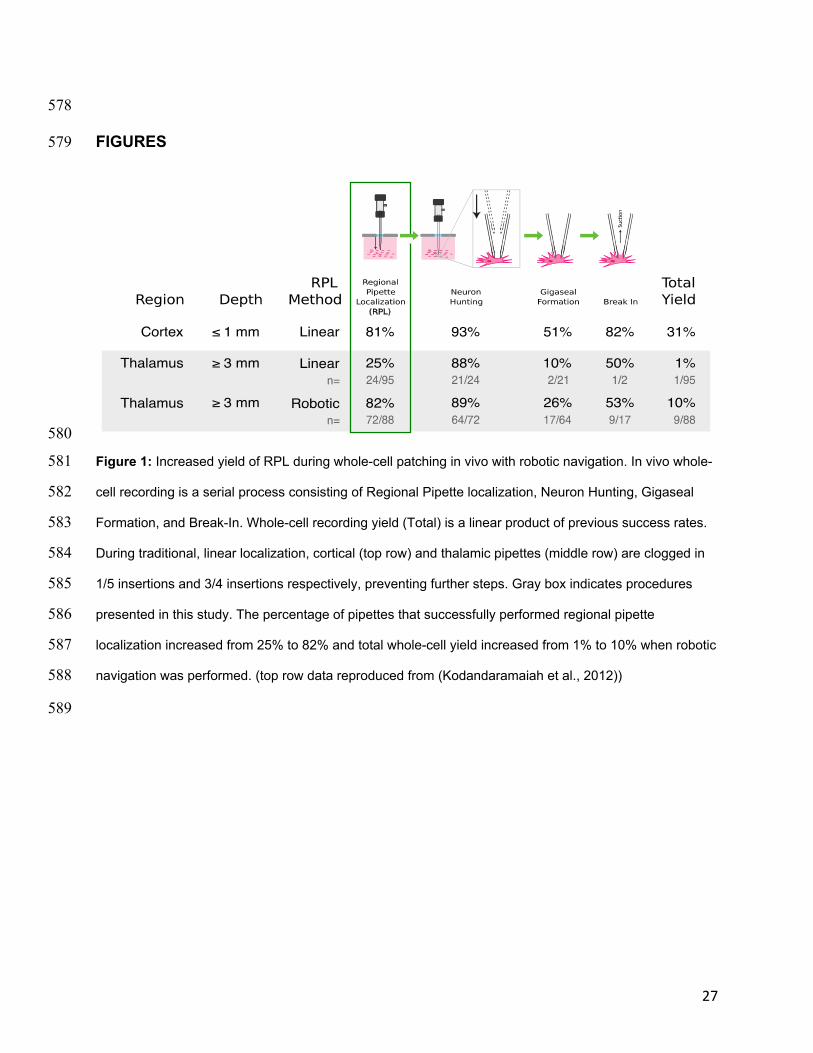
localization, and yield are shown in Fig. 1, top row, for depths less than 1 mm, 273
reproduced from Kodandaramaiah et al. 2012. 274
275
To explore the feasibility of using the Autopatcher for whole-cell recording in deep 276
subcortical nuclei we targeted the ventral posteromedial (VPM) nucleus and 277
surrounding nuclei of the thalamus. The resulting yield of whole-cell patching was far 278
below what we observed in our previous cortical patching efforts (See Fig. 1, top and 279
middle rows). In 95 trials, one successful whole-cell recording was achieved. Further, in 280
75% of trials (71/95), the pipette reached a depth of 3 mm with a tip resistance above 281
the threshold for removal and replacement. Thus 75% of trials were aborted without 282
attempting gigaseal formation. 283
284
To understand what was occurring during linear localization, we modified the 285
Autopatcher so that, as described in the Methods, resistance measurement was 286
performed throughout pipette translation, rather than only before and after translation as 287
in our prior work. Consequentially, during linear localization multiple high amplitude 288
14
fluctuations of the resistance were revealed, occasionally greater than 25 MΩ (Fig. 4 289
a,b). As the pipette advanced, resistance would often return near, but not exactly back 290
to baseline, indicating that the event was transient. Final pipette resistance (at the depth 291
of regional pipette localization) was on average 730 kΩ greater than the initial 292
resistance measured above the pial surface (Fig. 4e). High amplitude fluctuations were 293
observed in 91% of linear localization trials (n=86/95) (Fig. 4a). 294
295
Observations of obstacles 296
To understand the nature of these high amplitude resistance fluctuations, we 297
investigated them in vitro where we could visualize the interaction of the pipette tip with 298
the neural tissue under a microscope. After applying the requisite high positive pressure 299
(1000 mbar) to a patch pipette, we advanced its tip through the tissue. We noted that 300
the high positive pressure easily displaced neurons and glia, but some blood vessels 301
remained in the path of the pipette. Shown in Fig. 2a, as the pipette encountered one of 302
these blood vessels during manual, axial translation at approximately 15 µm/sec, the 303
pipette resistance was measured at 10 Hz. The pipette resistance, initially 4.3 MΩ, 304
increased to 26 MΩ within 35 µm as the pipette deformed the blood vessel (Fig. 2b). 305
The resistance then quickly decreased as the pipette pierced and passed through the 306
blood vessel. However, a residual blockage was noted, causing an increase in pipette 307
resistance of 3.7 MΩ that persisted until the end of the tissue slice was reached 308
(approximately 200 µm). The resistance signature of an obstruction in vitro appears 309
qualitatively similar to resistance fluctuations encountered in vivo (Compare Fig. 2a and 310

15
Fig. 4b)—a rapid increase in resistance, followed by a rapid decrease, resulting in a 311
residual resistance offset. 312
313
The previous result showed the uniaxial (z) signatures of a blood vessel encountered 314
with a pipette. We also mapped blood vessels in neural tissue laterally (x,y) by 315
performing scanning ion conductance microscopy. As shown in Fig. 2c-e, pipette 316
resistance increases were observed over a region that overlapped with the microscopy 317
image of the blood vessel. Concomitantly, regions of tissue adjacent to the blood 318
vessel, populated by neural cells and glia, showed only negligible resistance increases. 319
Additionally, without impaling the blood vessel, no residual increase in resistance was 320
noted even after 100 consecutive resistance measurements. Thus, we gained 321
confidence that a blood vessel was the predominant obstacle to pipette insertion in 322
neural tissue and that it could be avoided by moving the pipette laterally even after initial 323
contact if the vessel was not impaled. 324
325
Selection of Navigation Algorithm Parameters 326
We used SCIM to further optimize the parameters for encountering, and navigating 327
around obstacles. We set a threshold for obstacle encounters of 12.5% (approximately 328
500 kΩ); this is greater than the amplitude of baseline resistance variation during 329
localization (100 kΩ) but much less than observed during blood vessel impalement (See 330
Fig. 2a). 331
332

16
To determine the axial retraction distance necessary to attempt to dodge an obstacle, 333
zdodge, we first noted that the pipette can drag cells and blood vessels if it is positioned 334
too close to an obstacle. We moved the pipette above a blood vessel and determined 335
the minimum axial retraction distance required to allow a pipette to move laterally 336
without dragging an obstruction to be 15 µm (data not shown). We set a retraction 337
distance of up to z=50 µm in vivo to ensure that the pipette was safely away from the 338
obstacle before lateral motion, given that the in vivo environment is less predictable 339
than in vitro. 340
341
We next attempted to optimize the axial advancement distance necessary to bypass an 342
obstacle after lateral movement. Others have performed a rigorous study of blood 343
vessels in the mouse brain, showing sizes of 10-60 µm ((Santisakultarm et al., 2012), 344
Fig. 2c). From their data, we compute a mean blood vessel diameter of 28.1 ± 1.9 µm. 345
In our observations, capillaries 15 µm diameter and smaller were easily displaced under 346
high positive pressure. We set a distance of 30 µm to advance the pipette beyond the 347
blood vessel location (zobstruction) to ensure the obstacle had been passed. 348
349
Whereas we used 1000 mbar for RPL during linear localization, we chose a lower 350
pressure (800 mbar) for pipette insertion during robotic navigation to reduce the volume 351
of ejected intracellular solution during the longer time needed to perform RPL. 352
353
To minimize damage to the tissue in vivo, we designed the vessel avoidance algorithm 354
to make the smallest lateral and radial movements possible. Radial movement was 355

17
defined as the distance between the pipette position (xn,yn) and the original pipette 356
location at x,y = (0,0). Lateral distance traveled was defined as the sum of the distances 357
traveled between each point. Movements were made in small increments to minimize 358
radial distance, r, traveled. Additionally, because the orientation of the blood vessel’s 359
major axis with respect to manipulator’s x and y axes is unknown, we incremented the 360
angle, θ with each step resulting in a spiral search pattern. Similar search patterns have 361
been shown to minimize path length when searching for a line in a 2D plane, analogous 362
to finding the edge of a blood vessel in a plane (Finch 2016). 363
364
RPL using robotic navigation 365
Using robotic navigation algorithm to avoid obstructions during regional pipette 366
localization greatly improved the yield of successfully localized pipettes. In 88 trials, 367
82% of pipettes were localized successfully to a depth of 3000 µm when the dodging 368
algorithm was active (when compared to linear localization, n=95, p=7.8448e-15, 369
Fisher’s exact test). This high yield for RPL using robotic navigation for the thalamus is 370
comparable to rates achieved with RPL using linear trajectories in the cortex. 371
372
In addition, the final resistance increase (170 kΩ, n=88) was significantly lower than 373
when the pipette was localized without the algorithm (730 kΩ, n=95, p=0.0142, 374
Wilcoxon rank sum test) (See Fig. 4de). During robotic navigation, obstructions were 375
encountered in 95% of trials, and on average the dodging algorithm attempted to avoid 376
6.7 obstructions during each localization (See Fig. 4c). At a depth of 3000 µm, 377
obstructions were therefore encountered every 3000/6.7=445 µm on average. Each 378

18
obstruction that was successfully avoided in n=3.5 steps, resulting in a radial distance of 379
17.5 µm on average. Regional pipette localization under algorithmic guidance was 380
completed in an average of 75 ± 23 sec, significantly longer than 6 sec (3 mm at 500 381
µm/s) using the traditional localization method, due to the increased time to avoid 382
obstacles and the lower localization speed. Advantageously, this slow localization may 383
allow tissue to relax before attempting to patch. 384
385
The number of robotic navigation events did not have a significant effect on the success 386
of gigasealing. Trials that resulted in a gigaseal (n=17) underwent 3.65 navigation 387
events on average, while trials that failed to result in a gigaseal (n=71) underwent 7.24 388
navigation events on average (p=0.961 Wilcoxon rank sum test). Additionally, the 389
maximum resistance increase experienced by pipettes during localization did not have 390
an effect on the success of gigasealing. Trials that resulted in a gigaseal (n=17) had an 391
average maximum resistance during localization of 4.9 MOhms, while trials that failed to 392
result in a gigaseal (n=71) had an average maximum resistance during localization of 393
6.1 MOhms (p=0.19, Wilcoxon rank sum test). 394
395
Vertical descent to the thalamus requires penetration of the ventricular meninges, large, 396
relatively planar membranes that cannot be avoided using the algorithm described here. 397
The meninges were routinely detected at approximately 2.5 mm from the pial surface 398
(See Fig. 4h). The meninges were penetrated using, on average, 2.3 successive dodge 399
attempts (each resulting in a rapid 100 µm advancement of the pipette). 400
401

19
The yield of whole-cell recordings in thalamus improved when regional pipette 402
localization was performed using robotic navigation. In trials where whole-cell 403
recordings were attempted following RPL with robotic navigation, 10% of trials (n=9/88) 404
resulted in successful whole-cell recordings. In trials performed using RPL with linear 405
localization, 1% of trials (n=1/95) resulted in whole-cell recordings (p=0.0076, Fisher’s 406
exact test). 407
408
Whole-cell recordings performed in the thalamus with robotic navigation were of 409
comparable quality to those previously performed in the cortex using linear localization 410
(See Fig. 5, (Kodandaramaiah et al., 2012), (Margrie et al., 2002)). Our thalamic 411
recordings had similar access resistances (32.0 ± 4.1 MΩ) to cortical recordings 412
reported by Margrie et al. 2002 as well as our prior work, all in the range of 10-50 MΩ 413
((Margrie et al., 2002), (Kodandaramaiah et al., 2012)). Similarly, our thalamic 414
recordings had holding currents (-50.8 ± 8.9 pA) at -65 mV holding voltage and resting 415
membrane potentials (-62.9 ± 2.0 mV) that were not significantly different from our 416
previous cortical work, respectively, -23.5 ± 12.9 pA (p=0.1982, Wilcoxon rank-sum test) 417
and -61.54 ± 1.05 mV (p=0.1148, Wilcoxon rank-sum test). 418
419
Recorded neurons had electrophysiological properties consistent with ventrobasal 420
thalamic nucleus neurons (VB), consisting of the ventral posteromedial nucleus (VPM) 421
and the ventral posterolateral nucleus (VPL). In response to hyperpolarizing current 422
injection, sag potentials were observed (hyperpolarization induced depolarization, 423
indicative of H currents, IH (Kuisle et al., 2006; Leist et al., 2016), see Fig. 5b). Following 424

20
release of hyperpolarizing currents, burst firing was observed indicative of T-type 425
calcium channel activity, often followed by after (spike) depolarization (Kuisle et al., 426
2006; X. Wang et al., 2010b) (see Fig. 5c arrow). 427
428
Comparison of whole-cell recordings in the thalamus with robotic navigation to those 429
previously reported in the thalamus using linear localization is difficult. There are few 430
reports of successful whole-cell thalamic recordings ((Margrie et al., 2002), (Mease, 431
Sumser, Sakmann, & Groh, 2016), (Oberlaender et al., 2012a), (Brecht & Sakmann, 432
2002)) and the quality metrics are not reported consistently. However, Margrie et al 433
2002 reports that, “Despite the thalamic recordings being carried out on a younger 434
sample of animals the access resistance was consistently greater than that observed for 435
more superficial recordings.” In addition to numerous conversations with other labs 436
performing whole-cell recordings in vivo (personal communication), we assume that the 437
scarcity of published whole-cell recordings far below the cortex in vivo suggest that they 438
are very difficult to achieve. In our hands, the single cell that was recorded using linear 439
localization during RPL was of lower quality with an access resistance of 72 MΩ, resting 440
membrane potential of -45 mV, and holding current of -150 pA. 441
442
DISCUSSION 443
444
Here, we describe a method to robotically navigate whole-cell patch pipettes through 445
neural tissue in vivo in a way that significantly reduces clogging of the tips that occurs 446
commonly when blood vessels are pierced. Pipettes localized without robotic navigation 447

21
frequently encounter (91% of the trials) and impale obstructions during localization, as 448
others have previously noted ((Margrie et al., 2002), (Brecht & Sakmann, 2002), (A. K. 449
Lee, Epsztein, & Brecht, 2009)). This is tolerable for cortical recordings, but as we have 450
shown obstacles are encountered on average every 370 µm and therefore make deep, 451
sub-cortical recordings in vivo impractically low yield and quality. 452
453
Previous studies have noted the existence and detrimental effect of permanent 454
resistance increases during RPL ((Margrie et al., 2002), (D. Lee et al., 2014)), 455
speculating that electrode penetration of the vasculature was the cause (D. Lee et al., 456
2014). We have shown, through in vitro studies, that these obstructions are very likely 457
caused by encounters with blood vessels larger than 15 µm. Following these 458
encounters, vascular membrane residue adhering to the pipette tip obstructs the tip and 459
increases residual resistance. An efficient spiral navigation algorithm to find the edge of 460
the blood vessel with minimal tissue displacement (17.5 µm on average) enables high 461
yield regional pipette localization. 462
463
Robotic navigation enables one to localize pipettes in deep structures (e.g., mouse 464
thalamus at 3 mm) with yields similar to those reported in the cortex using linear 465
localization. Pipettes were successfully localized with robotic navigation to a depth of 3 466
mm below the pial surface in 82% of trials (n=72/88), comparable to linear localization in 467
the cortex from our previous study (81%, n=128/158, p=1, Fisher’s exact test) 468
(Kodandaramaiah et al., 2012). 469
470
22
For whole-cell recording yield, there are large ranges of reported yields that make 471
comparison more challenging. Whole-cell yield for blind in-vivo patching has been 472
reported between 20-50% ((Margrie et al., 2002), (A. K. Lee et al., 2009)), while the 473
yield for two photon targeted patching in mice in vivo is between 10-20% (Margrie et al., 474
2003). For blind, automated whole-cell recording in the mouse cortex, we have 475
previously reported a yield of 31% (Kodandaramaiah et al., 2012). Others have reported 476
yields of 17% (Desai et al., 2015) in mice using similar automation. Our yield of 10% in 477
the mouse thalamus makes recording there practical, although somewhat lower yield 478
than cortical whole-cell recording. Additionally, we believe that all subcortical nuclei are 479
now accessible using this method, as electrodes inserted to the VPM must traverse 480
white and gray matter. However, there may still be regions of the brain that may be 481
difficult to access due to their proximity to the ventricles. In this work, we did not 482
address the penetration of the thick ventricular membranes as such membranes are 483
likely impossible to navigate around, and would release CSF into the brain if punctured. 484
Penetration of such membranes remains a problem for maintaining the cleanliness of 485
the electrode, but might be mitigated with the application of a reversible, protective 486
coating (Singh, Zhu, & He, 2004). The advent of further automation strategies such as 487
pipette cleaning (Kolb et al., 2016) may further improve the throughput of these 488
experiments. 489
490
The whole-cell recording yield is the product of the yield of the four stages of the patch 491
algorithm (See Fig. 1). We note a decrease in gigaseal formation yield with deep 492
patching that is irrespective of localization method, linear or robotic. We have observed 493

23
higher amplitude heartbeat modulation during the preceding stage, neuron hunting, for 494
the thalamus relative to the cortex, which may indicate greater mechanical disturbances 495
at these depths affecting gigaseal yield. Identifying and overcoming gigaseal yield 496
issues would further advance deep whole-cell patch clamping efforts and motivate 497
further investigation. 498
499
One possible opportunity for improved gigaseal yield is to use the lateral steps 500
performed during SCIM to map target cells prior to gigaseal attempt to optimize the tip 501
placement with respect to the soma, both in vivo and in vitro. Blind in vitro whole-cell 502
recordings suffer from low yield (generally 50-80% in vitro, (Blanton, Loturco, & 503
Kriegstein, 1989)) when compared to image guided in vitro studies (> 80%) (Stuart, 504
Dodt, & Sakmann, 1993). Notably, in vitro resistance measurements alone are not 505
sufficient to identify cell membrane dimpling and cell shape, visual identifiers commonly 506
used to align pipettes with target cells for successful gigaseals (Desai et al., 2015). 507
Further, local membrane stiffness, a potential proxy for membrane dimpling, can be 508
estimated by modulating pressure and measuring the difference in tip resistance 509
(Sánchez et al., 2008). The combination of pressure modulation and scanning ion 510
conductance microscopy may improve pipette placement on cell membranes and 511
thereby increase the yield of single cell experiments in vivo and in vitro. 512
513
Leakage of biocytin-containing intracellular solution into the surrounding tissue during 514
RPL may cause background and off-target staining. Intracellular solution in the 515
extracellular space is also undesirable due to the osmotic pressure it places on 516

24
neurons. This problem may be compounded by the extended time (75 ± 23 sec vs 6 517
sec) spent navigating the pipette through tissue under high positive pressure towards 518
the region of interest. Intracellular solution leakage could be reduced by decreasing the 519
positive pressure during RPL. We hypothesize that a lower pipette pressure during RPL 520
will lead to an increase in obstacle detections as less debris, cells, and blood vessels 521
are displaced by the pipette pressure, however the optimal pipette pressure was not 522
investigated in this work. Alternatively, cellular contrasts that are not taken up by cells 523
from the extracellular space, such as DNA plasmids, may reduce off-target labeling 524
(Vélez-Fort et al., 2014). 525
526
The average number of navigation events (detected obstacles) for trials that eventually 527
successfully formed a gigaseal (3.65) was lower than for trials that eventually failed to 528
form a gigaseal (7.24). While we found that this difference was not statistically 529
significant, we believe the difference in the means is due to the skew of the navigation 530
event distribution. That is, several failed trials resulted from RPL where successive 531
obstacle detections were triggered throughout their descent, either because they were 532
clogged from internal debris or because accrued debris was not successfully dislodged 533
during the navigation events. Although the pipette resistance ultimately returned to 534
baseline in these trials, we suspect that the tip of the pipette may have become 535
contaminated, but not measurably clogged. Trials with high numbers of successive 536
obstacle detections may indicate unsuccessful navigation and the overall success rate 537
may improve by rejecting these trials. 538
539

25
Intracellular recording has remained as the gold standard electrophysiology technique 540
because of its high quality, mechanical stability, and resolution. Margrie et al. 541
hypothesized that pipette contamination results in higher access resistances, and thus 542
lower quality thalamic recordings. Previous efforts to reduce the access resistance of 543
recordings performed deep in the brain using higher pressures in the pipette or a guide 544
tube have not been successful (Brecht & Sakmann, 2002). Here, we demonstrate that 545
robotic navigation around blood vessels in vivo results not only in higher yield than with 546
linear localization but also higher-quality recordings. Critically, we demonstrate that 547
robotic navigation during regional pipette localization produces whole-cell recordings 3 548
mm below the pial surface with access resistances similar to those measured from cells 549
in the cortex ((Kodandaramaiah et al., 2012), (Margrie et al., 2002)). Other factors may 550
contribute to differences in yield and access resistance, namely pipette shape and tip 551
geometry, but these parameters are rarely reported or quantified, making comparison 552
difficult. Other parameters were also comparable to previous recordings in cortex, 553
including holding current (voltage clamp, used to keep the cell at -65mV) and resting 554
membrane potential (Kodandaramaiah et al., 2012). Thus, we are confident that in vivo 555
whole-cell recording quality is improved from previous efforts to perform whole-cell 556
recordings in the thalamus and are of equivalent quality to recordings in the cortex. 557
558
There are very few published studies that show in vivo whole-cell recordings at depths 3 559
mm or greater. In fact, to our knowledge, only 7 such studies have been published to 560
date ((Mease et al., 2016),(Groh et al., 2014),(Oberlaender et al., 2012a),(Oberlaender, 561
Ramirez, & Bruno, 2012b),(Kuo & Wu, 2012),(Brecht & Sakmann, 2002),(Margrie et al., 562
26
2002)). In contrast, in vitro whole-cell recordings in deep subcortical nuclei are abundant 563
((Kase, Inoue, & Imoto, 2012),(Neuhoff, Neu, Liss, & Roeper, 2002),(Benavides et al., 564
2007),(Guo et al., 2012),(Hu, Nasif, Zhang, & Xu, 2008),(Sosulina, Graebenitz, & Pape, 565
2010),(Porcello, Ho, Joho, & Huguenard, 2002)). This indicates that there is interest in 566
performing high-yield subcortical whole-cell recordings in vivo, while recording depth is 567
an impediment for whole-cell studies in these nuclei. Additionally, the whole-cell patch 568
clamp technique is uniquely positioned to investigate the structure-function-gene 569
relationship (Cadwell et al., 2016). This study opens the door for whole-cell 570
electrophysiology coupled with genetic or morphological profiling throughout the entire 571
brain, which is the focus of worldwide effort ((Oberlaender et al., 2012a), (Arkhipov et 572
al., 2016), (Vélez-Fort et al., 2014), (Cadwell et al., 2016)) and a major goal of the Brain 573
Research through Advancing Innovative Neurotechnologies (BRAIN) Initiative 574
(Bargmann et al., 2014). 575
576
577
27
578
FIGURES 579
580
Figure 1: Increased yield of RPL during whole-cell patching in vivo with robotic navigation. In vivo whole-581
cell recording is a serial process consisting of Regional Pipette localization, Neuron Hunting, Gigaseal 582
Formation, and Break-In. Whole-cell recording yield (Total) is a linear product of previous success rates. 583
During traditional, linear localization, cortical (top row) and thalamic pipettes (middle row) are clogged in 584
1/5 insertions and 3/4 insertions respectively, preventing further steps. Gray box indicates procedures 585
presented in this study. The percentage of pipettes that successfully performed regional pipette 586
localization increased from 25% to 82% and total whole-cell yield increased from 1% to 10% when robotic 587
navigation was performed. (top row data reproduced from (Kodandaramaiah et al., 2012)) 588
589

28
590
591
Figure 2: Lateral navigation around obstructions prevents persistent pipette resistance increase caused 592
by penetration of blood vessels in vitro. A) Resistance trace as a function of distance as a pipette pierces 593
a blood vessel under high positive pressure. A residual resistance increase of 3.7 MΩ remains after the 594
vessel is punctured. B) IR DIC images showing the pipette encountering and deforming the blood vessel 595
(scale bar, 50 µm). C) Schematic of Scanning Ion Conductance Microscopy (SCIM) mapping of a blood 596
vessel proceeds from a central point. Samples are collected randomly from a grid area 20 x 20 μm at 2 597
μm resolution D) The entire blood vessel and surrounding milieu is shown under IR DIC (scale bar, 20 598
μm) E) Resistances mapped as a function of grid position, clearly showing increased resistance when the 599
pipette is above the blood vessel (scale bar, 10 μm, 2x interpolation) 600
601

29
602
603
Figure 3: Robotic navigation algorithm for avoiding blood vessels during regional pipette localization in 604
vivo. A) Schematic showing vascular avoidance preparation. Brain outline from the Allen Institute’s Mouse 605
Brain Explorer program. B) Visual algorithm of vascular avoidance [1] Obstruction (here, a blood vessel) 606
is detected by an increase in pipette resistance. [2] The pipette is retracted to zdodge, [3] moved laterally, 607
and [4] advanced to the original zobstruction. If the difference in resistance at zobstruction and the resistance at 608
zdodge is < 200 kΩ, [5] the pipette is advanced through zobstruction and [6] the pipette is moved back to the 609
original x and y axis. C) The pipette is navigated around a blood vessel with sequential steps sampling 610
from a spiral pattern. Blood vessel in A and B shown in isometric view. Blood vessel in C shown in top 611
view (top) and cross section (bottom). 612
613

30
614
615
Figure 4: Pipette tip resistance increases during regional pipette localization in vivo due to accrued 616
debris, preventing whole cell recordings. Robotic navigation prevents this debris from accruing. A) 617
Recordings of change in pipette resistance during regional pipette localization reveal that obstructions are 618
encountered throughout the insertion path. B) When an obstruction is cleared by continuing linear pipette 619
advancement, debris may still be present at the pipette tip, reflected by the persistent resistance increase 620
of the pipette by 200kOhms. C) Using a robotic navigation algorithm, pipette debris is prevented from 621
accruing on the pipette tip, shown by the return of the pipette resistance to the baseline. Arrows indicate 622
locations of robotic navigation event. D) Detail of a single robotic navigation event. A-D) initial pipette 623
resistance was subtracted to show changes in resistance. Initial pipette resistances ranged from 4-7 MΩ. 624
E) The final resistance of the pipette is significantly lower after insertion to 3mm below the pial surface 625
when the robotic navigation algorithm to localize the pipette was used. F) The maximum resistance 626
measured during robotic navigation is not significantly different between trials that gigasealed 627
successfully (n=17) and trials that failed to seal (n=71), Wilcoxon rank sum test (p=0.19). G) The number 628
of navigation events was not significantly different between trials that gigasealed successfully (n=17) and 629
trials that failed to seal (n=71), Wilcoxon rank sum test (p=0.96) H) Histogram showing number of 630
navigation events as a function of depth. Note the slight increase in navigation events around 0 µm and 631
2500 µm from the pial surface, where the pia and ventricular meninges were encountered, respectively. 632
633

31
634
635
Figure 5: Neurons recorded in whole-cell configuration were of good quality. A) Example of spontaneous 636
activity from a neuron recorded 3.2 mm below the pial surface with detail of spontaneous burst (arrow 637
indicates burst shown in detail to the right). B) Example whole-cell traces recorded in the thalamus for 3 638
different neurons. Note the sag in membrane potential following hyperpolarizing current injection 639
(representing activity of Ih,(Leist et al., 2016)) and after-hyperpolarization rebound bursting in each trace, 640
indicative of thalamic neurons. Current injections lasted 0.5 sec and ranged from ± 50 pA (first recording) 641
to ± 100 pA (second and third recording). C) Following hyperpolarizing current injection, rebound bursts 642
exhibited after depolarization (ADP, see arrow, (X. Wang et al., 2010b)), consistent with ventrobasal 643
thalamic nucleus cells. Current injection was -150 pA. 644
645
646
32
647
REFERENCES 648
649
Arkhipov, A., Berg, J., Buice, M., Gouwens, N. W., Gratiy, S., Iyer, R., et al. (2016). Inferring cortical 650 function in the mouse visual system through large-scale systems neuroscience. Proceedings of the 651 National Academy of Sciences, 113(27), 7337–7344. http://doi.org/10.1073/pnas.1512901113 652
Bargmann, C., Newsome, W., & Anderson, A. (2014). BRAIN 2025: a scientific vision. … 653 /Science/Brain/2025/( …. 654
Benavides, D. R., Quinn, J. J., Zhong, P., Hawasli, A. H., DiLeone, R. J., Kansy, J. W., et al. (2007). Cdk5 655 modulates cocaine reward, motivation, and striatal neuron excitability. The Journal of Neuroscience : 656 the Official Journal of the Society for Neuroscience, 27(47), 12967–12976. 657 http://doi.org/10.1523/JNEUROSCI.4061-07.2007 658
Blanton, M. G., Loturco, J. J., & Kriegstein, A. R. (1989). Whole Cell Recording From Neurons in Slices of 659 Reptilian and Mammalian Cerebral-Cortex. Journal of Neuroscience Methods, 30(3), 203–210. 660
Brecht, M., & Sakmann, B. (2002). Whisker maps of neuronal subclasses of the rat ventral posterior 661 medial thalamus, identified by whole-cell voltage recording and morphological reconstruction. The 662 Journal of Physiology, 538(2), 495–515. http://doi.org/10.1113/jphysiol.2001.012334 663
Cadwell, C. R., Palasantza, A., Jiang, X., Berens, P., Deng, Q., Yilmaz, M., et al. (2016). 664 Electrophysiological, transcriptomic and morphologic profiling of single neurons using Patch-seq. 665 Nature Biotechnology, 34(2), 199–203. http://doi.org/10.1038/nbt.3445 666
Constantinople, C. M., & Bruno, R. M. (2013). Deep Cortical Layers Are Activated Directly by Thalamus. 667 Science, 340(6140), 1591–1594. http://doi.org/10.1126/science.1236425 668
Desai, N. S., Siegel, J. J., Taylor, W., Chitwood, R. A., & Johnston, D. (2015). MATLAB-based automated 669 patch-clamp system for awake behaving mice. Journal of Neurophysiology, 114(2), 1331–1345. 670 http://doi.org/10.1152/jn.00025.2015 671
Fee, M. S. (2000). Active stabilization of electrodes for intracellular recording in awake behaving animals. 672 Neuron, 27(3), 461–468. 673
Friedberg, M. H., Lee, S. M., & Ebner, F. F. (2004). The contribution of the principal and spinal trigeminal 674 nuclei to the receptive field properties of thalamic VPM neurons in the rat. Journal of Neurocytology, 675 33(1), 75–85. http://doi.org/10.1023/B:NEUR.0000029649.28599.a5 676
Groh, A., Bokor, H., Mease, R. A., Plattner, V. M., Hangya, B., Stroh, A., et al. (2014). Convergence of 677 cortical and sensory driver inputs on single thalamocortical cells. Cerebral Cortex, 24(12), 3167–678 3179. http://doi.org/10.1093/cercor/bht173 679
Guo, M.-L., Xue, B., Jin, D.-Z., Liu, Z.-G., Fibuch, E. E., Mao, L.-M., & Wang, J. Q. (2012). Upregulation of 680 Npas4 protein expression by chronic administration of amphetamine in rat nucleus accumbens in 681 vivo. Neuroscience Letters, 528(2), 210–214. http://doi.org/10.1016/j.neulet.2012.07.048 682
Harvey, C. D., Collman, F., Dombeck, D. A., & Tank, D. W. (2009). Intracellular dynamics of hippocampal 683 place cells during virtual navigation. Nature, 461(7266), 941–946. http://doi.org/10.1038/nature08499 684
Higley, M. J., & Contreras, D. (2007). Cellular Mechanisms of Suppressive Interactions Between 685 Somatosensory Responses In Vivo. Journal of Neurophysiology, 97(1), 647–658. 686 http://doi.org/10.1152/jn.00777.2006 687
Hu, X.-T., Nasif, F. J., Zhang, J., & Xu, M. (2008). Fos regulates neuronal activity in the nucleus 688 accumbens. Neuroscience Letters, 448(1), 157–160. http://doi.org/10.1016/j.neulet.2008.10.025 689
Kase, D., Inoue, T., & Imoto, K. (2012). Roles of the subthalamic nucleus and subthalamic HCN channels 690 in absence seizures. Journal of Neurophysiology, 107(1), 393–406. 691 http://doi.org/10.1152/jn.00937.2010 692
Kelly, S. T., Kremkow, J., Jin, J., Wang, Y., Wang, Q., Alonso, J.-M., & Stanley, G. B. (2014). The Role of 693 Thalamic Population Synchrony in the Emergence of Cortical Feature Selectivity. PLOS Comput Biol, 694 10(1), e1003418. http://doi.org/10.1371/journal.pcbi.1003418 695
Kodandaramaiah, S. B., Franzesi, G. T., Chow, B. Y., Boyden, E. S., & Forest, C. R. (2012). Automated 696 whole-cell patch-clamp electrophysiology of neurons in vivo. Nature Methods, 9(6), 585–587. 697 http://doi.org/10.1038/nmeth.1993 698

33
Kodandaramaiah, S. B., Holst, G. L., Wickersham, I. R., Singer, A. C., Franzesi, G. T., McKinnon, M. L., 699 et al. (2016). Assembly and operation of the autopatcher for automated intracellular neural recording 700 in vivo. Nature Protocols, 11(4), 634–654. http://doi.org/10.1038/nprot.2016.007 701
Kolb, I., Stoy, W. A., Rousseau, E. B., Moody, O. A., Jenkins, A., & Forest, C. R. (2016). Cleaning patch-702 clamp pipettes for immediate reuse. Scientific Reports, 6. http://doi.org/10.1038/srep35001 703
Kuisle, M., Wanaverbecq, N., Brewster, A. L., Frère, S. G. A., Pinault, D., Baram, T. Z., & Lüthi, A. (2006). 704 Functional stabilization of weakened thalamic pacemaker channel regulation in rat absence epilepsy. 705 The Journal of Physiology, 575(1), 83–100. http://doi.org/10.1113/jphysiol.2006.110486 706
Kuo, R. I., & Wu, G. K. (2012). The Generation of Direction Selectivity in the Auditory System. Neuron, 707 73(5), 1016–1027. http://doi.org/10.1016/j.neuron.2011.11.035 708
Lee, A. K., Epsztein, J. E. R. O. M., & Brecht, M. (2009). Head-anchored whole-cell recordings in freely 709 moving rats. Nature Protocols, 4(3), 385–392. http://doi.org/10.1038/nprot.2009.5 710
Lee, D., Shtengel, G., Osborne, J. E., & Lee, A. K. (2014). Anesthetized- and awake-patched whole-cell 711 recordings in freely moving rats using UV-cured collar-based electrode stabilization. Nature 712 Protocols, 9(12), 2784–2795. http://doi.org/10.1038/nprot.2014.190 713
Leist, M., Datunashvilli, M., Kanyshkova, T., Zobeiri, M., Aissaoui, A., Cerina, M., et al. (2016). Two types 714 of interneurons in the mouse lateral geniculate nucleus are characterized by different h-current 715 density. Scientific Reports, 6, 24904. http://doi.org/10.1038/srep24904 716
Lin, M. Z., & Schnitzer, M. J. (2016). Genetically encoded indicators of neuronal activity. Nature 717 Neuroscience, 19(9), 1142–1153. http://doi.org/10.1038/nn.4359 718
Llinás, R. R., & Steriade, M. (2006). Bursting of Thalamic Neurons and States of Vigilance. Journal of 719 Neurophysiology, 95(6), 3297–3308. http://doi.org/10.1152/jn.00166.2006 720
Margrie, T. W., Brecht, M., & Sakmann, B. (2002). In vivo, low-resistance, whole-cell recordings from 721 neurons in the anaesthetized and awake mammalian brain. Pflügers Archiv, 444(4), 491–498. 722 http://doi.org/10.1007/s00424-002-0831-z 723
Margrie, T. W., Meyer, A. H., Caputi, A., Monyer, H., Hasan, M. T., Schaefer, A. T., et al. (2003). Targeted 724 Whole-Cell Recordings in the Mammalian Brain In Vivo. Neuron, 39(6), 911–918. 725 http://doi.org/10.1016/j.neuron.2003.08.012 726
Mease, R. A., Sumser, A., Sakmann, B., & Groh, A. (2016). Cortical Dependence of Whisker Responses 727 in Posterior Medial Thalamus In Vivo. Cerebral Cortex, 26(8), bhw144–3543. 728 http://doi.org/10.1093/cercor/bhw144 729
Neher, E. (1995). … , ION CHANNELS FOR COMMUNICATION BETWEEN AND WITHIN CELLS, 730 DECEMBER 9, 1991 BY ERWIN NEHER AND ELEMENTARY STEPS IN …. Nobel Lectures. 731
Neuhoff, H., Neu, A., Liss, B., & Roeper, J. (2002). I(h) channels contribute to the different functional 732 properties of identified dopaminergic subpopulations in the midbrain. The Journal of Neuroscience : 733 the Official Journal of the Society for Neuroscience, 22(4), 1290–1302. 734
Oberlaender, M., de Kock, C. P. J., Bruno, R. M., Ramirez, A., Meyer, H. S., Dercksen, V. J., et al. 735 (2012a). Cell type-specific three-dimensional structure of thalamocortical circuits in a column of rat 736 vibrissal cortex. Cerebral Cortex, 22(10), 2375–2391. http://doi.org/10.1093/cercor/bhr317 737
Oberlaender, M., Ramirez, A., & Bruno, R. M. (2012b). Sensory experience restructures thalamocortical 738 axons during adulthood. Neuron, 74(4), 648–655. http://doi.org/10.1016/j.neuron.2012.03.022 739
Petersen, R. S., Brambilla, M., Bale, M. R., Alenda, A., Panzeri, S., Montemurro, M. A., & Maravall, M. 740 (2008). Diverse and temporally precise kinetic feature selectivity in the VPm thalamic nucleus. 741 Neuron, 60(5), 890–903. http://doi.org/10.1016/j.neuron.2008.09.041 742
Polack, P. O., & Charpier, S. (2006). Intracellular activity of cortical and thalamic neurones during 743 high‐voltage rhythmic spike discharge in Long‐Evans rats in vivo. The Journal of Physiology, 571(2), 744 461–476. http://doi.org/10.1113/jphysiol.2005.100925 745
Porcello, D. M., Ho, C. S., Joho, R. H., & Huguenard, J. R. (2002). Resilient RTN fast spiking in Kv3.1 null 746 mice suggests redundancy in the action potential repolarization mechanism. Journal of 747 Neurophysiology, 87(3), 1303–1310. 748
Rancz, E. A., Franks, K. M., Schwarz, M. K., Pichler, B., Schaefer, A. T., & Margrie, T. W. (2011). 749 Transfection via whole-cell recording in vivo: bridging single-cell physiology, genetics and 750 connectomics. Nature Neuroscience, 14(4), 527–532. http://doi.org/10.1038/nn.2765 751
Santisakultarm, T. P., Cornelius, N. R., Nishimura, N., Schafer, A. I., Silver, R. T., Doerschuk, P. C., et al. 752 (2012). In vivo two-photon excited fluorescence microscopy reveals cardiac- and respiration-753 dependent pulsatile blood flow in cortical blood vessels in mice. American Journal of Physiology-754
34
Heart and Circulatory Physiology, 302(7), H1367–H1377. http://doi.org/10.1152/ajpheart.00417.2011 755 Sánchez, D., Johnson, N., Li, C., Novak, P., Rheinlaender, J., Zhang, Y., et al. (2008). Noncontact 756
Measurement of the Local Mechanical Properties of Living Cells Using Pressure Applied via a 757 Pipette. Biophysical Journal, 95(6), 3017–3027. http://doi.org/10.1529/biophysj.108.129551 758
Sherman, S. M. (2005). The role of the thalamus in cortical function: not just a simple relay. 759 Singh, A., Zhu, H., & He, J. (2004). Improving mechanical stiffness of coated benzocyclobutene (BCB) 760
based neural implant. Conference Proceedings : ... Annual International Conference of the IEEE 761 Engineering in Medicine and Biology Society. IEEE Engineering in Medicine and Biology Society. 762 Annual Conference, 6, 4298–4301. http://doi.org/10.1109/IEMBS.2004.1404197 763
Sosulina, L., Graebenitz, S., & Pape, H.-C. (2010). GABAergic interneurons in the mouse lateral 764 amygdala: a classification study. Journal of Neurophysiology, 104(2), 617–626. 765 http://doi.org/10.1152/jn.00207.2010 766
Stuart, G. J., Dodt, H. U., & Sakmann, B. (1993). Patch-clamp recordings from the soma and dendrites of 767 neurons in brain slices using infrared video microscopy. Pflügers Archiv, 423(5-6), 511–518. 768 http://doi.org/10.1007/BF00374949 769
Vélez-Fort, M., Rousseau, C. V., Niedworok, C. J., Wickersham, I. R., Rancz, E. A., Brown, A. P. Y., et al. 770 (2014). The Stimulus Selectivity and Connectivity of Layer Six Principal Cells Reveals Cortical 771 Microcircuits Underlying Visual Processing. Neuron, 83(6), 1431–1443. 772 http://doi.org/10.1016/j.neuron.2014.08.001 773
Wang, Q., Webber, R. M., & Stanley, G. B. (2010a). Thalamic synchrony and the adaptive gating of 774 information flow to cortex. Nature Neuroscience, 13(12), 1534–1541. http://doi.org/10.1038/nn.2670 775
Wang, X., Vaingankar, V., Sanchez, C. S., Sommer, F. T., & Hirsch, J. A. (2011). Thalamic interneurons 776 and relay cells use complementary synaptic mechanisms for visual processing. Nature 777 Neuroscience, 14(2), 224–231. http://doi.org/10.1038/nn.2707 778
Wang, X., Wei, Y., Vaingankar, V., Wang, Q., Koepsell, K., Sommer, F. T., & Hirsch, J. A. (2007). 779 Feedforward excitation and inhibition evoke dual modes of firing in the cat's visual thalamus during 780 naturalistic viewing. Neuron, 55(3), 465–478. http://doi.org/10.1016/j.neuron.2007.06.039 781
Wang, X., Yu, G., Hou, X., Zhou, J., Yang, B., & Zhang, L. (2010b). Rebound bursts in GABAergic 782 neurons of the thalamic reticular nucleus in postnatal mice. 783
Wu 吴吴吴, Q., Kolb, I., Callahan, B. M., Su, Z., Stoy, W., Kodandaramaiah, S. B., et al. (2016). 784 Integration of autopatching with automated pipette and cell detection in vitro. Journal of 785 Neurophysiology, 116(4), 1564–1578. http://doi.org/10.1152/jn.00386.2016 786
Yu, Y.-Q., Xiong, Y., Chan, Y.-S., & He, J. (2004a). Corticofugal Gating of Auditory Information in the 787 Thalamus: An In Vivo Intracellular Recording Study. Journal of Neuroscience, 24(12), 3060–3069. 788 http://doi.org/10.1523/JNEUROSCI.4897-03.2004 789
Yu, Y.-Q., Xiong, Y., Chan, Y.-S., & He, J. (2004b). Corticofugal gating of auditory information in the 790 thalamus: an in vivo intracellular recording study. The Journal of Neuroscience : the Official Journal 791 of the Society for Neuroscience, 24(12), 3060–3069. http://doi.org/10.1523/JNEUROSCI.4897-792 03.2004 793
794


