robotic radiosurgery for liver metastases
DESCRIPTION
ROBOTIC RADIOSURGERY FOR LIVER METASTASESTRANSCRIPT

ROBOTIC RADIOSURGERY FOR LIVER METASTASES
Kayıhan Engina, Nadir Kucuk
a, Murat Dokdok
b, Tayfun Enunlu
c, Kezban
Berberoglud, Hande Ayata
a, Cemile Ceylan
a, Ayhan Kılıc
a, Metin Guden
a
aRadiation Oncology,
bRadiology,
cStatistic Department,
dNuclear Medicine,
Anadolu Medical Center, Kocaeli, Turkey
INTRODUCTION
The liver is one of largest organ in the body which is made of
hepatocytes, located right-hand portion of the abdominal cavity, divided into
two lobes. Main functions of the liver are to produce bile, which helps carry
away waste and break down fats in the small intestine during digestion and to
produce certain proteins for blood plasma.
Liver cancer is the one of the most common cancer worldwide, with an
estimated five hundred thousand new liver tumors diagnosed in 2008(1). Both
primary or metastatic tumors of the liver generally respond poorly to all types
of treatment modalities (2-3). The decision of the optimal treatment modality
in patients with liver metastases depends on the general clinical data such as
primary tumor type, imaging characteristics of the liver, number of the lesions,
size and location of the lesions etc. A mapping of metastases in the liver is also
very important to define volume of the uninvolved liver segments. The size of
the uninvolved liver volume is very critical before decision of treatment
modalities.
The prevalence of the liver metastases is very high for patients whose
primary diagnoses are colorectal, pancreas, lung and breast carcinomas. It has
been shown that up to about 50%-60% of patients with colorectal cancer will
develop metastases and one third of these are localized to the liver (4). Local
control of the disease may increase both quality of life and survival. Surgical
resection of the liver metastases is known to be a first therapeutic option in
patients with solitary and limited number of metastases (5,6). However,
resection cannot be carried out in the majority of these metastases and only
10-25% of those patients are candidates for curative resection (6-8). Morbidity
and mortality rates after hepatic resection were reported as high as 35% and
3% (9,10). Chemotherapy is also an efficient method to treat the liver lesions
which cannot be surgically removed. New chemotherapeutic agents and
targeted therapies have been commonly used over the past decade and
significant improvements in results for some primary sites have been observed
(11-13). Unfortunately, the results of systemic therapies need improvement for
the majority of primary sites and liver metastases still remain as a difficult
therapeutic challenge (5). Several local treatment modalities including
cryosurgery, radiofrequency ablation (RFA), external/internal beam irradiation,
laser induced thermal therapy and selective internal radiation therapy (SIRT)
have been used to treat these metastases. RFA is the most common method
used in practice for treatment of liver metastases. This method has some
limitations such as tumor size (smaller than 3 cm) and proximity of major biliary
/vascular structures (14,15).
Treatment of intrahepatic metastases by radiotherapy had a limited role in the
past because of the low tolerance of the whole liver to irradiation. New
radiotherapy techniques such as stereotactic body radiation therapy (SBRT)
allows delivering high dose of radiation in a single session or limited number of
sessions to a confined area while sparing surrounding critical organs and other
healty tissues. With the development of robotic stereotactic body radiation
therapy (rSBRT) device, it is possible to track tumor motion during in the
treatment by using implanted fiducial markers combined with respiratory
motion modeling. However, only a few number of studies have been published
to evaluate the efficiency of rSBRT for liver metastases (8,16-18,26). The aim of
this study is to show feasibility, efficiency and low morbidity combined with a
high local control probability in use of rSBRT treatment for liver metastases.
METHODS AND MATERIALS
Patient Selection
A total of 55 patients with 84 liver metastases from different primary
sites who underwent rSBRT in our center between July 2006 and December
2011 were reviewed for this study. Primary site of malignancy for patients are
listed in Table 1. Treatment decision of rSBRT was determined by evaluation of
a surgeon, a radiologist, a radiation oncologist and medical oncologist. Patients
who refused surgery or judged inoperable were included in this study. Patients

were required to have adequate liver function and more than 3 months
minimum life expectancy. Other inclusion criterias were maximum tumor
diameter<6 cm, Karnofsky performance status >60, no active liver infection
and no prior radiotherapy to the liver. Maximum three liver metastases from
any primary sites were treated and accepted for this study. Patients and tumor
characteristics are summarized in Table 2.
Table 1. Primary origins of tumors treated with rSBRT
Primary origin n
% of
patients
Breast 7 13%
Pancreas 8 14%
Colorectal 23 41%
Lung 6 11%
Bladder 3 5%
Stomach 2 4%
Ovary 1 2%
Cervix 1 2%
Head and Neck 2 4%
Liver
(Cholangiocarcinoma) 1 2%
Sarcoma 1 2%

Table 2. Patients and tumor characteristics
Characteristic Value
# of Patients 55
# of treated lesions 84
Single lesion 37 pts (67%)
2 lesions 7 pts (13%)
3 lesions 11 pts (20% )
Gender
Male 32 pts (58%)
Female 23 pts (42%)
Age
Median 59 years
Mean 60.7 years
Range 22-86 years
Time from primary diagnosis
to first metastasis
Median 17 months
Range 0-151months
Time from first liver
metastasis to rSBRT
Median 6 months
Range 0-15 months
Metastases except liver
during rSBRT
None 33 pts (60%)
Multiple (≥2 different
sites) 4 pts (7%)
Lung 9 pts (17%)
Bone 5 pts (9%)
Other sites (single
location) 4 pts (7%)
Received chemotherapy
during rSBRT
Yes 44 pts (80%)
No 11 pts (20%)
Tumor volume
Mean 40,48 cc
Range 3.8 cc-336 cc

Pre-treatment preperations
An average of 3 (3-5) fiducial markers (Civco Medical Solutions, Kalona,
Iowa) were implanted percutaneously under CT-guidance with local
anaesthesia. Fiducials were made of gold and shaped as cylindirical. It was
implanted in or near tumor volume by keeping minimum distance between
fiducials of 2 cm. After implementation, all patients were kept waiting
minimum a week to check the possibility of fiducial migration. Patients were
immobilized in supine position by using whole body vacuum cradle
immobilization device. Patients also were fitted with a special syncrony vest,
which is worn during Cyberknife treatment and enables the robot to correlate
chest motion and breathing patterns with the tumor position. Computed
tomography (CT) images were recorded with same immobilized position and
vest without using contrast material. CT images were taken in 1 mm slice
thickness using breat-holding technique. In addition, T1-weighted magnetic
resonance images were scanned with using contrast material for all patients.
Treatment planning and delivery
Treatment planning CT, magnetic resonance imaging (MRI) and positron
emission tomography-CT (PET-CT) were used for tumor delination. All these
scans were imported into the treatment planning system (Multiplan, version
2.2.0) and fused digitally, often with CT images, PET-CT scans or MRI images.
Contouring was done using only CT/MRI fusion in 6 (11%) patients. Treatment
planning CT was used for contouring in one patient. Both CT/PET-CT and
CT/MRI fusions were used for accurate identification of the location of the
gross tumor volume (GTV) in other 48 (87%) patients. The planning target
volume (PTV) was defined as a 5 mm uniform margin around GTV to account
for possible microscopic disease and targeting uncertanity. The liver, kidneys,
spinal cord, lungs, stomach, duodenum and intestine were contoured in
planning. Published reports have showed that minimum volume of 700 cc of
liver should remain uninjured by SBRT (19-20). We mandated that minimum
700 cc of normal liver had to receive at total dose less than 15 Gy in 3-5
fractions. Dose constraints for organ at risk (OARs) were applied in all
treatment plans as listed in Table 3. Treatment plan dose was prescribed at the
PTV with an isodose line range 70-85% which was covering the 95% volume of
PTV. Cyberknife (Accuray, Sunnyvale, CA) was used for rSBRT. Tumor tracking
was performed using the Synchrony system (Accuray) during rSBRT and the
total treatment time was approximately 50-180 min, with most patient treated
within 100 min.
Table 3. Dose constraints for OARs
Critical Structure Dose Constraint
Liver ≥700 cc ≤15 Gy
Kidneys V5<%75, Dmax<15 Gy
Spinal cord Dmax< 18 Gy
Stomach V20<%5, Dmax<25 Gy
Duedenum V20<%5, Dmax<25 Gy
Bowel V20<%5, Dmax<25 Gy
Lungs V15<%5, Dmax< 18 Gy
Heart Dmax<25 Gy
Vn-Organ volume that receives a dose of n
Gy or less,
Dmax- Maximum point dose in the organ
Follow up and toxicity
The patients were examined every 3 months after rSBRT by biochemical
examinations and imaging. PET-CT or MRI were performed at each follow-up.
Response was evaluated according to RECIST (Response Evaluation and Criteria
in Solid Tumors) criteria (21). Complete response (CR) was defined as the
complete disapperance of the target lesion. Partial response (PR) was defined
as at least a 30% decrease in the longest diameter of the target lesion.
Progressive disease (PD) was defined as at least 20% increase in the longest
diameter of the target lesion. Stable disease (SD) was defined as a tumor status
that no any changes in tumor dimensions or vascular modification. Local
control was defined as the absence of tumor progression within or at the
border of the radiation field based on imaging. Local recurrence was defined as
failure within treated volume or at the border of the radiation field. Toxicity
was also scored using National Cancer Institute (NCI) Common Terminology
Criteria for Adverse Events (CTCAE) v3.0 (22). Toxicity which was occured in

first 3 months after rSBRT called acute toxicity. Events which were occured
after 3 months from rSBRT called late toxicity.
Statistical analyses
SPSS 17 (SPSS Inc, Chicago, IL) software was used for the statistical
analyses. Fisher’s exact test and Log Rank were used to assess relation between
category variables. The rates of actuarial overall survival, actuarial local control,
actuarial survival by primary tumor types and actuarial survival by number of
treated lesion(s) were calculated by the Kaplan-Meier survival curve method.
All parameters were tested by univariable and multivariable regression model.
Cox regression model was used to express the factor effecting local control and
overall survival.
RESULTS
Patient Population
Fifty-five patients (32 males - 23 females) who had liver metastases from
various primary sites were treated with rSBRT using real time tumor tracking
system. Mean age of patients was 61 years (range 22 to 86 years). The primary
sites included colorectal (n=23), pancreas (n=8), breast (n=7), lung (n=6),
bladder (n=3) and others (n=8). Of the 3 patients who had two liver metastases
in the same lobe, single treatment plan was created and delivered. Of the 2
patients who had three liver metastases in the same lobe, single treatment
plan was created and delivered. Eighty-four lesions derived from different
primary sites in fifty-five patients were treated with totally seventy-seven
treatment plan. Median time from primary tumor diagnosis to first liver
metastases was 17 months (Ranging between 0 and 151 months). Median time
from first liver metastases to rSBRT was 6 months (Range of 0-15 months). Of
the 33 (60%) patients treated with rSBRT, there was no any other organ
metastases except liver. Nine (17%) patients had only lung and 5 (9%) patients
had only bone metastases except liver lesions during in rSBRT. Four (7%)
patient have only one metastases except lung or bone. Four (7%) patients had
multiple (≥2 different organ) metastases except liver lesions during treatment.

One fifth of patients 11 (20%) didn’t receive any concurrent systemic therapy
during irradiation (Table 2). Only two patients were received RFA treatment for
the same lesions before rSBRT.
Dose-volume characterictics
The dose-volume characteristics of treatments are listed in Table 4.
Median collimator size was 20 mm (Range: 10 mm to 35 mm). The total
prescribed dose was 45 Gy (Range: 24 Gy to 45 Gy) in median 3 (Range: 3 to 6)
fractions. The radiation dose was prescribed to median 80% (Range: 70% to
85%) isodose line. The median percent of the PTV covered by the prescription
isodose line was 96.6% (Range: 90% to 99%). Mean number of beam used for
rSBRT was 224 (Range: 117 to 314). The minimum normal liver volume (700 cc)
received mean 7.8 Gy (Range: 2 Gy to 14.6 Gy).
Table 4. Dose-volume characteristics of rSBRT
Characteristic Value
Total liver volume
Mean 1571,8 cc
Collimator size
Median 20 mm
Range 10 mm-35 mm
Prescription dose
Median 45 Gy
Range 24 Gy-45 Gy
# of fraction
Median 3
Range 3-6
Prescibed isodose line
Median 80%
Range 70%-85%
Conformality index
Median 1.22
Range 1.06-1.94
Homogenity index
Median 1.25
Range 1.18-1.43
Tumor Coverage
Median 96.6%
Range 89.8%-99.5%
# of beam
Median 224
Range 117-314
700 cc uninvolved liver dose
Mean 7.8 Gy
Range 2 Gy-14.6 Gy
Clinical outcome
Fifty-five patients was accepted and evaluated for this study. Six patients
had no follow up images in our system. Tumor response rates, local control and
survival analysis for 49 (89%) patients are listed in Table 5. Twenty-seven (55%)
patients died and 22 (45%) were alive at the time of our evaluation. The
median follow up was 12 months (Range: 4 to 39 months). CR was seen in 25
(51%) patients, PR in 12 (25%) patients and SD in 2 (4%) patients at first follow
up. 10 (20%) patients progressed inside the treated volume at first follow-up.
The mean tumor volumes in patients with CR, PR and SD were 36,5 cc, 39,4 cc
and 43,2 cc, respectively. PD was seen in 17 (35.4%) patients at second follow
up. Percentage of tumor response (CR, PR or SD) rate was decrease from 80%
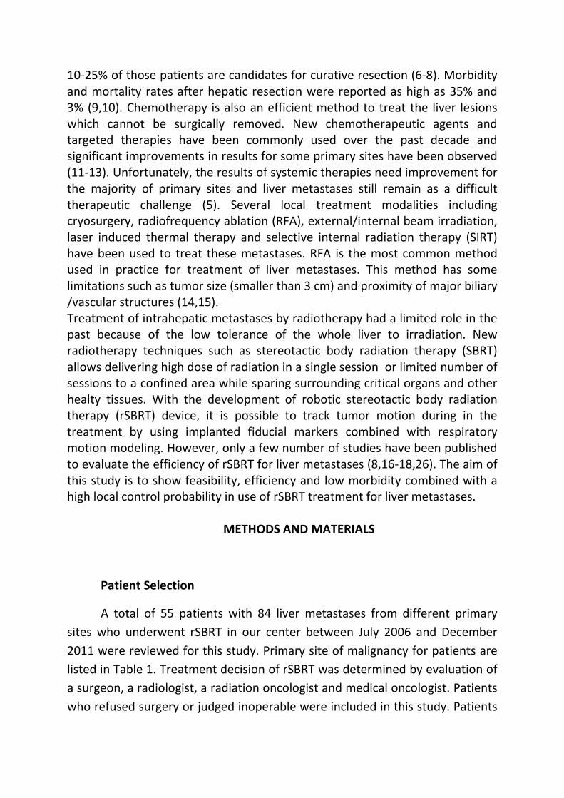
to 63% at the time of second follow-up. An example of CR with continued local
control at second follow-up is shown in Figure 1. Logistic regression model was
used for evaluating effect of different parameters to tumor response. Age,
gender, location of primary tumor, tumor size, number of fraction, total dose,
receiving concurrent chemotherapy and rSBRT, presence of other metastases
except liver and number of treated lesion were analysis using logistic regression
model. Location of primary tumor is only parameter that effect tumor
response. Colorectal tumor metastases had better results than other tumor
metastases in the first follow-up (p=0.05). The effect of other organ metastases
to tumor response was evaluated. Of the 19 (39%) patients who had other
organ metastases except liver, progression was seen in 2 patients. Of the 30
(61%) patients who had no any other organ metastases except liver,
progression was seen in 8 patients. No correlation was found statistically
(p=0.16, Fisher’s Exact Test) between tumor response and presence of other

organ metastases except liver. The effect of receiving chemotherapy during
rSBRT to tumor response was observed. No progression was seen in all 8 (16%)
patients who had no any systemic therapy during rSBRT. Of the 41 (84%)
patients who had received chemotherapy during rSBRT, progression was seen
in 10 patients. There was no significant correlation statistically (p=0.14,
Fisher’s Exact Test) between tumor response and receiving chemotherapy
during rSBRT.
Table 5. Clinical evaluation of patients after rSBRT
Variable Value
Patients (n) 49
Alive(n) 22
Dead(n) 27
Follow up (mo)
Median 12
Range 4-39
Response in first follow
up
CR 25(51%)
PR 12(25%)
SD 2(4%)
PD 10(20%)
Local recurrence
interval (mo)
Median 10
Range 6-23
Overall survival (mo)
Median 16
(a)
(b)
(c)
Figure 1. An example of CR with continued local control in a patient with
metastatic colorectal cancer (a) axial view of lesions in PET before rSBRT, (b)
treatment planning showing dose distrubition, (c) PET image after 6 months
from rSBRT

The actuarial local control rate for patients is shown in Figure 2. The
actuarial 1-year and 2-year local control rates were 61% (%95 confidence
interval (CI), 53%-69%) and 39% (95% CI, 28%-49%),respectively. Of the
complete responding patients at the first follow up, developed local
recurrence in 12(48%) of them, with a median time to relaps of 10 months
(range 6 months to 23 months). In 2 patients who had PR at first follow up, CR
was seen at second follow up. Kaplan Meier univariate analysis and Cox
regression multivariate analysis were used for evaluating local control. Results
are seen in Table 6. Age, number of treated lesion(s) and number of fraction
was statically important for local control according to Cox regression
multivariate analysis. Patients with >60 years old and 1 or 2 metastases and
patients who received ≤3 fractions had better local control according to our
statical results.
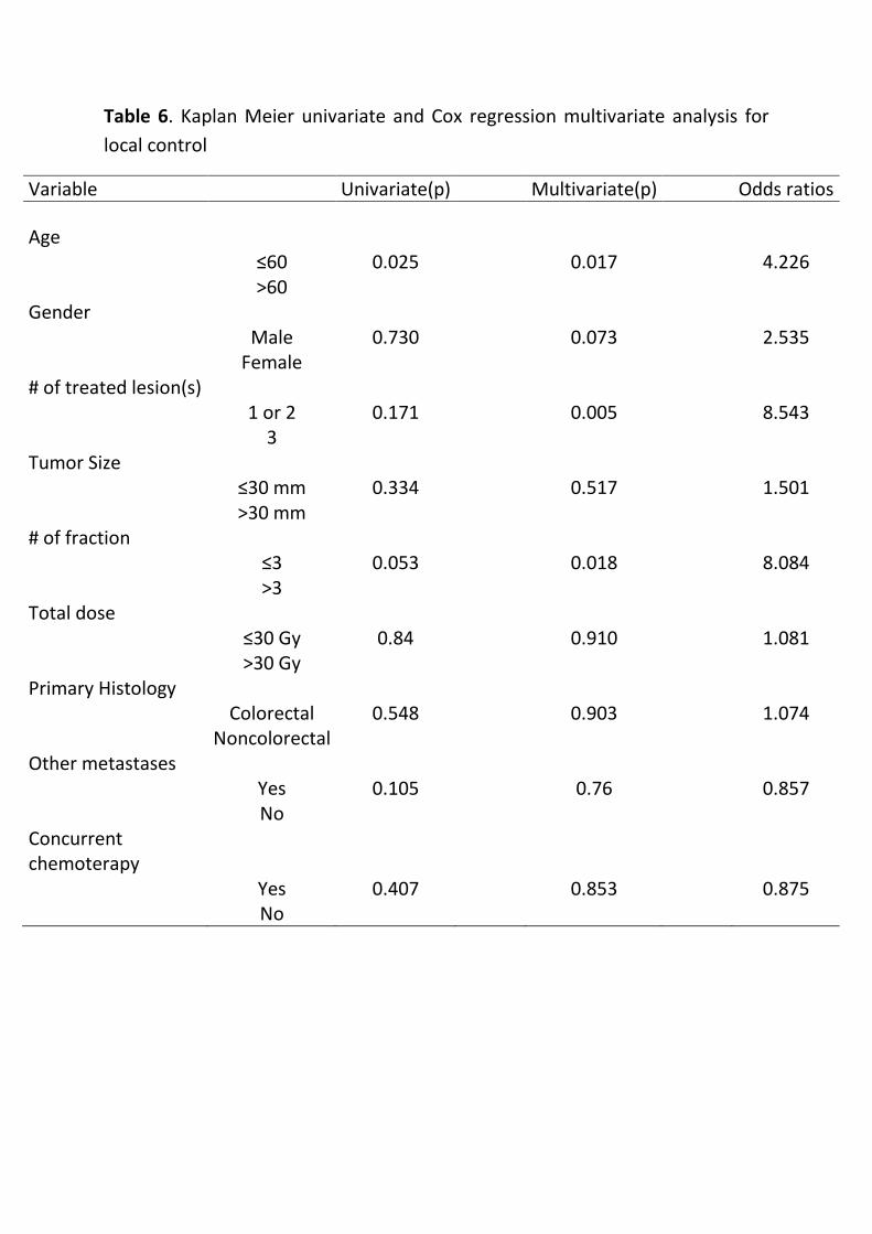
Table 6. Kaplan Meier univariate and Cox regression multivariate analysis for
local control
Variable Univariate(p) Multivariate(p) Odds ratios
Age
≤60 0.025 0.017 4.226
>60
Gender
Male 0.730 0.073 2.535
Female
# of treated lesion(s)
1 or 2 0.171 0.005 8.543
3
Tumor Size
≤30 mm 0.334 0.517 1.501
>30 mm
# of fraction
≤3 0.053 0.018 8.084
>3
Total dose
≤30 Gy 0.84 0.910 1.081
>30 Gy
Primary Histology
Colorectal 0.548 0.903 1.074
Noncolorectal
Other metastases
Yes 0.105 0.76 0.857
No
Concurrent
chemoterapy
Yes 0.407 0.853 0.875
No

Figure 2. Actuarial local control rate for patients evaluated by the Kaplan-
Meier method.
The median overall survival was 16 months. The actuarial overall survival
rates for patients are shown Figure 3. The actuarial 1-year and 2-year overall
survival rates were 79% (%95 (CI), 73%-85%) and 56% (95% CI, 48%-63%),
respectively. The overall survival rates were evaluated according to primary
tumor histology and statically no difference was found between colorectal liver
metastases and other primary tumor metastases (p=0,085 Log rank). The
actuarial 1-year overall survival rates were 70% (%95 (CI), 59%-81%) and 67%
(95% (CI), 56%-77%) and 2–year were 52% (%95 (CI), 38%-66%) and 37% (95%
(CI), 25%-50%) were for colorectal liver metastases and other primary tumor
metastases, respectively (Fig 4). Kaplan Meier univariate analysis and Cox
regression multivariate analysis were used for evaluating overall survival.
Results are shown in Table 7. Number of treated metastases was statically
important for overall survival according to Cox regression multivariate analysis.
Treated one or two metastases had better survival than three metastases
according to our statical results as shown Figure 5.
Table 7. Kaplan Meier univariate and Cox regression multivariate analysis for
overall survival
Variable Univariate (p) Multivariate (p) Odds ratios
Age
≤60 0.425 0.339 0.623
>60
Gender
Male 0.827 0.839 1.115
Female
# of treated lesion
1 or 2 0.127 0.011 6.085
3
Tumor Size
≤30 mm 0.266 0.940 1.054
>30 mm
# of fraction
≤3 0.293 0.072 3.817
>3
Total dose
≤30 Gy 0.485 0.547 1.428
>30 Gy
Primary Histology
Colorectal 0.398 0.287 1.713
Noncolorectal
Other metastases
Yes 0.290 0.230 0.485
No
Concurrent
chemoterapy
Yes 0.852 0.737 0.782
No
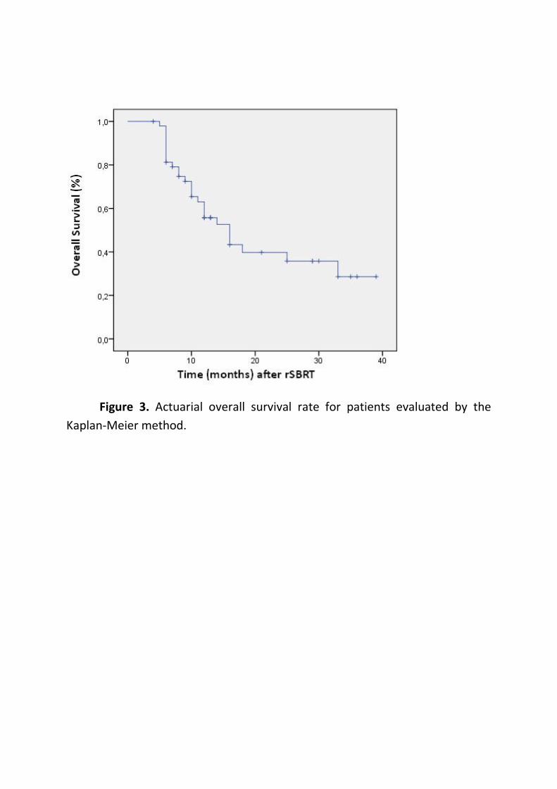
Figure 3. Actuarial overall survival rate for patients evaluated by the
Kaplan-Meier method.
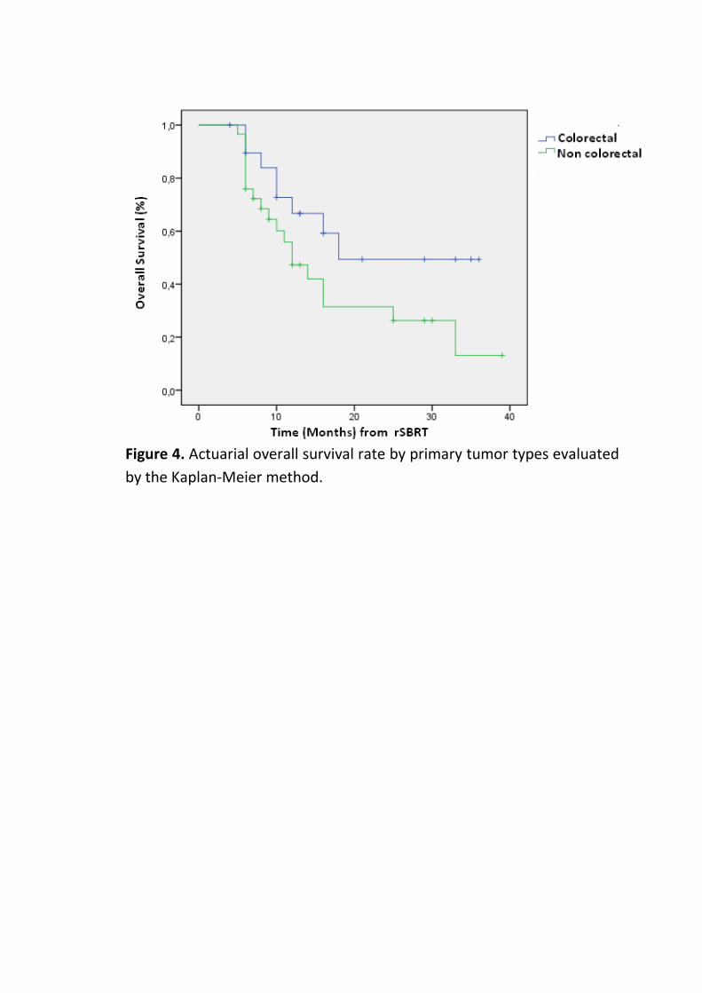
Figure 4. Actuarial overall survival rate by primary tumor types evaluated
by the Kaplan-Meier method.
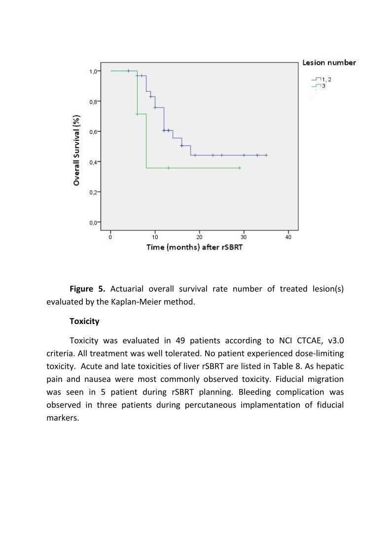
Figure 5. Actuarial overall survival rate number of treated lesion(s)
evaluated by the Kaplan-Meier method.
Toxicity
Toxicity was evaluated in 49 patients according to NCI CTCAE, v3.0
criteria. All treatment was well tolerated. No patient experienced dose-limiting
toxicity. Acute and late toxicities of liver rSBRT are listed in Table 8. As hepatic
pain and nausea were most commonly observed toxicity. Fiducial migration
was seen in 5 patient during rSBRT planning. Bleeding complication was
observed in three patients during percutaneous implamentation of fiducial
markers.

Table 8. Toxicity evaluation of rSBRT based on NCI CTCAE, v3.0 criteria.
Follow up time
During
rSBRT
Acute
Late
Toxicity Grade
Grade
1,2
Grade
3,4
Grade
1,2
Grade
3,4
Grade
1,2
Grade
3,4
Vomiting 3 1 4 2
Nausea 5 1 1
Diarrhea 3 1
Dermatitis 3 1
Bleeding 3
Pain 5 4 3
Gastritis 1
Gastro duodenal 1
Fiducial
migration 5
Evaluated # of
patients 41 32 25
Acute toxicity= Toxicity ≤3 months, Late toxicity=Toxicity >3 months
DISCUSSION
Liver is a common site of metastatic disease. The local control of
oligometastatic disease might lead to improved systemic control. Several series
have reported the outcomes after surgical resection of liver metastases (6,7,
23). But only a small portion of patients with liver metastases are candidates
for surgical resection. In surgical series, resection of metastases increases
median survival to approximately one year(4). Patients with central lesions
have worst prognosis and the fewest therapeutic options. Other local ablative
techniques or systemic therapies provide for a number of treatment options
(11-15).
SBRT for liver metastases has been described in only a few studies (16-
19,24). rSBRT techniques have been increasingly applied to the treatment of
liver metastases within the last decade, further refining the ability to target the
tumor and minimize toxicity to normal tissue. It is possible to decrease the rate
of toxic liver complications/radiation-induced liver disease and increase the
radiation dose to cancerous tissue, thereby allowing for better local control.
These studies show that high local control rates can be achieved in patients
with multiple liver metastases. In our study, the actuarial 1-year and 2-year
local control rates were 61% (%95 confidence interval (CI), 53%-69%) and 39%
(95% CI, 28%-49%), respectively. These results are quite similar to previously
published series (5,17-19). Different dose and fraction schema was tested in all
these studies(16-19). At the beginning of our rSBRT liver experience, our
prescribed dose to the tumor was 24 Gy or 30 Gy in three fraction, according to
number of the tumor and it’s dimensions. After 2007, we increased prescribed
dose to the tumor and used 45 Gy in three fraction. We could say that,
expecially infield recurrence rate was reduced and our local control rate was
slightly improve. Also, in our study show that, age, number of treated lesion
and number of fraction was statically important for local control according to
Cox regression multivariate analysis. Patients with >60 years old and 1 or 2
metastases and patients who received ≤3 fractions had better local control
according to our statical results.
In this study, the median overall survival was 16 months and this result is
similar to published series (17-19). The patients in our series treated with rSBRT
had an actuarial 1-year and 2-year overall survival rates were 79% (%95 (CI),
73%-85%) and 56% (95% CI, 48%-63%), respectively. These results agree with
literature which was reported by Wulf et al (25) (1-year and 2-year overall
survival rates were 76% and 61%, respectively). Some factors such as patient
selection, additional metastases to other organs, etc. could adversely affect the
local control and survival. Both Cox regression multivariate analysis for local
control and survival show that number of treated lesion(s) is one the important
parameter effecting treatment result. Data from this study show that tumor
dimension is not effect local control or survival. There were no significant
difference between tumors which were ≤30 mm or >30 mm in local control
and survical rates. The effect of primary tumor histology and receiving systemic
therapy were also observed in this study but these results were not significant.
These parameters maybe need re-evaluation with larger patient populations in
further studies.
There are now a number of Phase I and II studies available for patients
with liver metastases. These results are comparable to the best results
achieved studies on RFA. Also advances in imaging technologies are improving
our ability to deliver rSBRT more safety and increase effectiveness. Using these
imaging techniques such as MRI or PET CT during in contouring process could
decrease geographic miss and reduce infield or tumor border recurrence. One
of the negative points of this study is having small number of patients.
Therefore, results which were present here need confirmation in a larger
number of patient series.
CONCLUSION
The results of our study have show that rSBRT for the treatment of liver
metastases is feasible and safe with minimal side effects. Further studies are
required with larger patient series to compare rSBRT with other local ablative
techniques and systemic therapies.
REFERENCES
1.Ferlay J.,Shin H.R., Bray F., et al.,Estimates worldwide burden of cancer in
2008, International Journal of Cancer, vol 127, no.12, 2893-2917
2.Lau W.Y, Lai E.C., Hepatocellular carcinoma:current management and recent
advances, Hepatobiliary Pancreat Dis Int 7, 237-257, 2008
3. Wei A.C, Greig P.D., Grand D., et.al, Survival after hepatic resection for
colorectal metastases: a 10-year experience, Ann Surg Oncol,13,668-676, 2006
4. Yoon S.S., Tanabe K.K., Surgical treatment and other regional treatments for
colorectal cancer liver metastases. Oncologist, 4(3),197–208, 1999
5. Alan W.K.,Madeline C.S.,Ann G.M.,et al, Hypofractioned stereotactic body
radiation therapy (SBRT) for limited hepatic metastases, Int J Radiation
Oncology Biol Phys, 67 (3), 793-798, 2007
6. Adam R, Vinet E, Regional treatment of metastasis surgery of colorectal liver
metastases, Annals of Oncology 15 (Supp 4), iv 103-iv 106, 2004

7. Scheele J, Stang R, Altendorf-Hofmann A, et al, Resection of colorectal liver
metastases, World J Surg, 19, 59-71, 1995
8.Claire V.D.,Sylvain D., Francois B, et al, Image-guided robotic stereotactic
body radiation therapy for liver metastases: Is there a dose response
relationship?, Int J Radiation Oncology Biol Phys, 81 (3), e39-e47, 2011
9.Doci R.,Gennari L.,Bignami P., et al, Morbidity and mortality after hepatic
resection of metastases from colorectal cancer, Br J Surg, 82, 377-381, 1995
10. Fong Y., Fortner J., Sun R.L., et al, Clinical score for predicting,recurrence
after hepatic resection for metastatic colorectal cancer: analysis of 1001
consecutive cases, Ann Surg, 230, 309-318, 1999
11. Bokemeyer C., Bondarenko I., Makhson A., et al, Fluorouracil, leucovorin
and oxaliplatin with and without cetuximab in the first-line treatment of
metastatic colorectal cancer, J Clin Oncol, 27(5), 663-671, 2009
12. Meyerhardt J.A.,Mayer R.J., Drug therapy: Systemic therapy for colorectal
cancer, N Engl J Med , 352, 476-487, 2005
13. Hurwitz H., Fehrenbacher L., Novotny W., et al, Bevacizumab plus
irinotecan, fluorouracil and leucovorin for metastatic colorectal cancer, New
Engl J Med, 350, 2335-2342, 2004
14. de Baere T., Elias D., Dromain C., et al, Radiofrequency ablation of 100
hepatic metastates with a mean follow-up of more than 1 year, AJR Am J
Roentgenol, 175, 1619-1625, 2000
15. Shibata T., Niinobu T., Ogata N., et al, Microwave coagulation therapy for
multiple hepatic metastases from colorectal carcinoma, Cancer, 89(2), 276-284,
2000
16. Karyn A.G., Ellen A.W., Katherine E.M., et al, Dose-escalation study of
single-fraction stereotactic body radiotherapy for liver malignancies, Int J
Radiation Oncology Biol Phys, 78 (2), 486-493, 2010
17. Giovanni A., Francesco P., Giorgio C., et al, Image-guided robotic
stereotactic radiosurgery for unresectable liver metastases: Preliminary results,
Anticancer Research, 29, 3381-3384, 2009

18. Stintzing S., Hoffman R.T., Heinemann V., et al, Frameless single-session
robotic radiosurgery of liver metastases in colorectal cancer patients, Eur J
Cancer, 46(6),1026-1032, 2010
19. Schefter T.E., Kavanagh B.D., Timmerman R.D., et al, A phase I trial of
stereotactic body radiaition therapy (SBRT) for liver metastases, Int J Radiation
Oncology Biol Phys, 62 (5), 1371-1378, 2005
20. Pan C.C., Kavanagh B.D., Dawson L.A., et al, Radiation-associated liver
injury, Int J Radiation Oncology Biol Phys, 76 (3), supp. S94-100, 2010
21. Therasse P., Arbuck S.G., Eisenhauer E.A., et al, New guidelines to evaluate
the response to treatment in solid tumors, J Natl Cancer Inst, 92(3), 205-216,
2000
22. Common Terminology Criteria for Adverse Events (CTCAE) v3.0
http://www.eortc.be/services/doc/ctc/ctcaev3.pdf
23. Bozzetti F., Cozzaglio L., Baracci P., et al, Comparing surgical resection of
limited hepatic metastases from colorectal cancer to non-operative treatment.,
Eur J Surg Oncol, 19, 162-167, 1993
24. Wulf J., Guckenberger M., Haedinger U., et al, Stereotactic radiotherapy of
primary liver cancer and hepatic metastases, Acta Oncol, 45, 838-847, 2006
25. Wulf J., Hadinger U., et al, Stereotactic radiotherapy of targets in the lung
and liver, Strahlenther Onkol, 177, 645-655, 2001
26. Stintzing S., Hoffmann R.T., Heinemann V., et al, Radiosurgery of liver
tumors: value of robotic radiosurgical device to treat liver tumors., Ann Surg
Oncol. 2010 Nov,17(11),2877-2883, Epub 2010 Jun 24.