robust iris recognition in unconstrained environments
TRANSCRIPT

Journal of AI and Data Mining
Vol 7, No 4, 2019, 495-506 DOI: 10.22044/JADM.2019.7434.1884
Robust Iris Recognition in Unconstrained Environments
A. Noruzi1, M. Mahlouji2,*and A. Shahidinejad 1
1. Department of Computer Engineering, Qom Branch, Islamic Azad University, Qom, Iran.
2. Department of Electrical & Computer Engineering, Kashan Branch, Islamic Azad University, Kashan, Iran.
Received 06 September 2018; Revised 22 February 2019; Accepted 08 May 2019
*Corresponding author: [email protected] (M. Mahlouji).
Abstract
A biometric system provides the automatic identification of an individual based on a unique feature or
characteristic possessed by him/her. Iris recognition (IR) is known to be the most reliable and accurate
biometric identification system. The iris recognition system (IRS) consists of an automatic segmentation
mechanism, which is based on the Hough transform (HT). The IRS is divided into six stages including imaging,
pre-processing, segmentation, normalization, feature extraction and matching. Through this method, first, a
photo is taken from the iris, and then edge detection is done. Later, on a contrast, adjustment is persecuted in
the pre-processing stage. Circular HT is subsequently utilized for localizing circular area of the iris inner and
outer boundaries. Also, through applying parabolic HT, boundaries are localized between the upper and lower
eyelids. The proposed method, in comparison with the available IRSs, not only enjoys a higher accuracy, but
also competes with them in terms of the processing time. Experimental results on the images available in the
UBIRIS, BATH, CASIA, and MMUI databases show that the proposed method has an accuracy rate of
99.12%, 97, 98%, 98.80%, and 98.34%, respectively.
Keywords: Hough Transform, Biometric Identification, Segmentation, Normalization, Matching.
1. Introduction
The human iris contains rich texture, which is
highly stable and distinctive. Iris recognition (IR)
is a biometric technology that utilizes pattern
recognition techniques on the basis of iris high
quality images, and has become one of the most
promising technologies for biometric
authentication. The existing state-of-the-art IR
algorithms have achieved remarkable
performances, since in comparison with other
features utilized in biometric systems, iris patterns
are more stable and reliable, and IR is known to be
one of the most outstanding biometric technologies
[1]. IR for iris images, which are taken distantly
from the sensor, is a major challenge of the
biometric platform. Additionally, in an
unconstrained environment, iris may have
occlusions caused by the upper or lower eyelids or
eyes may be rolling left or right. In this paper, we
try to address such issues. The algorithm has been
proposed in [2], where one method stage accurately
localizes the iris by a model designed on the basis
of the Histograms of Oriented Gradient (HOG)
descriptor and Support Vector Machine (SVM),
namely HOG-SVM. Based on this localization, iris
texture is automatically extracted by means of a
cellular automata that evolved through the Grow-
Cut technique. Daugman’s [3] and Wildes’ [4]
systems are the two earliest and most famous iris
recognition systems (IRSs) containing all IR stages.
In the Daugman’s algorithm, an iris with two
circles that are not necessarily concentric forms the
model. Each circle is defined by three parameters
(𝑥0, 𝑦0, 𝑟) in a way that (𝑥0, 𝑦0) determines the
center of a circle with the radius of 𝑟. An integro-
differential operator is used to estimate the values
of the three parameters for each circular boundary,
and the whole image is searched in relation to the
increment of radius 𝑟. In Wildes’ system, gradient-
based Hough transform (HT) has been used to
localize two iris circular boundaries. This system
consists of two stages. First, a binary map is
produced from the image edges by a Gaussian
filter. Then an the analysis is performed in a
circular Hough space in order to estimate the three

Noruzi et al. / Journal of AI and Data Mining, Vol 7, No 4, 2019.
496
parameters (𝑥0, 𝑦0, 𝑟) for a circle. In [5], iris
images are projected vertically and horizontally to
estimate the center of the iris. IRS usually consists
of three component stages: iris segmentation,
feature extraction, and iris matching. Currently, iris
segmentation and feature extraction for the
captures in less-constrained environments have
been extensively investigated in [6]. The algorithm
in [7] proposes a computationally efficient iris
segmentation approach for segmenting iris images
acquired from at-a-distance and under less-
constrained imaging conditions. The proposed
segmentation approach is developed based on the
cellular automata that evolve through using Grow-
Cut algorithm. Also an IRS has been proposed in
[8], which is used for frontal iris images and for an
iris image that is not taken from a frontal view. In
this system, when a frontal image is not available
for a particular individual, the issue is addressed
via maximizing Hamming distance (HD) between
the two mentioned images or by minimizing
Daugman’s integra-differential operator, and then
the image is transformed into a frontal image. An
algorithm is presented to find eyelash and eyelids
occlusions on iris in a completely close-up image
similar to Daugman’s method in [9]. The algorithm
proposed in [10] describes an efficient way for IR
by characterizing the key local variations. The
basic idea is that local sharp variation points, which
denote the appearing or vanishing of an important
image structure, should be utilized to represent the
characteristics of an iris. The paper [11] proposed
a novel texture feature for IR. During pre-
processing, iris is segmented using constrained
circular HT, which reduces both time-and space-
complexity. Through this, a novel texture code
matrix is generated, which is then used to obtain a
co-occurrence matrix. The algorithm in [12]
proposed a reliable iris localization algorithm. It
includes localizing a coarse iris location in the eye
image using the HT and image statistics; localizing
the pupillary boundary using a bi-valued adaptive
threshold and the two-dimensional (2D) shape
properties; localizing the limbic boundary by
reusing the Hough accumulator and image
statistics; and regularizing these boundaries using a
technique based on the Fourier series and radial
gradients. In [13], it is suggested that data reduction
has the highest impact on the iris segmentation.
Since segmentation is also the first step in the
pipeline, this potentially affects the performance of
later steps as well, and is therefore of particular
importance. Reference [14] introduces a noise-
resistant and computational efficient segmentation
approach towards less constrained IR. The
proposed segmentation approach is based on a
modified and fast HT augmented with a newly
developed strategy to define iris boundaries with
multi-arcs and multi-lines.
The remainder of this paper is organized as
follows. In Section 2, the proposed method of IR is
introduced, and next, in Section 3, experimental
results of the proposed method on several
databases are presented, and finally, in Section 4,
conclusions are drawn.
2. Proposed method for iris recognition
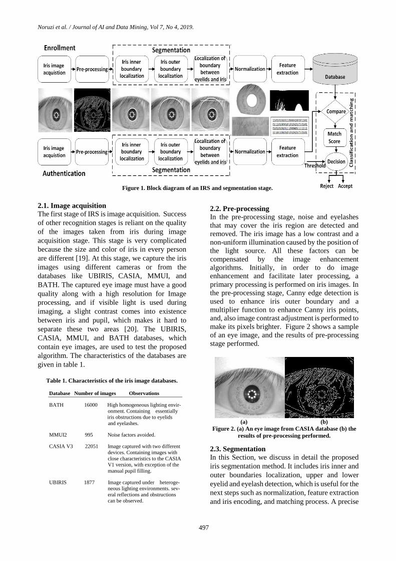
Figure 1 shows the block diagram for a biometric
system of IR in unconstrained environments in
which the function of each block is briefly
discussed as follows:
1) Image acquisition
At this stage, we capture the iris images
using different cameras or from the
databases like UBIRIS [15], CASIA [16],
MMUI [17], and BATH [18].
2) Pre-processing
Involving edge detection, contrast
adjustment, and multiplier.
3) Segmentation
The iris part is detected by eliminating the
upper and lower boundaries of eyelids, and
eyelashes and central part of pupil
boundary is done using the HT and Canny
edge detection technique.
4) Normalization
Segmented iris converted from the circular
region (polar) into a rectangular region
(cartesian) and normalization of iris image.
5) Feature extraction
The normalized iris is converted into
binary bit format using the Gabor filter
technique and noise removal from iris
image.
6) Classification and matching
The difference between the 2 codes (iris
input code and database eye image
template code) is calculated using HD, and
left and right shift operations are
performed in order to get similarity
between two images, and decision is made
like Match/No match based on the
matching score.
Noruzi et al. / Journal of AI and Data Mining, Vol 7, No 4, 2019.
497
2.1. Image acquisition The first stage of IRS is image acquisition. Success
of other recognition stages is reliant on the quality
of the images taken from iris during image
acquisition stage. This stage is very complicated
because the size and color of iris in every person
are different [19]. At this stage, we capture the iris
images using different cameras or from the
databases like UBIRIS, CASIA, MMUI, and
BATH. The captured eye image must have a good
quality along with a high resolution for Image
processing, and if visible light is used during
imaging, a slight contrast comes into existence
between iris and pupil, which makes it hard to
separate these two areas [20]. The UBIRIS,
CASIA, MMUI, and BATH databases, which
contain eye images, are used to test the proposed
algorithm. The characteristics of the databases are
given in table 1.
Table 1. Characteristics of the iris image databases.
Database Number of images Observations
BATH 16000 High homogeneous lighting envir-
onment. Containing essentially iris obstructions due to eyelids
and eyelashes.
MMUI2 995 Noise factors avoided.
CASIA V3 22051 Image captured with two different
devices. Containing images with
close characteristics to the CASIA V1 version, with exception of the
manual pupil filling.
UBIRIS 1877 Image captured under heteroge- neous lighting environments. sev-
eral reflections and obstructions
can be observed.
2.2. Pre-processing
In the pre-processing stage, noise and eyelashes
that may cover the iris region are detected and
removed. The iris image has a low contrast and a
non-uniform illumination caused by the position of
the light source. All these factors can be
compensated by the image enhancement
algorithms. Initially, in order to do image
enhancement and facilitate later processing, a
primary processing is performed on iris images. In
the pre-processing stage, Canny edge detection is
used to enhance iris outer boundary and a
multiplier function to enhance Canny iris points,
and, also image contrast adjustment is performed to
make its pixels brighter. Figure 2 shows a sample
of an eye image, and the results of pre-processing
stage performed.
(a) (b)
Figure 2. (a) An eye image from CASIA database (b) the
results of pre-processing performed.
2.3. Segmentation
In this Section, we discuss in detail the proposed
iris segmentation method. It includes iris inner and
outer boundaries localization, upper and lower
eyelid and eyelash detection, which is useful for the
next steps such as normalization, feature extraction
and iris encoding, and matching process. A precise
Figure 1. Block diagram of an IRS and segmentation stage.

Noruzi et al. / Journal of AI and Data Mining, Vol 7, No 4, 2019.
498
iris image segmentation plays an important role in
an IRS since success of the system in upcoming
stages is directly dependent on the accuracy of this
stage [21]. Figure 1 shows block of the
segmentation stage and includes the 3 following
stages:
1) Localization of iris inner boundary.
2) Localization of iris outer boundary.
3) Localization of the boundary between
eyelids and iris.
HT is a standard computer vision algorithm that
can be used to determine the parameters of simple
geometric objects, such as lines and circles present
in an image. The circular HT can be employed to
deduce the radius and center coordinates of the
pupil and iris regions [22]. These parameters are
the center coordinates 𝑥𝑐 and 𝑦𝑐, and the radius 𝑟,
which are able to define any circle according to the
following equation:
𝑥𝑐2 + 𝑦𝑐
2 − 𝑟2 = 0 (1)
The parabolic HT is used to detect the eyelids. The
mathematical relationship used to localize the
upper and lower eyelids is gained with parabolic
arcs, which is represented as:
(−(𝑥 − ℎ𝑗)𝑠𝑖𝑛𝜃𝑗 + (𝑦 − 𝑘𝑗)𝑐𝑜𝑠𝜃𝑗)𝑛 = 𝑅 (2)
where in
𝑅 = 𝑎𝑗 ((𝑥 − ℎ𝑗)𝑐𝑜𝑠𝜃𝑗 + (𝑦 − 𝑘𝑗)𝑠𝑖𝑛𝜃𝑗) (3)
where 𝑎𝑗 controls the curvature, (ℎ𝑗, 𝑘𝑗) is the peak
coordinates of the parabola, and 𝜃𝑗 is the angle of
rotation relative to the 𝑥 –axis.
2.3.1. Iris inner boundary localization
Since pupil is a black circular region and darker
compared with the iris, it is easy to detect the pupil
inside an eye image. Firstly, pupil is detected using
the thresholding operation. An appropriate
threshold is selected to generate the binary image
that contains pupil only. Since the inner boundary
of an iris can be approximately modelled as circles,
circular HT is used to localize the iris. Firstly,
Canny edge detection is applied to binary image to
generate the edge map. Figure 2 shows the results
of performing Canny edge detection on an eye
image as the pre-processing output. Figure 3 shows
the iris inner boundary that has been achieved via
this method for three eye images. As it is
observable in figure 3, the method yields a
boundary localized with a high accuracy.
Figure 3. Iris inner boundary localized for three eye
images.
2.3.2. Iris outer boundary localization
Too much blurring may dilate the boundaries
of the edge or may make it difficult to detect
the outer iris boundary, separating the eyeball
and sclera. Thus a special smoothing filter such as
the median filter is used on the original intensity
image. This type of filtering eliminates sparse
noise, while preserving image boundaries. After
filtering, the contrast of image is enhanced to
have sharp variation at image boundaries using
histogram equalization. As a result, edge detection
algorithms, which are able to detect outer iris
edges, identify those points as edge. Therefore, in
order to detect the iris outer boundary, these points
have to be identified and eliminated. In this work,
the available boundaries are initially enhanced, and
then extra edge points are identified and
eliminated. At the end, through circular HT, the
outer iris boundary is obtained. In order to enhance
iris the outer boundary edges, Canny edge
detection is performed on eye image in the pre-
processing stage. By performing such edge
detection, a matrix is obtained with the same
dimensions as of the image itself whose elements
are high in areas where there is a definite boundary
and the elements are low in areas where there is no
perfectly definite boundary, such as iris outer
boundary. Through multiplying of 2.76 in the
matrix of pixel values of iris image and intensifying
light in eye image, the edges are enhanced.
Applying Canny edge detection and multiplying
that to the constant value of 2.76 result in a better
revelation of iris outer boundary edge points. It
gains number 2.76 by trial and error, and by a little
change, there is no accurate boundary recognition.
Table 2 displays the accuracy rate of iris outer
boundary localization for different values
multiplying on three databases. As it is illustrated,
with the multiplying value of 2.76, the most
optimum accuracy rate in the iris outer boundary
localization will be evident. Results of such
application on three eye images are shown in figure
4. As it is observable in figure 4, the method yields
a boundary localized with a high accuracy.

Noruzi et al. / Journal of AI and Data Mining, Vol 7, No 4, 2019.
499
Table 2. Accuracy rate of iris outer boundary localization
for different values multiplying on three databases.
Value CASIA V1 (%) CASIA V3 (%) BATH (%)
1.50 87.12 86 87.10
2.00 93.37 92.12 93
2.76 99 98.19 98.17
3.00 94.12 94 92.38
3.50 91.73 90.67 89.34
Figure 4. Iris outer boundary localized for three eye
images.
The only issue of this method is sclera boundary
not being circular, which is the result of angled or
sideward imaging, and in these cases, some
information is lost or clutter comes into existence.
In this stage, after identifying the iris inner and
outer boundaries, the results of these three stages
are combined. Figure 5 shows the results obtained.
As it could be seen in this figure, the iris inner and
outer boundaries are correctly identified in the
CASIA iris image-interval database.
Figure 5. Iris inner and outer boundaries localized for
three eye images.
2.3.3. Localization of boundary between eyelids
and iris
Similar to the iris outer boundary localization, the
proposed method selects two search regions to
detect the upper and lower eyelids. The pupil
center, iris inner, and outer boundaries are used as
reference to select the two search regions. Canny
edge detection is applied to the search regions to
detect the eyelids. In order to reduce the false edges
detection caused by eyelashes, Canny filter is tuned
to the linear direction. After the edge detection
step, the edge image is generated. The eyelids are
detected using the parabolic HT method. The
method calculates the total number of edge points
in every linear row inside the search regions. The
linear row with maximum number of edge points is
selected as the eyelid boundary. If the maximum
number of edge points is less than a predefined
threshold, it is assumed that eyelid is not presented
in the search regions. As it could be seen in figure
2, there are only pupillary edge points between the
two eyelids, and since pupillary boundary has
already been obtained, these points are eliminated.
Figure 6 shows few boundaries localized through
this method for some eye images. This method
could result in a false outcome only for some
images that have too many patterns in iris tissue
when the edges of these patterns are detected by
Canny edge detection. As it is observable in figure
6, the method localizes eyelids with a relatively
high accuracy. Figure 6 exhibits samples of eye
images in which eyelids are parabolic and linearly
shaped. The boundaries of eyelids and iris are
recognized properly but for those images in which
eyelids are parabola shaped, this boundary is
recognized with a slight discrepancy. The accuracy
rate of the proposed method for the segmentation
stage on different databases is presented in table 3.
As the results presented in this table show, the
method has an accuracy rate of between 97.6% and
99.63% for iris boundary localization.
Figure 6. Boundaries between iris and eyelids localized
for three eye images.
For the CASIA V1 and V3 databases, the
segmentation technique managed to correctly
segment the iris region from 747 out of 756 eye
images, which corresponds to a success average
rate of around 98.80%. The BATH images proved
problematic and the segmentation process correctly
identified iris and pupil boundaries for only 117 out
of 120 eye images, which corresponds to a success
average rate of around 97.98%. It is difficult to
recognize the inner and outer iris boundaries for
those images with small illumination intensity
differences between the iris region, and the pupil
region as shown in figure 7. For solving the
problem, important parameters in imaging such as
minimum and maximum radius of iris and pupil to
search, threshold values for creating edge maps,
camera hardware, imaging distance, and lighting
conditions for each database, must be adjusted.
The eyelid detection system also proved quite
successful, and managed to isolate the most
occluding eyelid regions. One problem was that it
would sometimes isolate too much of the iris
region, which could make the recognition process
less accurate, since there is less iris information.
However, this is preferred over including too much

Noruzi et al. / Journal of AI and Data Mining, Vol 7, No 4, 2019.
500
of the iris region, if there is a high chance it would
also include undetected eyelash and eyelid regions.
The eyelash detection system implemented for the
CASIA database also proved to be successful in
isolating most of the eyelashes occurring within the
iris region, as shown in figure 6.
Table 3. Accuracy rate (AR) of iris boundary localization
(BL) for three databases. Iris Database AR in pupil AR in sclera AR in eyelids BL (%) BL (%) BL (%)
CASIA Iris
Image V1 98.13 99 99.31
CASIA Iris
INTERVAL V3 99.63 98.19 98.58
University of BATH 98.18 98.17 97.60
Figure 7. An example where segmentation fails for three
eye images from BATH database
A slight problem was that areas where the
eyelashes were light, such as at the tips that were
not detected. However, these undetected areas were
small when compared with the size of the iris
region. Isolation of specular reflections from eye
images in the MMUI database also proved to be
successful. Numerous examples of their isolation
are shown in figure 8.
Figure 8. Automatic segmentation of various images from
the MMUI database.
2.4. Normalization
For normalization of iris regions, a technique based
on Daugman’s rubber sheet model was employed.
The center of the pupil was considered as the
reference point, and radial vectors passed through
the iris region. A number of data points are selected
along each radial line, and this is defined as the
radial resolution. The number of radial lines
existing around the iris region is defined as the
angular resolution. Since the pupil can be non-
concentric to the iris, a remapping formula is
required to rescale points depending on the angle
around the circle. This is given by:
𝑟′ = √𝛼𝛽 ± √𝛼𝛽2 − 𝛼 − 𝑟𝐼2 (4)
where in
𝛼 = 𝑜𝑥2 + 𝑜𝑦
2 (5)
β= cos (𝜋 − arctan (𝑂𝑦
𝑂𝑥)− θ) (6)
where displacement of the center of the pupil
relative to the center of the iris is given by 𝑜𝑥, 𝑜𝑦,
and 𝑟′ is the distance between the edge of the pupil
and edge of the iris at an angle, θ around the region,
and 𝑟𝐼 is the radius of the iris. The remapping
formula first gives the radius of the iris region
‘doughnut’ as a function of the angle θ. Figure 9
shows transforming iris area from the polar to
Cartesian coordinates. Therefore, iris area is
obtained as a normalized strip with regard to iris
boundaries and pupillary center. In this work, iris
area is illustrated on a rectangular strip of 8*512
[23][26-27].
θ , 512 pixels
r ,
8 p
ixe
lsr
θ
OXC-Pupil
C-Iris
Oy
Figure 9. Transforming polar to Cartesian coordinates.
In order to transform iris area from polar to
Cartesian coordinates, 128 pupils-centered perfect
circles are chosen starting from iris-pupil
boundary, and then the pixels are located on these
circles that are next mapped into a rectangle (to
change polar coordinates into the Cartesian). As a
result, iris area, which looks like a circular strip, is
converted into a rectangular strip. Choosing these
128 perfect circles normalizes iris in terms of size
as well. Then illumination intensity was adjusted in
segmented iris tissue, i.e. image contrast was
applied to bring more clarity into iris tissue. Figure
10 shows a sample of normalized iris tissue [3].
Figure 10. Transforming iris area into normalized
rectangular strip.

Noruzi et al. / Journal of AI and Data Mining, Vol 7, No 4, 2019.
501
2.5. Feature extraction and iris encoding
2D Gabor filters are used to extract iris features
from the normalized iris image. A 2D Gabor filters
is a Gaussian transfer function on a logarithmic
scale [23]. It has strictly band pass filter to remove
the caused background brightness and an ultimate
feature vector is obtained. In this stage, the
normalized iris is encoded in the form of binary bits
(0, 1). In the coding system, the normalization
image is converted to Gabor filter stage and Fourier
for gaining iris image of feature extraction. Then
iris image tissues become quantization, and finally,
Quantization parameters are converted to binary.
The dimensions of the feature vector extracted
from iris area have to be as small as possible. Since
vectors with large size, in respect of spatial, have
high mass for saving, moreover, they have very
high calculation mass in feature extraction and
matching stage forcing system and decelerate. It
regarding high dimensions of the image drawn,
Gabor wavelet transform was performed in order to
decrease the dimensions in the way that important
information existing in tissue can be preserved in
spite of downsizing image dimensions [14]. To do
this, 2D Gabor wavelet transforms were conducted.
Then the encoding obtained in this stage would be
saved in dimensions of 80*240, and then enters the
next stage of the system, namely the matching
stage. Regarding that some sections of the area
chosen for feature extraction may have occlusions
caused by eyelids and eyelashes and since it is
possible that because of error in segmentation stage
some parts of sclera are subjected to be detected as
iris area, it is required that a measure be taken to
remove these points from the feature extraction
stage. To resolve the latter issue that is caused by
error when detecting iris outer boundary, 20% of
the lower section of the image is eliminated, and to
resolve the first issue, points of the image that are
placed in this section are eliminated from encoding.
To do this, a binary encoding that detects occlusion
points is produced. This encoding is implied in the
matching stage, and these points are eliminated in
that stage [25].
2.6. Classification and matching
For matching, HD was chosen as a metric for
recognition, since bit-wise comparisons were
necessary. The HD algorithm includes calculation
of differences for the 2 codes and the noise masking
in a way that only significant bits are used in
calculating the HD between two iris template
codes. Now when taking HD, only those bits in the
iris pattern that correspond to ‘0’ bits in noise
masks of both iris patterns will be used in the
calculation. HD will be calculated using only the
bits generated from the true iris region, and this
modified HD formula is given as
𝐻𝐷 =1
𝑁 − ∑ 𝑋𝑛𝑘(𝑂𝑅)𝑁
𝐾=1 𝑌𝑛𝑘
∗ 𝑀 (7)
where in
𝑀 = ∑ 𝑋𝑗(𝑋𝑂𝑅)
𝑁
𝑗=1
𝑌𝑗(𝐴𝑁𝐷)𝑋′𝑛𝑗
(𝐴𝑁𝐷)𝑌′𝑛𝑗
(8)
where, 𝑋𝑗 and 𝑌𝑗 are the two bit-wise template
codes to compare, 𝑋′𝑛𝑗
and 𝑌′𝑛𝑗
are the
corresponding noise masks for 𝑋𝑗 and 𝑌𝑗, while N
is the number of bits represented by each template
code. If the value for feature vector in point (𝑋, 𝑌)
is equal to the value for other feature vector in that
point, digit 1 is allotted to that point, and if they are
not equal, digit 0 is done. Then the values allotted
to the pixels are summed up and similarity criterion
of the two images is attained. One important factor
in the matching stage is rotation of the incoming
image. With respect to the position of the
individual’s head at the time of imaging, it may be
that the individual’s head and eye direction are in
different positions each time, and that causes the
imbalance of the images taken from eyes. To
resolve rotation issue, feature vector is rotated and
relocated, and encoding is done for several images
obtained through rotating original image instead of
just one image, and matching is done for all of
them, and then the lowest value, among other
calculated values, is considered as the matching
criterion.
Because iris circular strip in the normalization
stage is transformed to the horizontal strip,
movement is on the horizontal direction on
encoding equivalent with iris area rotation, and
rotation direction is related to movement direction
on the encoding. Since the 360 degrees of iris
section are registered as a 512 strip, movement of
2 bits on horizontally- registered strip equals
rotation of 90 degrees in iris section. Figure 11
displays iris encoding transference to the left and
right sides, and finding the best matching of HD as
well.
3. Experimental results
In various databases, a perfect recognition is not
possible due to the overlapping distributions.
Hence, at first, it must mark value threshold. As it
is clear in table 4, for value threshold 0.4, false
accept rate (FAR) and false reject rate (FRR)
0.12%, 1.08% are obtained, respectively. This
value among the values in table 4 in respect of

Noruzi et al. / Journal of AI and Data Mining, Vol 7, No 4, 2019.
502
FAR, FRR is the best value threshold. Therefore, if
two irises are identical, their HD value must be
below 0.4 and if two irises are distinctive, their HD
value must approximate or exceed 0.4. Efficiency
of a biometric system is usually evaluated by taking
into account FAR and FRR.
Figure 11. Transference of iris encoding to left and right
side and finding best matching of HD.
Table 4. FAR and FRR for the CASIA database with
different value threshold using the optimum parameters.
Threshold FAR (%) FRR (%)
0.20 0.000 99.047
0.25 0.000 82.787
0.30 0.000 37.880
0.35 0.000 5.181
0.40 0.12 1.08
0.45 7.599 0.000
0.50 99.499 0.000
Evaluated as more efficient, FAR means how many
people the system mistakenly accepts when they
introduce someone instead of others. FRR means
how many people with entrance allowance are not
recognized by the system and are announced as
errors.
The FAR rate is calculated in off-class distribution
of 𝑃𝑑𝑖𝑓𝑓 regarding the normalized area between
zero and K value threshold. The FRR rate equals
the normalized area between value threshold and a
point in in-class distribution of 𝑃𝑠𝑎𝑚𝑒.
𝐹𝐴𝑅 =∫ 𝑃𝑑𝑖𝑓𝑓
𝐾
0(𝑥)𝑑𝑥
∫ 𝑃𝑑𝑖𝑓𝑓 1
0(𝑥)𝑑𝑥
(9)
𝐹𝑅𝑅 =∫ 𝑃𝑠𝑎𝑚𝑒
1
𝑘(𝑥)𝑑𝑥
∫ 𝑃𝑠𝑎𝑚𝑒1
0(𝑥)𝑑𝑥
(10)
In table 5, the results of the proposed method for
two different databases have gained base on
Equations (9) and (10). According to the table,
accuracy rate of the proposed method on the
CASIA database is 98.8% that is a rather proper
accuracy. The reason the low accuracy attained for
the BATH database is the very low illumination
intensity differences in iris and pupil boundary in
their images.
Table 5. FRR and FAR for two different databases with
threshold of 0.4. Iris Database FAR FRR System Accuracy (%) (%) (%)
CASIA Iris Interval 0.12 1.08 98.80
University of BATH 2.02 0 98.80
The segmentation error rate, 𝐸𝑖, per image is given
by the fraction of disagreeing pixels between the
ground-truth image and the output image produced
by the proposed method:
𝐸𝑖 =1
𝑚 ∗ 𝑛∑ ∑ 𝐶(𝑥, 𝑦) ⊗ 𝑂(𝑥, 𝑦)
𝑛
𝑦=1
𝑚
𝑥=1
(11)
where, 𝐸𝑖 is the segmentation error rate per image
𝑖, 𝐶(𝑥, 𝑦) and 𝑂(𝑥, 𝑦) are two pixels from the
ground-truth and the output images, the operator ⊗
refers to the Boolean XOR, and 𝑚 and 𝑛 are the
height and width of the image, respectively. The
overall segmentation error rate, 𝐸, is then
calculated as the average segmentation error rates
over all images:
𝐸 =1
𝑁∑ 𝐸𝑖 (12)
𝑁
𝑖=1
where, 𝑁 is the number of tested images, and 𝐸𝑖 is
the segmentation error rate per image 𝑖, which is
calculated by Equation (11). Mainly, 𝐸 ranges
between 0 and 1 interval, value 0 represents the
optimal error rate, and the worst error rate occurs
when 𝐸 equals 1. In table 6, the performance results
of several popular algorithms of IR with the
proposed method on the MMUI, UBIRIS, and
CASIA database images are presented.
As seen in the table, the accuracy rate of the
proposed method is higher than the Ma algorithm
and it is very close to the accuracy rate of the Yahya
algorithm [20]. It should be mentioned that the
reason for very high accuracy of the Daugman’s
method is their strict standards regarded at the time
of imaging. Parabolic HT is used for eyelids
localization; therefore, the speed of the proposed
0 0 1 1 0 0 1 0 0 1 1 0
0 0 1 1 0 0 1 0 0 1 1 0
1 0 0 0 1 1 0 0 1 0 0 1
0 0 1 1 0 0 1 0 0 1 1 0
0 1 1 0 0 0 1 1 0 0 1 0
0 0 1 1 0 0 1 0 0 1 1 0
Template code 1
HD = 0.83
HD=0.00
HD=0.33
2 bit Shift rotate left
2 bit Shift 2 bit Right
Template code 2
Template code 1
Template code 2
Template code 1
Template code 2
Noruzi et al. / Journal of AI and Data Mining, Vol 7, No 4, 2019.
503
algorithm is more than the speed of others such as
parabolic HT in the stage of iris localization [24].
The proposed approach is better than all the recent
methods used with the MMUI and UBIRIS
databases, and compared to the CASIA database,
only the Daugman’s method is a bit better, since
the conditions that Daugman’s considers for
imaging is a very restrictive and specific condition.
However, recent IRSS have focused on images
acquired in unconstrained environments. These
imaging environments allow the capture of iris
images at a distance, in motion, and under visible
wavelength illumination, which lead to more noise
factors such as off-focus, gaze deviation, and
obstruction by eyelids, eyeglasses, hair, lighting,
and specular reflections.
As it is evident in table 5, the proposed algorithm
on 386 different images from equal databases was
conducted and the researchers' algorithm results
were elicited from referenced tables [2, 28-30]. The
number of images and databases are equal
throughout the present contrastive comparison but
the images are not. The major purpose is to
determine the state and the rate of satisfaction of
the proposed method among the recognized
comparative algorithms. As it is evident, the
proposed method, has a significant satisfiable state
in this algorithm.
Table 6. Efficiency comparison on MMUI, UBIRIS and
CASIA database images for popular algorithms (Overall
Accuracy (OA), Segmentation Error Rate (E), UBIRIS).
Algorithm OA (%) OA (%) OA (%) E (%) E (%)
CASIA UBIRIS MMUI UBIRIS CASIA
Umer [11] 97.92 97.51 97.78 1.10 1
Ma [10] 87.27 95.79 95.11 1.63 1.75
Jan [12] 96.75 97.65 96.30 2.50 3.00
Tan [7] 98.20 93.6 ---- 1.90 1.09
Radman[2] 98.40 ---- 92.67 1.60 ----
Daugman’s[3] 99.90 97.29 90.59 1.52 0.77
Proposed
method 98.80 99.12 98.34 1.35 0.83
Provided that N number of images in the system are
registered, that is N number of images be used for
education, the accuracy of identity recognition will
differ. Table 7 presents the accuracy of the system
per number of images registered for each person.
As it is evident, with 10 images for each person, the
accuracy of identity recognition increases.
The proposed method of identity recognition was
done on 386 iris images from databases BATH and
CASIA.
Table 7. Comparing identity distinction precise of
proposed method with Daugman's method based on
register teaching picture number (N=1, 3, 5, 7, 10).
Picture Daugman’s proposed
Number (N) method (%) method (%)
1 96 96.3
3 96 96.8
5 96 97
7 96 97.6
10 96 98.80
Due to probability equality for each extracted bit
for iris being 0 or 1, if 2 codes are produced for 2
virtual irises, HD value difference will probably
equal 0.4. Therefore, if 2 irises are identical, the
HD value should be lower than 0.4 and if they are
distinctive, their HD value will approximate or
even exceed 0.4. The bigger the HD value than 0.4,
the more safely a decision could be made.
Consequently, the system success rate equals
98.80% on the CASIA database and is comparable
with other methods of IR available utilizing iris
images. The failure of 1.2% in iris segmentation
stage is due to those images in the database that
bear an extremely low color contrast intensity at the
border of iris and pupil. The system success rate
equaled 97.98% on the BATH database. The
reason for this lower accuracy for the BATH
database is that the difference of light intensity is
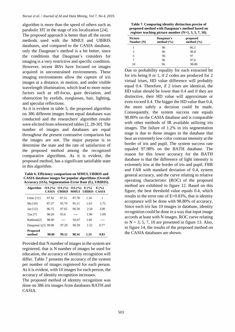
extremely low at the border of iris and pupil. FRR
and FAR with standard deviation of 0.4, system
general accuracy, and the curve relating to relative
operating characteristic (ROC) of the proposed
method are exhibited in figure 12. Based on this
figure, the best threshold value equals 0.4, which
results in the error rate of E=0.83%, that is identity
acceptance will be done with 98.80% of accuracy.
Since each iris has 10 images in database, identity
recognition could be done in a way that input image
accords at least with N images. ROC curve relating
to N = 3, 5, 7, 10 are presented in figure 13. Also,
in figure 14, the results of the proposed method on
the CASIA databases are shown.
Noruzi et al. / Journal of AI and Data Mining, Vol 7, No 4, 2019.
504
Figure 12. Curve ROC relateds to proposed method.
Figure 13. Curve ROC relateds to proposed method with
regard to adoptions numbers (N= 3, 5, 7, 10).
4. Conclusion
The accuracy of IRS is dependent on the
performance of the iris segmentation method. This
paper presented an effective and robust method of
iris segmentation for IR in unconstrained
environments by performing HT. In this method,
the pupil's boundary and the iris boundary were
localized with high accuracy. Despite variations of
illumination intensity in iris outer boundary
compared with other sections of the eye, a very
high accuracy rate was achieved for the proposed
method. Also, after recognizing the boundaries, the
iris section is separated from the human eye and
entered the normalization and feature extraction
and iris encoding. The wrong determination of iris
images will affect the normalization results. This is
due to a non-uniform distance between the inner
and outer iris boundaries. The results of examining
the method on the UBIRIS, CASIA, MMUI, and
BATH database images indicated the efficiency
and high accuracy of the proposed method, which
is comparable with other existing methods for
identity recognition using iris images. The
recognition time will not change when the iris
determination is correctly or wrongly evaluated
because it is an independent variable in the iris size.
Researchers interested in following the lines drawn
in this paper, may utilize oval-shaped model in
IRSs, utilize parabolic model in eyelid recognition,
or project IRS on GPUs with CUDA in order to
accelerate the execution.
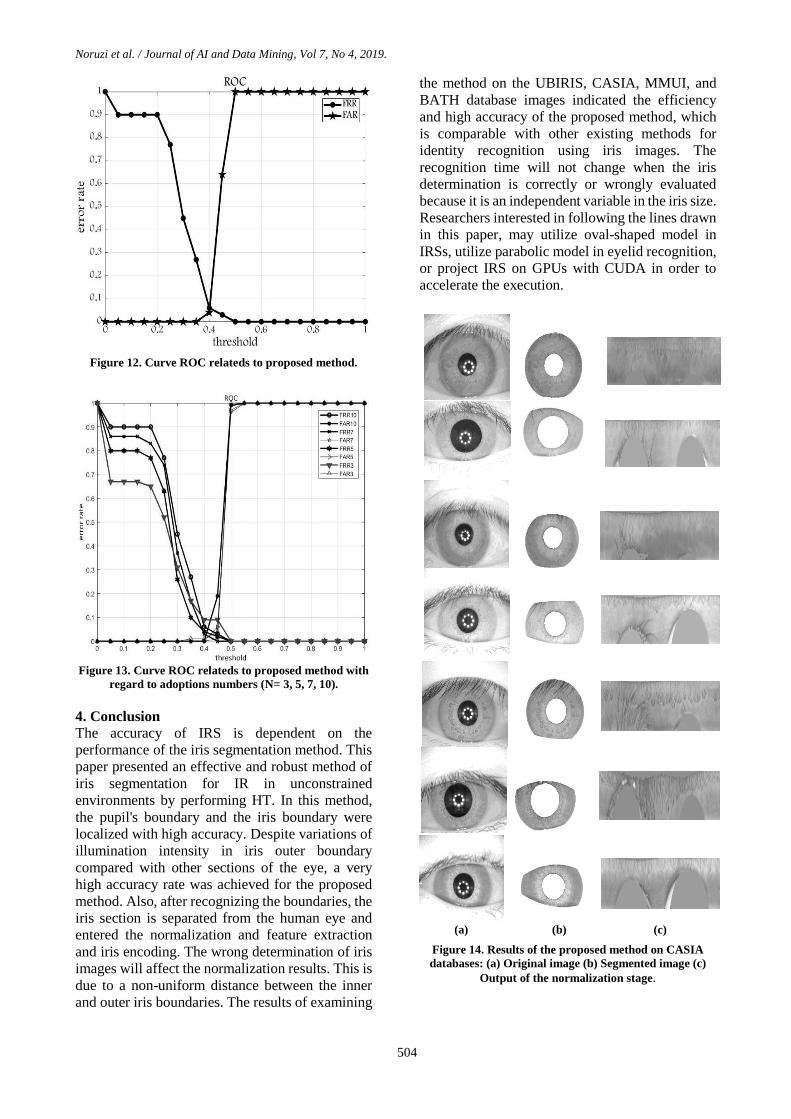
(a) (b) (c)
Figure 14. Results of the proposed method on CASIA
databases: (a) Original image (b) Segmented image (c)
Output of the normalization stage.

Noruzi et al. / Journal of AI and Data Mining, Vol 7, No 4, 2019.
505
References [1] Jain, A. K., Ross, A. & Pankanti, S. (2006). A Tool
for Information Security. IEEE Transactions on
Information Forensics and Security, vol. 1, pp. 125-143.
[2] Radman, A., Zainal, N. & Suandi, S. (2017).
Automated segmentation of iris images acquired in an
unconstrained environment using HOG-SVM and
GrowCut. Journal of Digital Signal Processing, vol. 64,
pp. 60-70.
[3] Daugman, J. G. (2007). New methods in IR. IEEE
Transactions on Systems, Man and Cybernetics, vol. 37,
pp. 1167-1175.
[4] Wildes, R. (1997). IR: an emerging biometric
technology. Proceedings of the IEEE, vol. 85, pp. 1348-
1363.
[5] Kong, W. & Zhang, D. (2001). Accurate iris
segmentation based on novel reection and eyelash
detection model. In International Symposium on
Intelligent Multimedia, Video and Speech Processing,
pp. 263-266.
[6] Hu, Y., Sirlantzis, K. & Howells, G. (2015).
Improving colour iris segmentation using a model
selection technique. Journal of Pattern Recognition, vol.
57, pp. 24-32.
[7] Tan, C. W. & Kumar, A. (2012). Efficient iris
segmentation using grow-cut algorithm for remotely
acquired iris images. In 5th IEEE International
Conference on Biometrics: Theory. Applications and
Systems (BTAS), pp. 99-104.
[8] Dorairaj, V., Schmid, A. & Fahmy, G. (2005).
Performance evaluation of iris based recognition system
implementing PCA and ICA encoding techniques. In
Proceedings of SPIE 5779, pp. 51-58.
[9] Fancourt, C., Bogoni, L., Hanna, K., Guo, Y.,
Wildes, R., Takahashi, N. & Jain, U. (2005). IR at a
distance. In Proceedings of the International Conference
on Audio and Video-Based Biometric Person
Authentication, pp. 1-13.
[10] Ma, L., Tan, T., Wang, Y. & Zhang, M. (2004).
Effcent iris recognition by characterizing key local
variations. IEEE Transactions on Image Processing vol.
13, pp. 739-750.
[11] Umer, S., Dhara, B. C. & Chanda, B. (2016).
Texture code matrix-based multi-instance IR. Springer
Pattern Ana, vol. 19, pp. 283-295.
[12] Jan, F., Usman, I. & Agha, S. (2013). Reliable iris
localization using HT. histogram-bisection and
eccentricity, Springer Signal Process, vol. 93, pp. 230-
241.
[13] Rathgeb, C., Uhl, A. & Wild, P. (2014). Effects of
severe image compression on iris segmentation
performance. Proceedings of the IEEE/IAPR
International Joint Conference on Biometrics (IJCB14),
pp. 22-28.
[14] Chen, Y., Adjouadi, M., Barreto, A., Rishe, N. &
Andrian, J. (2009). A computational efficient iris
extraction approach in unconstrained environments. in
BTAS09 Proceedings of the IEEE International
Conference on Biometrics, Applications and Systems,
pp. 17-23.
[15] UBIRIS dataset obtained from Department of
Computer Science, University of Beira Interior,
Portugal. Available from: http://iris.di.ubi.pt/.
[16] CASIA-IrisV3 Interval database. Available from:
http://www.cbsr.ia.ac.cn/english/IrisDatabase.asp.
[17] MMU1 and MMU2 iris databases. Available from:
http://pesona.mmu.edu.my/∼ccteo/.
[18] Bath iris image database. Available from:
http://www.smartsensors.co.uk/products/iris-database/.
[19] Subha, R. & Pushpa Rani, M. (2017). Wavefront
Coding for Iris Recognition, International Journal of
Scientific Research in Computer Science, Engineering
and Information Technology, vol. 2, pp. 89-92.
[20] Yahya, A. E. & Nordin, M. J. (2008), A new
technique for iris localization in IR system. Information
Technology Journal, vol. 7, pp. 924-928.
[21] Masek, L. (2003), Recognition of human iris
patterns for biometric identification. B. S. Dissertation,
The School of Computer Science and Software
Engineering, The University of Western Australia,
Crawley WA, Perth, Australia.
[22] Memar Zadeh, S. & Harimi, A. (2017). Iris
localization by means of adaptive thresholding and
circular Hough transform. Journal of Artificial
Intelligence & Data Mining (JAIDM), vol. 5, pp. 21-28.
[23] Turner, M. R. (2013). Texture discrimination by
Gabor functions, Cybernetics, vol. 55, pp. 71-82.
[24] Shah, S. & Ross, A. (2009). Iris segmentation using
geodesic active contours. IEEE Transactions on
Information Forensics and Security, vol. 4, pp. 824-836.
[25] Yang, H., Konstantinos, S. & Gareth, H. (2017). A
novel iris weight map method for less constrained IR
based on bit stability and discriminability. Image and
Vision Computing, Elsevier, vol. 58, pp. 168-180.
[26] Trokielewicz, M., Czajka, A. & Maciejewicz, P.
(2017). Implications of ocular pathologies for IR
reliability. Image and Vision Computing, Elsevier, vol.
58, pp. 158-167.
[27] Bergmller, T., Christopoulos, E., Fehrenbach, K.,
Schnll, M. & Uhl, A. (2017). Recompression effiects in
IR. Image and Vision Computing, Elsevier, vol. 58, pp.
142-157.
[28] Ouabida, E., Essadique, A. & Bouzid, A. (2017).
Vander lugt correlator based active contours for iris
segmentation and tracking, Expert Systems with
Applications, vol. 71, pp. 383-395.
[29] Mahlouji, M. & Noruzi, A. (2012). Human Iris
Segmentation for Iris Recognition in Unconstrained

Noruzi et al. / Journal of AI and Data Mining, Vol 7, No 4, 2019.
506
Environments. IJCSI International Journal of Computer
Science Issues, vol. 9, pp.149-155.
[30] Umer, S., Chandra Dhara, B. & Chanda, B. (2017).
Novel cancelable iris recognition system based on
feature learning techniques, Information Sciences,
Elsevier, vol. 406, pp. 102-118.
[31] Kaushik, R., Prabir, B. & Ching, Y. (2011). Iris
segmentation using variation level set method, Optics
and Lasers in Engineering, Elsevier, vol. 49, pp. 578-
588.
[32] Jan, F. (2016). Segmentation and localization
schemes for non-ideal iris biometric systems, Signal
Processing, Elsevier, vol. 133, pp. 192-212.

نشریه هوش مصنوعی و داده کاوی
هاي بدون محدوديتسیستم شناسايي عنبیه چشم انسان در محیط
1علي شهیدي نژاد و ،*2محمود محلوجي، 1علي نوروزي
ايران. ،قم ،دانشگاه آزاد اسلامي ،واحد قم ،گروه کامپیوتر ،فني و مهندسيدانشکده 1
ايران. ،کاشان ،دانشگاه آزاد اسلامي ،واحد کاشان ،گروه برق ،دانشکده برق و کامپیوتر 2
62/60/8600 ؛ پذیرش88/68/8600بازنگری ؛60/60/8602 ارسال
چکیده:
کند. يزمان تغییر نمکند و با گذشت ر به فرد، متعلق به فرد فراهم مي، شناسایي خودکار یک فرد را بر اساس ویژگي منحصبیومتریکسیستم یک
خودکار است که بر بندیشناسایي عنبیه شامل مکانیزم قطعهسیستم .باشدبیومتریک ميشناسایي ترین سیستم ترین و صریحقطعيشناسایي عنبیه
تخراج سازی، اسنرمالبندی، پردازش، قطعهشود، تصویربرداری، پیشنبیه اساسا به شش مرحله تقسیم ميتشخیص عکند. ار ميمبنای تبدیل هاف ک
پردازش ن یک تنظیم کنتراست در مرحله پیششود، بعد از آشود، سپس تشخیص لبه انجام ميوش، ابتدا عکس از عنبیه گرفته ميدر این رویژگي و انطباق.
است. همچنین شده عنبیه استفاده خارجي و داخلي ای شکل مرزهاییابي بخش دایرهبرای مکان ایهاف دایره تبدیل گیرد. سپس ازاده قرار ميمورد استف
روش پیشنهادی، در های بالا و پایین چشم که با بخش عنبیه تداخل دارد انجام شده است.یابي مرز پلک، مکانسهمي گونهبا به کارگیری تبدیل هاف
نتایج بلکه از لحاظ زمان پردازش با آنها رقابت مي کند. بالاتری بوده نسبتا دارای دقت درستي نه تنها موجود، مقایسه با سیستم های شناسایي عنبیه
دهد که روش پیشنهادی دارای دقت نشان مي MMUI و UBIRIS ،BATH ،CASIAهایموجود در پایگاه داده روی تصاویر فوق سازی روشپیاده
باشد.مي ٪02،89و ٪02،26، ٪09،02، ٪00،08درستي به ترتیب
شناسایي بیومتریک، قطعه بندی، نرمال سازی، انطباق.تبدیل هاف، :کلمات کلیدي