rock magnetic and geochemical evidence for authigenic ... oxides like (titano)magnetite, hematite,...
TRANSCRIPT

RESEARCH ARTICLE10.1002/2017GC006943
Rock magnetic and geochemical evidence for authigenicmagnetite formation via iron reduction in coal-bearingsediments offshore Shimokita Peninsula, Japan (IODP SiteC0020)Stephen C. Phillips1,2 , Joel E. Johnson1 , William C. Clyde1 , Jacob B. Setera1,3,Daniel P. Maxbauer4,5, Silke Severmann6, and Natascha Riedinger7,8
1Department of Earth Sciences, University of New Hampshire, Durham, New Hampshire, USA, 2Now at Institute forGeophysics, Jackson School of Geosciences, University of Texas at Austin, Austin, Texas, USA, 3Now at Department ofEarth and Planetary Sciences, Rutgers University, Piscataway, New Jersey, USA, 4Department of Earth Sciences, Universityof Minnesota, Minneapolis, Minnesota, USA, 5Institute for Rock Magnetism, University of Minnesota, Minneapolis,Minnesota, USA, 6Department of Marine and Coastal Sciences, Rutgers University, New Brunswick, New Jersey, USA,7Department of Earth Sciences, University of California-Riverside, California, USA, 8Now at Boone Pickens School ofGeology, Oklahoma State University, Stillwater, Oklahoma, USA
Abstract Sediments recovered at Integrated Ocean Drilling Program (IODP) Site C0020, in a fore-arcbasin offshore Shimokita Peninsula, Japan, include numerous coal beds (0.3–7 m thick) that are associatedwith a transition from a terrestrial to marine depositional environment. Within the primary coal-bearing unit(�2 km depth below seafloor) there are sharp increases in magnetic susceptibility in close proximity to thecoal beds, superimposed on a background of consistently low magnetic susceptibility throughout theremainder of the recovered stratigraphic sequence. We investigate the source of the magnetic susceptibilityvariability and characterize the dominant magnetic assemblage throughout the entire cored record, usingisothermal remanent magnetization (IRM), thermal demagnetization, anhysteretic remanent magnetization(ARM), iron speciation, and iron isotopes. Magnetic mineral assemblages in all samples are dominated byvery low-coercivity minerals with unblocking temperatures between 350 and 5808C that are interpreted tobe magnetite. Samples with lower unblocking temperatures (300–4008C), higher ARM, higher-frequencydependence, and isotopically heavy d56Fe across a range of lithologies in the coal-bearing unit (between1925 and 1995 mbsf) indicate the presence of fine-grained authigenic magnetite. We suggest that iron-reducing bacteria facilitated the production of fine-grained magnetite within the coal-bearing unit duringburial and interaction with pore waters. The coal/peat acted as a source of electron donors during burial,mediated by humic acids, to supply iron-reducing bacteria in the surrounding siliciclastic sediments. Theseresults indicate that coal-bearing sediments may play an important role in iron cycling in subsiding peatenvironments and if buried deeply through time, within the subsequent deep biosphere.
1. Introduction
Iron cycling is thought to be an important component within the deep biosphere and the microbial pro-cesses and iron phase transformations occurring within deep sedimentary sequences remain an area ofactive investigation [e.g., Lovley and Chapelle, 1995; D’Hondt et al., 2004; Riedinger et al., 2010]. Rock mag-netic studies of buried petroleum hydrocarbon systems indicate iron oxide and sulfide precipitation associ-ated with these deposits [e.g., McCabe et al., 1987; Sassen et al., 1989; Elmore et al., 1997]. However, ironcycling and authigenic magnetic mineral assemblages in deeply buried coal systems are less understood.Peat precursors to coal formation are host to active iron cycling during early burial [e.g., Altschuler et al.,1983; Holmer et al., 1994; Steinmann and Shotyk, 1997], but after burial, coal beds are generally considereddevoid of bioavailable iron [Strapoc et al., 2011]; however, coal bed methane production may be influencedby iron availability [€Unal et al., 2012]. Application of rock magnetic techniques to scientific drilling recordscan provide insight into iron cycling within subsiding peat/coal sequences, and the evolution of authigeniciron oxides and sulfides in these systems during burial.
Key Points:� Increased magnetic susceptibility was
observed associated with a deeplyburied (�2 km) coal-bearing unit� Rock magnetic/geochemical
properties indicate that fine-grainedmagnetite within the coal-bearingunit increases magnetic susceptibility� Peat/lignite was likely source of
electron donors during burial to fuelproduction of fine-grained,authigenic magnetite via ironreduction
Supporting Information:� Supporting Information S1� Table S1� Table S2� Table S3� Table S4
Correspondence to:S. C. Phillips,[email protected]
Citation:Phillips, S. C., J. E. Johnson, W. C. Clyde,J. B. Setera, D. P. Maxbauer,S. Severmann, and N. Riedinger (2017),Rock magnetic and geochemicalevidence for authigenic magnetiteformation via iron reduction incoal-bearing sediments offshoreShimokita Peninsula, Japan (IODP SiteC0020), Geochem. Geophys. Geosyst.,18, 2076–2098, doi:10.1002/2017GC006943.
Received 31 MAR 2017
Accepted 8 MAY 2017
Accepted article online 22 MAY 2017
Published online 14 JUN 2017
VC 2017. American Geophysical Union.
All Rights Reserved.
PHILLIPS ET AL. AUTHIGENIC MAGNETITE OFFSHORE SHIMOKITA 2076
Geochemistry, Geophysics, Geosystems
PUBLICATIONS

Rock magnetic properties of sedimentary sequences can reveal information about the composition andgrain size of magnetic mineral assemblages, which often facilitates the interpretation of depositional and/ordiagenetic processes [Verosub and Roberts, 1995; Liu et al., 2012; Roberts, 2015]. In marine deposits, commonmagnetic minerals include (titano)magnetite (Fe3–xTixO4), hematite (Fe2O3), goethite (FeOOH), greigite(Fe3S4), and pyrrhotite (Fe7S8). Each of these minerals is suggestive of different depositional and/or diage-netic conditions and each can be identified using common rock magnetic techniques. For instance, detritaliron oxides like (titano)magnetite, hematite, and goethite are often formed on continents and then trans-ported to marine sediments via fluvial [e.g., Canfield, 1997], eolian [e.g., Robinson, 1986; Bloemendal et al.,1993; Mahowald et al., 2005; Fan et al., 2006], and ice-rafted debris [e.g., Hall and King, 1989; Richter et al.,2001] transport. Fine-grained authigenic magnetite can also be produced directly by magnetotactic bacteriain deep marine and coastal environments [e.g., Kirschvink and Chang, 1984; Karlin et al., 1987; Roberts et al.,2011]. In anoxic sediments, magnetic iron oxides are common and subject to dissolution and replacementby pyrite [e.g., Canfield and Berner, 1987; Karlin, 1990; Canfield et al., 1992; Poulton et al., 2004; Garming et al.,2005; Riedinger et al., 2005]. Iron sulfides like greigite and pyrrhotite form as an intermediate step duringpyrite formation in anoxic environments [e.g., Sweeney and Kaplan, 1973; Furukawa and Barnes, 1995; Nere-tin et al., 2004] and are generally indicative of sulfur-limiting conditions, such as those present in gashydrate-bearing settings [e.g., Housen and Musgrave, 1996; Larrasoa~na et al., 2006, 2007; Musgrave et al.,2006; Fu et al., 2008; Kars and Kodama, 2015]. Microbial reduction of amorphous ferric iron oxides in anoxicenvironments often results in the precipitation of extracellular magnetite [Lovley et al., 1987]. Additionally,crystalline magnetic Fe(III)-bearing oxides, such as hematite, goethite, and magnetite, may directly serve asa source of Fe(III) electron acceptors for iron-reducing bacteria [e.g., Arnold et al., 1988; Lovley, 1991a; Kostkaand Nealson, 1995; Byrne et al., 2015].
Often, marine sediments contain multiple populations of detrital magnetic minerals that record sources anddiagenetic processes that affect the preserved magnetic mineral assemblage [e.g., Larrasoa~na et al., 2007;Just et al., 2012; Ludwig et al., 2013; Kars and Kodama, 2015]. Magnetic mineral diagenesis, however, is gen-erally influenced by depositional setting. For example, in marine environments, the diffusion of seawatersulfate into near-seafloor sediments serves as an electron acceptor for sulfate reduction, and this increasesthe potential for the formation of pyrite and/or greigite. Similarly, iron oxides are also consumed by reactionwith hydrogen sulfide or microbial iron reduction. In contrast, in freshwater environments, where sulfateconcentrations are typically 2–3 orders of magnitude lower, iron is a more prevalent electron acceptor[Nealson and Saffarini, 1994].
In this study, we use the rock magnetic properties to characterize ferrimagnetic mineral phases within adeeply buried (1.2–2.5 km) interval of sediment recovered by the D/V Chikyu in an ocean drilling record(IODP Hole C0020A) in the Hidaka Trough offshore Shimokita Peninsula, Japan (Figure 1). Within this record,we utilize magnetic susceptibility, coercivity, unblocking temperature, iron speciation, and iron isotopicmeasurements to identify dominant magnetic mineral assemblages and discuss their origin, while also eval-uating their potential role in iron and carbon cycling during burial at this site in association with the subsur-face coal beds [Inagaki et al., 2015].
2. Geological Setting
IODP Site C0020A (Figure 1) is located in a fore-arc basin formed as a result of the subduction of the Pacificplate beneath northeast Honshu [Von Huene et al., 1982; Sacks and Suyehiro, 2003]. Fore-arc basin subsi-dence offshore Shimokita has been occurring since the Cretaceous, and through time the interactionbetween subsidence and eustatic sea level change has significantly modified the continental margin depo-sitional environment [Von Huene et al., 1982]. Rates of fore-arc subsidence in northeast Honshu since theMiocene correspond to changes in regional plate boundary dynamics [Regalla et al., 2013]. The late Oligo-cene to early Miocene sediments within the basin record a broad transition in the depositional environmentfrom a terrestrial to marginal marine setting and the sediments from the Neogene to present represent amarginal marine to open marine transition [Arthur et al., 1980; Von Huene et al, 1982]. Previous drilling dur-ing Deep Sea Drilling Project Legs 56, 57, and 58 and Ocean Drilling Program (ODP) Leg 186 along theJapan Trench offshore northern Honshu (seaward of IODP Site C0020A) revealed Cretaceous to Holocene
Geochemistry, Geophysics, Geosystems 10.1002/2017GC006943
PHILLIPS ET AL. AUTHIGENIC MAGNETITE OFFSHORE SHIMOKITA 2077

sediments primarily composed of hemipelagic clay containing lithic fragments, siliceous/calcareous micro-fossils, volcanic ash/pumice, and turbidites [Arthur et al., 1980].
3. Site C0020 Lithostratigraphy
The cores recovered onboard the D/V Chikyu at IODP Site C0020 are subdivided into four main lithostrati-graphic units that are defined from macroscopic and microscopic description of core cuttings and sedimentcores. The sediments in these strata record a transition from a late Oligocene/early Miocene terrestrial wet-land environment to a Pleistocene marine continental slope sedimentary environment (Figure 2) [Expedition337 Scientists, 2013; Phillips et al., 2016]. Unit I (636.5–1256.5 mbsf; late Pliocene to Miocene) is composed ofdiatom-rich silty clay consistent with a continental slope hemipelagic environment. No cores were recov-ered from Unit I and all descriptions were made from cuttings. Unit II (1256.5–1826.5 mbsf; Miocene) com-prises lithified silty shale, siltstone, sandstone, and unconsolidated sand. Observations of Cruzianaichnofacies and symmetric wave-formed ripples, along with an increase in glauconite and plant material,suggest the transition to a continental shelf and nearshore environment within Unit II [Expedition 337 Scien-tists, 2013]. Lignite fragments were observed in cuttings in Unit II between 1526.5 and 1546.5 mbsf anddownhole logging identified three coal beds, ranging from 0.3 to 0.9 m in thickness suggesting a nearshoreenvironment [Expedition 337 Scientists, 2013]. Unit III (1826.5–2046.5 mbsf; early Miocene to late Oligocene)contains numerous coal beds interbedded with sandstones (some cemented), siltstones, and coaly shalewith common authigenic siderite nodules (1–9 cm thick). These coal beds range from 0.3 to 7.3 m in thick-ness and comprise low-maturity lignite to subbituminous coal, increasing in maturity with depth [Grosset al., 2015]. Geochemical and petrological analyses of Unit III coal indicate a shift from peat deposited in a
Figure 1. Bathymetric map with the location of Site C0020A along with the location of previous Deep Sea Drilling Project (DSDP) drilledduring the International Phase of Ocean Drilling (IPOD Sites 438, 439, 584) and industry holes (Miti Sanriku Oki, Hachinohe Oki, Kuji Oki)drilled offshore Shimokita. Inset shows plate tectonic configuration and plate motions. Open arrows indicate plate motions, pink arrowsindicate modern warm currents, and the blue arrow indicates modern cold currents. Modified from Expedition 337 Scientists [2013].
Geochemistry, Geophysics, Geosystems 10.1002/2017GC006943
PHILLIPS ET AL. AUTHIGENIC MAGNETITE OFFSHORE SHIMOKITA 2078

freshwater, neutral-to-slightly-acidic environment at the bottom of Unit III to a brackish, and alkaline envi-ronment at the top of Unit III [Gross et al., 2015]. Dark-colored, organic-rich mm-scale laminations occurwithin sandstone and siltstone within Unit III. Flaser and lenticular bedding, cross-bedding, and extensivebioturbation present within Unit III suggest a nearshore to estuarine/intertidal environment [Expedition 337Scientists, 2013]. Unit IV (2046.5–2466 mbsf; early Miocene) is comprised of silty shale, sandstone, and silt-stone, with common siderite nodules (1–3 cm thickness). Fluctuations between fine and coarse-grainedbeds suggest tidal flats and channels within a fluviodeltaic system [Expedition 337 Scientists, 2013]. Unit IV ismostly devoid of coal beds, except for a 0.9 m thick coal bed with a pyrite vein at 2448 mbsf, near the bot-tom of the hole.
There are cm-scale increases in volume-specific magnetic susceptibility (j, SI) (see section 4.2) in Unit IIIbetween 1919 and 1970 mbsf (Figure 2). The increases in j, up to 975 3 1026 SI from a background of15.5 6 31.9 3 1026 SI occur in sediments adjacent to, but rarely within, the massive coal beds. The increasesin j within this interval occur over intervals that are several-to-tens of cm thick and often are associatedwith intervals containing dark, organic-rich laminations (Figure 3). There is an additional sharp increase in jin Unit II at 1599.0–1599.16 mbsf associated with a coarse-grained gravel interval containing rounded peb-bles and cobbles of igneous rock [Expedition 337 Scientists, 2013].
4. Methods
4.1. Sample PreparationOnboard D/V Chikyu, bulk sediment and rock samples between 10 and 20 cm3 were sampled for magneticanalyses either from working half sections or whole round core sections from microbiology sampling [seeExpedition 337 Scientists, 2013], flushed with nitrogen and vacuum-sealed. No samples were collected fromdrill cuttings. The working half samples were chosen based on the downhole pattern in shipboard j meas-urements and to be representative of recovered lithologies. Whole round samples were recovered at regularintervals down core. Samples were stored at sea and shipped at 28C, and then stored in the laboratory at
Figure 2. Shipboard measurements of (a) volume-specific magnetic susceptibility, (b) coal thickness, (c) depositional environment, and (d)biostratigraphic age for IODP Hole C0020A. (e) Expanded view of shipboard measurements of volume-specific magnetic susceptibility inUnit III with a simplified stratigraphic column of Unit III (f). Data from Expedition 337 Scientists [2013] with updated biostratigraphy fromPhillips et al. [2016].
Geochemistry, Geophysics, Geosystems 10.1002/2017GC006943
PHILLIPS ET AL. AUTHIGENIC MAGNETITE OFFSHORE SHIMOKITA 2079

2208C. Six additional samples were sampled from archived sediment cores after Expedition 337. A 1 cm3
subsample was cut from within each sample and measured for mass-specific magnetic susceptibility (v, m3
kg21), isothermal remanent magnetization (IRM) acquisition, and three-axis thermal demagnetization analy-sis. Semiconsolidated samples were wrapped in aluminum foil before IRM analysis to prevent loss of mate-rial during heating. In total, 144 discrete samples were analyzed for magnetic susceptibility and IRM analysisat the University of New Hampshire. To better constrain magnetic mineralogies, a subset of 10 samples thatspan a range of magnetic susceptibilities and represent all three cored lithostratigraphic units were furthercharacterized at the Institute for Rock Magnetism, University of Minnesota. To constrain magnetic mineralcompositions and grain size, diagnostic Curie temperature transitions at high temperature, the frequencydependence of magnetic susceptibility (vfd, %) and anhysteretic remanent magnetization (ARM, Am2 kg21)were measured. In addition, three samples from high j sandstones in Unit III were prepared as thin sectionsand analyzed with an electron microprobe at the University of Texas at Austin.
For sequential iron extraction analyses, sediment samples were taken after contamination screening fromwhole round cores designated for microbiology analyses [see also Expedition 337 Scientists, 2013; Glombitzaet al., 2016]. Sample splits were collected under anoxic conditions in a glove box, sealed in airtight bagsunder N2 atmosphere, and stored (and shipped) frozen until further processing for geochemical analyses. Atotal of 47 samples were measured for total and extractable iron at the University of California at Riverside.Of these 47 samples, 41 were taken adjacent to whole round samples collected for magnetic analyses toallow for comparison of geochemical and magnetic records.
4.2. Magnetic SusceptibilityMagnetic susceptibility provides a general measure of the magnetic mineral concentration in sediments,regardless of grain size or composition. Here we report two variations of magnetic susceptibility. Bothvolume-specific and mass-specific magnetic susceptibility measurements were conducted using a Barting-ton MS2 magnetic susceptibility meter calibrated to water. Volume-specific susceptibility (j) is reportedfrom shipboard measurements conducted using a Bartington MS2C meter mounted on a Geotek wholeround multisensor core logger. j is a dimensionless ratio of an induced magnetization (M, here normalized
Figure 3. Shipboard measurements of magnetic susceptibility next to linescan core photos for two intervals within Unit III: (a) 1937.45–1937.95 mbsf in core 17R-2, (b) 1940.15–1940.84 mbsf in core 17R-5. These spikes in magnetic susceptibility occur in multiple lithologiesbut often occur within intervals containing organic-rich laminations. Typical background magnetic susceptibility throughout the hole is<100 (SI 3 1026).
Geochemistry, Geophysics, Geosystems 10.1002/2017GC006943
PHILLIPS ET AL. AUTHIGENIC MAGNETITE OFFSHORE SHIMOKITA 2080

to volume in units of Am21) to the applied field (H, also in units of Am21) and is reported here in SIunits. Mass-specific magnetic susceptibility (v, m3 kg21) is related to the volume susceptibility by thefollowing expression: v 5 j/q where q is the density of the sample. v was measured on all discrete coresamples using a Bartington MS2B in low-frequency (LF) mode by recording the average of threemeasurements.
The frequency dependence of magnetic susceptibility (vfd) was determined for the subset of 10 samplesthat capture the range in magnetic susceptibility (both j and v). In-phase magnetic susceptibility was mea-sured at low-frequency (465 Hz) and high-frequency (4650 Hz) susceptibility was measured in an alternatingcurrent field of 300 Am using a MAGNON variable frequency susceptibility meter at the Institute for RockMagnetism. The reported vfd values are percentages, where vfd 5 [(v465Hz – v4650Hz)/v465Hz] 3 100 [Dearinget al., 1996].
4.3. Remanent MagnetizationRemanent magnetization is the relict magnetization of a material that exists in the absence of an appliedfield and reflects the sum of all magnetizations acquired over the history of the material. Remanent magne-tization was measured in three positions using an HSM2 SQUID spinner magnetometer or a 2G 755 super-conducting rock magnetometer at the UNH Paleomagnetism Laboratory. Prior to any manipulation ofremanence with applied laboratory fields, each sample was measured for natural remanent magnetization(NRM), which represents permanent magnetization acquired by sediments during deposition in the Earth’smagnetic field. Subsequently, samples were subjected to stepwise acquisition of IRM. IRM varies from NRMin that the magnetizing field is produced using laboratory instruments and is often much stronger (byorders of magnitude) than the Earth’s magnetic field. IRM was imparted using an ASC IM-10 impulse mag-netizer over sixteen steps from background to 1.1 T (see supporting information Table S1), and measuredafter each step. An induced magnetic field of 1.1 T is sufficient to approximately reach saturation IRM (SIRM)for lower coercivity minerals like magnetite, titanomagnetite, greigite, and pyrrhotite, but not for hematiteor goethite. 1.1 T was sufficient to reach saturation in all samples from Hole C0020A. Coercivity was approxi-mated from the IRM acquisition curves using a linear acquisition plot [Kruiver et al., 2001] to obtain B1/2, thefield that imparts one-half of SIRM. A backfield IRM of 2100 and 2300 mT, in the opposite direction of theacquisition curve, were applied for the calculation of an S ratio (S100 and S300) [e.g., Verosub and Roberts,1995; Quinton et al., 2011],
SxmT 5
IRM2xMTSIRM
:
This approach allows for the determination of whether a magnetic assemblage is dominated by low coer-civity (e.g., titanomagnetite and greigite) or high coercivity (e.g., goethite and hematite) minerals. Sampleswere not demagnetized prior to IRM acquisition; however, NRM is a small fraction of IRM in the vast majorityof samples (�2% in noncoal lithologies). Coal samples with low IRM were excluded from S-ratio and B1/2 cal-culations. After acquisition of IRM at 1.1 T along a primary axis, fields of 400 and 120 mT were imparted atright angles to the primary axis [Lowrie, 1990]. We refer to IRM along the 1.1 T, 400 mT, and 120 mT axes asthe hard, medium, and soft axes, respectively. Samples were then stepwise thermally demagnetized usingan ASC TD48-SC magnetically shielded oven from room temperature to 6808C over 21 steps (see supportinginformation Table S2).
Measurements of anhysteretic remanent magnetization (ARM) were conducted on the same 10 samplesmeasured for vfd, using a 2G Enterprises 760-R SQUID magnetometer with track system and inline ARM atthe Institute for Rock Magnetism. ARM was acquired in a peak alternating field of 100 mT in the presence ofa DC bias field of 0.05 mT. For these same 10 samples, an IRM of 100 mT was imparted using a pulse magne-tizer and measured immediately afterward using the 2G magnetometer.
4.4. Curie Temperature MeasurementsDifferent specimens from the same subset of 10 samples analyzed for ARM and vfd were measured for Curietemperatures using a high-temperature Geofyzika KLY-2 KappaBridge AC Susceptibility Bridge. For eachsample, v was monitored during heating from room temperature to 7008C and then back to 508C. Replicatesof each sample were run in either ambient atmosphere or under argon atmosphere.
Geochemistry, Geophysics, Geosystems 10.1002/2017GC006943
PHILLIPS ET AL. AUTHIGENIC MAGNETITE OFFSHORE SHIMOKITA 2081

4.5. Iron Speciation and Isotope AnalysesSamples from a range of lithologies across Units II, III, and IV were analyzed for sequentially extracted ironat the University of California, Riverside. To determine the fractions of amorphous Fe-(oxyhydr)oxides, crys-talline Fe-oxides (e.g., hematite and goethite), and magnetite in the sediment a three-step chemical extrac-tion method was applied, using ascorbate (Feasc), dithionite (Fedith), and oxalate (Feoxa), respectively [e.g.,Ferdelman, 1988; Kostka and Luther, 1994; Poulton and Canfield, 2005; Raiswell et al., 2010]. Each extractionstep was carried out under anoxic conditions; frozen wet samples splits were taken in an anoxic (N2) glovebox. Each solution was deoxygenated with N2 (for >30 min) prior to transfer into the glove box and subse-quent addition to the sample. Extraction steps were performed in sequential order according to the follow-ing procedure: (1) ascorbate solution at pH 7.5 for 24 h, (2) dithionite at pH 4.8 for 2 h, and (3) oxalate a pH3.2 for 6 h. Samples were agitated on a shaking table at room temperature during each extraction step.After centrifugation the supernatant was removed, and residues were washed with deoxygenated ultrapurewater between each extraction step. The extracted iron from each phase was analyzed via a quadrupoleinductively coupled plasma-mass spectrometer (ICP-MS; Agilent 7500c) upon dilution with 0.3M nitric acid,and reproducibility was monitored using an in-house mixed mineral standard. The reproducibility of thisextraction method was determined to be better than 9%. To determine the water content, a separate freshsample split was taken at the same time as the split for the Fe sequential extraction scheme and dried at508C. All Fe content data are reported on a dry sediment basis.
The highly reactive Fe pool in sediments (FeHR) is considered the part of FeT that is readily reactive with H2Son timescales of 103–104 years. FeHR is operationally defined through carefully calibrated sequential extrac-tion protocols [e.g., Raiswell et al., 1994; Kostka and Luther, 1994; Raiswell and Canfield, 1998; Poulton andCanfield, 2005] and includes all three sequentially extracted Fe pools measured in this study as well as pyriteFe (Fepy). We calculated the Fepy fraction by converting previously measured chromium-reducible sulfur(CRS) concentrations [Glombitza et al., 2016] using pyrite stoichiometry FeS2. Standard ascorbate extractionhas been shown to target amorphous Fe-(oxyhydr)oxides as well as less-crystalline Fe-carbonates [Tessieret al., 1979; Kostka and Luther, 1994]. Poulton and Canfield [2005], however, have shown that a 24 h extrac-tion at room temperature, as performed here, may be ineffective at extracting the crystalline forms of sider-ite and ankerite.
Total digestions for Fe (FeT) and Al were performed on approximately 100 mg of dry sediment applying amultiacid total digestion procedure (hydrofluoric acid, HF; hydrochloric acid, HCl; and nitric acid, HNO3). Ironisotope compositions were determined on total iron and easily HCl-extractable iron phases. The latter wereextracted using a cold 0.5 M HCl solution for 1 h (modified after Kostka and Luther [1994]), which leaches Fefrom amorphous oxyhydr(oxides), ferrihydrite, Fe-monosulfides, Fe-carbonate, and some Fe from alumino-silicates (e.g., chlorite and glauconite), but not crystalline Fe-oxides or pyrite.
Sample solutions were purified for iron isotopes analysis following a standard anion-exchange chromatog-raphy method [e.g., Severmann et al., 2006]. In brief, a sample aliquot from total digests or HCl extracts weretransferred to 6 M HCl and loaded onto �200 ml Bio-Rad AG1X8 anion exchange resin. The sample matrixwas removed with a 5 ml 6 M HCl rinse. Iron was eluted with 2 ml 0.5 M HCl, evaporated and redigested byadding a few drops of concentrated HNO3. Purified Fe samples were measured via a multi collector-ICP-MS(MC-ICP-MS, ThermoScientific NeptunePlus) at Rutgers University. Samples were introduced into the MC-ICP-MS through an Apex desolvating nebulizer as 100 ppb solutions in 2% nitric acid and Fe isotopes weremeasured in medium resolution mode following the method of Arnold et al. [2004]. For correction of instru-mental mass bias, a copper solution of known isotope composition (NIST-976 copper isotope standard) wasadded at the same concentration to the purified sample solution. In addition, the Fe isotope standard refer-ence material IRMM-014 was measured before and after each two sample measurements. Precision andaccuracy were monitored by measuring a geological standard reference material of known isotope compo-sition (shale SDO-1, USGS), as well as a solution of IRMM-014 that had been spiked with purified 54Fe isotopespike to lower its isotope composition by 1& [see Arnold et al., 2004]. Fe isotope compositions are reportedin standard delta notation (d56Fe 5 ([56Fe/54Fesample]/[56Fe/54Festandard] – 1) 3 1000) relative to average igne-ous rock, which has an isotope composition of d56Fe 5 0 6 0.05& [Beard et al., 2003]. The Fe isotope refer-ence standard IRMM-014 has a d56Fe of 20.09& on this scale. The average external precision for replicatechemical processing and isotope analysis of five samples was 0.08& (2-SD) for d56Fe. SDO-1 was measuredas 0.06 6 0.12& (n 5 5) and 54Fe-spiked IRMM-14 was measured as 1.04 6 0.10& (n 5 13).
Geochemistry, Geophysics, Geosystems 10.1002/2017GC006943
PHILLIPS ET AL. AUTHIGENIC MAGNETITE OFFSHORE SHIMOKITA 2082

4.6. Electron Microprobe AnalysisThree samples from 1937.655, 1941.085, and 1955.90 mbsf within high v intervals in Unit III were preparedas thin sections and subjected to electron microprobe analysis (EMPA) for the elements Fe, Ti, Al, Mn, Cr,and S in an attempt to directly measure iron oxide or iron sulfide composition. EMPA measurements wereperformed using a JEOL 8200 Superprobe at the University of Texas at Austin Electron Microbeam Labora-tory and calibrated with ilmenite, chromite, and celestite. Each of these samples was collected from highmagnetic susceptibility intervals to maximize the likelihood of measuring an individual grain of titanomag-netite. EMPA was conducted using a 15 kV beam with a beam width of 5 mm. Results were generated asweight percent (wt %) oxide and converted to the percent of each element using the atomic weight ofeach element. Samples were first probed with energy-dispersive X-ray spectrometry (EDS) to view anapproximate composition and then probed with the wavelength-dispersive X-ray spectrometry (WDS) forquantitative analysis.
5. Results
5.1. Magnetic SusceptibilityLaboratory v closely matches the relative changes in the downcore record observed from the shipboard jmeasurements suggesting little to no alteration of the magnetic mineral assemblage after core collection/sampling (Figure 4 and supporting information Table S3). The Pearson correlation coefficient between thesetwo data sets is 0.93. LF v ranges from 27.7 to 265.6 3 1028 kg/m3 (mean: 18.3 3 1028 kg/m3, median:9.0 3 1028 kg/m3). vfd ranges from 2.2 to 12.3%, and notably increases in samples with higher v (Figure 4).Measurements of vfd in high v samples of Unit III approach the theoretical maximum for ferrimagneticmaterials (�15%) [Dearing et al., 1996].
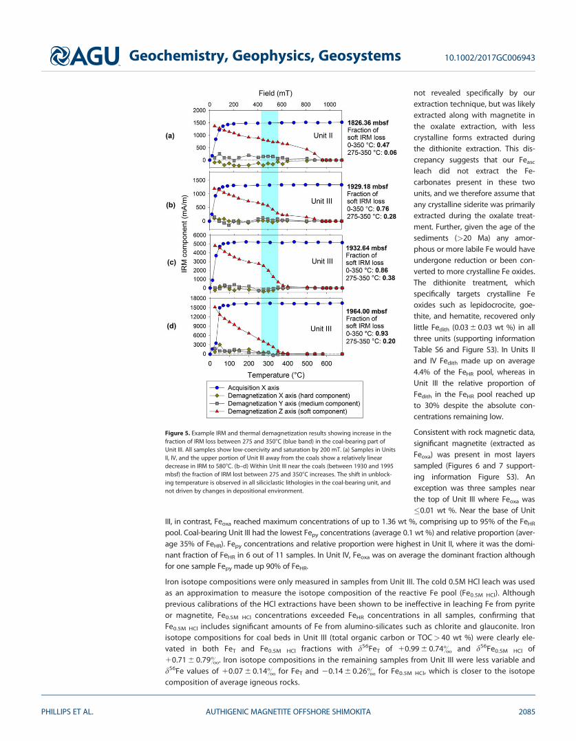
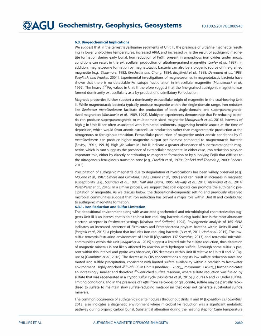
5.2. IRM Acquisition and Backfield IRMAll samples show acquisition curves typical of low-coercivity minerals reaching SIRM below 200 mT(Figure 5). Likewise, the soft axis (120 mT) contains the dominant fraction of IRM after three-axis magnetiza-tion. B1/2 ranges between 23 and 98 mT (mean: 48 mT), with the highest coercivities in Unit II at approxi-mately 1500 mbsf and decreasing in Unit III (Figure 6). S300 varies from 0.76 to 1.04 with a mean of 0.92. S100
and S300 are lower in Units II and IV compared to Unit III (Figure 6).
NRM intensity follows trends in magnetic susceptibility ranging from 0.3 to 68,600 mA/m (median: 12 mA/m)(Figure 6). Similarly, SIRM follows a pattern similar to v and ranges from 31 to 221,100 mA/m (median:1262 mA/m). SIRM/v ratios are elevated in a substantial portion of samples in Unit II, and several samplesin Units III and IV (Figure 6).
5.3. Thermal DemagnetizationThermal demagnetization removed all IRM by 5808C or below in all samples, and in all samples the primarycarrier of IRM was the Z (soft) axis throughout demagnetization. Demagnetization curves in samples fromUnit II, Unit III between 1920–1925 and 1995–2002 mbsf, and in Unit IV are characterized by a lineardecrease to 5808C (Figure 5a). Samples between 1925 and 1973 mbsf have demagnetization curves inwhich soft IRM decreases overall to 5808C but with a pronounced decrease between 275 and 3508C (Figures5b and 5c). In the interval 1979–1993 mbsf, demagnetization curves decrease linearly from room tempera-ture to 350–4008C (Figure 5d). The fraction of soft IRM lost between 0 and 3508C and 275 and 3508Cincreases in Unit III. There is a pronounced increase in the fraction of soft IRM lost below 350–4008C atdepths between 1925 and 1973 mbsf (Figures 6 and 7 and supporting information Figure S1). Between1979 and 1993 mbsf, and at 1826, 2110, 2307, 2309, and 2463 mbsf, there is an enhanced fraction of softIRM lost between 0 and 3508C but no distinct drop at 2758C. The shift in unblocking temperature isobserved in all siliciclastic lithologies in the coal-bearing unit, and not driven by changes in depositionalenvironment.
5.4. Anhysteretic Remanent MagnetizationTen samples measured for ARM range from 8.6 3 1026 to 1.07 3 1023 Am2/kg. The highest ARM values arein Unit III (average: 3.8 3 1024 Am2/kg) compared to Units II and IV (average: 5.5 3 1025 Am2/kg) (Figure 6and supporting information Figure S1 and Table S4). ARM/IRM ratios are relatively consistent throughoutthe record (0.01–0.11) with one high value in Unit III (0.58).
Geochemistry, Geophysics, Geosystems 10.1002/2017GC006943
PHILLIPS ET AL. AUTHIGENIC MAGNETITE OFFSHORE SHIMOKITA 2083

5.5. Curie Temperature MeasurementsAll measurements for Curie temperature show an increase in v when heated above 3008C and then a rapidremoval of v below 6008C (supporting information Figure S2 and Table S5). The increase in v is observed insamples both under open atmosphere and argon, but in some cases is reduced under argon. The increasein v is generally most pronounced in samples from Unit III and IV.
5.6. Iron Speciation and Isotope CompositionsTotal Fe concentrations were lowest, and variability highest, in the coal bearing Unit III (average FeT
2.3 6 1.5 wt %, Figures 6 and 7 supporting information Table S6). The low concentrations and high variabil-ity of Fe are also reflected in the FeT/Al ratios (average FeT/Al 0.3 6 0.16) in this unit. In Units II and IV, con-centrations are in the range typical for continental margin sediments (average FeT 3.5 6 0.8 wt %) with FeT/Al ratios (0.41 6 0.08) closely resembling that of average continental crust (0.44) [Taylor and McLennan,1985]. Low FeT and FeT/Al in Unit III coincide with low sulfur concentrations [Glombitza et al., 2016] and lowFeHR concentrations.
Feasc is largely absent throughout all three units (supporting information Table S6 and Figure S3) with con-centrations below detection limit (2 mg/kg) except for two samples that are <50 mg/kg. Previous mineral-ogical investigations of sediments from Unit III and Unit IV have shown the presence of siderite, which was
Figure 4. (a) Correlation of shipboard volume-specific magnetic susceptibility (j) from multisensor core logging (MSCL) and postcruiseMS2 mass-specific low-frequency magnetic susceptibility measurements (v). Correlation between data indicates little to no alteration tothe magnetic mineral assemblage since sampling. (b) Zoomed-in view of the lower magnetic susceptibility samples. (c) Correlation of ship-board and postcruise MAGNON measurements. (d) Relationship of vfd to v. Frequency dependence increases in the high magnetic suscep-tibility samples of Unit III.
Geochemistry, Geophysics, Geosystems 10.1002/2017GC006943
PHILLIPS ET AL. AUTHIGENIC MAGNETITE OFFSHORE SHIMOKITA 2084
not revealed specifically by ourextraction technique, but was likelyextracted along with magnetite inthe oxalate extraction, with lesscrystalline forms extracted duringthe dithionite extraction. This dis-crepancy suggests that our Feasc
leach did not extract the Fe-carbonates present in these twounits, and we therefore assume thatany crystalline siderite was primarilyextracted during the oxalate treat-ment. Further, given the age of thesediments (>20 Ma) any amor-phous or more labile Fe would haveundergone reduction or been con-verted to more crystalline Fe oxides.The dithionite treatment, whichspecifically targets crystalline Feoxides such as lepidocrocite, goe-thite, and hematite, recovered onlylittle Fedith (0.03 6 0.03 wt %) in allthree units (supporting informationTable S6 and Figure S3). In Units IIand IV Fedith made up on average4.4% of the FeHR pool, whereas inUnit III the relative proportion ofFedith in the FeHR pool reached upto 30% despite the absolute con-centrations remaining low.
Consistent with rock magnetic data,significant magnetite (extracted asFeoxa) was present in most layerssampled (Figures 6 and 7 support-ing information Figure S3). Anexception was three samples nearthe top of Unit III where Feoxa was�0.01 wt %. Near the base of Unit
III, in contrast, Feoxa reached maximum concentrations of up to 1.36 wt %, comprising up to 95% of the FeHR
pool. Coal-bearing Unit III had the lowest Fepy concentrations (average 0.1 wt %) and relative proportion (aver-age 35% of FeHR). Fepy concentrations and relative proportion were highest in Unit II, where it was the domi-nant fraction of FeHR in 6 out of 11 samples. In Unit IV, Feoxa was on average the dominant fraction althoughfor one sample Fepy made up 90% of FeHR.
Iron isotope compositions were only measured in samples from Unit III. The cold 0.5M HCl leach was usedas an approximation to measure the isotope composition of the reactive Fe pool (Fe0.5M HCl). Althoughprevious calibrations of the HCl extractions have been shown to be ineffective in leaching Fe from pyriteor magnetite, Fe0.5M HCl concentrations exceeded FeHR concentrations in all samples, confirming thatFe0.5M HCl includes significant amounts of Fe from alumino-silicates such as chlorite and glauconite. Ironisotope compositions for coal beds in Unit III (total organic carbon or TOC> 40 wt %) were clearly ele-vated in both FeT and Fe0.5M HCl fractions with d56FeT of 10.99 6 0.74& and d56Fe0.5M HCl of10.71 6 0.79&. Iron isotope compositions in the remaining samples from Unit III were less variable andd56Fe values of 10.07 6 0.14& for FeT and 20.14 6 0.26& for Fe0.5M HCl, which is closer to the isotopecomposition of average igneous rocks.
Figure 5. Example IRM and thermal demagnetization results showing increase in thefraction of IRM loss between 275 and 3508C (blue band) in the coal-bearing part ofUnit III. All samples show low-coercivity and saturation by 200 mT. (a) Samples in UnitsII, IV, and the upper portion of Unit III away from the coals show a relatively lineardecrease in IRM to 5808C. (b–d) Within Unit III near the coals (between 1930 and 1995mbsf) the fraction of IRM lost between 275 and 3508C increases. The shift in unblock-ing temperature is observed in all siliciclastic lithologies in the coal-bearing unit, andnot driven by changes in depositional environment.
Geochemistry, Geophysics, Geosystems 10.1002/2017GC006943
PHILLIPS ET AL. AUTHIGENIC MAGNETITE OFFSHORE SHIMOKITA 2085

5.7. Electron Microprobe AnalysisWithin the three high magnetic susceptibility sand/sandstone samples collected from Unit III for EMPA, allopaque/high backscatter minerals analyzed with EDS showed peaks that were suggestive of ilmenite, pyrite,rutile, apatite, or siderite. Very few grains showed EDS peaks suggestive of magnetite, and these were gen-erally in grains that were too small for analysis. WDS analysis of grains large enough for analysis confirmedthe dominant presence of ilmenite and pyrite, with minor rutile (supporting information Table S7). All grainsthat were suspected to be (titano)magnetite were less than 5 mm and produced poor WDS results (40–60wt % total elemental content) that contained considerable Si and Al content suggesting the beam over-lapped from high-backscatter potential iron oxide grains to the surrounding low-backscatter grains ofquartz and clays due to the small grain size of the target grains.
6. Discussion
6.1. Magnetic Mineral AssemblageResults of IRM acquisition (low-coercivity) and unblocking temperatures (5808C) suggest a magnetic mineralassemblage dominated by detrital magnetite for most samples in Unit II and Unit IV. Our measurementsshow little evidence for contribution from hematite, goethite, greigite, or pyrrhotite to the magnetic mineralassemblage. All samples at Site C0020A saturated below 200 mT suggesting low-coercivity minerals domi-nate the magnetic properties reported here. Plots of SIRM/v and B1/2 are consistent with titanomagnetitefor all samples but three (Figure 8). Two samples in Unit II and one sample in Unit III show high SIRM/v ratiostypical of magnetic iron sulfides [Dekkers, 1988; Roberts, 1995; Dekkers et al., 2000]. However, these samplesdo not show a characteristic unblocking temperature for greigite or pyrrhotite [Lowrie, 1990; Roberts, 1995].These samples possibly represent a minor component of authigenic mineral formation associated with
Figure 6. Key down core rock magnetic and geochemical results. (a) Magnetic susceptibility, (b) frequency dependence of magnetic susceptibility, isothermal remanent magnetization(IRM) at 1.1 T, and natural remanent magnetization (c) fraction of soft isothermal remanent magnetization (IRM) removed during thermal demagnetization (0–350 and 275–3508C),(d) anhysteretic remanent magnetization (ARM) and ARM/IRM ratios, (e) 2100 and 2300 mT S-ratios (f) coercivity (B1/2), (g) IRM at 0.9T/v, range of magnetite, magnetitic iron sulfides,and mixed magnetite and magnetic iron sulfides from Larrasoa~na et al. [2006]. Chromium-reducible sulfur (CRS) [Glombitza et al., 2016], (h) d34S of the CRS fraction [Glombitza et al.,2016], (i) highly reactive (FeHR) and total (FeT) iron, (j) Fe/Al ratios (vertical green bar represents average continental Fe/Al) (0.44) [Taylor and McLennan, 1985] and oxalate-extractable ironas a fraction of FeHR (i) coal-bed thickness [Expedition 337 Scientists, 2013], and (k) total organic carbon (TOC) and headspace methane [Expedition 337 Scientists, 2013].
Geochemistry, Geophysics, Geosystems 10.1002/2017GC006943
PHILLIPS ET AL. AUTHIGENIC MAGNETITE OFFSHORE SHIMOKITA 2086

diagenesis of marine sediments [Larrasoa~na et al., 2007] or rapid burial of detrital pyrrhotite [Horng and Rob-erts, 2006].
Samples in Unit III that show a significant portion of soft IRM with an unblocking temperature at approxi-mately 3508C are consistent with the demagnetization of magnetic iron sulfides, maghemite, or (titano)-magnetite (either fine-grained magnetite or TM30 titanomagnetite). However, a few samples have SIRM/vand B1/2 that are consistent with at least a partial greigite or pyrrhotite component [Peters and Dekkers,2003] but do not correspond to samples with a 3508C unblocking temperature. Additional measurements ofv after long exposure to oxygen (approximately 2 years) do not show a decrease relative to the shipboard
measurements, indicating that authi-genic magnetic iron sulfides vulnerableto oxidation are not likely a significantcomponent. A magnetic mineral assem-blage with a large, metastable mag-netic iron sulfide component wouldlikely experience a loss in v after pro-longed exposure to oxygen [Hungerand Benning, 2007]. EMPA analysis indi-cated that iron sulfides present in UnitIII are dominantly pyrite, which gener-ally is characterized by very low mag-netic susceptibility [Waters et al., 2008].
IRM acquisition and v results are con-sistent with the dominant presenceof magnetite but a possible minormaghemite (c-Fe2O3) component can-not be ruled out. Maghemetization of
Figure 7. Detailed view of down core rock magnetic and geochemical results for the key coal-bearing interval of record shown in Figure 6. (a) Magnetic susceptibility, (b) frequency depen-dence of magnetic susceptibility, (c) fraction of soft IRM removed during thermal demagnetization (0–350 and 275–3508C), (d) ARM and ARM/IRM ratios, (e) chromium-reducible sulfur (CRS)[Glombitza et al., 2016], (f) d34S of the CRS fraction [Glombitza et al., 2016], (g) highly reactive (FeHR), HCl-extractable (FeHCl) and total (FeT) iron, (h) Fe/Al ratios (vertical green bar represents aver-age continental Fe/Al) [Taylor and McLennan, 1985] and oxalate-extractable iron as a fraction of FeHR, (i) d56Fe of total (d56FeT) and HCl-extractable (d56FeHCl) iron, vertical purple bar representsaverage igneous d56Fe from Beard et al. [2003], and (j) total organic carbon (TOC) and headspace methane [Expedition 337 Scientists, 2013]. Shaded horizontal bars indicate coal intervals.
Figure 8. Crossplot of SIRM/v and B1/2 for all samples from Units II, III, and IV. Min-eral ranges from Peters and Dekkers [2003].
Geochemistry, Geophysics, Geosystems 10.1002/2017GC006943
PHILLIPS ET AL. AUTHIGENIC MAGNETITE OFFSHORE SHIMOKITA 2087

titanomagnetite can occur in oxygenated sediments, but is a more common component in pelagic set-tings where low sedimentation rates yield long exposure times to oxygenated bottom waters [Smirnovand Tarduno, 2000; Xu et al., 1997]. In this record, sediments from nonwetland environments were likelyexposed to oxygenated bottom waters, but due to high sedimentation rates (�100 m/Ma over the entirehole) [Phillips et al., 2016] and high TOC throughout Site C0020 (up to 50 wt % in coal layers) [Expedition337 Scientists, 2013], magnetite, probably of detrital origin, was likely buried quickly into anoxicconditions.
In our record, magnetite is the mineral most likely associated with the �300–4008C unblocking tempera-ture, due to its decreased grain size compared to the background coarser-grained magnetite with anunblocking temperature of �5808C, close to the Curie temperature. The unblocking temperature of magne-tite can deviate from the Curie temperature decreasing below 5758C in cases where the particle size is lessthan 50 nm [Dunlop, 1973a; Winklhofer et al., 1997]. Our Curie temperature measurements show an increasein v due to alteration at 300–3508C and a rapid decrease below 6008C. Our results show a consistent behav-ior in Curie temperature measurements in both types of samples with unblocking temperatures of 580 and300–4008C. In Unit III, between 1925 and 1975 mbsf demagnetization curves suggest that there is a mix ofdetrital and authigenic magnetite, while between 1975 and 2000 mbsf the magnetic mineral assemblagemay be dominated by authigenic magnetite. Increased ARM in Unit III (Figure 6) also suggests fine-grainedmagnetite is present. Increased vfd in Unit III indicates the increased presence of superparamagnetic mag-netite at room temperature suggesting magnetite grains <30 nm [Dunlop, 1973b]. In summary, rock mag-netic measurements suggest detrital magnetite composes the primary magnetic mineral assemblagethroughout C0020A with an additional fine-grained secondary phase (authigenic) of magnetite with lowerunblocking temperatures within Unit III.
6.2. Depositional EnvironmentIn our data set, there is no clear change in magnetic mineralogy or magnetic susceptibility that is associ-ated with a particular major change in lithology (supporting information Figure S4). Sandstone/sandsamples have an average v of 18.9 3 1028 m3 kg21, siltstone 26.8 3 1028 m3 kg21, and shale 15.7 3
1028 m3 kg21. The ultimate source for detrital magnetite at Site C0020A is most likely from weatheringof rocks within northern Honshu. Subangular fragments were commonly observed within Unit III [Expedi-tion 337 Scientists, 2013] indicating minimal transport from source to deposition. Titanomagnetite is acommon constituent in volcanic rocks [Akimoto and Katsura, 1959; Sakuyama and Nesbitt, 1986; Hoshiand Teranishi, 2007; Ohba et al., 2007; Suzuki, 2008], magnetite-series granitic rocks [Takagi, 2004], andhornfels facies metamorphic rocks [Tsusue, 1962] from Honshu. Magnetite has also been observed as theprimary detrital magnetic mineral in the Nankai accretionary complex [Kanamatsu et al., 2012; Zhaoet al., 2013; Kars and Kodama, 2015] and Japan Sea [Razjigaeva and Naumova, 1992; Vigliotti, 1997].Paleomagnetic studies of sediments from DSDP and ODP sites in the Japan Trench and fore-arc basinshow stable magnetic remanance [Hall and Smeltzer, 1980; Niitsuma, 1986; Kanamatsu and Niitsuma,2004], but do not directly address magnetic mineral assemblage. The titanomagnetite-dominant mag-netic mineral assemblage at C0020A is consistent with sites and lithologic sources around the Japanesemargin.
Magnetite has a density (5.20 g/cm3) nearly double that of quartz (2.65 g/cm3) [Sch€on, 2004], and commonlyshows hydraulic sorting in beach and fluvial environments [e.g., Slingerland and Smith, 1986; Komar, 1989].The anomalous increases in v (to �100–400 3 1028 kg/m3) are similar to the range observed in moderntitanomagnetite placer deposits (�50–2000 3 1028 kg/m3) [Badesab et al., 2012]. However, the presence offine-grained SP magnetite and the fact that increases in v also occur in fine-grained rocks at Site C0020 sug-gest placer sorting is not the cause of the observed increases in v.
The increases in magnetic susceptibility within Unit III are likely a result of additional authigenic precipita-tion of magnetite rather than a rapid additional influx of detrital magnetite. Given the high sedimentationrate of site C0020, cm-scale intervals of high magnetic susceptibility likely do not correspond to changes inprovenance. These cm-scale increases in magnetic susceptibility occur in multiple lithologies: sand, silt-stone, and shale, but only in the coal-bearing unit and often associated with organic-rich laminations. Inaddition, the high v samples correspond to increased vfd suggesting an increase of superparamagnetic fine-grained material within high v intervals.
Geochemistry, Geophysics, Geosystems 10.1002/2017GC006943
PHILLIPS ET AL. AUTHIGENIC MAGNETITE OFFSHORE SHIMOKITA 2088
6.3. Biogeochemical ImplicationsWe suggest that in the terrestrial/estuarine sediments of Unit III, the presence of ultrafine magnetite result-ing in lower unblocking temperatures, increased ARM, and increased vfd is the result of authigenic magne-tite formation during early burial. Iron reduction of Fe(III) present in amorphous iron oxides under anoxicconditions can result in the extracellular production of ultrafine-grained magnetite [Lovley et al., 1987]. Inaddition, magnetosome formation by magnetotactic bacteria can also be a biogenic source of fine-grainedmagnetite [e.g., Blakemore, 1982; Kirschvink and Chang, 1984; Bazylinski et al., 1988; Devouard et al., 1988;Bazylinski and Frankel, 2004]. Experimental investigations of magnetosomes in magnetotactic bacteria haveshown that there is no detectable Fe isotope fractionation in intracellular magnetite [Mandernack et al.,1999]. The heavy d56FeT values in Unit III therefore suggest that the fine-grained authigenic magnetite wasformed dominantly extracellularly as a by-product of dissimilatory Fe reduction.
Magnetic properties further support a dominantly extracellular origin of magnetite in the coal-bearing UnitIII. While magnetotactic bacteria typically produce magnetite within the single-domain range, iron reducerslike Geobacter metallireducens facilitate the production of both single-domain- and superparamagnetic-sized magnetites [Moskowitz et al., 1989, 1993]. Multiyear experiments demonstrate that Fe-reducing bacte-ria can produce superparamagnetic to multidomain-sized magnetite [Abrajevitch et al., 2016]. Intervals ofhigh v in Unit III are often associated with laminated sediments, suggesting benthic anoxia at the time ofdeposition, which would favor anoxic extracellular production rather than magnetotactic production at thenitrogenous to ferruginous transition. Extracellular production of magnetite under anoxic conditions by G.metallireducens can produce higher magnetite output per biomass compared to magnetotactic bacteria[Lovley, 1991a, 1991b]. High vfd values in Unit III indicate a greater abundance of superparamagnetic mag-netite, which in turn suggests the presence of extracellular magnetite. In either case, iron reduction plays animportant role, either by directly contributing to magnetite formation or by supplying Fe(II) that diffuses tothe nitrogenous-ferruginous transition zone [e.g., Froelich et al., 1979; Canfield and Thamdrup, 2009; Roberts,2015].
Precipitation of authigenic magnetite due to degradation of hydrocarbons has been widely observed [e.g.,McCabe et al., 1987; Elmore and Crawford, 1990; Elmore et al., 1997] and can result in increases in magneticsusceptibility [e.g., Saunders et al., 1991; Hall and Evans, 1995; Mewafy et al., 2011; Atekwana et al., 2014;P�erez-P�erez et al., 2016]. In a similar process, we suggest that coal deposits can promote the authigenic pre-cipitation of magnetite. As we discuss below, the depositional/diagenetic setting and previously observedmicrobial communities suggest that iron reduction has played a major role within Unit III and contributedto authigenic magnetite formation.6.3.1. Iron Reduction and Sulfur LimitationThe depositional environment along with associated geochemical and microbiological characterization sug-gests Unit III is an interval that is able to host iron-reducing bacteria during burial. Iron is the most abundantelectron acceptor in freshwater settings [Nealson and Saffarini, 1994]. Phylogenetic analysis of 16S rRNAindicates an increased presence of Firmicutes and Proteobacteria phylum bacteria within Units III and IV[Inagaki et al., 2015], a phylum that includes iron-reducing bacteria [Li et al., 2011; Hori et al., 2015]. The low-sulfur terrestrial/estuarine environment of Unit III [Expedition 337 Scientists, 2013] and terrestrial microbialcommunities within this unit [Inagaki et al., 2015] suggest a limited role for sulfate reduction, thus alterationof magnetic minerals is not likely affected by reaction with hydrogen sulfide. Although some sulfur is pre-sent within this interval and pyrite was observed, CRS decreases within Unit III relative to Units II and IV (Fig-ure 6) [Glombitza et al., 2016]. The decrease in CRS concentrations suggests low sulfate reduction rates andmuted iron sulfide precipitation, consistent with limited sulfate availability within a brackish-to-freshwaterenvironment. Highly enriched d34S of CRS in Unit III (median: 126.9&, maximum: 145.6&) further indicatesan increasingly smaller and therefore 34S-enriched sulfate reservoir, where sulfate reduction was fueled bysulfate that was regenerated in a cryptic sulfur cycle [Glombitza et al., 2016] (Figures 6 and 7). Under sulfate-limiting conditions, and in the presence of Fe(III) from Fe-oxides or glauconite, sulfide may be partially reoxi-dized to sulfate to maintain slow sulfate-reducing metabolism that does not generate substantial sulfideminerals.
The common occurrence of authigenic siderite nodules throughout Units III and IV [Expedition 337 Scientists,2013] also indicates a diagenetic environment where microbial Fe reduction was a significant metabolicpathway during organic carbon burial. Substantial alteration during the heating step for Curie temperature
Geochemistry, Geophysics, Geosystems 10.1002/2017GC006943
PHILLIPS ET AL. AUTHIGENIC MAGNETITE OFFSHORE SHIMOKITA 2089
measurements of Unit III sediments (supporting information Figure S2) is consistent with alteration of sider-ite to magnetite and maghemite during heating [Pan et al., 1999, 2000, 2002]. Laboratory experiments andequilibrium calculations by Bell et al. [1987] suggest that magnetite formation is favored under more alka-line conditions, siderite formation is favored under more acidic conditions, and both magnetite and sideritewould be present at pH 7.0 if H2S is not present. The upward transition from a neutral-to-acidic, freshwaterenvironment to alkaline, brackish environment in Unit III [Gross et al., 2015] are consistent with our observa-tions of authigenic magnetite that overly and overlap with the abundant siderite nodules in the base ofUnit III and in Unit IV [Expedition 337 Scientists, 337]. The Fe(II) produced via iron reduction is available toreact with bicarbonate within the methanic zone when H2S is not present [Berner, 1981; Maynard, 1982;Postma, 1982] or in an environment in which rates of iron reduction are greater than sulfate reduction [Pyeet al., 1990]. Fe(III)-reducing bacteria can produce microbially derived siderite as a direct by-product of dis-similatory iron reduction [Lovley and Phillips, 1986; Mortimer and Coleman, 1997]. The decrease in CRS withinsiliciclastic sediments of Unit III [Expedition 337 Scientists, 2013; Glombitza et al., 2016] indicates sulfur limita-tion and an environment in which the sink for Fe(II) produced during iron reduction are siderite or magne-tite rather than pyrite. Overall, geochemical and mineralogical observations from Site C0020 suggest thatthe terrestrial to marine transition Unit III provided a diagenetic environment favorable for microbial mag-netite formation.
Magnetite is typically low or absent in anoxic coastal margin sediments for two reasons: (1) In marine sedi-ments where sulfate concentrations are relatively high, bacterial sulfate reduction usually dominates overmicrobial Fe reduction, suppressing authigenic magnetite formation. (2) Magnetite of detrital origin dis-solves during burial in the presence of H2S [e.g., Canfield and Berner, 1987; Karlin, 1990]. A slight decrease inS100 and increase in B1/2 in the marine Unit II relative to Units III and IV (Figures 6e and 6f) may indicate anyminor presence of high coercivity minerals resistant to H2S dissolution (hematite and goethite) becomemore concentrated in sulfidic sediments as low coercivity magnetite is preferentially removed [e.g., Garminget al., 2005]. With increasing burial depth magnetite and other crystalline Fe oxides are replaced by pyrite orits precursor greigite. Previous studies have shown that greigite formed during sulfidization may contributeto late stage remanence acquisition long after the original deposition [Rowan and Roberts, 2006]; however,we found little evidence for the presence of greigite. [email protected]/v values possibly suggestive of a minor pres-ence of magnetic iron sulfides are largely restricted to Unit II (Figure 6g). Magnetite concentrations of up to1.36 wt % in Unit III suggest that either magnetite has been added or that detrital magnetite has escapedsulfidization during burial diagnesis.
Collectively the rock magnetic, Fe isotope, and Fe speciation data suggest that the coal-bearing Unit III hasundergone postdepositional diagenetic alteration that has led to the removal or replacement of the originalmineral assemblage. FeT and FeHR are markedly decreased in Unit III, but the large variability in FeT/Al rang-ing between 0.11 and 0.61 in this unit suggests that Fe has been removed from some intervals, and addedto others, during burial diagenesis. Bulk Fe isotope data are consistent with this interpretation: a shift ind56FeT cannot be achieved by internal redistribution of reactive Fe into different minerals during early dia-genesis. Elevated d56FeT values of up to 2& can only be achieved through significant loss of light Fe oraddition of heavy Fe. All except one sample with d56FeT> 0.3& are associated with FeT/Al values that aresignificantly below the detrital baseline (supporting information Figure S5), suggesting that loss of FeHR wasthe primary control on bulk Fe isotope compositions. The Fe isotope compositions for the 0.5M HCl extrac-tions show a trend very similar to the bulk isotope values (Figure 7). The slightly lower d56Fe0.5M HCl values,closer to the detrital baseline, confirm that this extraction likely included significant Fe from silicates whileat the same time being ineffective at dissolving other phases of the FeHR pool that may have a heavier iso-tope composition. The increase in d56FeT at the base of Unit III corresponds to a shift from a mixed detritaland authigenic magnetite (a pronounced drop in soft IRM from 275 to 3508C superimposed on a slowerdrop from 0 to 5758C) to mostly authigenic magnetite (linear decrease in soft IRM from 0 to 3508C). Whereauthigenic magnetite composes a higher fraction of total magnetite, the near-zero d56Fe values from igne-ous magnetite contribute less to masking the heavy d56Fe composition of authigenic magnetite.
Vertical fluid advection, such as during groundwater fluid flow, has been shown to alter the remanant mag-netization [Rowan and Roberts, 2006] as well as the Fe isotope composition [Rouxel et al., 2008] of the origi-nal mineral assemblage. Low-salinity groundwater may stimulate microbial Fe-oxide reduction as well asoxidative precipitation of Fe-oxides. The low CRS concentrations in Unit III suggest the lack of pyrite
Geochemistry, Geophysics, Geosystems 10.1002/2017GC006943
PHILLIPS ET AL. AUTHIGENIC MAGNETITE OFFSHORE SHIMOKITA 2090
precipitation and/or the removal of pyrite through oxidation that could occur through alteration by ground-water. Magnetite that is produced during microbial Fe reduction typically has a heavy isotope composition[Johnson et al., 2005], while the pore water Fe(II) that is being generated during redox recycling will be iso-topically light [Severmann et al., 2006]. Rouxel et al. [2008] have observed a wide range (–2 to 11.5&) in theFeHR isotope composition of sediments that have been affected by groundwater alteration in a subterra-nean estuary. Although the sediments in Unit III only show near-detrital or positive d56FeT values it is likelythat alteration was caused by a similar combination of processes. These processes may include dissolutionof the original mineral assemblage, conversion of Fe-oxide to authigenic magnetite and potentially also for-mation of new authigenic mineral phases such as glauconite and siderite. We propose that the combinedeffect of these processes was the observed decrease in FeT and FeHR, variable FeT/Al, ratios and heavy FeT
and Fe0.5M HCl isotope compositions.6.3.2. Peat/Lignite As Source of Electron DonorsThe presence of fine-grained biogenic magnetite occurs over a broad interval containing numerous coalbeds, and coal may serve as an important source of organic substrates to the surrounding sediments.Coal can serve as a bioreactor in which complex coal macerals are degraded into simple, more labilemolecules (e.g., acetate, H2, and CO2) that become mobile as electron donors that can fuel methanogen-esis [Strapoc et al., 2008, 2011]. Peat prior to coal formation is a major source of dissolved organic carbon(DOC) into underlying sediments as well [Dalva and Moore, 1991]. During burial and through early coalifi-cation, the peat/lignite intervals at Site C0020 were a likely source of DOC to the surrounding low TOCsediments. Lignite within Unit III promotes methanogenesis and H2 production [Inagaki et al., 2015] andH2 can serve as an electron donor for iron-reducing bacteria commonly found in estuaries [Caccavoet al., 1992]. Within the C0020A sediments, average TOC of coal is 41 wt % while mudrocks, siltstone, andsandstone have 1.4, 0.43, and 0.26 wt %, respectively, [Expedition 337 Scientists, 2013]. Most coal bedswithin Unit III have little to no ferrimagnetic fraction, and likely had little Fe(III) present, thus presentinga physical separation between electron donors and electron acceptors necessary to fuel dissimilatoryiron reduction. These coal beds are interbedded among siliciclastic sediments including massive fine-to-medium sand layers in the upper half of Unit III indicating an adjacent proximity of high-permeabilitysediments, initially (during early burial) likely to contain Fe(III) oxides, directly adjacent to a DOC and CH4
sources.
In addition to the lignite as a source of electron donors, humic acids can facilitate iron reduction [Lovleyet al., 1996; Lovley and Blunt-Harris, 1999; Kappler et al., 2004; Kl€upfel et al., 2014]. Humic acids comprise a sig-nificant fraction of lignite [e.g., Ibarra and Juan, 1985; Gonzalez-Vila, 1992, 1994; Cavani et al., 2003; Allard,2006] and can transfer electrons to iron oxides during acetate oxidation, alleviating the necessity of directcontact between Fe(III)-reducing bacteria and iron oxides [Lovley et al., 1996]. The presence of peat/lignitein the subsiding environment at Site C0020A likely acted as a significant source of humic acids in the DOCto the surrounding sediments, thus enhancing iron reduction within intervals in pore water contact withthe peat/coal intervals (Figure 9). Maximum methane content occurs within the coal beds (Figures 6 and 7)[Expedition 337 Scientists, 2013], including production by present-day methanogenesis [Inagaki et al., 2015].Methane oxidation may be coupled to iron reduction [Konhauser et al., 2005; Thauer and Shima, 2008; Bealet al., 2009; Segarra et al., 2013; Riedinger et al., 2014], suggesting methane exported from peat/lignite as apotential electron donor for iron reduction. Although both Units III and IV were deposited within a brackish/freshwater environment, only Unit III has numerous coal beds that can allow for a considerable source ofhumic acids and methane to the Fe(III)-bearing sediments. The presence of numerous coal beds in Unit IIIcan explain why fine-grained authigenic magnetite is present in this unit, but not in Unit IV.
Within Unit III, the presence of unconsolidated sands suggests that fluid connectivity between coal bedsand the surrounding sediments is maintained, but may be increasingly constricted with depth. Porositydecreases with depth from >0.8% at the top of overlying Hole C9001C to an average of 0.26% in Unit III[Aoike, 2007; Expedition 337 Scientists, 2013]. Within carbonate-cemented intervals of Unit III, porosity is fur-ther reduced to <0.15%. The decrease in porosity with depth due to compaction and diagenetic cementsalmost certainly limits permeability, thus impeding the connection between magnetite-sourced electronacceptors and coal-sourced electron donors. This presumed decrease in permeability, along with thedecrease in cell concentration with depth [Inagaki et al., 2015], suggests that the maximum rates for ironreduction likely occurred during shallow burial and declined with further burial depth.
Geochemistry, Geophysics, Geosystems 10.1002/2017GC006943
PHILLIPS ET AL. AUTHIGENIC MAGNETITE OFFSHORE SHIMOKITA 2091

6.3.3. Potential for Continued Iron Reduction in the Deep BiosphereAs we discuss above, electron donors and Fe(III) electron acceptors were likely more readily available dur-ing early burial; however, the presence of indigenous terrestrial microbial communities within Unit III [Ina-gaki et al., 2015] suggests the possibility that there may be continued iron reduction at depth, albeit atlow rates. If iron reduction continues within the modern buried coal bed it is likely not a dominant pro-cess. Elevated H2 concentrations at Site C0020 [Inagaki et al., 2015] are consistent with observations thatH2 concentrations are lowest in sediments dominated by Fe(III) reduction and highest in those dominatedby methanogenesis [Lovley and Goodwin, 1988; Lovley et al., 1994], suggesting that methanogenesis is adominant process.
Iron may play a role in the deep biosphere as a potential source of Fe(III) for iron reduction, possibly cou-pled to methane oxidation [Riedinger et al., 2014]. Although iron reduction is thermodynamically morefavorable than sulfate reduction and methanogenesis [Froelich et al., 1979], Fe(III)-bearing iron oxides canpersist during burial of sediments through the sulfidic and methanic zones [e.g., Kasten et al., 1998; Riedingeret al., 2005]. While the original depositional environment for Unit III is likely to have been rich in Fe(III)oxides, the lack of ascorbate extractable iron suggests that this source of Fe(III) has been depleted. Fe(III) isrelatively immobile within a crystalline structure, and the presence of Fe(II) can limit the accessibility ofFe(III) thus limiting the bioavailability of magnetite Fe(III) for iron reducers [Roden and Zachara, 1996; Roden
Surface peatland
Buried peat
Buried lignite
Buried lignite
Siliciclastic sediments
Siliciclastic sediments
Fe(III)-oxidee- acceptors
CH4, acetate, H2, CO2, humic acids
Magnetitee- acceptors
Decreasing porosity and
microbial activity
Late Oligocene/Early Miocene peatland
Present dayafter ~2 km burial
Porosity: ~0.4Cell counts: ~107 to ~109 cells cm-3
Porosity: ~0.2Cell counts: <101 to ~104 cells cm-3
(a)
(b)
Loss of Fe(III) oxides
CH4, acetate, H2, CO2, humic acids
CH4, acetate, H2, CO2, humic acids
CH4, acetate, H2, CO2, humic acids
Figure 9. Schematic diagram illustrating the role that organic-rich peat/coal beds may play in promoting microbial iron reduction inthe surrounding siliciclastic sediments of Unit III during burial. The coal can serve as an electron donor to the Fe(III) electron acceptors inthe surrounding sediments via export of methane, DOC, and hydrogen, causing the formation of magnetite at the highest rate duringearly burial. With continued burial, depletion of amorphous iron oxides along with decreasing microbial activity and permeability likelyslowed the rate of iron reduction. In the modern sequence, magnetite may be the main source of Fe(III) potentially available for ironreduction.
Geochemistry, Geophysics, Geosystems 10.1002/2017GC006943
PHILLIPS ET AL. AUTHIGENIC MAGNETITE OFFSHORE SHIMOKITA 2092

and Urrutia, 1999]. However, Fe(III) and Fe(II) within magnetite have been shown to be bioavailable to bothiron-reducing and iron-oxidizing bacteria, respectively [Byrne et al., 2015].
Despite its crystalline structure, the presence of magnetite as the dominant magnetic mineral in the SiteC0020A record suggests this mixed-valence iron oxide is a potential source of Fe(III) for present iron reduc-tion in sediments offshore Shimokita within the deep biosphere. Reduction of magnetite-bound Fe(III) as anelectron acceptor and magnetite dissolution have been observed in modern cultures of iron-reducing bac-teria, and are shown to be energetically favorable under pH conditions typical of freshwater [Kostka andNealson, 1995; Hori et al., 2015]. In modern cultures of iron reducing bacteria with glucose or lactate as elec-tron donors, magnetite loss occurs at rate on the order of 1029 to 1027 mmol h21 cell21 [Nealson and Saffar-ini, 1994; Kostka and Nealson, 1995]. This rate of magnetite dissolution would consume all magnetite at SiteC0020 over a period of tens of thousands to several million years. The fact that magnetite, even ultrafinemagnetite, has remained the dominant magnetic mineral in these sediments since the Miocene, suggeststhat if magnetite is a source of Fe(III), iron reduction is proceeding at extremely low rates potentially limitedby the accessibility of electron donors.
Other possible Fe(III)-bearing minerals present at Site C0020 include glauconite and smectite-group clays.Glauconite was commonly observed in smear slides in cores from Unit II and IV but was less common inUnit III [Expedition 337 Scientists, 2013]. Glombitza et al. [2016] suggest Fe(III) in glauconite may drive slowrates of sulfate recycling at Site C0020A. Clay mineralogy has not been characterized at C0020A, but smec-tite clay minerals are abundant in late Oligocene-early Miocene sediments at DSDP sites further offshorenorthern Honshu [Chamley et al., 1986]. Iron-reducing bacteria (as well as sulfate-reducing bacteria andmethanogens) have been shown to reduce Fe(III)-bearing clay minerals (e.g., nontronite) [e.g., Kostka et al.,1999; Dong et al., 2009; Pentr�akov�a et al., 2013]. Furthermore, humic acids can stimulate the reduction ofFe(III)-bearing clays [Liu et al., 2017]. Characterization of clay mineralogy at Site C0020 may further help elu-cidate the role of iron reduction in the deep biosphere.
7. Summary and Conclusions
We investigated changes in magnetic mineral assemblages and iron speciation across a transition indepositional and diagenetic environments at IODP Hole C0020A in order to identify the source of anoma-lous increases in magnetic susceptibility associated with a unit containing 0.3–7.3 m thick coal beds. Mag-netic assemblages throughout the core are dominated by low-coercivity magnetite. In this record, cm-scale magnetic susceptibility increases are associated with organic-rich laminations in sandstone, silt-stone, and shale lithologies within Unit III, a nearshore-to-intertidal coal-bearing depositional sequence.In Units II and IV, the linear loss of low-coercivity IRM during thermal demagnetization to 5808C suggeststhe presence of magnetite. Within the primary coal bearing intervals of Unit III (1925–1975 mbsf and1979–1993 mbsf) partial or complete loss of low-coercivity IRM by 350–4008C indicates a secondary mag-netite assemblage with a lower unblocking temperature due to decreased grain size. ARM and frequencydependence measurements support the presence of fine-grained SD and ultrafine SP magnetite. Meas-urements of d56Fe from total iron extractions also indicate the precipitation of authigenic magnetite. Thisphase of fine-grained magnetite is likely formed as extracellular precipitates during iron reduction. Inter-pretation of our rock magnetic record in the context of geochemical and microbiological results fromIODP Expedition 337 [Expedition 337 Scientists, 2013; Inagaki et al., 2015; Gross et al., 2015] suggest thatthe depositional/diagenetic environment in which we observe fine-grained magnetite is conducive tomicrobial iron reduction. Peat/lignite in Unit III likely served as a source of electron donors (e.g., CH4 andH2) for iron reduction. Beginning early after deposition in this nearshore environment, microbial ironreduction most likely resulted in precipitation of authigenic magnetite by consumption of amorphousiron oxides. The proximity of the coalbeds as a source of electron acceptors and humic acid electron shut-tles further enhanced iron reduction in Unit III. This process of microbial iron reduction may continue tooccur with deeper burial and could continue today in the deep biosphere off Shimokita but at reducedrates due decreased microbial activity, reduced permeability, and the presence of only crystalline sourcesof Fe(III). Overall, these results suggest that peat/coal can facilitate iron reduction and magnetite forma-tion in subsiding sediments.
Geochemistry, Geophysics, Geosystems 10.1002/2017GC006943
PHILLIPS ET AL. AUTHIGENIC MAGNETITE OFFSHORE SHIMOKITA 2093

ReferencesAbrajevitch, A., L. M. Kondratyeva, E. M. Golubeva, K. Kodama, and R. S. Hori (2016), Magnetic properties of iron minerals produced by natu-
ral iron- and manganese-reducing groundwater bacteria, Geophys. J. Int., 206, 1340–1351.Akimoto, S., and T. Katsura (1959), Magneto-chemical study of generalized titanomagnetite in volcanic rocks, J. Geomagn. Geoelectr., 10,
69–90.Allard, B. (2006), A comparative study on the chemical composition of humic acids from forest soil, agricultural soil, and lignite deposit
bound lipid, carbohydrate, and amino acid distributions, Geoderma, 130, 77–96.Altschuler, Z. S., M. M. Schnepfe, C. C. Silber, and F. O. Simon (1983), Sulfur diagenesis in Everglades peat and origin of pyrite in coal, Sci-
ence, 221, 221–227.Aoike, K. (Ed.) (2007), CDEX laboratory operation report: CK06–06 D/V Chikyu shakedown cruise offshore Shimokita: Yokohama (CDEX-JAM-
STEC), CDEX. [Available at http://sio7.jamstec.go.jp/JAMSTEC-exp-report/902/CK06-06_CR.pdf.]Arnold, R. G., T. J. DiChristina, and M. R. Hoffman (1988), Reductive dissolution of Fe(III) oxides by Pseudomonas sp. 2000, Biotechnol. Bioeng.,
32, 1081–1096.Arnold, G. L., S. Weyer, and A. D. Anbar (2004), Fe isotope variations in natural materials measured using high mass resolution multiple col-
lector ICPMS, Anal. Chem., 76, 322–327.Arthur, M. A., R. Von Huene, and C. G. Adelseck (1980), Sedimentary evolution of the Japan fore-arc region off northern Honshu, Legs 56
and 57, Deep Sea Drilling Project, in Initial Reports of the Deep Sea Drilling Project, vol. 56/57, pp. 521–568, U.S. Gov. Print. Off., Washing-ton, D. C.
Atekwana, E. A., F. M. Mewafy, G. A. Aal, D. D. Werkema, A. Revil, and L. D. Slater (2014), High-resolution magnetic susceptibility measure-ments for investigating magnetic mineral formation during microbial mediated iron reduction, J. Geophys. Res. Biogeosci., 119, 80–94.
Badesab, F., T. von Dobeneck, K. Bryan, H. M€uller, R. M. Briggs, T. Frederichs, and E. Kwoll (2012), Formation of magnetite-enriched zones inand offshore of a mesotidal estuarine lagoon: An environmental magnetic study of Tauranga Harbor and Bay of Plenty, New Zealand,Geochem. Geophys. Geosyst., 13, Q06012, doi:10.1029/2012GC004125.
Bazylinski, D. A., and R. B. Frankel (2004), Magnetosome formation in prokaryotes, Nat. Rev., 2, 217–230.Bazylinski, D. A., R. B. Frankel, and H. W. Jannasch (1988), Anaerobic magnetite production by a marine, magnetotactic bacterium, Nature,
334, 518–519.Beal, E. J., C. H. House, and V. J. Orphan (2009), Manganese- and iron dependent marine methane oxidation, Science, 325, 184–187.Beard, B. L., C. M. Johnson, J. L. Skulan, K. H. Nealson, L. Cox, and H. Sun (2003), Application of Fe isotopes to tracing the geochemical and
biological cycling of Fe, Chem. Geol., 195, 87–117.Bell, P. E., A. L. Mills, and J. S. Herman (1987), Biogeochemical conditions favoring magnetite formation during anaerobic iron reduction,
Appl. Environ. Microbiol., 53, 2610–2616.Berner, R. A. (1981), A new geochemical classification of sedimentary environments, J. Sediment. Petrol., 51, 359–365.Blakemore, R. P. (1982), Magnetotactic bacteria, Annu. Rev. Microbiol., 36, 217–238.Bloemendal, J., J. W. King, A. Hunt, P. B. Demenocal, and A. Hayashida (1993), Origin of the sedimentary magnetic record at Ocean Drilling
Program sites on the Owen Ridge, Western Arabian Sea, J. Geophys. Res., 98, 4199–4219.Byrne, J. M., N. Klueglein, C. Pearce, K. M. Rosso, E. Appel, and A. Kappler (2015), Redox cycling of Fe(II) and Fe(III) in magnetite by Fe-
metabolizing bacteria, Science, 347, 1473–1476.Caccavo, F., R. P. Blakemore, and D. R. Lovley (1992), A hydrogen-oxidizing, Fe(III)-reducing microorganism from the Great Bay Estuary,
New Hampshire, Appl. Environ. Microbiol., 58, 3211–3216.Canfield, D. E. (1997), The geochemistry of river particulates from the continental USA: Major elements, Geochim. Cosmochim. Acta, 61,
3349–3365.Canfield, D. E., and R. A. Berner (1987), Dissolution and pyritization of magnetite in anoxic marine sediments, Geochim. Cosmochim. Acta,
51, 645–659.Canfield, D. E., and B. Thamdrup (2009), Towards a consistent classification scheme for geochemical environments, or, why we wish the
term ‘‘suboxic’’ would go away, Geobiology, 7, 385–392.Canfield, D. E., R. Raiswell, and S. Bottrell (1992), The reactivity of sedimentary iron minerals toward sulfide, Am. J. Sci., 292, 659–683.Cavani, L., C. Ciavatta, and C. Gessa (2003), Identification of organic matter from peat, leonardite, and lignite fertilisers using humification
parameters and electrofocusing, Bioresour. Technol., 86, 45–52.Chamley, H., J.-P. Cadet, and J. Charvet (1986), Nankai Trough and Japan Trench late Cenozoic paleoenvironments deduced from clay min-
eralogic data, in Initial Reports of the Deep Sea Drilling Project, vol. 87, pp. 633–642, U.S. Gov. Print. Off., Washington, D. C.Dalva, M., and T. R. Moore (1991), Sources and sinks of dissolved organic carbon in a forested swamp catchment, Biogeochem., 15, 1–19.Dearing, J. A., R. J. L. Dann, K. Hay, J. A. Lees, P. J. Loveland, B. A. Maher, and K. O. Grady (1996), Frequency-dependent susceptibility meas-
urements of environmental materials, Geophys. J. Int., 124, 228–240.Dekkers, M. J. (1988), Magnetic properties of natural pyrrhotite Part I: Behaviour of initial susceptibility and saturation-magnetization-
related rock-magnetic parameters in a grain-size dependent framework, Phys. Earth Planet. Inter., 57, 266–283.Dekkers, M. J., H. F. Passier, and M. A. A. Schoonen (2000), Magnetic properties of hydrothermally synthesized greigite (Fe3S4): II. High- and
low-temperature characteristics, Geophys. J. Int., 141, 809–819.Devouard, B., M. P�osfai, X. Hua, D. A. Bazylinski, R. B. Frankel, and P. R. Buseck (1998), Magnetite from magnetotactic bacteria: Size distribu-
tions and twinning, Am. Mineral., 83, 1387–1398.D’Hondt, S., et al. (2004), Distributions of microbial activities in deep subseafloor sediments, Science, 306, 2216–2221.Dong, H., D. P. Jaisi, J. Kim, and G. Zhang (2009), Microbe-clay interactions, Am. Mineral., 94, 1505–1519.Dunlop, D. J. (1973a), Thermoremanent magnetization in submicroscopic magnetite, J. Geophys. Res., 78, 7602–7613.Dunlop, D. J. (1973b), Superparamagnetic and single-domain threshold sizes in magnetite, J. Geophys. Res., 78, 1780–1793.Elmore, R. D., and L. Crawford (1990), Remanence in authigenic magnetite: Testing the hydrocarbon-magnetite hypotheses, J. Geophys.
Res., 95, 4539–4549.Elmore, R. D., M. H. Engel, L. Crawford, K. Nick, S. Imbus, and Z. Sofer (1997), Evidence for a relationship between hydrocarbons and authi-
genic magnetite, Nature, 325, 428–430.Expedition 337 Scientists (2013), Site C0020, Proc. Integrated Ocean Drill. Program, 337, 1–164, doi:10.2204/iodp.proc.337.103.2013.Fan, S.-M., W. J. Moxim, and H. Levy II (2006), Aeolian input of bioavailable iron to the ocean, Geophys. Res. Lett., 33, L07602, doi:10.1029/
2005GL024852.
AcknowledgmentsWe thank the Captain and crew of D/VChikyu and the IODP Expedition 337Scientists and technicians for corecollection and initial onboard analysis.We are grateful for the support ofIODP and the Ministry of Education,Culture, Sports, Science andTechnology of Japan for supportingExpedition 337. We thank Lallan Guptaand the Kochi Core Center forcollecting additional samples. Thisresearch was supported by two Post-Expedition Awards from theConsortium for Ocean Leadership-U.S.Science Support Program for S.P. andN.R., and this work was part of thedissertation of S.P. at the University ofNew Hampshire. We are grateful forhelpful discussions with John Pohlman(USGS, Woods Hole), Jo Laird (UNH),and Julie Bryce (UNH) as part of thedissertation of S.P. We also thank JoLaird for assistance with reflected lightpetrography. We thank Donggao Zhaoand Brent Jackson at the UT-AustinElectron Microbeam Laboratory forassistance with EMPA analysis. Wethank Christian M€arz and twoanonymous reviewers for theircomments and suggestions thatimproved this paper. All data from thispaper are available in the supportinginformation.
Geochemistry, Geophysics, Geosystems 10.1002/2017GC006943
PHILLIPS ET AL. AUTHIGENIC MAGNETITE OFFSHORE SHIMOKITA 2094
Ferdelman, T. G. (1988), The distribution of sulfur, iron, manganese, copper and uranium in a salt marsh sediment core as determined by asequential extraction method, MS thesis, 244 p., University of Delaware, Newark, Del.
Froelich, P. N., G. P. Klinkhammer, M. L. Bender, N. A. Luetke, G. R. Heath, D. Cullen, P. Dauphin, D. Hammond, B. Hartman, and V. Maynard(1979), Early oxidation of organic matter in pelagic sediments of the eastern equatorial Atlantic: suboxic diagenesis, Geochim. Cosmo-chim. Acta, 43, 1075–1090.
Fu, Y., T. von Dobeneck, C. Franke, D. Heslop, and S. Kasten (2008), Rock magnetic identification and geochemical process models of grei-gite formation in Quaternary marine sediments from the Gulf of Mexico (IODP Hole U1319A), Earth Planet. Sci. Lett., 275, 233–245.
Furukawa, Y., and H. L. Barnes (1995), Reactions forming pyrite from precipitated amorphous ferrous sulfide, in Geochemical Transformationof Sedimentary Sulfur, ACS Symp. Ser., edited by M. A. Vairavamurthy and M. A. A. Schoonen, vol. 612, pp. 194–205, American ChemicalSociety, Washington, D. C.
Garming, J. F. L., U. Bleil, and N. Riedinger (2005), Alteration of magnetic mineralogy at the sulfat-methane transition: Analysis of sedimentsfrom the Argentine continental slope, Phys. Earth Planet. Inter., 151, 290–308.
Glombitza, C., R. R. Adhikari, N. Riedinger, W. P. Gilhooly, K.-U. Hinrichs, and F. Inagaki (2016), Microbial sulfate reduction potential in coal-bearing sediments down to �2.5 km below the seafloor off Shimokita Peninsula, Japan, Front. Microbiol., 7, 1–15.
Gonzalez-Vila, F. J., F. Martin, J. C. Del Rio, and R. Fr€und (1992), Structural characteristics and geochemical significance of humic acids iso-lated from three Spanish lignite deposits, Sci. Total Environ., 117/118, 335–343.
Gonzalez-Vila, F. J., J. C. del Rio, G. Almendros, and F. Martin (1994), Structural relationship between humic fractions from peat and lignitesfrom the Miocene Granada basin, Fuel, 73, 215–221.
Gross, D., A. Bechtel, and G. Harrington (2015), Variability in coal facies as reflected by organic petrological and geochemical data in Ceno-zoic coal beds offshore Shimokita (Japan) – IODP 337, Int. J. Coal Geol., 152B, 63–79, doi:10.1016/j.coal.2015.10.007.
Hall, F. R., and J. W. King (1989), Rock-magnetic stratigraphy of Site 645 (Baffin Bay) from ODP Leg 105, in Proceedings of the Ocean DrillingProgram, Scientific Results, edited by S. P. Srivastava et al., vol. 105, pp. 843–859, Ocean Drill. Program, College Station, Tex.
Hall, J. M., and D. Smeltzer (1980), Paleomagnetism of sediments, Sites 438 and 440, Deep Sea Drilling Project, in Initial Reports of the DeepSea Drilling Project, vol. 57, pp. 1159–1166, U.S. Gov. Print. Off., Washington, D. C.
Hall, S. A., and I. Evans (1995), Palaeomagnetic and rock magnetic properties of hydrocarbon reservoir rocks from the Permian Basin, south-eastern New Mexico, USA, Geol. Soc. Spec. Publ., 98, 79–95.
Holmer, M., E. Kristensen, G. Banta, K. Hansen, M. H. Jensen, and N. Bussawarit (1994), Biogeochemical cycling of sulfur and iron in sedi-ments of a south-east Asian mangrove, Phuket Island, Thailand, Biogeochemistry, 26, 145–161.
Hori, T., T. Aoyagi, H. Itoh, T. Narihiro, A. Oikawa, K. Suzuki, A. Ogata, M. W. Friedrich, R. Conrad, and Y. Kamagata (2015), Isolation of micro-organisms involved in reduction of crystalline iron(III) oxides in natural environments, Front. Microbiol., 6, 386, 1–16, doi:10.3389/fmicb.2015.00386.
Horng, C. S., and A. P. Roberts (2006), Authigenic or detrital origin of pyrrhotite in sediments?: Resolving a paleomagnetic conundrum,Earth Planet. Sci. Lett., 241, 750–762.
Hoshi, H., and Y. Teranishi (2007), Paleomagnetism of the Ishikoshi Andesite: A middle Miocene paleomagnetic pole for northeastern Japanand tectonic implications, Earth Planets Space, 59, 871–878.
Housen, B. A., and R. J. Musgrave (1996), Rock-magnetic signature of gas hydrates in accretionary prism sediments, Earth Planet. Sci. Lett.,139, 509–519.
Hunger, S., and L. G. Benning (2007), Greigite: A true intermediate on the polysulfide pathway to pyrite, Geochem. Trans., 8, 1, doi:10.1186/1467-4866-8-1.
Ibarra, J. V., and R. Juan (1985), Structural changes in humic acids during the coalification process, Fuel, 64, 650–656.Inagaki, F., et al. (2015), Exploring the bottom of the deep biosphere �2.5 km below the seafloor, Science, 349, 420–424.Johnson, C. M., E. E. Roden, S. A. Welch, and B. L. Beard (2005), Experimental constraints on Fe isotope fractionation during magnetite and
Fe carbonate formation coupled to dissimilatory hydrous ferric oxide reduction, Geochim. Cosmochim. Acta, 69, 963–993.Just, J., M. J. Dekkers, T. vonDobeneck, A. vanHoesel, and T. Bickert (2012), Signatures and significance of aeolian, fluvial, bacterial and dia-
genetic magnetic mineral fractions in Late Quaternary marine sediments off Gambia, NW Africa, Geochem. Geophys. Geosyst., 13,Q0AO02, doi:10.1029/2012GC004146.
Kanamatsu, T., and N. Niitsuma (2004), Rock magnetism and paleomagnetic stratigraphy of forearc sediments of the Japan Trench, ODPSites 1150 and 1151, Island Arc, 13, 180–190.
Kanamatsu, T., J. M. Par�es, and Y. Kitamura (2012), Pliocene shortening direction in Nankai Trough off Kumano, southwest Japan, SitesIODP C0001 and C0002, Expedition 315: Anisotropy of magnetic susceptibility analysis for paleostress, Geochem. Geophys. Geosyst., 13,Q0AD22, doi:10.1029/2011GC003782.
Kappler, A., M. Benz, B. Schink, and A. Brune (2004), Electron shuttling via humic acids in microbial iron(III) reduction in a freshwater sedi-ment, FEMS Microbiol. Ecol., 47, 85–92.
Karlin, R. (1990), Magnetite diagenesis in marine sediments from the Oregon continental margin, J. Geophys. Res., 95, 4405–4419.Karlin, R., M. Lyle, and G. R. Heath (1987), Authigenic magnetite formation in suboxic marine sediments, Nature, 326, 490–493.Kars, M., and K. Kodama (2015), Authigenesis of magnetic minerals in gas hydrate-bearing sediments in the Nankai Trough, offshore Japan,
Geochem. Geophys. Geosyst., 16, 947–961, doi:10.1002/2014GC005614.Kasten, S., T. Freudenthal, F. X. Gingele, and H. D. Schulz (1998), Simultaneous formation of iron-rich layers at different redox boundaries in
sediments of the Amazon deep-sea fan, Geochim. Cosmochim. Acta, 62, 2253–2264.Kirschvink, J. L., and S.-B. R. Chang (1984), Ultrafine magnetite in deep-sea sediments: Possible bacterial magnetofossils, Geology, 12, 559–
562.Kl€upfel, L., A. Piepenbrock, A. Kappler, and M. Sander (2014), Humic substances as fully regenerable electron acceptors in recurrently anoxic
environments, Nat. Geosci., 7, 195–200.Komar, P. D. (1989), Physical processes of waves and currents and the formation of marine placers, Rev. Aquat. Sci., 1, 393–423.Konhauser, K. O., D. K. Newman, and A. Kappler (2005), The potential significance of microbial Fe(III) reduction during deposition of Pre-
cambrian banded iron formations, Geobiology, 3, 167–177.Kostka, J. E., and G. W. Luther III (1994), Partitioning and speciation of solid phase iron in saltmarsh sediments, Geochim. Cosmochim. Acta,
58, 1701–1710.Kostka, J. E., and K. H. Nealson (1995), Dissolution and reduction of magnetite by bacteria, Environ. Sci. Technol., 29, 2535–2540.Kostka, J. E., E. Haefele, R. Viehweger, and J. W. Stucki (1999), Respiration and dissolution of iron(III)-containing clay minerals by bacteria,
Environ. Sci. Technol., 33, 3127–3133.
Geochemistry, Geophysics, Geosystems 10.1002/2017GC006943
PHILLIPS ET AL. AUTHIGENIC MAGNETITE OFFSHORE SHIMOKITA 2095

Kruiver, P. P., M. J. Dekkers, and D. Heslop (2001), Quantification of magnetic coercivity components by analysis of acquisition curves of iso-thermal remanent magnetization, Earth Planet. Sci. Lett., 189, 269–276.
Larrasoa~na, J. C., E. Gr�acia, M. Garc�es, R. J. Musgrave, E. Pi~nero, F. Mart�ınez-Ruiz, and M. E. Vega (2006), Rock magnetic identification of mag-netic iron sulfides and its bearing on the occurrence of gas hydrates, ODP Leg 204 (Hydrate Ridge), in Proceedings of Ocean Drilling Pro-gram, Scientific Results, edited by A. M. Tr�ehu et al., vol. 204, pp. 1–33, Ocean Drill. Program, College Station, Tex. [Available at http://www-odp.tamu.edu/publications/204_SR/VOLUME/CHAPTERS/111.PDF]
Larrasoa~na, J. C., A. P. Roberts, R. J. Musgrave, E. Gr�acia, E. Pi~nero, M. Vega, and F. Mart�ınez-Ruiz (2007), Diagenetic formation of greigiteand pyrrhotite in gas hydrate marine sedimentary systems, Earth Planet. Sci. Lett., 261, 350–366.
Li, H., J. Peng, K. A. Weber, and Y. Zhu (2011), Phylogenetic diversity of Fe(III)-reducing microogranisms in rice paddy soil: Enrichment cul-tures with different short-chain fatty acids as electron acceptors, J. Soils Sediments, 11, 1234–1242.
Liu, G., S. Qiu, B. Liu, Y. Pu, Z. Gao, J. Wang, R. Jin, and J. Zhou (2017), Microbial reduction of Fe(III)-bearing clay minerals in the presence ofhumic acids, Sci. Rep., 7, 45354, 1–9, doi:10.1038/srep45354.
Liu, Q., A. P. Roberts, J. C. Larrasoa~na, S. K. Banerjee, Y. Guyodo, L. Tauxe, and F. Oldfield (2012), Environmental magnetism: Principles andapplications, Rev. Geophys., 50, RG4002, doi:10.1029/2012RG000393.
Lovley, D. R. (1991a), Dissimilatory Fe(III) and Mn(IV) reduction, Microbiol. Rev., 55, 259–287.Lovley, D. R. (1991b), Magnetite formation during microbial dissimilatory iron reduction, in Iron Biominerals, edited by R. B. Frankel and
R. P. Blakemore, pp. 155–166, Plenum, New York.Lovely, D. R., and F. H. Chapelle (1995), Deep surface microbial processes, Rev. Geophys., 33, 365–381.Lovley, D. R., and E. L. Blunt-Harris (1999). Role of humic-bound iron as an electron transfer agent in dissimilatory Fe(III) reduction, Appl.
Environ. Microbiol., 65, 4252–4254.Lovley, D. R., and S. Goodwin (1988), Hydrogen concentrations as an indicator of the predominant terminal electron accepting reactions in
aquatic sediments, Geochim. Cosmochim. Acta, 52, 2993–3003.Lovley, D. R., and E. J. P. Phillips (1986), Availability of ferric iron for microbial reduction in bottom sediments of the freshwater tidal Poto-
mac River, Appl. Environ. Microbiol., 52, 751–757.Lovley, D. R., J. F. Stoltz, G. L. Nord, and E. J. P. Phillips (1987), Anaerobic production of magnetite by a dissimilatory iron-reducing microor-
ganism, Nature, 330, 252–254.Lovley, D. R., F. H. Chapelle, and J. C. Woodward (1994), Use of dissolved H2 concentrations to determine the distribution of microbially cat-
alyzed redox reactions in anoxic ground water, Environ. Sci. Technolol., 28, 1005–1210.Lovley, D. R., J. D. Coates, E. L. Blunt-Harris, E. J. P. Phillips, and J. C. Woodward (1996), Humic substances as electron acceptors for microbial
respiration, Nature, 382, 445–448.Lowrie, W. (1990), Identification of ferromagnetic minerals in a rock by coercivity and unblocking temperature properties, Geophys. Res.
Lett., 17, 159–162.Ludwig, P., R. Egli, S. Bishop, V. Chernenko, T. Frederichs, G. Rugel, S. Merchel, and M. J. Orgeira (2013), Characterization of primary and sec-
ondary magnetite in marine sediment by combining chemical and magnetic unmixing techniques, Global Planet. Change, 110, 321–329.
Mahowald, N. M., A. R. Baker, G. Bergametti, N. Brooks, R. A. Duce, T. D. Jickells, N. J. Kubilay, J. M. Prospero, and I. Tegen (2005), Atmo-spheric global dust cycle and iron inputs to the ocean, Global Biogeochem. Cycles, 19, GB4025, doi:10.1029/2004GB002402.
Mandernack, K. W., D. A. Bazylinski, W. A. Shanks, and T. D. Bullen (1999), Oxygen and iron isotope studies of magnetite produced by mag-netotactic bacteria, Science, 285, 1892–1896.
Maynard, J. B. (1982), Extension of Berner’s "new geochemical classification of sedimentary environments" to ancient environments, J. Sedi-ment. Petrol., 52, 1325–1331.
McCabe, C., R. Sassen, and B. Saffer (1987), Occurrence of secondary magnetite within biodegraded oil, Geology, 15, 7–10.Mewafy, F. M., E. A. Atekwana, D. D. Werkema, L. D. Slater, D. Ntarlagiannis, A. Revil, M. Skold, and G. N. Delin (2011), Magnetic susceptibility
as a proxy for investigating microbially mediated iron reduction, Geophys. Res. Lett., 38, L21402, doi:10.1029/2011GL049271.Mortimer, R. J. G., and M. L. Coleman (1997), Microbial influence on the oxygen isotopic composition of diagenetic siderite, Geochim.
Cosmochim. Acta, 61, 1705–1711.Moskowitz, B. M., R. B. Frankel, D. A. Bazylinski, H. W. Jannasch, and D. R. Lovley (1989), A comparison of magnetite particles produced
anaerobically by magnetotactic and dissimilatory iron-reducing bacteria, Geophys. Res. Lett., 16, 665–668.Moskowitz, B. M., R. B. Frankel, and D. A. Bazylinski (1993), Rock magnetic criteria for the detection of biogenic magnetite, Earth Planet. Sci.
Lett., 120, 283–300.Musgrave, R. J., N. L. Bangs, J. C. Larrasoa~na, E. Gr�acia, J. A. Hollamby, and M. E. Vega (2006), Rise of the base of the gas hydrate zone since
the last glacial recorded by rock magnetism, Geology, 34, 117–120.Nealson, K. H., and D. Saffarini (1994), Iron and manganese in anaerobic respiration: Environmental significance, physiology, and regula-
tion, Annu. Rev. Microbiol., 48, 311–343.Neretin, L. N., M. E. B€ottcher, B. B. Jørgensen, I. I. Volkov, H. L€uschen, and K. Higenfeldt (2004), Pyritization processes and greigite formation
in the advancing sulfidization front in the Upper Pleistocene sediments of the Black Sea, Geochim. Cosmochim. Acta, 68, 2081–2093.Niitsuma, N. (1986), Paleomagnetic resuls, Nankai Trough and Hapan Trench, Deep Sea Drilling Project Leg 87, in Initial Reports of the Deep
Sea Drilling Project, vol. 87, pp. 757–786, U.S. Gov. Print. Off., Washington, D. C.Ohba, T., Y. Kimura, and H. Fujimaki (2007), High-magnesian andesite produced by two-stage magma mixing: A case study from Hachiman-
tai, northern Honshu, Japan, J. Petrol., 48, 627–645.Pan, Y., R. Zhu, and J. Ping (1999), Mineralogical alteration of thermally treated siderite in air: M€ossbauer spectroscopy results, Chin. Sci.
Bull., 44, 1712–1717.Pan, Y., R. Zhu, S. K. Banerjee, J. Gill, and Q. Williams (2000), Rock magnetic properties related to thermal treatment of siderite: Behavior
and interpretation, J. Geophys. Res., 105, 783–794.Pan, Y., R. Zhu, Q. Liu, and M. Jackson (2002), Low-temperature magnetic behavior related to thermal alteration of siderite, Geophys. Res.
Lett., 29(3), 2087, doi:10.1029/2002GL016021.Pentr�akov�a, L., K. Su, M. Pentr�ak, and J. W. Stucki (2013), A review of microbial redox interactions with structural Fe in clay minerals, Clay
Minerals, 48, 543–560.P�erez-P�erez, A. R., H. E. Romero, and L. D’Onofrio (2016), A statistical and graphical workflow for the analysis of magnetic susceptibility
data from drill cuttings in a hydrocarbon exploration setting, Geophysics, 81, J35–J46.Peters, C., and M. J. Dekkers (2003), Selected room temperature magnetic parameters as a function of mineralogy, concentration and grain
size, Phys. Chem. Earth, 28, 659–667.
Geochemistry, Geophysics, Geosystems 10.1002/2017GC006943
PHILLIPS ET AL. AUTHIGENIC MAGNETITE OFFSHORE SHIMOKITA 2096
Phillips, M. P., D. M. Harwood, and G. J. Harrington (2016), Neogene and Early Pleistocene diatom biostratigraphy and age synthesis of SiteC9001/C0020, Northwest Pacific, Mar. Micropaleontol., 128, 39–49.
Postma, D. (1982), Pyrite and siderite formation in brackish and swamp sediments, Am. J. Sci., 282, 1151–1183.Poulton, S. W., and D. E. Canfield (2005), Development of a sequential extraction procedure for iron: Implications for iron partitioning in
continentally derived particulates, Chem. Geol., 214, 3703–3713.Poulton, S. W., M. D. Krom, and R. Raiswell (2004), A revised scheme for the reactivity of iron (oxyhydr)oxide minerals towards dissolved sul-
fide, Geochim. Cosmochim. Acta, 68, 3703–3715.Pye, K., J. A. D. Dickson, N. Schiavon, M. L. Coleman, and M. Cox (1990), Formation of siderite-Mg-calcite-iron sulphide concretions in inter-
tidal marsh and sandflat sediments, north Norfolk, England, Sedimentology, 37, 325–343.Quinton, E. E., D. E. Dahms, and C. E. Geiss (2011), Magnetic analyses of soils from Wyoming, constrain rates and pathways of magnetic
enhancement for soils from semiarid climates, Geochem. Geophys. Geosyst., 12, Q07Z30, doi:10.1029/2011GC003728.Raiswell, R., and D. E. Canfield (1998), Sources of iron for pyrite formation in marine sediments, Am. J. Sci., 298, 219–245.Raiswell, R., D. E. Canfield, and R. A. Berner (1994), A comparison of iron extraction methods for the determination of degree of pyritisation
and the recognition of iron-limited pyrite formation, Chem. Geol., 111, 101–110.Raiswell, R., H. P. Vu, L. Brinza, and L. G. Benning (2010), The determination of labile Fe in ferrihydrite by ascorbic acid extraction: Methodol-
ogy, dissolution kinetics and loss of solubility with age and dewatering, Chem. Geol., 278, 70–79.Razjigaeva, N. G., and V. V. Naumova (1992), Trace element composition of detrital magnetite from coastal sediments of northwestern
Japan Sea for provenance study, J. Sediment. Res., 62, 802–809.Regalla, C., D. M. Fisher, E. Kirby, and K. P. Furlong (2013), Relationship between outer forearc subsidence and plate boundary kinematics
along the Northeast Japan convergent margin, Geochem. Geophys. Geosyst., 14, 5227–5243.Richter, T. O., S. Lassen, T. C. E. van Weering, and H. de Haas (2001), Magnetic susceptibility patterns and provenance of ice-rafted material
at Feni Drift, Rockall Trough: Implications for the history of the British-Irish ice sheet, Mar. Geol., 173, 37–54.Riedinger, N., K. Pfeifer, S. Kasten, J. F. L. Garming, C. Vogt, and C. Hensen (2005), Diagenetic alteration of magnetic signals by anaerobic
oxidation of methane related to a change in sedimentation rate, Geochim. Cosmochim. Acta, 69, 4117–4126.Riedinger, N., B. Brunner, M. J. Formolo, E. Solomon, S. Kasten, M. Strasser, and T. G. Ferdelman (2010), Oxidative sulfur cycling in the deep
biosphere of the Nankai Trough, Japan, Geology, 38, 851–854.Riedinger, N., M. J. Formolo, T. W. Lyons, S. Henkel, A. Beck, and S. Kasten (2014), An inorganic geochemical argument for coupled anaero-
bic oxidation of methane and iron reduction in marine sediments, Geobiology, 12, 172–181.Roberts, A. P. (1995), Magnetic properties of sedimentary greigite (Fe3S4), Earth Planet. Sci. Lett., 134, 227–236.Roberts, A. P. (2015), Magnetic mineral diagenesis, Earth Sci. Rev., 151, 1–47, doi:10.1016/j.earscirev.2015.09.010.Roberts, A. P., F. Florindo, G. Villa, L. Chang, L. Jovane, S. M. Bohaty, J. C. Larrasoa~na, D. Heslop, and J. D. FitzGerald (2011), Magnetotactic
bacterial abundance in pelagic marine environments is limited by organic carbon flux and availability of dissolved iron, Earth Planet.Sci. Lett., 310, 441–442.
Robinson, S. G. (1986), The late Pleistocene palaeoclimatic record of North Atlantic deep-sea sediments revealed by mineral-magneticmeasurements, Phys. Earth Planet. Inter., 42, 22–47.
Roden, E. E., and J. M. Zachara (1996), Microbial reduction of crystalline iron(III) oxides: Influence of oxide surface area and potential for cellgrowth, Environ. Sci. Technol., 30, 1618–1628.
Roden, E. E., and M. M. Urrutia (1999), Ferrous iron removal promotes microbial reduction of crystalline iron(III) oxides, Environ. Sci. Technol.,33, 1847–1853.
Rouxel, O., E. Sholkovitz, M. Charette, and K. J. Edwards (2008), Iron isotope fractionation in subterranean estuaries, Geochim. Cosmochim.Acta, 72, 3413–3430.
Rowan, C. J., and A. P. Roberts (2006), Magnetite dissolution, diachronous greigite formation, and secondary magnetizations from pyriteoxidation: Unravelling complex magnetizations in Neogene marine sediments from New Zealand, Earth Planet. Sci. Lett., 241, 119–137.
Sacks, I. S., and K. Suyehiro (2003), Leg 186 synthesis: Drilling the forearc of the northeast Japan arc—Causes and effects of subductionplate coupling over 20 m.y., in Proceedings of the Ocean Drilling Program, Scientific Results, edited by K. Suyehiro et al., vol. 186, pp. 1–27,Ocean Drill. Program, College Station, Tex. [Available at http://www-odp.tamu.edu/publications/186_SR/VOLUME/SYNTH/SYNTH.PDF.]
Sakuyama, M., and R. W. Nesbitt (1986), Geochemistry of the Quaternary volcanic rocks of the northeast Japan Arc, J. Volcanol. Geotherm.Res., 29, 413–450.
Sassen, R., C. McCabe, J. R. Kyle, and E. W. Chinn (1989), Deposition of magnetic pyrrhotite during alteration of crude oil and reduction ofsulfate, Org. Geochem., 14, 381–392.
Saunders, D. F., K. R. Burson, and C. K. Thompson (1991), Observed relation of soil magnetic susceptibility and soil gas hydrocarbon analy-ses to subsurface hydrocarbon accumulations, Am. Assoc. Pet. Geol. Bull., 75, 389–408.
Sch€on, J. H. (2004), Physical Properties of Rocks: Fundamentals and Principles of Petrophysics, 600 p., Pergamon, Oxford, U. K.Segarra, K. E. A., C. Comerford, J. Slaughter, and S. B. Joye (2013), Impact of electron acceptor availability on the anaerobic oxidation of
methane in coastal freshwater and brackish wetland sediments, Geochim. Cosmochim. Acta, 115, 15–30.Severmann, S., C. M. Johnson, B. L. Beard, and J. McManus (2006), The effect of early diagenesis on the Fe isotope compositions of pore-
waters and authigenic minerals in continental margin sediments, Geochim. Cosmochim. Acta, 70, 2006–2022.Slingerland, R., and N. D. Smith (1986), Occurrence and formation of water-laid placers, Annu. Rev. Earth Planet. Sci., 14, 113–147.Smirnov, A. V., and J. A. Tarduno (2000), Low-temperature magnetic properties of pelagic sediments (Ocean Drilling Program Site 805C):
Tracers of maghemetization and magnetic mineral reduction, J. Geophys. Res., 105, 16,457–16,471.Steinmann, P., and W. Shotyk (1997), Chemical composition, pH, and redox state of sulfur and iron in complete vertical porewater profiles
from two Sphagnum peat bogs, Jura Mountains, Switzerland, Geochim. Cosmochim. Acta, 61, 1143–1163.Strapoc, D., et al. (2008), Methane-producing microbial community in a coal bed of the Illinois Basin, Appl. Envrion. Microbiol., 74, 2424–
2432.Strapoc, D., M. Mastalerz, K. Dawson, J. Macalady, A. V. Callaghan, B. Wawrik, C. Turich, and M. Ashby (2011), Biogeochemistry of microbial
coal-bed methane, Annu. Rev. Earth Planet. Sci., 39, 617–656.Suzuki, T. (2008), Analysis of titanomagnetite within weathered middle Pleistocene KMT tephra and its application for fluvial terrace chro-
nology, Kanto Plain, central Japan, Quat. Int., 178, 119–127.Sweeney, R. E., and I. R. Kaplan (1973), Pyrite framboid formation: Laboratory synthesis and marine sediments, Econ. Geol., 68, 618–634.Takagi, T. (2004), Origin of magnetite- and ilmenite- series granitic rocks in the Japan Arc, Am. J. Sci., 304, 169–202.Taylor, S. R., and S. M. McLennan (1985), The Continental Crust: Its Composition and Evolution: An Examination of the Geochemical Record Pre-
served in Sedimentary Rocks, 312 p., Blackwell, Oxford.
Geochemistry, Geophysics, Geosystems 10.1002/2017GC006943
PHILLIPS ET AL. AUTHIGENIC MAGNETITE OFFSHORE SHIMOKITA 2097

Tessier, A., P. G. C. Campbell, and M. Bisson (1979), Sequential extraction procedure for the speciation of particulate trace metals, Anal.Chem., 51, 844–851.
Thauer, R. K., and S. Shima (2008), Methane as fuel for anaerobic microorganisms, Ann. New York Acad. Sci., 1125, 158–170.Tsusue, A. (1962), Genetic considerations of magnetite and pyrrhotite in pyritic deposits, Yanahara District, southwestern Japan, Econ.
Geol., 57, 696–977.€Unal, B., V. R. Perry, M. Sheth, V. Gomez-Alvarez, K.-J. Chin, and K. N€usslein (2012), Trace elements affect methanogenic activity and diver-
sity in enrichments from subsurface coal bed produced water, Front. Microbiol., 3, 1–14.Verosub, K. L., and A. P. Roberts (1995), Environmental magnetism: Past, present, and future, J. Geophys. Res., 100, 2175–2192.Vigliotti, L. (1997), Magnetic properties of light and dark sediment layers from the Japan Sea: Diagenetic and paleoclimatic implications,
Quat. Sci. Rev., 16, 1093–1114.Von Huene, R., M. Langseth, N. Nasu, and H. Okada (1982), A summary of Cenozoic tectonic history along the IPOD Japan Trench transect,
Geol. Soc. Am. Bull., 93, 829–846.Waters, K. E., N. A. Rowson, R. W. Greenwood, and A. J. Williams (2008), The effect of heat treatment on the magnetic properties of pyrite,
Miner. Eng., 21, 679–682.Winklhofer, M., K. Fabian, and F. Heider (1997), Magnetic blocking temperatures of magnetite calculated with a three-dimensional micro-
magnetic model, J. Geophys. Res., 102, 22,695–22,709.Xu, W., D. R. Peacor, W. A. Dollase, R. Van Der Voo, and R. Beaubouef (1997), Transformation of titanomagnetite to titanomaghemite: A
slow, two-step oxidation-ordering process in MORB, Am. Mineral., 82, 1101–1110.Zhao, X., H. Oda, H. Wu, T. Yamamoto, Y. Yamamoto, Y. Yamamoto, T. Nakajima, Y. Kitamura, and T. Kanamatsu (2013), Magnetostrati-
graphic results from sedimentary rocks of IODP’s Nankai Trough Seismogenic Zone Experiment (NanTroSEIZE) Expedition 322, Geol.Soc. Spec. Publ., 373, 191–243.
Geochemistry, Geophysics, Geosystems 10.1002/2017GC006943
PHILLIPS ET AL. AUTHIGENIC MAGNETITE OFFSHORE SHIMOKITA 2098