role of a18 in vaccinia virus post-replicative gene
TRANSCRIPT
1
ROLE OF A18 IN VACCINIA VIRUS POST-REPLICATIVE GENE TRANSCRIPTION TERMINATION
By
APARNA MANOHARAN
A DISSERTATION PRESENTED TO THE GRADUATE SCHOOL OF THE UNIVERSITY OF FLORIDA IN PARTIAL FULFILLMENT
OF THE REQUIREMENTS FOR THE DEGREE OF DOCTOR OF PHILOSOPHY
UNIVERSITY OF FLORIDA
2010

2
© 2010 Aparna Manoharan

3
To my brother, Aroon, who inspires me to reach for everything I want; my dad, who shows me everyday what it is to learn and grow; and my mom, who humbles me with her
quiet strength and fortitude

4
ACKNOWLEDGMENTS
To begin with, I thank the many people I’ve worked with in the Condit lab - Baron
McFadden, Brad Dilling, Carson Rodeffer, Desyree Jesus, Dr. Hendrik Nollens, Nicole
Kay, Olga Boyd, Ricky Antonia, Dr. Sayuri Kato, Dr. Steve Cresawn, Travis Bainbridge
and Dr. Amber Shatzer – for the science, the good music and great times. They have
been my home away from home. I also thank Dr. Cari Lackner from whom I inherited
this project for her ready answers to all my questions, Dr. Cindy Prins for holding my
hands and helping me get started on this project, and especially Dr. Susan D’Costa and
Dr. Nissin Moussatche for their constant help with my experiments, for letting me pick
their brains incessantly, their kindness and generosity.
I owe my deepest gratitude to my mentor, Dr. Richard Condit, without his help this
dissertation would not have been possible. He has been an incredible source of support
and guidance through the ups and downs of this project. I have learnt, as much from his
words as his actions, valuable lessons in science, work ethics, conduct, temperament
and life. I thank him for the opportunity to learn and grow. I am greatly indebted to the
members of my dissertation committee - Dr. David Bloom, Dr. Jorg Bungert and Dr.
James B. Flanegan for their valuable time and guidance and most importantly the
helpful and interactive discussions.
I would like to acknowledge the efforts of the support staff at the Department of
Molecular Genetics and Microbiology - Joyce Conners, Michele Ramsey, Julie Dillard,
Dorcas Ortiz, Steve Howard, Kristyn Minkoff and Deborah Burgess.
It is my pleasure to thank the various friends and family, far too many to name,
who have been a source of great strength and encouragement through the difficult
phases of this journey.

5
TABLE OF CONTENTS
ACKNOWLEDGMENTS .................................................................................................. 4
page
LIST OF FIGURES .......................................................................................................... 7
ABSTRACT ..................................................................................................................... 8
CHAPTER
1 INTRODUCTION .................................................................................................... 10
Transcription & Gene Expression ........................................................................... 10 Prokaryotic Transcription ........................................................................................ 11
RNA Polymerase .............................................................................................. 12 Initiation ............................................................................................................ 13 Elongation ........................................................................................................ 14 Termination ...................................................................................................... 15
Rho-dependent termination........................................................................ 15 Intrinsic termination .................................................................................... 17 Mfd-mediated termination .......................................................................... 18
Eukaryotic Transcription ......................................................................................... 20 RNA Polymerase .............................................................................................. 21 Initiation ............................................................................................................ 22 Elongation ........................................................................................................ 26 Termination ...................................................................................................... 28
Vaccinia Virus Biology ............................................................................................ 32 Vaccinia Life Cycle ........................................................................................... 33 Vaccinia Virus Transcription ............................................................................. 35
Early gene expression ............................................................................... 38 Post-replicative gene expression ............................................................... 44 A18 ............................................................................................................ 48
Significance of this Study ........................................................................................ 50
2 MATERIALS AND METHODS ................................................................................ 53
Eukaryotic Cells, Prokaryotic Hosts, Viruses and Extracts ..................................... 53 Plasmids ................................................................................................................. 53 Transcription Competent Lysolecithin Extracts ....................................................... 54 HeLa Cytoplasmic Extracts ..................................................................................... 54 Preparation of His-A18............................................................................................ 55
E.coli Lysate Preparation.................................................................................. 55 Purification on a HisTrap Column ..................................................................... 56
Templates Used in In Vitro Transcription Assays ................................................... 56 Preparation of pG8G Template ........................................................................ 56 Preparation of pG8GU Template ...................................................................... 57

6
In Vitro Transcription Release of Stalled Ternary Complexes ................................. 57 In Vitro Transcription Release of Paused Elongation Complexes ........................... 58
3 PROPERTIES OF THE RELEASE ASSAY AND HOST FACTOR ......................... 60
Specific Aim 1: Is A18 Mediated Termination Functionally Dependent on Elongation? .......................................................................................................... 60
Isolation of the Stalled Ternary Complex .......................................................... 61 Are A18 and the Host Extracts Required During Elongation In Order to
Mediate Termination? ................................................................................... 62 Specific Aim 2: Characterization of the Host Factor Activity ................................... 63
Non-specific Substitution of Host Factor Activity .............................................. 64 Detergent Stimulation of A18-mediated Termination ........................................ 65 Heat Stability of the Host Factor Activity .......................................................... 66 Host Factor is a Protein .................................................................................... 66 Host Factor is a Ubiquitous Eukaryotic Protein ................................................ 67
4 BIOCHEMICAL CHARACTERISTICS OF A18 MEDIATED TERMINATION .......... 75
Specific Aim 3: Properties of A18 Mediated Termination of Stalled Ternary Complexes ........................................................................................................... 75
Kinetics of Termination of STC ......................................................................... 75 Salt Optima for Termination .............................................................................. 76 Divalent Metal Ion Optima for Termination ....................................................... 77 Energy Requirement for Termination ............................................................... 78
Specific Aim 4: A18 Mediated Termination of Paused Complexes ......................... 79 Termination of Paused Elongation Complexes ................................................. 80 Early Time Course of Elongation ...................................................................... 81 A18-specific Termination of Paused Complexes .............................................. 82
5 DISCUSSION ......................................................................................................... 91
Host Factor Requirement for A18 Mediated Termination ........................................ 91 Properties of A18 Mediated Termination of STC .................................................... 92 A18 Mediated Termination and Transcriptional Pausing......................................... 93 Model for A18 Mediated Termination ...................................................................... 94 Role of A18 in Viral Post-replicative Transcription Regulation ................................ 98 Future Directions .................................................................................................. 100
LIST OF REFERENCES ............................................................................................. 108
BIOGRAPHICAL SKETCH .......................................................................................... 124

7
LIST OF FIGURES
Figure
page
3-1 Termination of stalled ternary complexes (STC)................................................. 69
3-2 Non-specific substitution of host factor activity ................................................... 70
3-3 Detergent stimulation of A18 mediated termination ............................................ 71
3-4 Heat stability of the host factor activity ............................................................... 72
3-5 Host factor is a protein ........................................................................................ 73
3-6 Host factor is a ubiquitous eukaryotic protein ..................................................... 74
4-1 Kinetics of A18 mediated termination ................................................................. 83
4-2 Salt optima for A18 mediated termination ........................................................... 84
4-3 Metal ion optima for termination ......................................................................... 85
4-4 Energy requirement for termination .................................................................... 86
4-5 Transcription of paused complexes .................................................................... 87
4-6 Termination of paused complexes ...................................................................... 88
4-7 Early time course of elongation .......................................................................... 89
4-8 A18 specific termination ..................................................................................... 90
5-1 Model for A18 mediated termination ................................................................. 106
5-2 Schematic representation of the A18 protein .................................................... 107

8
Abstract of Dissertation Presented to the Graduate School of the University of Florida in Partial Fulfillment of the Requirements for the Degree of Doctor of Philosophy
ROLE OF A18 IN VACCINIA VIRUS POST-REPLICATIVE GENE TRANSCRIPTION
TERMINATION
By
Aparna Manoharan
August 2010
Chair: Richard C. Condit Major: Medical Sciences – Immunology and Microbiology
Vaccinia virus is a large double stranded DNA virus, unique among DNA viruses
because it replicates entirely in the host cytoplasm and hence encodes for its own
transcriptional machinery. Elucidation of the process of transcription in vaccinia has
served as an excellent model to understand different aspects of eukaryotic transcription
biology. Transcription in vaccinia virus is temporally regulated, with the genome being
transcribed as early, intermediate and late genes. Intermediate and late gene
expressions require the onset of genome replication and hence are referred to as post-
replicative gene expression. Our lab is specifically interested in the process of
transcription regulation of post-replicative genes in vaccinia virus. Several viral proteins
have been identified to play a role in post-replicative transcription regulation. This
dissertation is based on work done with one such protein, A18, and its role in viral
transcription termination.
The vaccinia virus (VV) protein A18 was identified in our lab to play a role in late
gene transcription regulation, from studies done with temperature sensitive mutant
viruses. The mutant viruses made late transcripts that were longer than wildtype virus
transcripts, implying that the mutant gene product, A18, functioned as a negative

9
elongation factor. Purified histidine-tagged A18 shows DNA-dependent ATPase activity
and a weak helicase activity for dsDNA. An in vitro transcription assay shows that A18
functions as a transcript release factor in the presence of additional host factors from
uninfected HeLa extracts.
This dissertation involves studying A18 in the context of an in vitro transcription
assay to understand the role of A18 as a transcription termination factor. Attempts made
to characterize the nature of the host factor indicated that a specific, ubiquitous
eukaryotic protein component of the HeLa extracts aided in A18 mediated termination.
Stalled transcription ternary complexes generated from a vaccinia intermediate gene
promoter that were isolated and used to study A18 mediated termination showed that
neither A18 nor the host factor were required to interact with the elongating RNA
polymerase complex in order to mediate termination.
These stalled ternary complexes proved an important tool to delve into the nature
of A18 mediated termination and study the reaction kinetics, define salt and divalent
metal ion optima and energy requirements for this enzymatic process. An important
aspect of the in vitro studies with A18 involves looking at the role of the enzyme in the
context of transcriptional pausing. We have been able to show that the enzyme
mediates termination of ternary complexes paused at natural pause sites, proving that
pausing is a requirement for A18 mediated termination. In the context of transcriptional
pausing, we have also identified that A18 has to a certain degree host factor
independent termination potential.

10
CHAPTER 1 INTRODUCTION
Transcription & Gene Expression
The primary step in the process of gene expression is transcription, which can be
simplistically defined as a process where a DNA molecule is used as a template by
enzymes to synthesize the complementary RNA product. The central molecule that
drives the process of transcription is the DNA-dependent RNA polymerase. RNA
polymerases vary in complexity from a single subunit enzyme used by the
bacteriophages to the multisubunit enzyme used by prokaryotes, archea and
eukaryotes, possessing up to 12 subunits and an overall molecular mass of 500 KDa.
Despite the differences in the complexity of the transcription apparatus across the
different realms of life, the process of transcription has generally been divided into three
stages – initiation, elongation and termination. Transcription initiation begins with the
formation of a binary complex which consists of the double-stranded DNA template and
the DNA-dependent RNA polymerase. It involves, along with the RNAP and the DNA
template; other associated transcription factors. The process is initiated by the
recognition of promoter sequences that drive transcription from a specific gene by
transcription factors. These factors also recruit the RNA polymerase to the DNA
template and help initiate RNA synthesis. Elongation involves the productive synthesis
of the newly formed RNA transcript along the length of the gene and involves regulation
by transcription factors. The final stage of transcription, termination, is the least
understood of the stages. In the simplest sense it involves the cessation of transcription,
mediated by the dissociation of the ternary complex which consists of the DNA
template, the RNA transcript and the RNA polymerase. Our lab is involved in

11
understanding the process of transcription regulation, especially with respect to
termination, in the late stages of vaccinia virus transcription. In the following sections of
the introduction, the process of transcription is discussed in prokaryotes, eukaryotes
and vaccinia virus, with an emphasis on the various mechanisms of termination
employed in the different organisms.
Prokaryotic Transcription
Prokaryotic genomes are organized differently and are smaller than even the
smallest eukaryotic genome. While in E.coli, which has been historically studied
extensively as a prototypical representative of the prokaryotic world, the genome is
organized as a unipartite, circular DNA molecule, some prokaryotes also exhibit
multipartite genomes. The individual genes in prokaryotes are transcribed, in most
cases, as multi-gene transcription units, also referred to as operons, which were
identified by Jacob and Monod and described as a bacterial phenomenon where
expression of functionally related genes was co-regulated. Evidence accrued over the
years suggests that operons no longer necessarily include functionally related genes
and any given operon is a dynamic unit that can either form or disintegrate or
reconfigure over many generations either due to sequence or gene deletion, insertion,
translocation or transfer (Osbourn & Field, 2009).
Given the high degree of conservation in their gene products, the variations
among the different prokaryotic species are mediated mainly through the regulation of
gene expression. The expression of a gene or an operon is highly regulated at the level
of transcription, specifically during initiation, by external factors like growth conditions.
The specific mechanism through which gene expression is regulated involves three
12
main components, the RNA polymerase complex, cis-acting promoter sequences and
transcription factors, which are discussed in the following paragraphs.
RNA Polymerase
The prokaryotic RNA polymerase (RNAP) is a multisubunit enzyme complex that
can be isolated as either a core complex or a holoenzyme made up of the core complex
in association with additional subunits or factors. The E.coli core enzyme subunits were
identified by purifying the complex on a phosphocellulose column followed by size
exclusion on a sephadex G200 column. The enzyme was shown to be made up of five
subunits, two α subunits, and one each of β, β’, ω subunit, with a total mass of about
400 KDa (Burgess, 1969). The α subunits that serve as a scaffold on which the rest of
the polymerase complex is assembled, have identical amino acid sequences but
functionally one binds the β subunit while the other binds β’. The α subunits have two
domains: an amino terminal domain (NTD) that interacts with the rest of RNAP complex,
and a carboxyl terminal domain (CTD) that interacts with transcription factors and the
promoter DNA, separated by a flexible linker region (Ebright & Busby, 1995). The β and
β’ subunits which are the catalytic centers are arranged like the pincers of a crab claw.
The ω subunit, though identified with the rest of the subunits was largely ignored as an
associated impurity of purification that was not necessary for the functional RNAP.
Subsequent work done in the late 1990s validated its presence as part of the core
complex and indicated a requirement for the ω subunit in the assembly of the RNA
polymerase core by sequestering the β’ and recruiting it to the α2β subassembly
(Mukherjee & Chatterji, 1997; Ghosh et al., 2001; Mathew & Chatterji, 2006). The RNAP
holoenzyme comprises of the core polymerase in conjunction with one of the many

13
available σ factors, which determine the specificity of the polymerase in promoter
binding.
Initiation
The promoter region is a major determinant of gene regulation and was identified
initially as cis-acting DNA sequences which when mutated affects the rate of initiation of
transcription (Scaife & Beckwith, 1966). Classical mutational analyses of the DNA
template, biochemical analyses using techniques such as primer extension, s1
nuclease, in vitro transcription, gel shift assays and eventually sequence comparison
helped define recognition elements in the promoter region that were crucial in
transcription initiation (Hawley & McClure, 1983; Mulligan et al., 1984; Lisser & Margalit,
1993). The promoter region has been determined to be a 70-80 bp region spanning
from -60 to +20 with respect to the initiation site, +1. The most common and conserved
elements of the promoter are the -35 TTGACA and -10 TATAAT hexamers. Additional
elements include the UP element, an AT rich region with A and T tracts found between -
57 and-38, an extended -10 element (ext) immediately upstream of the -10 and a
discriminator element (dis) downstream from the -10 (Shultzaberger et al., 2007;
Haugen et al., 2008; Ross & Gourse, 2009).
Basal transcription, defined as the level of transcription seen at a given promoter
in the absence of any trans-activating factors, is initiated when the holoenzyme is
recruited to and positioned at the promoter by the presence of the multi-domain σ factor.
The holoenzyme makes sequence specific contacts with the promoter with the α-CTDs
binding the UP element and the σ factor binding the -10, - 35 and ext elements, thus
forming a transcriptionally inactive closed complex that is positioned at the promoter. An
isomerization of the σ subunit unwinds ~12-15 nucleotides of the double-stranded DNA

14
at the start site, thus forming an open complex which initiates transcription. Additional
stability is provided to the promoter-RNA polymerase complex by the β and β’ subunits,
which can nonspecifically bind the promoter downstream from the initiation site and also
in the spacer region between -10 and -35 elements (Haugen et al., 2008). Additional
promoter proximal and distant DNA binding regions also serve as binding sites for
transcription associated factors.
A final layer of regulation is added to the basal interaction between the promoter
and the RNA polymerase complex by the presence of transcription factors. The factors
can function as regulators by either binding DNA elements or by interacting with the
holoenzyme. Transcriptional activators bind to regions upstream of the promoter or to
the α-CTD and σ subunits and recruit the holoenzyme to the promoter. In contrast, the
repressors inhibit either RNAP-DNA interaction by binding regions surrounding the -35
and -10 regions or by competing for specific elements, or inhibit various intermediates
during initiation. Other repressors bind and sequester either the sigma or alpha subunits
and prevent RNAP holoenzyme formation or RNAP-DNA binding (Browning & Busby,
2004; Beck et al., 2007).
Elongation
The transition from an initiation complex to a competent elongation complex
involves conformation changes in the RNA polymerase, associated with the process of
abortive initiation, where the holoenzyme undergoes a repetitive cycle of RNA chain
synthesis and release. When the RNA chain length reaches ~12 nucleotides the
polymerase forms a stable ternary complex and clears the promoter (Hsu et al., 2003).
This transition is accompanied by but does not require the release of the σ subunit. The
elongating polymerase maintains a constant size with a 12-13 nucleotide bubble

15
housing an 8–9 nucleotide RNA-DNA hybrid at its catalytic center (Borukhov & Nudler,
2008). The ternary elongation complex is also subject to regulation in the form of
template sequence or sequence-directed nucleic acid structures or DNA binding
proteins that force the RNA polymerase to pause or backtrack. Productive re-elongation
of the ternary complex is mediated by elongation or antitermination factors such as the
bacterial GreA, GreB, Nus factors or the bacteriophage N and Q antiterminators through
transient interaction with the ternary complex (Borukhov et al., 2005).
Termination
As with both initiation and elongation the process of transcription termination is a
regulated process that has been the focus of extensive research. The process is
complex and seems to be mediated through several different mechanisms. The
prokaryotic system has served as an excellent model to decipher the final stage of
transcription. Evidence accrued over the years suggests three major mechanisms of
transcription termination that are conserved among the prokaryotes. The following sub-
sections are reviews of our current understanding of the various mechanisms.
Rho-dependent termination
The rho-dependent termination mechanism, which accounts for half the
termination events seen in prokaryotes, involves a multisubunit bacterial protein rho (ρ).
The rho factor has the distinction of being the first known protein to play a role in the
phenomenon of transcription termination. It was discovered in 1969 by Jeffrey Roberts
when a partially purified fraction of crude E.coli extracts seemed to depress the net RNA
synthesis in an in vitro synthesis reaction using phage λ DNA as template. The
multisubunit factor seemed not only to produce discrete transcripts, indicating an

16
involvement of a specific site or sequence on the template, but also released these
transcripts from the DNA template RNA polymerase complex (Roberts, 1969).
Over the span of the next forty years emphasis was laid on the biochemical and
structural analysis of rho in order to determine a mechanism for its termination activity.
The enzyme binds nucleic acid in the absence of NTP (Richardson, 1970). An essential
component of its function in termination is its RNA-dependent beta-gamma phosphate
hydrolyzing ATPase activity with a marked specificity towards poly-cytidine substrates
(Lowery-Goldhammer & Richardson, 1974; Howard & de Crombrugghe, 1976; Lowery &
Richardson, 1977a; Lowery & Richardson, 1977b). Rho-dependent terminator
sequences identified in both bacterial and phage transcript RNA has two specific
regions, a 5’ rho utilization (rut) site where rho is thought to bind and load onto the RNA
and downstream transcription stop (tsp) region that has a cluster of sites where the
elongating RNAP can pause and is made to terminate (Richardson & Richardson,
1996). When provided RNA with the appropriate loading sequence the enzyme has
been shown in vitro to translocate along the RNA in the 5’-3’ direction and utilize energy
derived from ATP and dATP hydrolysis to unwind downstream RNA-RNA and RNA-
DNA hybrids (Brennan et al., 1987), and also displace streptavidin molecules placed at
the 3’ end of the RNA (Schwartz et al., 2007).
The biochemical data are well supported by the structural characterization of the
enzyme. Rho is a homohexamer with individual protomers shaped and assembled like
the wedges of an orange. Each protomer has 419 residues and a molecular weight of
46.8 KDa with three functional domains; an N-terminal primary RNA binding domain that
can also bind single stranded DNA, a central ATP binding domain and a C-terminal

17
secondary RNA binding domain (Dolan et al., 1990; Wei & Richardson, 2001). Rho
exists predominantly as homohexamer, but can be found in different states of assembly
depending on the ionic environment and the presence of additional cofactors
(Geiselmann et al., 1992). The hexameric structure has an open, five subunit
conformation that upon RNA binding forms a closed six unit ring conformation.
A classical model for the mechanism of rho mediated termination was proposed
based on the above observations and also evidence that while rho-mediated termination
is kinetically linked to the elongating polymerase (Jin et al., 1992), rho did not directly
bind the core RNAP complex (Schmidt & Chamberlin, 1984). The enzyme was
postulated to scan for and load onto the rut site of the newly transcribed mRNA, and
track along the transcript in search of the paused ternary complex and dissociate the
complex. An alternate model for rho mediated termination has been proposed based on
recent evidence (Epshtein et al., 2010) of rho interacting early on with the RNAP and
conformational changes to the RNAP paused for dissociation. This model has the
enzyme persistently bound to the transcribing RNAP, loading onto the extruding RNA
forming a loop that tightens as it pulls on the RNA thereby trapping the elongation
complex and ultimately dissociating it by invading the main channel and unwinding the
RNA:DNA hybrid.
Intrinsic termination
Intrinsic termination is a factor independent mechanism of termination seen in
prokaryotes. In vitro studies using rho in transcription termination showed that a portion
of the transcripts terminated in the absence of additional factors (Roberts, 1969).
Termination by this mechanism is thought to be mediated by a conformational change in
the transcribing polymerase induced by regions in the DNA template. Analysis of the
18
rho-dependent and rho-independent or intrinsic termination sites within a given DNA
template revealed that the transcribing polymerase pauses at either of these sites and
terminates only the intrinsic sites in the absence of rho (Adhya & Gottesman, 1978).
Two key features were identified in the intrinsic transcription termination sites, an
RNA:RNA interaction region transcribed from CG rich regions of the DNA with a dyad
symmetry and downstream from it an RNA:DNA interaction region that is AT rich. It was
surmised that the dyad CG-rich region gave rise to an RNA stem loop structure that in
conjunction with the instability of the DNA:RNA hybrid in the AT-rich region caused the
elongating ternary complex to pause, alter its conformation and ultimately dissociate
(Farnham & Platt, 1981). These features are seen in all rho-independent and some rho-
dependent sites. Intrinsic termination sites have been identified at the ends of bacterial
and phage operons and are sometimes also seen within operons between two cistrons.
The orientation of the stem loop followed by the AT rich region is important and
termination occurs heterogeneously past the stem loop region resulting in RNAs with 3’
udridylate residues (Holmes et al., 1983).
Mfd-mediated termination
Mutation frequency decline (mfd) is a protein that has emerged as an alternate
mechanism of termination in bacterial systems. Along with rho-dependent release and
intrinsic terminator mediated release, mfd mediated release is one of the three main
termination mechanisms in bacterial cells. Mfd is highly conserved across the bacterial
genome. Mfd was originally discovered as a protein that decreased the mutations in
cells that were subjected to UV irradiation; Mfd mutant cells had an increased sensitivity
to UV damage. Mfd is involved in the process of transcription coupled repair (TCR) and
hence is also referred to as the transcription repair coupling factor (TRCF). It is capable

19
of identifying transcription complexes that have stalled due to damaged DNA and
recruiting DNA repair enzymes to the site of damage while causing the dissociation of
the stalled complex.
Mfd is a monomeric 130 KDa protein. Its structure has been resolved into various
functional domains (Roberts & Park, 2004). In the C-terminal end it has seven helicase
motifs, a translocase domain and the C-terminal domain. This region of the protein
bears a strong homology to the superfamily II helicases, especially to RecG which is
involved in DNA repair. Based on the helicase domains the protein was tested for
helicase activity and found to contain none. However the helicase motifs account for the
ATPase activity and DNA binding activity. Adjacent to the helicase domain towards the
C-terminus is a motif homologous to the RecG translocase, the translocase (TRG) motif
that might provide the translocase motor functions. The N-terminal region of the protein
is involved in recruiting DNA repair enzymes to the site of DNA damage and shares
homology with the UvrB region that is responsible for recruiting the repair enzyme UvrA.
The central domain of the protein is the RNA polymerase interacting domain (RID). This
domain has been shown to bind the β subunit of the RNA polymerase that is present in
the upstream region of the ternary complex. Consistent with these are experimental
results that show that mfd needs access to about 25 base pairs of DNA upstream of the
ternary complex in order to bind it and moreover mfd does not bind the σ70 subunit
containing polymerase complex (Selby & Sancar, 1993).
Not only is mfd involved in DNA damage repair but it can bind stalled elongation
complexes and depending on the environment can either promote productive elongation
or cause dissociation of the stalled complex. Mfd binds and dissociates from the

20
elongation complex repeatedly in an ATP dependent fashion. While its binding does not
affect normally transcribing elongation complexes, it targets slow moving or stalled,
backtracked complexes. Mfd is recruited to the ternary complex by the RID domain and
in the presence of ATP binds dsDNA. The translocase and helicase domains help in the
forward translocation of the enzyme. The enzyme pushes backtracked complexes into
its active state by applying a force on the DNA and the polymerase in opposite
directions. If the active complex is no longer hindered it results in productive re-
elongation of the complex in vitro. However, in the presence of either a roadblock or
insufficient nucleotides or DNA damage, the complex is incapable of further elongation
and the force generated by the mfd action results in dissociation of the ternary complex
(Park et al., 2002).
Eukaryotic Transcription
The eukaryotic genome is transcribed to produce a variety of different RNA
products; mRNA, tRNA, rRNA, etc. The protein coding sequences are embedded in the
mRNA, which as opposed to the prokaryotic mRNA is monocistronic, each mRNA
coding for a single protein. Eukaryotic mRNA undergo extensive processing: splicing to
produce variations of the primary transcript as dictated by the environment and 5` and
3` end processing to maintain stability of the primary transcripts within the nucleus.
Eukaryotic transcription is carried out by three species of related RNA polymerases.
RNA polymerase II (RNAPII) is responsible for transcription of protein-coding genes and
many noncoding RNAs, including spliceosomal small nuclear RNAs (snRNAs), small
nucleolar RNAs (snoRNAs), microRNA (miRNA) precursors, and cryptic unstable
transcripts (CUTs). RNA polymerase I (RNAPI) transcribes the abundant ribosomal
RNAs (rRNAs), and RNA polymerase III (RNAPIII) transcribes noncoding RNAs such as
21
transfer RNAs (tRNAs), 5S rRNA, and U6 spliceosomal snRNA. In this section only
RNA pol II transcription will be detailed.
RNA Polymerase
The DNA-dependent RNA polymerase II (Pol II) in eukaryotic cells is a 12 subunit,
514 KDa enzyme highly conserved between yeast, human and drosophila which are the
model eukaryotic organisms. The yeast core enzyme has 10 subunits: Rpb1, Rpb2,
Rpb3, Rpb5, Rpb6, Rpb8, Rpb9, Rpb10, Rpb11 and Rpb12. The peripheral heterodimer
is made up of Rpb4 and Rpb7. Core bacterial polymerase shares sequence, structure
and functional homology with subunits of the Pol II polymerase in that the largest and
second largest prokaryotic subunits, β’ and β, that are involved in catalysis are similar to
the Pol II Rpb1 and Rpb2 respectively, the two α subunits involved in polymerase
assembly and regulation are homologous to Rpb3 and Rpb11 and the ω subunit
involved in assembly has a counterpart in Rpb6 (Allison et al., 1985; Sweetser et al.,
1987; Larkin & Guilfoyle, 1997; Minakhin et al., 2001). The rest of the core subunits are
either shared between or have homologues within eukaryotic Pol I and Pol III. The
Rpb4/7 subcomplex is peripherally bound to the core enzyme, required for promoter-
specific initiation and capable of dissociating from and not required for but associated
with the elongating polymerase (Edwards et al., 1991; Jasiak et al., 2008). Pol II is also
unique among the polymerases in that the largest subunit contains a flexibly linked C-
terminal domain (CTD) heptapeptide sequence Tyr-Ser-Pro-Thr-Ser-Pro-Ser, varying
between 27 repeats in yeast to 52 repeats in humans, whose phosphorylation status
determines the position of the polymerase during transcription, by interacting with
various binding partners (Buratowski, 2003).
22
Initiation
Eukaryotic class II genes’ core promoter region can either be focused with a single
transcription start site or dispersed with multiple start sites, with the latter being more
predominant in vertebrate systems. The core promoter region spans a length of 80 base
pairs between -40 and +40 nucleotides with respect to the transcription start site (TSS).
Core promoters contain distinct elements, only a subset of which is seen in any given
promoter. The most common element is the initiator (Inr) motif which starts at -2 and
contains an A that often is the transcription start. The earliest identified core element
includes the TATA box consensus sequence TATAWAAR at the -31 position with
respect to transcription start site. B recognition elements (BRE) are found either
upstream or downstream from the TATA box. A conserved downstream promoter
element (DPE) found between +28 to +33 plays an important role in basal transcription.
A motif ten element (MTE) found between +18 and +27 acts in synergy with the TATA
box and DPE (Yang et al., 2007; Juven-Gershon et al., 2008; Juven-Gershon &
Kadonaga, 2010). There are promoter proximal regions and distant gene regions, called
enhancers, sometimes several kilobases away that also regulate transcription from a
specific promoter.
Pol II by itself is capable only of promoter non-specific transcription. While the
eukaryotic polymerase, Pol II, lacks a bacterial σ factor homolog, the promoter specific
recruitment and regulation of transcription initiation of Pol II is mediated by a host of
transcription factors. Five distinct activities purified from yeast transcription extracts
necessary for promoter specific transcription of Pol II (Sayre et al., 1992), were
attributed to a conserved group of eukaryotic factors referred to as basal or general
transcription factors (GTF), comprising of TFIIB, TFIID, TFIIE, TFIIF and TFIIH. The

23
GTFs in association with the Pol II can contain as many as 30 subunits with a total
mass over 2 MDa. TFIID is a multisubunit complex comprising of a TATA-binding
protein (TBP) and 14 TBP associated factors (TAFs). In promoters containing TATA
elements, transcription is initiated by the recognition and binding of the element by the
saddle-shapped molecule, TBP. Similarly, each member of the GTF and other co-
activators such as TFIIA and TBP related factors (TRFs) function to assemble the
polymerase at the core promoter to form a pre-initiation complex (PIC), similar to the
closed complex in prokaryotes, by binding specific core elements, each other and
binding pol II (Dvir et al., 2001; Hahn, 2004).
The process of eukaryotic transcription initiation is also regulated by a host of
proteins that function as activators or repressors, with some factors capable of either
function depending on the transcriptional environment. Most regulators bind promoter
proximal DNA or distant enhancers or bind other regulators, but some have functional
kinase, helicase or acetyl transferase activities (Hahn, 2004). In order for the regulators
to interact with the GTFs or the polymerase, a higher order complex called mediator is
required. The mediator complex, first identified in yeast, is a large multisubunit complex
with more than 20 subunits which also acts as a GTF by interacting directly with CTD of
the polymerase (Kelleher et al., 1990; Kim et al., 1994; Kornberg, 2005; Malik & Roeder,
2005). A final dimension is added to the complexity of transcription regulation in
eukaryotes by genome packaging. DNA is packaged as nucleosomes, which are basic
units of the chromatin with ~147 nucleotides of DNA wrapped around a histone octamer.
Nucleosomes act as natural repressors of transcription, making the promoter region
inaccessible for DNA binding by the GTFs and the polymerase. Depletion of a histone

24
molecule in yeast has been shown to deplete nucleosomes and activate several genes
(Han et al., 1988). Constitutively active genes have open promoters, where the region
upstream from the transcription start site (TSS) is depleted of nucleosomes. Highly
regulated genes have closed promoters where the region upstream of the TSS is
covered with nucleosomes. Transcriptional activation of these covered promoters
involves chromatin modifications or chromatin remodeling mediated by transcription
activators that recruit modifiers like histone acetyltransferases or remodelers like the
SWI/SNF family of proteins (Boeger et al., 2003; Kornberg, 2007; Cairns, 2009).
The combined activity of the GTFs and other activators results in the up
regulation of assembly of stable pre-initiation complexes (PIC). PIC assembly occurs in
an ordered fashion beginning with the binding of TFIID to the DNA and ends with the
binding of TFIIE and TFIIH to form a completed PIC. TFIIH is a multifunctional, 10
subunit enzyme that catalyzes the unwinding of the ~11-15 bp of downstream DNA in
an ATP-dependent manner by virtue of its helicase domain, to form an open complex
and allow for transcription initiation (Svejstrup et al., 1996). Transcription initiates with
the formation of the first phosphodiester bond and proceeds to a process of abortive
initiation within 3-10 nucleotides due to the instability of the newly formed DNA-RNA
hybrid and the inability of the polymerase to dissociate from the GTFs. TFIIH in addition
to other TFs aids the polymerase in promoter clearance by unwinding downstream DNA
and phosphorylating the Pol II CTD at the Ser5 residue, thus dissociating the
polymerase from the promoter associated factors (Conaway et al., 2000; Fuda et al.,
2009). Hyperphosphorylation of the CTD by factors such as P-TEFb pushes the ternary
complex into productive elongation.

25
While mutational and biochemical analyses set the stage for examining the
transcription complex, an overall understanding of the topology of the transcription
complex and the catalytic mechanisms involved in the various stages of transcription,
comes from structural studies. Initial structural studies of both the bacterial and yeast
polymerases involved analyses of electron micrographs of negatively stained 2D
crystals constrained on a lipid layer, at a relatively low resolution of 30 Ǻ (Darst et al.,
1988; Edwards et al., 1990). Subsequent advances that allowed for formation of 3D
crystals and heavy atom derivatives helped solve the structure of the polymerase at
higher resolutions (Darst et al., 1991; Fu et al., 1999; Cramer et al., 2001). The core
prokaryotic and eukaryotic polymerases were found to have similar structural basis.
Analyses of the polymerases in various functional states, alone and in conjunction with
transcription factors, inhibitors, nucleic acid template and NTP substrates helped
determine the architecture of the transcription complex.
An atomic model for the evolutionarily conserved core polymerase involves a
positively charged active center contained in a cleft formed by the interaction of the two
largest subunits, β’ and β in prokaryotes and Rpb1 and Rpb2 in eukaryotes. These
flanking subunits resemble the pincers of a crab-claw and have subdomain regions and
flanking structures with different functional roles. The largest subunit forms a mobile
clamp that can swing over the cleft during transcription to form a closed structure in the
presence of DNA and RNA. This subunit also has a bridge helix along the length of this
main channel or cleft. An additional helix forms a wall-like structure to bifurcate the main
channel (27 Ǻ) into secondary channels (11 Ǻ) near the downstream end. The second
largest subunit forms a protein wall that blocks upstream end of the cleft and also

26
contains a lid and rudder loop regions involved in DNA-RNA strand separation. These
loops interact with each other and other protein elements to form exit channels for the
newly transcribed RNA and exiting DNA (Westover et al., 2004; Chen et al., 2009). The
other subunits bind on the external surface of the catalytic core resulting in an overall
size of about 100-150 Ǻ. Transcribing complexes have a two-metal-ion requirement,
with one metal ion held in the active center by three aspartate side chains, bound to the
3` end of the growing RNA and another that is associated with the incoming NTP. The
active center has two NTP binding sites; i and i+1, a newly formed 3` end binds at i
while the incoming NTP binds at i+1 to initiate phosphodiester bond formation between
the residues at i and i+1 (Zhang et al., 1999; Gnatt et al., 2001).
During initiation, promoter DNA binds outside the cleft and upon DNA melting to
form open complexes, the unwound template strand enters the cleft and is positioned at
the active site to initiate RNA synthesis, leading to the formation of a ~15 nucleotide
transcription bubble containing a 1-3 nucleotide unwound DNA strand in the leading
edge followed by an 8-9 nucleotide DNA-RNA hybrid at the active center. Footprinting
analyses have concluded that an overall 14 base region of the newly transcribed mRNA
is protected by the elongating complex; the 9 nucleotide hybrid region in the main
channel and an upstream 5 nucleotide protected from nucleases within the exit channel
(Kettenberger et al., 2004; Cramer et al., 2008).
Elongation
Eukaryotic pol II elongation complexes have been shown to transcribe at the rate
of 20-70 nucleotides per second (Darzacq et al., 2007). However, the movement of the
elongating Pol II complex along the genome is regulated to be discontinuous due to
several factors that are discussed in this paragraph. The ratchet like movement of the
27
elongating complex in conjunction with template sequence, transcript or protein factors
cause the elongation complex (EC) to form paused complexes. Paused complexes are
formed when the elongation complex is unstable and at times backtracks leading to the
extrusion of one or two nucleotides of the 3` end of the RNA transcript past the catalytic
site. Paused complexes resume normal elongation by forward translocation on their
own or due to the direct binding or action of elongation factors, without any catalytic
modification to the elongation complex. Transcription of AT rich regions leading to
unstable hybrids, DNA lesions, nucleosomes and DNA binding road block proteins have
been shown to result in paused complexes. Genome wide analysis of density of
transcribing complexes has revealed a phenomenon of promoter proximal pausing in
eukaryotic genes, where transcriptionally active complexes that have cleared the
promoter form paused complexes 20-50 nucleotides downstream from the initiation site
(Price, 2008; Core & Lis, 2008). In the presence of a nucleotide analog repressor of
transcription, DRB, the activity of negative elongation factors like DSIF which interacts
with pol II and NELF which interacts with the pol II-DSIF complex have been shown to
coincide with promoter proximally paused complexes (Yamaguchi et al., 1999). Release
from pausing is regulated by elongation factors like TFIIF, PTEF-b, Fcp1, ELL proteins,
CSB or the elongins that are seen in higher eukaryotes. Although these factors promote
re-elongation, depending upon the transcription requirement or the environment, some
of these factors can target the paused polymerase for degradation (Sims et al., 2004).
Paused complexes can over time decay into arrested complexes, where many
more nucleotides of the 3’ end of the nascent transcript extend past the catalytic site
and the polymerase can no longer resume elongation. Re-elongation requires the

28
cleavage of the extruding 3’ end of the mRNA. This cleavage function is inherent to the
polymerase but additional stimulation in the presence of TFIIS which functions similar to
the prokaryotic Gre factors has shown to be critical for cell viability (Sigurdsson et al.,
2010). An important component of regulation of the eukaryotic elongation complex is the
CTD of the largest subunit, Rpb1. The CTD is hypophosphorylated in the PIC and its
hyperphosphorylation coincides with promoter clearance. Promoter proximally paused
complexes are mostly phosphorylated at Ser5 and as the polymerase transcribes
through the gene the CTD becomes dually phosphorylated at Ser5 and Ser2. The
phosphorylation status of the CTD determines its binding partners and plays a major
role in recruiting positive and negative elongation factors to the polymerase. Since
mRNA processing activities such as mRNA capping and splicing has been shown to
occur co-transcriptionally in eukaryotes, the CTD in promoter proximally paused
complexes serves to recruit binding partners involved in mRNA processing (Glover-
Cutter et al., 2008). They also recruit histone modification enzymes such as methylases
and acetyltransferases and chromatin remodeling complexes to regulate elongation
(Sims et al., 2004).
Termination
As the elongating ternary complex progresses towards the final stage of
transcription, namely termination, the CTD of the large subunit of RNAP acquires a
different signature. These elongation complexes are predominantly phosphorylated in
their Ser2 position and lose Ser5 phosphorylation and thereby recruit new regulatory
molecules. Most of these molecules play a role in 3` end processing. In addition to the
changes in the ternary complex mediated by the CTD, another facet of transcription of
the 3` ends of genes that plays an important role in termination is the polyA signal. The

29
role of the polyA signal was clearly elucidated from experiments that show that when 3`
ends of the eukaryotic mouse beta-globin gene were ligated into an in vitro template,
termination only occurred in cases where the 3` regions had the polyA signal site
included. In addition, experiments where the polyA sequence was altered in vitro
showed inactivation of their 3` end processing, clearly linking both 3` end processing
and termination to the polyA signal (Proudfoot, 1989). However, given the 3`end
processing and relative instability of the primary transcript within the nucleus, mapping
the 3` ends of the primary transcript to distinct termination sites, as with prokaryotes, is
not possible. In eukaryotes, normal termination of genes occurs at regions downstream
of the polyA site and occurs with great heterogeneity, in some cases terminating at
various sites over a kilobase region downstream from the polyA signal. While this may
hint at termination being a random mechanism, evidence for the presence of various
factors that regulate termination have accrued over the years and in the following
paragraphs the models postulated based on these data will be discussed.
The primary model for termination among eukaryotes is the allosteric or anti-
terminator mechanism, which is based upon two key evidences; the change in the
phosphorylation status of the CTD and the polyA signal. The polyA signal in eukaryotes
consists of a conserved hexanucleotide, AAUAAA followed downstream by a GU rich
region, which are recognized by multisubunit cleavage and polyadenylation specificity
factors (CPSF) and cleavage stimulation factors (CstF), respectively (Venkataraman et
al., 2005). Recognition of the polyA signal by these factors results in the recruitment of
other 3` end processing factors, some among which are the polyA polymerase (PAP),
polyA polymerase binding protein (PAPB) and Pcf11. Pcf11 is an important

30
polyadenylation factor, shown to be necessary for cleavage and polyadenylation and
termination. Pcf11 also has been shown to be recruited by the Ser2 phosphorylated
CTD, thereby providing a bridge between the processing factors and the ternary
complex. Cleavage and polyadenylation of the mRNA transcript occurs at a region
between the hexanucleotide and the GU region. The allosteric or anti-terminator model
suggests that as the elongating ternary complex transcribes past the polyA signal in the
template, conformational changes to the complex lead to the release of elongation or
processivity factors and an association of termination factors, leading to termination of
the complex in regions downstream of the polyA site (Richard & Manley, 2009).
Cleavage of the mRNA transcript has been shown to occur co-transcriptionally
(West et al., 2008), resulting in two molecules of RNA, one that has its 5` end capped
and 3` end associated with polyA factors and another with an uncapped 5` end that is
still associated with the transcribing polymerase. The second model for transcription
termination among eukaryotes, torpedo model, is based on the observation that these
5` uncapped RNA species were being degraded. Purification of this factor, Rat1,
identified a 5`-3` exonuclease seen in abundance in the 3` ends of genes (Kim et al.,
2004). Rat1 has not been shown to interact with the CTD and is suspected to interact
via the 3` processing factors. While deletion of Rat1 does not affect cleavage at the
polyA site, it does inhibit termination. Rat1 chews up the uncapped RNA and in
association with Sen1, an associated RNA/DNA helicase, mediates dissociation of the
ternary complex (Kawauchi et al., 2008).
An emerging idea in the field is one where the process of eukaryotic termination
is a combination of both these models involving changes that occur to the ternary

31
complex on transcription past the polyA site, thereby mediating dissociation of the
ternary complex (Rosonina et al., 2006). While in the prokaryotic world termination has
been attributed to three major mechanisms, the mechanism of termination in eukaryotes
is still debated upon. The mechanistic details of termination remain unclear and possibly
involve a complex interaction between several multi-functional factors. Among the
various factors investigated in the context of eukaryotic termination only three have
been shown to be capable of dissociating the ternary complex in vitro. TTF2, the best
characterized of the three proteins, was identified as a eukaryotic helicase capable of
terminating the ternary complex in vitro. It shares similarities with mfd in that its activity
is based on energy derived from dsDNA dependent ATPase activity (Xie & Price, 1997).
TTF2 has ATP independent nucleic acid binding capacity and no in vitro helicase
activity. TTF2 regulates transcription during mitotic repression and is not affected by the
phosphorylation status of the CTD. Sen1, another eukaryotic helicase, has been shown
to dissociate ternary complexes in vitro. The protein is a superfamily I RNA helicase
shown to unwind nucleic acid hybrids. It is a multifunctional protein involved in the
termination of snRNA and snoRNA and capable of multiple protein-protein interactions,
notably with the CTD of the RNA polymerase (Finkel et al., 2010). Pcf11 the only other
eukaryotic protein with the ability to dismantle the ternary complex, has no helicase
activity, but is part of the 3’ mRNA end processing factors. Its ability to mediate
termination is independent of nucleotides and dependent of its interaction with the CTD
(Zhang & Gilmour, 2006).
As with initiation and elongation, the process of termination is also regulated by
the presence of pause sites and chromatin remodeling. Even though these mechanisms

32
have been proposed based on general observations in different genes, across different
eukaryotic model organisms, there exist exceptions to both these mechanisms. For
example, in the beta-globin gene, cleavage of the mRNA transcript to generate a new
uncapped 5` end does not take place at the polyA site but at a specific cleavage site
downstream from the polyA signal. Another example of the variations is the 3` end
processing of histone mRNAs (Richard & Manley, 2009). These mRNAs lack a polyA
tail and are processed by an alternate mechanism. Recent evidence also suggests the
possibility of a third mechanism of termination for mRNAs involving complexes involved
in the termination of snRNAs and snoRNAs (Rondon et al., 2009).
Vaccinia Virus Biology
Vaccinia virus is the prototypic member of the Poxviridae family of large DNA
viruses. Poxviruses belong to a group of nucleocytoplasmic large DNA viruses (NCLDV)
and replicate entirely in the host cytoplasm. The family can be subdivided into two
subfamilies, seven genera, and a dozen species of poxviruses infecting a variety of host
species including mammals, birds, and insects. Vaccinia is subgrouped under the
Chordopoxvirinae which have a vertebrate host range. The most notorious member of
this group is variola, the causative agent for smallpox. Vaccinia, due to its serological
cross reactivity to variola and limited pathology in humans was the primary active
component in the live vaccine used in the long campaign by the WHO for the
eradication of smallpox. It has been used as the common lab workhorse in the efforts to
study the various aspects of poxvirus life cycle. In addition to this, vaccinia enjoys
significance as a model used to elucidate various aspects of gene expression in higher
eukaryotes, as a recombinant viral vector in vaccine therapy and as an oncolytic viral
agent.

33
The infectious virus is a brick-shaped structure with slightly rounded edges and
recently reported dimensions of about 360 x 270 x 250 nm (Cyrklaff et al., 2005). Intact
virions are presumed to have surface tubule elements as seen from various imaging
studies (Heuser, 2005). The virus particle is encapsidated by a lipid bilayer that upon
controlled degradation in the presence of a non-ionic detergent and reducing agent
leaves behind intact internal structures; a barrel shaped core flanked on either side by a
lateral body. The bilayered core wall encloses a tightly packed nucleo-protein genome
that is 192 Kb long and condensed in the form of a tubular structure. The lateral bodies
are proteinaceous in nature, however their significance is yet to be determined (Condit
et al., 2006).
Vaccinia Life Cycle
Viral life cycles begin with the entry of the infectious particles into the host cell
using one among a variety of mechanisms. Vaccinia viral entry into host cells has been
shown to differ based on the type of cell, type of infectious viral particle and even the
strain of the virus (Whitbeck et al., 2009; Bengali et al., 2009). The major infectious form
of the virus has been shown to enter cells by direct fusion with the plasma membrane,
mediated by a group of eight or more proteins that form part of the entry fusion complex
(EFC), by direct interactions with either the cell surface glycoseaminoglycans (GAGs) or
laminins (Carter et al., 2005). Infectious virions have also been shown to enter through
a low-pH dependent endosomal pathway (Townsley et al., 2006). A novel mechanism of
entry has been defined via macropinocytosis, where the virus particles by virtue of
interactions mediated by the phospholipid molecules on their membrane mimic
apoptotic debris and are therefore engulfed into the cell (Mercer & Helenius, 2008).

34
Upon entry into the cytoplasm, early gene expression within the viral cores is
initiated by the virally encoded transcription enzymes packaged within the core and
peaks within 1-3 hours post infection, resulting in the transcription of half the viral
genome. The newly synthesized viral mRNA is extruded in an ATP-dependent manner
into the cytoplasm where it is translated. Early genes trigger the expression of
intermediate class of genes which in turn help transcribe late genes leading to a
temporal pattern of gene expression. Transcription of the viral genome is described in
detail in the following section. A key trigger in the transfer from early to intermediate
gene expression is genome replication. Early proteins mediate the uncoating and
release of the viral genome, which is then replicated by viral factors in distinct
membrane-bound cytoplasmic viral factories. The viral genome is a linear, double-
stranded molecule with covalently closed ends and terminal inverted repeat regions.
The final stage of the viral life cycle, virus assembly, is initiated within viral
factories by the late gene products at about 5 hours post infection. The earliest
identifiable components of viral assembly are crescent shaped structures which grow
with time to form closed circular forms called immature virions (IV), with enclosed
viroplasm. The viroplasm condenses to form internal nucleoid subdomains to transition
from IVs to particles referred to as IVNs. Morphogenesis of IVNs into mature viral
particles MVs is associated with the proteolytic cleavage of several viral proteins. MVs
are mostly found outside of the viral factories and are released upon cell lysis. Some
MVs acquire golgi derived outer membranes to become wrapped virions (WV) and
eventually exit the cell via exocytosis losing one layer of the wrapped memebrane in the
process and keeping one additional membrane more than the MVs to form particles

35
referred to as enveloped virions (EVs). MVs and EVs are antigenically different as they
contain different sets of viral proteins on their surface membrane and re-enter viral life
cycle via different pathways. MVs are the most abundant and most stable of the two
infectious forms and hence the most widely studied (Condit et al., 2006).
Vaccinia Virus Transcription
Vaccinia virus transcription is interlinked with the genome organization. Two key
features of the viral transcription, the bidirectionally transcribed viral genome and the
temporal regulation of transcription aids in the efficient packaging of the viral genes. The
genes are tightly packed within the genome, for example in the HindIII D fragment there
is less than 42 bp between adjacent genes (Lee-Chen & Niles, 1988), leading to
scenarios where the promoter regions of adjacent genes overlap or the termination
signal of one gene is present within the coding region of the adjacent gene. These
problems are addressed at the level of transcription by dividing the genes into different
temporal classes and transcribing both strands of the genome, and at the level of
genome organization by placing different gene classes adjacent to each other. The
temporal regulation of gene expression also helps decrease incidences of dsRNA
formed from converging transcripts.
The temporal regulation of viral gene expression is commonly used among
viruses to regulate the various stages of the viral life cycle. In vaccinia virus, the
genome is transcribed as early, intermediate and late classes of genes or in some
cases throughout the viral life cycle. The early genes are transcribed within the host
cytoplasm inside the infectious viral cores and encode for proteins involved in host
immune evasion, viral DNA replication and intermediate gene transcription. Intermediate
and late gene transcriptions occur post initiation of and concurrent with genome

36
replication and are classified as post-replicative gene transcription. Intermediate gene
classes encode proteins involved in regulation of late gene transcription and also viral
morphogenesis and late transcription in turn encodes for factors involved in
morphogenesis and also early transcription factors that are packaged into the newly
formed virions.
Transcription among the different classes share many common facets that are
described in the succeeding paragraphs, the most important of them being the virus
encoded RNA polymerase complex. While the cytoplasm had been recognized as the
site of vaccinia virus infection in 1960, it was not until 1967 the existence of the viral
RNA polymerase was demonstrated by purifying the virus cores and studying the core-
associated RNA synthesis and NTP incorporation activities in vitro (Munyon et al., 1967;
Kates & McAuslan, 1967). The viral polymerase complex was isolated in 1977 from the
cytoplasm of infected HeLa cells as a multisubunit enzyme with distinct
chromatographic properties from the host polymerase (Nevins & Joklik, 1977b). It also
differed from the bacterial and host polymerases in that unlike the bacterial and
mammalian polymerases the viral counterpart was resistant to rifampicin and α-
amanitin, respectively (Costanzo et al., 1970). The virion associated RNA polymerase
was purified and determined to have eight distinct subunits with an overall weight of 500
KDa (Spencer et al., 1980; Baroudy & Moss, 1980). Immunoprecipitaion and mapping
studies identified that the RNA polymerase subunits were encoded in the central region
of the viral genome (Jones et al., 1987). Sequence analysis of the subunits indicated
that the two largest subunits of the viral RNA polymerase, J6 and A24, 147 KDa and
132 KDa respectively, shared sequence similarity with both the prokaryotic and
37
eukaryotic two largest subunits (Broyles & Moss, 1986; Patel & Pickup, 1989). Among
the other subunits, the smallest G5.5 was shown to be homologous to a eukaryotic RNA
polymerase II subunit and E4 shared homology with the mammalian transcription factor
TFIIS. Structural and biochemical analyses of the functional polymerase complex were
done in the context of the early gene specific template and are discussed later on in this
section.
Viral mRNAs behave like eukaryotic mRNA in that they have polyadenylated 3’
ends. However the viral polyA polymerase (PAP) bears very little structural and
mechanistic resemblance, if any to the cellular PAP. Viral mRNAs containing 3` polyA
tails were seen in 1970 and was determined to be a template independent phenomenon
(Kates & Beeson, 1970). The activity was attributed to a stable viral heterodimeric
enzyme purified both from cores and infected cytoplasm, with a molecular weight of 80
KDa (Brakel & Kates, 1974a; Brakel & Kates, 1974b; Moss et al., 1975; Nevins & Joklik,
1977a). In vitro activity studies concluded that the heterodimer was capable of adding
polyA stretches to oligonucleotide primers at a rate of 1000 adenylate residues per
primer at 37oC, the activity being inhibited by dATP and ddATP (Shuman & Moss,
1988). The larger 55 KDa subunit, VP55 was determined to be the polyA polymerase
capable of polymerizing 30-35 nucleotide stretches of polyA beyond which its
processivity decreases (Gershon et al., 1991; Gershon & Moss, 1992). The smaller 39
KDa subunit, J3 was determined to act as the processivity factor, increasing the
processivity of the polyA polymerase by up to 40 fold (Gershon & Moss, 1993). J3 which
is present in molar excess and can be isolated as both heterodimers and monomers,
plays other roles in viral transcription regulation as listed below. Although the

38
heterodimer purifies independently of the RNA polymerase, it could interact with the
RNAP through associations with other polymerase associated factors (Mohamed et al.,
2001).
The 5` ends of viral mRNA are also processed similar to eukaryotic mRNA. The
viral capping enzyme was purified from disrupted viral cores and identified by its ability
to modify the 5` ends of uncapped mRNA (Ensinger et al., 1975; Martin et al., 1975).
The enzyme has been shown to have three catalytic functions in mRNA capping that
have been attributed to individual domains of this heterodimeric protein, encoded by the
95 KDa, D1 subunit and the 33 KDa, D12 subunit. The amino terminus of D1 has been
shown to be responsible for an RNA triphosphatase function that acts on the 5`
triphosphate of the nascent mRNA to produce a diphosphate end (Shuman & Morham,
1990). Also contained in the amino terminus of D1 is the guanylyltransferase activity
that forms a bridge between the 5` end of guanosine monophosphate with the
previously formed 5` diphoaphate end (Cong & Shuman, 1995). The carboxyl terminus
of D1 in conjunction with D12 has the methyltransferase activity that methylates the N7
position of the terminal guanine residue (Cong & Shuman, 1992; Higman et al., 1994).
Early gene expression
With the purification of the vaccinia RNA polymerase in 1980, experiments
conducted in the following decade helped decipher the cis-acting template sequences
important for early transcription. Mapping of the 5` termini of several early genes, both
in the terminal repeat and the central regions of the genome, by S1 nuclease analysis
and primer extension revealed that regions about 60 bp upstream from the transcription
start sites were 88% AT rich and the transcribed early mRNAs had a 50 bp 5’ UTR
sequence (Venkatesan et al., 1981; Venkatesan et al., 1982; Cochran et al., 1985;

39
Rosel et al., 1986). Recombinant viruses generated by linking reporter cassettes with
different lengths of the upstream regions of the early gene TK helped define the 5’ and
3` ends of the early promoters as being between -31 and -8 bp respectively (Coupar et
al., 1987; Wei & Richardson, 2001). Mutational analysis of a 33 bp segment of the
promoter region was carried out by creating 99 mutations in the 33 bp region, annealed
to a lacZ reporter. The consensus drawn from these results and other studies indicate
that vaccinia early promoter is highly flexible and can be divided into three elements; an
A rich critical core region separated from a 7 bp initiation region by an 11 bp T rich
spacer region. The high level of flexibility seen in early promoters is defined by the lack
of a consensus sequence and the most conserved nucleotides seem to be at the A on -
20,-19 and -13. The initiator is defined at position +1 and requires a purine nucleotide.
The spacer region is less important for transcription and is an 11 bp long T rich region.
The core region, which can be compared to the TATA box in eukaryotic pol II
transcription, is 15 bp long and critical for recognition and even single substitution
mutations in this region can have a drastic effect on transcription. Multiple copies of the
critical region in a given promoter, even when they overlap are capable of initiating
transcription from the appropriate independent downstream sites leading to a
hypothesis where the critical region serves as a recognition site to bind TFs and RNAP
and initiation begins at a downstream region as dictated by the stereochemistry of the
ternary complex at that site (Davison & Moss, 1989a).
The transcription factor associated with early initiation in the virus is the virally
encoded heterodimer VETF, vaccinia early transcription factor, identified during the
purification of the polymerase from vaccinia virus cores (Broyles et al., 1988). VETF

40
comprises of an 82 KDa subunit encoded by the late gene A8L and a 70 KDa subunit
encoded by the late gene D6R, both subunits sharing no homology with any known
prokaryotic or eukaryotic transcription factor (Broyles & Fesler, 1990; Gershon & Moss,
1990). The D6 subunit has a helicase motif with a functional ATPase domain in the
amino terminus, contributing to the DNA-dependent ATPase function of VETF (Broyles
& Moss, 1988). VETF has been shown to bind early promoters independent of its
ATPase activity, with A8 interacting with regions -12 to -29 in a sequence specific
manner and D6 interacting sequence-independently with regions +8 to +10, relative to
the initiation site thereby inducing a structural alteration in the promoter region that
promotes transcription (Broyles et al., 1991; Cassetti & Moss, 1996). The ATPase
activity of VETF although not required for DNA binding is required for its dissociation
from the promoter region and also transcriptional activation (Broyles, 1991; Li & Broyles,
1993; Li et al., 1994). VETF has been shown to recruit the viral early RNA polymerase
to the early promoter region forming a functionally stable ternary complex with the
formation of a tetrameric RNA (Hagler & Shuman, 1992d; Baldick, Jr. et al., 1994).
The interaction between the VETF and the early vaccinia polymerase is mediated
by an important component of the early vaccinia polymerase that sets it apart from the
post-replicative complex, the presence of the RNA polymerase associated protein,
RAP94, the 94 KDa product of the late gene H4L (Ahn & Moss, 1992). RAP94 is found
associated with 30-40% of all polymerase complexes isolated from the core and only
this fraction of the polymerase can interact with VETF and is active in transcription (Ahn
et al., 1994). Recent evidence for the RAP94 mediated interaction of the RNA
polymerase and VETF demonstrated that both the subunits of VETF were involved in an

41
interaction with the amino terminal end of RAP94, while the polymerase complex was
associated with the carboxyl terminus (Yang & Moss, 2009).
Initiation of early gene transcription during vaccinia virus infection seems to
involve a step-wise efficient mechanism that involves the formation of the preinitiation
complex (PIC) and promoter clearance. VETF, the promoter recognition unit of the PIC
binds the DNA and recruits early polymerase holoenzyme by virtue of its interaction with
RAP94 to form a functional PIC. Although ATP is not required for the formation of the
PIC, binding of the holoenzyme to form the PIC has a divalent metal ion requirement
(Baldick, Jr. et al., 1994). Promoter clearance is achieved with the ATP-dependent
dissociation of the VETF from DNA both in the promoter region and downstream from
the initiator, allowing for the holoenzyme to proceed into elongation.
The well defined components of the early core vaccinia transcription initiation
have allowed for in vitro experiments involving purified factors. In vitro transcription
assays with paramagnetic bead bound templates containing viral early promoters
followed by a G-less stretch can be transcribed in the presence purified early vaccinia
polymerase, VETF and added NTPs to form an elongation competent ternary complex.
The ternary elongation complex (TEC) once formed is stable in the presence of salt,
sarkosyl and heparin concentrations that inhibit complex formation during initiation
(Hagler & Shuman, 1992d). Elongation is accompanied by co-transcriptional mRNA
processing, as shown by the presence of 5` caps on mRNAs greater than 30
nucleotides (Hagler & Shuman, 1992a). The capping enzyme associates with the
elongation complex via interaction with the carboxyl terminus of RAP94 (Yang & Moss,
2009). Structural analysis of the TEC by DNase I and chemical footprinting revealed an

42
overall footprint of 48-49 nucleotides on the non-template strand and 40-41 nucleotides
on the template strand. The leading edge of the polymerase was 17 nucleotide from the
catalytic site on the template strand and the concurrent lagging edge about 23-24
nucleotides. The transcription bubble was identified to stretch to 17-18 nucleotides, with
a leading edge 3 nucleotides downstream from the catalytic start site and a 9 nucleotide
long DNA-RNA hybrid (Hagler & Shuman, 1992c). RNA footprinting analysis of the
elongation complex reveal 18 nucleotides of mRNA from the 3`end protected within the
ternary complex (Hagler & Shuman, 1992a). Elongation complexes have been shown to
transcribe at the rate of 20-50 nucleotides per second (Hagler & Shuman, 1992b),
however like their counterparts in the prokaryote and eukaryote systems the rate of
elongation is regulated by template and NTP concentration driven pausing (Deng &
Shuman, 1997). The vaccinia RNA polymerase also possesses inherent cleavage
activity to help regulate backtracking during elongation (Hagler & Shuman, 1993).
Termination of early vaccinia transcription takes place in a sequence and factor
dependent manner in the presence of energy derived from ATP or dATP hydrolysis. The
TTTTTNT sequence, present about 50 nucleotides upstream from the 3` ends,
functioning as the minimum sequence required for termination was identified by
analyzing 3` ends of early mRNA and confirmed by mutagenesis (Yuen & Moss, 1987;
Lee-Chen et al., 1988). Modification of the T5NT DNA sequence and its cognate U5NU
RNA sequence determined that termination was mediated by the RNA (Shuman &
Moss, 1989). The two factors involved in early termination are the Vaccinia Termination
factor (VTF) and the Nucleosidetraiphosphate Phosphohydrolase I (NPH I). VTF was
identified to be the viral capping enzyme (Shuman et al., 1987). Although both D1 and

43
D12 subunits of the capping enzyme were required for termination, the termination
function of the enzyme is independent of the capping activities including its ATPase
activity (Luo et al., 1995; Yu & Shuman, 1996). However VTF was shown to recognize
and bind the U5NU sequence provided it was 30 nucleotides away from the 3` end.
Recognition of the termination signal and energy dependent termination of the ternary
complex is kinetically linked by the presence of NPH I, a ssDNA-dependent ATPase
(Deng & Shuman, 1998). NPH I was found to be activated by ssDNA binding,
suggesting the ssDNA in the transcription bubble is an appropriate binding area for the
NPH I (Condit & Niles, 2002). NPH I associates with the transcribing early RNA
polymerase by virtue of its carboxyl terminus interaction with the amino terminus of
RAP94 (Mohamed & Niles, 2000).
Based on the work done on the individual factors involved in vaccinia early
transcription termination and their interactions with each other the model proposed for
early termination is discussed below. The vaccinia early elongation complex consists of
the early polymerase in association with the RAP94 subunit. RAP94 recruits NPH I to
the polymerase via its amino terminal end. RAP94 has been shown capable of
recruiting mRNA processing factors such as the capping enzyme and the J3 polyA
polymerase via its amino terminal end, but neither of these factors is required during
elongation (Mohamed et al., 2001). Termination of early transcription requires both VTF
and NPH I and in the absence of VTF NPH I has been shown to stimulate elongation
(Deng & Shuman, 1998). Efficient termination has been shown to be inversely
proportional to the rate of elongation and the distance between the 3` end and the
termination sequence is dependent on the NTP concentration. Mechanistically,

44
termination is initiated by the recognition of the U5NU motif in the nascent mRNA about
30-50 nucleotides from the catalytic site as it exits the transcription complex, by RAP94
and VTF. This interaction serves to signal the NPH I molecule associated with the
ternary complex to mediate termination via energy acquired through ATP hydrolysis
(Christen et al., 2008; Piacente et al., 2008).
Post-replicative gene expression
Intermediate and late promoters were identified in similar fashion as the early
promoters by mapping the 5` ends of intermediate and late gene products. The
upstream regions were compared and characterized by mutational analysis to reveal
the key functional elements. Intermediate promoters appear similar to early promoters in
size and % AT content. The promoter regions of five intermediate genes annealed to a
reporter cassette were analyzed to identify the three key elements; a 14 bp core
element separated from a short initiator by a 10-11 by spacer that is equivalent to one
turn of the DNA helix. The core and spacer regions did not show any sequence
specificity, however the initiator region had a common TAAA element between -1 to +3
with the first A residue serving as the transcription start site (Hirschmann et al., 1990;
Baldick, Jr. et al., 1992). Some but not all intermediate promoters had GG residues
downstream from the initiator that seemed to play a role in transcription factor binding
(Broyles, 2003). Late promoters also possessed the same three key elements, however
the core region was found to be a 20bp AT rich region with T-tracts crucial in deciding
the promoter strength, the spacer element was 6 bp and the initiator had a TAAAT
element containing the transcription start site (Hanggi et al., 1986; Davison & Moss,
1989b).

45
Post-replicative gene transcription takes place in the host cytoplasm and hence
is complicated by its requirement for host proteins in addition to viral factors. This has
made it difficult to study in detail the post-replicative ternary complexes and their
characteristics. While the viral proteins involved in intermediate and late transcription
initiation have been identified, it is still unclear if the list of host proteins identified so far
is a comprehensive one. The first attempt made to reconstitute intermediate gene
specific transcription in vitro identified the need for two fractions of virus infected cell
extracts, in addition to the eight subunit viral RNA polymerase. Both these fractions
were absent in the core extracts indicating that they were early viral proteins or host
proteins (Vos et al., 1991a). Subsequent purifications helped characterize the factors
involved in intermediate gene transcription to be the viral capping enzyme (Vos et al.,
1991b), VITF-1 which is encoded by the viral protein E4 (Rosales et al., 1994), VITF-3
which is a heterodimer of the viral proteins A8 and A23 (Sanz & Moss, 1999) and VITF-
2 which is a host heterodimer (Katsafanas & Moss, 2004). The viral capping enzyme
was found to be associated with an affinity tagged purified RNA polymerase used in
these assays. The E4 protein also functions as a subunit of the viral RNA polymerase
and shares homology with the eukaryotic cleavage factor, TFIIS. The A8, A23
heterodimer are conserved among poxviruses but have no cellular homologs. VITF-2 is
a heterodimer of p137 and G3BP, involved in cell activation but have no identified role
in transcription.
Late gene transcription factors were identified by co-transfecting candidate
intermediate viral genes along with DNA containing a late promoter under infection
conditions where DNA replication was inhibited (Keck et al., 1990). The three viral gene
46
products G8, A1 and A2 were and later a cellular factor VLTF-X was identified to be
required for late gene transcription. VLTF-X activity was identified as belonging to
heterogeneous nuclear ribonucleoproteins (hnRNP) A2 and RBM3 (Wright et al., 2001).
In addition, the TAAA sequence in the initiator of intermediate and late gene promoters
have been reported to be targets of host TATA-binding protein (TBP) and YY1 (Knutson
et al., 2006; Knutson et al., 2009). While a clear mechanism for intermediate and late
transcription initiation is yet to be defined, the body of evidence proves that transcription
in both these classes initiates at the A residue of the TAAA initiator and undergoes
abortive initiation at this site resulting in the addition of a 30 nucleotide polyA segment
at the 5` ends of the mRNA transcripts.
Post-replicative transcription elongation is regulated in a sequence and factor
dependent manner. Elongation of the post-replicative ternary complex has been shown
to be affected by sequence or NTP concentration dependent pausing (Prins et al.,
2004). Factors involved in regulating post-replicative transcription complexes were
identified through genetic analysis and interaction studies. Phenotypic characterization
of temperature sensitive mutants revealed an interesting post-replicative transcription
associated phenotype at non-permissive temperatures in a virus mutant in the A18R
gene. The mutants synthesized normal early transcripts but longer than normal
intermediate and late transcripts implicating a role for the protein as a negative
elongation factor (Pacha & Condit, 1985). A similar phenotype was seen when
infections were carried out in the presence of the anti-poxviral drug IBT, indicating the
drug targeted a factor(s) associated with transcription regulation, which was later
identified to be the second largest subunit of the viral RNA polymerase, A24.
47
Subsequent work done with IBT resistant and dependent mutants revealed viral
mutants in two different genes G2R, which encodes for a 26 KDa protein and J3R, the
viral polyA polymerase, with the same elongation defect phenotype in postreplicative
gene transcription (Black & Condit, 1996; Latner et al., 2000). These observations
suggest a model of a post-replicative ternary complex that is regulated by positive
elongation factors such as G2 and J3 and negative elongation factors such as A18. This
model is further strengthened by evidence that these factors interact with each other. It
has been shown that mutations in the A18R gene can rescue G2R mutants (Condit et
al., 1996b). In vitro interaction studies have shown that A18 immunoprecipitates with
G2. Moreover, G2 co-purifies and interacts in vivo with a viral protein, H5, which has
been shown to stimulate late gene transcription. In vitro interaction of H5 with A18 and
evidence that H5 mutants were IBT-resistant includes this factor among those that are
presumed to regulate post-replicative ternary complexes (Black et al., 1998; Cresawn et
al., 2007).
Vaccinia post-replicative transcripts possess 5` caps and 3` polyA tails much like
their early counterparts. However, they differ from the early transcripts in that they have
polyA heads at their 5` ends and are highly heterogeneous in their 3`ends, resulting in
transcripts from each gene that vary in length by a few kilobases (Mahr & Roberts,
1984). This indicates the lack of a discrete termination sequence for post-replicative
termination. In the event that termination does involve DNA sequence, it must involve
an element that is ubiquitous or degenerate or both. Two viral factors have been
implicated to play a role in transcription termination; A18 and H5. A18 is a 56 KDa viral
protein that has been shown to release nascent mRNA from stalled elongation

48
complexes, in vitro. The characteristics of the protein will be discussed in detail in the
following section. Evidence for the role of host protein(s) in post-replicative transcription
termination have been reported in studies with A18 (Lackner & Condit, 2000).
Despite the apparent heterogeneity among post-replicative transcripts, there
exists evidence for distinct 3` end formation by site-specific cleavage among a few
genes; A24R, F17R, ATI (Patel & Pickup, 1989; D'Costa et al., 2004). The cleavage
factor responsible for cleavage of F17 transcripts was identified by conventional
purification to be H5. H5 is a multifunctional tetramer with an apparent molecular mass
of 440 KDa on a gel filtration column (D'Costa et al., 2008). The protein has been
implicated in DNA replication, virion morphogenesis and post-replicative transcription
regulation (D'Costa et al., 2010). Given their association with each other and their role in
post-replicative transcription regulation, the model for post-replicative transcription
termination will likely involve a concerted mechanism of action between both H5, in its
role as a transcript cleavage factor and A18, in its role as a transcript release factor.
A18
The vaccinia virus A18R gene product is a 56KDa protein that has been shown to
function in vitro as a transcript release factor. Based on its sequence homology to the
DEXH subfamily of DNA and RNA superfamily II helicases, the protein was tested for
ATPase activity (Bayliss & Condit, 1995). A18 was shown to have a DNA-dependent
ATPase activity. It has the capacity to cleave the β-γ phosphodiester bond in ATP,
dATP and to a lesser extent in the other ribonucleotides using ssDNA, dsDNA or
DNA:RNA hybrids, as short as 17 nucleotides, as cofactors. However the ATPase
activity is not stimulated in the presence of either ssRNA or dsRNA. Similarly the
purified histidine tagged protein product was tested for helicase activity and is known to

49
be capable of effectively unwinding in the 3’-5’ direction a DNA:DNA hybrid no greater
than 20-22nt in length in the presence of ATP or dATP (Simpson & Condit, 1995).
A18 was initially identified during the characterization of temperature sensitive
mutants of the virus. Infections with the three non-complementing ts mutants –
cts22,cts23,cts4 – which produce thermolabile A18, revealed an interesting abortive late
phenotype where infections with the mutants at non-permissive temperatures have
protein synthesis comparable to the wild-type infections up to 8 hours post infection.
Between 8-10 hours post-infection all protein synthesis comes to an abrupt irreversible
stop in the mutant infections at non-permissive temperatures. Viral mRNA and host
rRNA was found degraded under these conditions. Since the pattern of RNA
degradation was similar to that seen during RNase L degradation in viral infections
(Pacha & Condit, 1985; Cohrs et al., 1989), the role of A18 in maintaining RNA stability
was investigated. RNase L degradation is the downstream function of the 2-5A pathway
in which dsRNA-activated-2-5A synthetase uses ATP to form 2’-5’-linked adenylate
oligomers which activate RNase L. Mutant infections at nonpermissive termperatures
showed a higher level of both dsRNA and 2-5A during late infection. However, since
A18 does not possess RNA helicase activity and does not form a stable complex with
RNA (Simpson & Condit, 1995) it seems unlikely that A18 by itself functions to inhibit
the 2-5A pathway (Xiang et al., 1998).
The A18 ts mutants also showed a promiscuous transcription phenotype where
early genes that are transcriptionally silent become activated late during infection. This
along with A18’s shared homology with members of the helicase family especially
human EERC3 and yeast RAD25 which are involved in transcription, prompted
50
investigations into the role of A18 in vaccinia transcription. The protein is expressed
throughout the virus lifecycle and also found packaged in the virions (Simpson & Condit,
1994).In vivo studies show that the A18 mutants have normal early transcripts and
defective post-replicative transcription phenotype. The mutants produced longer than
normal later transcripts that explained the activation of silent genes since these genes
were present downstream of transcribed late genes. Moreover mutations in an early
gene G2R that produced shorter-than-normal transcripts were shown to suppress the
A18R ts mutants. In vivo immunoprecipitation data showed the interactions between
A18 and G2 and also with H5 (Black et al., 1998), yet another protein involved in late
gene transcription. These results implied a possible role played by A18 in post-
replicative transcription termination, the mechanism of which is not completely
understood.
An in vitro assay with immobilized templates containing vaccinia virus promoters
concluded that A18 is a transcript release factor (Lackner & Condit, 2000). A18 required
the presence of uninfected cell extracts and ATP hydrolysis to release nascent
transcripts from stalled elongation complexes. A18 mediated release of transcripts
apparently required the protein to be present during transcription elongation. Release
was mediated from templates having early, intermediate or late promoters. However,
the exact mechanism by which A18 induces release in vitro or its role in post-replicative
transcription termination in vivo is unknown.
Significance of this Study
Termination of post-replicative genes in vaccinia virus is distinct from early gene
termination in the virus in that it lacks a distinct termination sequence leading to the
formation of post-replicative transcripts with heterogeneous 3’ ends. However, this does
51
not mean that the process of post-replicative transcription termination is a spontaneous
process. It is in fact a factor-mediated process, regulated by the presence of the viral
post-replicative transcription termination factor, A18. Although the protein has been
shown to mediate release of nascent mRNA transcript in vitro, the mechanism by which
termination is mediated by this enzyme is still unknown.
Transcription termination in prokaryotes is a mechanistically easier process to
study given the simpler experimental system and lack of 3’ end processing. Eukaryotic
termination is a more complicated scenario that is poorly understood, complicated by
the 3` end processing reactions. Moreover eukaryotic termination shares similarities
such as the lack of cis-acting termination signals and heterogeneous 3’ ends with
vaccinia post-replicative transcription. Vaccinia post-replicative transcription termination
proves a relatively simpler model to study termination with the overall goal of
understanding transcription regulation in eukaryotes. This project was aimed at studying
A18 mediated termination in the context of its in vitro function as a transcript release
factor. Experiments were proposed to investigate the termination properties of the
enzyme that would lead to an understanding of the mechanism of vaccinia virus post-
replicative termination. In addition we aimed to probe the nature of the host factor
requirement in A18 mediated termination. The experiments described in chapter three
of this dissertation describe the efforts at a partial characterization of the host factor
component. As part of this chapter, is described the isolation of stalled ternary
complexes which give an insight into the role of A18 during elongation. Chapter four
contains the results from experiments using these stalled complexes to investigate the
biochemical properties of A18 mediated termination. In addition this chapter also

52
contains experiments studying A18 mediated termination in the context of transcriptional
pausing.

53
CHAPTER 2 MATERIALS AND METHODS
Eukaryotic Cells, Prokaryotic Hosts, Viruses and Extracts
A549 cells, human lung carcinoma adherent cultures were maintained in
Dulbecos’s Modified Eagle’s Medium with 10% FBS as described earlier (Condit et al.,
1996a). Frozen HeLa cell pellets were obtained from NCCC. Conditions for wild-type
vaccinia virus strain WR growth, infection and plaque titration have been described
(Condit & Motyczka, 1981). Bacterial expression host used was BL21 pLysS.
Drosophila nuclear extracts were a gift from Dr. David Price, University of Iowa. Yeast
extracts were obtained through Dr. Paul Gollnick, Univeristy of Buffalo.
Plasmids
The templates used in the in vitro transcription assays were from the plasmids
pG8G and pG8GU. Both plasmids were derivatives of the pC2AT19 described
previously (Sawadogo & Roeder, 1985) to have been constructed from an insertion of a
375 bp G-less segment with an average composition of bases 2C,A,T, into pUC13. G-
less cassettes are a common feature of transcription templates; transcription through a
G-less cassette in the absence of GTP or the presence of 3’OMeGTP, a GTP analogue,
stalls the ternary complex at the end of the cassette. pG8G had a vaccinia virus
intermediate promoter, G8, with a 3` A in place of its 3` G, inserted 5` of the G-less
cassette (Condit et al., 1996a). pG8U plasmid was derived from pG8G and contains the
G8 promoter followed by a 37 nucleotide G-less cassette and then a 41 nucleotide A
less cassette and finally by a stretch of 9 Ts (Condit et al., 1996a; Prins et al., 2004).
Both plasmids were extracted using Qiagen plasmid megaprep kits.

54
The plasmid p16A18 contains a amino terminally polyhistidine tagged A18
inserted into the pET16b vector (Bayliss & Condit, 1995).
Transcription Competent Lysolecithin Extracts
Intermediate gene transcription competent extracts are made using lysolecithin
as described previously (Condit et al., 1996a). A549 cells grown to confluence in 100
mm dishes in DME media containing 10% FBS were infected with wild-type vaccinia
virus strain WR at a multiplicity of infection of 10-15 plaque forming units in PBS/AM at
37oC. Infection was allowed to proceed for 14-16 hours in the presence of 10 mM
hydroxyurea at 37oC. The cells were washed and permeabilized on ice with lysolecithin
in a buffer containing 150 mM sucrose, 30 mM Hepes pH 7.4, 50 mM KoAc, 4.5 mM
Mg(OAc)2. The cells were then harvested and proteins extracted in a buffer containing
25 mM Hepes pH 7.4, 50 mM KoAc, 1 mM dithiothreitol, 7.5% glycerol by scraping the
cells off the dish, triturating them with a Pasteur pipette followed by micrococcal
nuclease (Worthington) treatment and collecting the clarified supernatant from a
centrifugal spin designed to mimic the removal of the nuclei. The total protein
concentration was determined for each preparation of extracts and aliquots of this
supernatant were stored at -80oC and used in the transcription assays described below.
HeLa Cytoplasmic Extracts
A ten liter volume of HeLa cells, obtained from the National Cell Culture Center,
Minneapolis as frozen cell pellets, were resuspended in two volume of buffer A (25 mM
Tris-HCl, pH 8, 1 mM MgCl2, 0.1 mM EDTA, 2 mM dithiothreitol, protease inhibitor
cocktail (Sigma)) containing 10 mM NaCl, and allowed to swell for 10 minutes on ice.
Cells were dounce homogenized and checked for cell lysis, and the nuclei were
separated from the cytoplasmic fractions by centrifugation at 32000 x g for 30 minutes

55
at 4oC. The cytoplasmic fractions in the supernatant were resuspended with an equal
volume of Buffer A containing 800 mM NaCl and 20 % glycerol was added drop wise
with stirring such that the cytoplasmic fractions were in a solution containing Buffer A
with 400 mM NaCl and 10% glycerol. The extracts were centrifuged at 150,000 x g for 2
hours in a swinging bucket rotor to derive a clarified supernatant of the HeLa
Cytoplasmic extracts.
The clarified supernatant was applied to a 5 ml DEAE column that had been
equilibrated with Buffer A containing 400 mM NaCl, 10 % glycerol in the same buffer.
The DEAE flow-through was collected and dialyzed overnight in the cold into a buffer
containing 25 mM Hepes pH 7.5, 80 mM KoAc, 1 mM dithiothreitol, 10 % glycerol and
protease inhibitor cocktail (Sigma). The total protein concentration was determined
using the Bradford protein assay and the aliquots stored at -80oC without loss of activity.
Preparation of His-A18
E.coli Lysate Preparation
BL21 pLysS cells containing pET16b A18 vector, expressing A18 tagged with
polyhistidine at its amino terminal end, was used to inoculate a 100 ml of Luria Bertini
(LB) broth containing 100 µg/ml ampicillin and 34 µg/ml chloramphenicol and incubated
overnight in a shaker at 37oC. The starter culture was used to inoculate 5 L of LB media
with ampicilin and chloramphenicol and incubator in a shaker at 37oC with frequent OD
measurements taken at 600 nm. At OD 0.6-0.8, the cultures were induced with 1 mM
IPTG and allowed to incubate for an additional four hours. The cells were pelleted at
5000 x g at 4oC and the pellets stored overnight at -80oC. The following day the pellets
were thawed and resuspended in 250 ml of lysis buffer containing 25 mM Tris pH 7.0, 1
mM MgCl2, 500 mM NaCl, 20 mM immidazole and 10 % glycerol. Fresh lysozyme and
56
triton-x 100 were added to a final concentration of 50 µg/ml and 0.1 %, respectively. The
mixture was allowed to incubate on ice for 30 minutes and then sonicated on ice for ten
sequences with a microtip with 15 seconds on and 45 seconds off. The insoluble
material was removed by centrifuging at 39,000 x g for 30 minutes at 4oC. The
supernatant was filtered through a 0.45 µm filter.
Purification on a HisTrap Column
A 2 ml HisTrap column prepacked with nickel (GE Healthcare) was equilibrated
and the lysates were applied to the column at a flowrate of 2 mls per minute. The
column was washed with 20 column volumes of Buffer I (25 mM Tris pH 7.0, 1 mM
MgCl2, 500 mM NaCl, 1 mM dithiothreitol, 10 % glycerol and protease inhibitor cocktail)
containing 20 mM immidazole, followed by a second wash with 20 column volumes of
Buffer I containing 50 mM immidazole. The protein was eluted in 20 column volumes of
Buffer I, using a 50 mM – 500 mM immidazole gradient and fractions collected. The
peak fractions were collected and dialyzed overnight in the cold into an A18 dilution
buffer containing 40 mM Tris pH 7.0, 20 mM KCl, 1 mM dithiothreitol, 40 % glycerol and
protease inhibitor cocktail. The fractions were assayed by SDS PAGE and western blot
analysis to identify the protein and concentration determined by Bradford analysis. A
total of 4.2 mg of protein was stored at -80oC.
Templates Used in In Vitro Transcription Assays
Preparation of pG8G Template
Templates were prepared by linearizing 100 µg of pG8G plasmid with the
restriction enzyme, Nde1 (NEB), resulting in a 220 bp non-specific DNA region
upstream from the G8R promoter. The templates were then end filled in the presence of
klenow, dATP, dGTP, dCTP and biotin-16-dUTP. The products were purified using

57
Roche high pure PCR purification kit to separate out the biotinylated DNA molecule
from the dNTPs. The template was then bound to 1.2 ml of M280 streptavidin-coated
paramagnetic beads (Invitrogen). The average template per µl of bead was calculated
to be at 50 fmoles. Prior to binding the beads were washed in equal volume PBS
containing 0.1 % BSA, followed by an equal volume to Tris-EDTA. Binding is carried out
in Tris-EDTA buffer containing 1 M NaCl, at 37oC for 30 minutes.
Preparation of pG8GU Template
Templates were prepared by PCR amplification of a 522 bp region from the
pG8GU plasmid. The forward primer, TCGACACCTGCGGTGTGAA, was biotinylated at
the 5` end and binds a region 229 bp upstream from the G8R promoter. The reverse
primer, GCACCCCAGGCTTTACACTTT, binds a region 169 bp downstream from the
9T site. The biotinylated PCR fragment was purified from the unincorporated free biotin
using Roche high pure PCR purification kit. The template was then bound to an
appropriate volume of M280 streptavidin-coated paramagnetic beads (Invitrogen).
Binding was carried out as described above.
In Vitro Transcription Release of Stalled Ternary Complexes
Transcription was initiated by incubating 5 µl of beadbound pG8G template with
10 µl of A549 extracts in the presence of transcription buffer (25 mM Hepes pH 7.4, 80
mM KoAc, 5 mM MgCl2, 1 mM DTT) containing 1 mM ATP, 1 mM UTP, 6 µCi (80 µM) α-
32P labeled CTP in a final reaction volume of 25 µl at 30oC for 10 minutes. This
generated ternary complexes with 5` radiolabeled transcripts. The ternary complexes
were isolated on ice by applying the reaction tube to a magnetic stand and removing the
clear supernatant. The beadbound ternary complexes were washed with 1.2 volumes of
high stringency HSSW (25 mM Hepes pH 7.4, 1 M KoAc, 0.1 % sarkosyl, 5 mM MgCl2,
58
1.6 mM DTT and 7.5 % glycerol) followed by three washes with 1.2 volumes of LSW (25
mM Hepes pH 7.4, 80 mM KoAc, 5 mM MgCl2, 200 µg/ml acetylated BSA, 1.6 mM DTT
and 7.5 % glycerol). The complexes were resuspended and allowed to chase at 30oC
for 10 minutes in transcription buffer containing 1 mM ATP, 1 mM CTP, 1 mM UTP, 200
µM 3`OMe-GTP, 40 U/µl RNasin (Promega). The ternary complexes elongate to the
end of the G-less cassette in the template where they are blocked from further
elongation in the absence of GTP, to form stalled ternary complexes (STC). STC were
isolated on ice and washed once with 1.2 volumes of LSW. The complex was
resuspended into transcription buffer containing 40U/µl RNasin. For formation of STC,
the reactions were carried out in multiples depending on the requirements of each
assay, generating a master mix. 18 µl of the STC containing master mix was aliquoted
into individual reaction tubes containing reagents as required by each experiment, such
that the final volume remained 25 µl and the final buffer concentration was that of the
transcription buffer. The reactions were incubated at 30oC for 10 minutes. The reaction
tubes were applied to a magnet on ice and the components separated into beadbound
fractions and supernatant fractions. The fractions were resuspended in 25 µl formamide
loading dye, denatured at 96oC for 2 minutes and loaded onto a 6 % denaturing urea-
PAGE. Gels were imaged and quantified by autoradiography and phosphorimager
analysis.
In Vitro Transcription Release of Paused Elongation Complexes
Transcription was initiated by incubating 5 µl of beadbound pG8GU template with
20 µl of A549 extracts in the presence of transcription buffer (25 mM Hepes pH 7.4, 80
mM KoAc, 5 mM MgCl2, 1 mM DTT) containing 1 mM ATP, 1 mM UTP, 1.25 µM CTP, 6
µCi (80 µM) α-32P labeled CTP, 200 µM 3`OMe-GTP in a final reaction volume of 25 µl

59
at 30oC for 20 minutes. This generated ternary complexes with 5` radiolabeled
transcripts that were stalled at the end of the 37 nucleotide G-less cassette. The ternary
complexes were isolated as above and washed with 1.2 volumes of HSSW followed by
three washes with 1.2 volumes of LSW. The complex was resuspended in transcription
buffer containing 40U/µl RNasin. The reactions were carried out in multiples depending
on the requirements of each assay, generating a master mix. 18 µl of the master mix
was aliquoted into individual reaction tubes containing NTPs and other reagents as
required by each experiment, such that the final volume remained 25 µl and the final
buffer concentration was that of the transcription buffer. The reactions were allowed to
chase at 30oC for 10 minutes. The reaction tubes were applied to a magnet on ice and
the components split into beadbound fractions and supernatant fractions. The fractions
were resuspended with 25 µl formamide loading dye, denatured at 96oC for 2 minutes
and loaded onto a 10 % denaturing urea-PAGE. Gels were imaged and quantified by
autoradiography and phosphorimager analysis.

60
CHAPTER 3 PROPERTIES OF THE RELEASE ASSAY AND HOST FACTOR
A18 was identified during the characterization of temperature sensitive mutants to
function in vivo as a negative elongation factor during post-replicative vaccinia
transcription. An in vitro function for the purified protein was described using an in vitro
transcription assay which demonstrated the release of nascent mRNA transcripts during
transcription in the presence of A18 and additional uninfected cellular extracts (Lackner
& Condit, 2000). In this chapter of the dissertation, I will describe the specific aims that
pertain to experiments done to understand the basic components of the in vitro assay:
the role of A18 and the host extracts in mediating release of the mRNA transcripts and
the nature of the host activity that aids in transcript release.
Specific Aim 1: Is A18 Mediated Termination Functionally Dependent on Elongation?
The in vitro assay that was previously used to study A18 mediated release activity
was based upon in vitro transcription assays. The template used in these assays is a
streptavidin coated paramagnetic bead bound DNA containing a vaccinia intermediate
gene specific promoter, followed by a 375 nucleotide G-less cassette. Transcription was
initiated by incubating the templates in the presence of vaccinia virus infected cell
extracts and limiting radiolabeled nucleotide to form ternary elongation complexes
containing transcripts radiolabeled in the 5’ region. These complexes were washed at a
maximum stringency of 1M salt to remove unincorporated nucleotides and accessory
factors and allowed to re-elongate in the presence of ATP, CTP, UTP and 3’OMeGTP
and varying combinations of A18 and host extracts. The reactions were then separated
as bead bound and supernatant fractions and assessed for the presence of mRNA
transcripts and classified as ternary complex associated (bound) transcripts or

61
transcripts released into the supernatant, respectively. Based on this assay it was
previously determined that A18 and additional host extracts were required for the
release of RNA from bead bound templates implying the dissociation of the vaccinia
ternary complexes. This release of the nascent mRNA transcripts required energy in the
form of ATP and the activity was independent of the presence of A18 during initiation of
transcription. However the requirement for A18 during elongation in order to mediate
termination of the ternary complex, or the optimal reaction conditions for termination
independent of elongation could not be determined.
Isolation of the Stalled Ternary Complex
The study of the properties of termination of a ternary complex independent of its
elongation function involves the isolation of a stable stalled ternary complex (STC).
Previous attempts at isolating such a complex proved unsuccessful due to the high
background release of transcripts, due possibly to the inherent complex instability or the
presence of termination factors still associated with the elongating ternary complex. Our
efforts at isolating a stable stalled ternary complex began with experiments that sought
to strip the ternary complex by processing it under high stringency wash conditions.
Stalled ternary complexes are isolated using in vitro transcription assays. The
DNA template, pG8G, was bound to streptavidin coated paramagnetic beads. This
allowed for easy isolation of the template and any complex associated with it at various
points during transcription by applying the reaction tube to a magnetic stand; the beads
stuck to the walls of the tube leaving behind a clear, bead free supernatant. The pG8G
template possesses a vaccinia virus intermediate promoter, G8, followed by a 375
nucleotide G-less cassette. As described in the materials and methods, transcription
through this cassette in the absence of GTP or presence of 3’OMeGTP stalled the

62
ternary complex at the end of the G-less cassette. Transcription was initiated by
incubating the templates with extracts prepared from infection of A549 cells with wild
type vaccinia virus in the presence of hydroxyurea to enrich for intermediate
transcription factors. The reactions were incubated with saturating amounts of ATP and
UTP and limiting α-32P labeled CTP at 30oC for 10 minutes to generate transcription
complexes with mRNA transcripts labeled in the 5` regions. The complexes were
washed under high stringency wash conditions in buffers containing 1 M salt in the
presence of 0.1 % sarkosyl. The complexes were then further elongated to the end of
the G-less cassette in the presence of ATP, UTP, CTP and 3’OMeGTP. This stalled
ternary complex could be successfully isolated and washed under transcription
conditions to recover a stable complex that could be used in further experiments (Fig. 3-
1A).
Are A18 and the Host Extracts Required During Elongation In Order to Mediate Termination?
The stalled ternary complex was used to determine an important feature of A18
mediated termination of vaccinia intermediate gene specific ternary complexes. A18 has
been shown to be unnecessary during transcription initiation. The nature of its
interaction with the elongating ternary complex and the importance of such an
interaction for its function during termination were undetermined. We incubated the STC
either by itself as a control to test its stability or in the presence of A18 or the uninfected
cellular extracts or both at 30oC. The reactions were then applied to a magnetic stand
on ice and separated into beadbound fractions or released fractions and probed for the
presence of the radiolabeled transcript by electrophoresis of the fractions on a
denaturing polyacrylamide gel (Fig. 3-1B). The autoradiogram shows that in the

63
absence of any additional factors, the STC is stable under the reaction conditions. The
percent of released transcripts when quantified and graphed indicate that while the
termination of a stalled ternary complex requires the presence of both purified A18 and
uninfected cellular extracts, neither of these termination components are not required to
associate with the ternary complex during elongation in order to mediate termination.
Specific Aim 2: Characterization of the Host Factor Activity
A18 mediated termination in vitro requires a factor present in uninfected host cells.
Initial experiments identifying A18’s role in termination involved comparison of activity in
wild-type virus infected extracts to that of extracts infected with a temperature-sensitive
virus defective in the A18R gene product. The purified protein although active in in vitro
nucleic acid binding, ATPase and helicase assays could not mimic the termination
properties of the wild-type virus infected extracts. Termination by the purified protein
required the presence of additional uninfected cellular extracts. Attempts at purifying the
factor(s) showed that while the activity had defined chromatographic binding properties,
in that it bound anion exchange columns and hydroxyapatite columns but not cation
exchange columns, however the activity eluted under different conditions with a broad
profile and no apparent increase in specific activity thus preventing purification of the
factor. The host factor(s) are present in uninfected HeLa cellular extracts, both nuclear
and cytoplasmic. In the experiments described in this dissertation we use as “host
factor” a high speed supernatant of HeLa cytoplasmic extracts that have been passed
over a DEAE column in 0.4 M salt to remove nucleic acids. The experiments in this
specific aim describe our attempts at understanding and defining the nature of the host
factor activity in the cellular extracts.
64
Non-specific Substitution of Host Factor Activity
Given that the host activity that aided A18 mediated termination could not be
fractionated to homogeneity under different investigator non-specific purification
protocols, we questioned the validity of host factor activity in the in vitro termination
assay. In an effort to determine if the host factor activity was due to a non-specific
component of the uninfected extracts that did not specifically affect termination, but
affected the in vitro assay artifactually we attempted to substitute for the host activity in
the in vitro termination assays. Stalled ternary complexes were isolated as previously
described and incubated in the presence of ATP with A18 and host factor extracts to
serve as a positive control. Incubation of STC under these conditions with the absence
of the host factor served as a negative control. Termination was tested by substituting
the host factor with non-specific nucleic acids (poly dI/dC, tRNA), nucleic acid binding
proteins (TRAP - a bacterial RNA binding protein, H5 – a vaccinia viral multi functional
protein with nucleic acid binding properties, ssDNA binding protein), non-specific
proteins (BSA, actin), a polyamine (spermidine), polysaccharide (glycogen, heparin), a
phospholipid (lysolecithin), a non-ionic detergent (NP-40 substitute), phosphocreatinine
and translation initiation factor fractions, The host factor activity could be partially
substituted by the presence of NP-40 substitute (NP-40 s) and completely substituted
by intiation factor (IF) fractions (Fig. 3-2). Initiation factor fractions (a gift from Dr. James
B. Flanegan, University of Florida) are ribosome associated translation initiation factors
isolated from uninfected HeLa cellular extracts. Substitution with IF was not unexpected
as attempts at fractionating the host factor activity by differential centrifugation had
shown that the activity did not partition with a specific cellular compartment and was

65
present in both soluble and insoluble fractions. Partial substitution of the host activity by
NP-40 substitute suggested a possible non-specific effect and was further investigated.
Detergent Stimulation of A18-mediated Termination
We investigated to ability of detergents to substitute for the host factor activity
during in vitro A18 mediated termination. Based on the evidence that 0.1 % NP-40
substitute (NP-40s) partly substituted for the host factor activity, we titrated the
detergent into the termination assay in place of the host factor(s) (HF) (data not shown).
The assay was conducted by incubating STC with varying amounts of NP-40s between
0.01 % - 0.2 % NP-40 substitute in the presence of A18 and ATP. The same reactions
were carried out in the absence of A18 to exclude the possibility that detergent affected
the stability of the complex thus resulting in dissociation of the complex. Although the
detergent effect on A18 mediated termination was specific, there was no discernible
difference in the activity between the different concentrations (data not shown). We
investigated the effect of other detergents such as tween 20, triton x-100 and CHAPS
on the termination assay. STCs were allowed to terminate under the four control
reaction conditions, in the absence of any factors and in the presence of A18 and HF
added alone and in combination. The four conditions were tested also in the presence
of each of the detergent (Fig. 3-3). The results indicated that while the detergents
stimulated A18 termination activity, it could neither completely substitute for the host
factor extracts nor synergistically affect the termination potential of A18 in combination
with the host factor(s). The spike in termination potential of A18 in the presence of the
detergents could possibly be attributed to a stabilizing effect of the detergent on the
enzyme.

66
Heat Stability of the Host Factor Activity
The heat stability of the host factor activity was investigated. Host factor extracts
were heated at 37, 45, 60 or 90oC for up to 1.5 hours. The extracts were tested for their
ability to aid A18 mediated termination by incubating them with the STC in the presence
of A18 and ATP at 30oC (Fig. 3-4). Extended incubations at 37 or 45oC did not
compromise termination activity. However at temperatures above 60oC, the host factor
activity was rapidly inactivated.
Host Factor is a Protein
The heat sensitivity of the host factor activity suggested that the active component
in the cellular extracts could be a protein factor(s). However as the factor has proved
inaccessible to conventional purification protocols, we investigated the nature of the
active components of the cellular extracts. The extracts were treated with immobilized
TPCK treated trypsin (Piercenet). Briefly, extracts were incubated in an equal volume of
20 mM Tris pH 8.3 containing no trypsin or a minimum of 7.5-75 U of immobilized
trypsin. The reactions were incubated at 37oC for an hour, centrifuged to remove trypsin
and the supernatant applied to a STC in the presence of A18 and ATP (Fig. 3-5A). The
trypsin treated extracts when analyzed by SDS gel electrophoresis showed a decrease
in the protein profile in comparison to untreated controls (Fig. 3-5B), demonstrating
successful degradation. Termination of the STC decreased with increasing
concentrations of trypsin treatment. The mock treated reaction containing no trypsin
retained its activity indicating that incubation did not compromise the extracts. In order
to confirm that the reactions were unaffected by any residual trypsin in the extracts we
added trypsin treated extracts to the positive control and noted no changes to the

67
activity. These data indicate that a protein component of the extracts was responsible
for the host factor activity.
The host factor activity was also tested for its sensitivity to nuclease treatment.
Briefly, extracts were treated with 0-48 ug/ml of micrococcal nuclease in the presence of
calcium chloride at 30oC for 15 minutes. The samples were chilled and treated with
EGTA, the precipitates were removed by centrifugation and the supernatant applied to
STC in the presence of A18 and ATP (Fig. 3-5C). Effective nuclease treatment of the
samples was confirmed by agarose gel analysis (Fig. 3-5D). The results showed an
absence of nucleic acid specific decrease in termination activity, indicating the host
factor activity in the extracts is nuclease resistant.
Host Factor is a Ubiquitous Eukaryotic Protein
The host protein(s) required for A18 mediated termination in the in vitro assays
was ubiquitous in that upon fractionation of the cellular components, the activity was
present in both soluble and insoluble fractions. Moreover the inability to substitute for
the factors indicated that specific cellular factors were involved in A18 mediated
termination. We were interested in testing if these cellular factors were specific to
mammalian extracts. The presence of the host proteins required for A18 mediated
termination, in yeast and drosophila extracts were determined by titrating in these
extracts in reactions containing the STC and A18 in the presence of ATP and assessing
the percentage of terminated complexes (Fig. 3-6). The results indicate that the host
protein(s) involved in A18 mediated termination is a ubiquitous eukaryotic protein(s). We
tested bacterial lysates to determine if the host factors were present in prokaryotic
extracts. The results were inconclusive due to inherent nucleases in the extracts. In

68
reactions containing the bacterial extracts the labeled transcripts were either degraded
or an increased background release of transcripts in the absence of A18 was seen.

69
Figure 3-1. Termination of stalled ternary complexes (STC). A) pG8G template has a vaccinia intermediate gene G8R promoter, shown as the yellow box, followed by a G-less cassette, shown as the blue box. The template was bound to a streptavidin coated paramagnetic bead, depicted by the brown circle. Transcription of the beadbound template in the presence of vaccinia infected cell extracts and radiolabeled nucleotide, followed by high stringency washes generated a pulse labeled complex. This complex was then chased to the end of the G-less cassette to form a functionally stable stalled ternary complex that can be further manipulated to study termination. B) STC was incubated in the presence of A18 and host factor (HF), as indicated in the autoradiogram of the separated bead bound ( ) and released ( ) fractions. The results were quantified and graphed as percentage released.
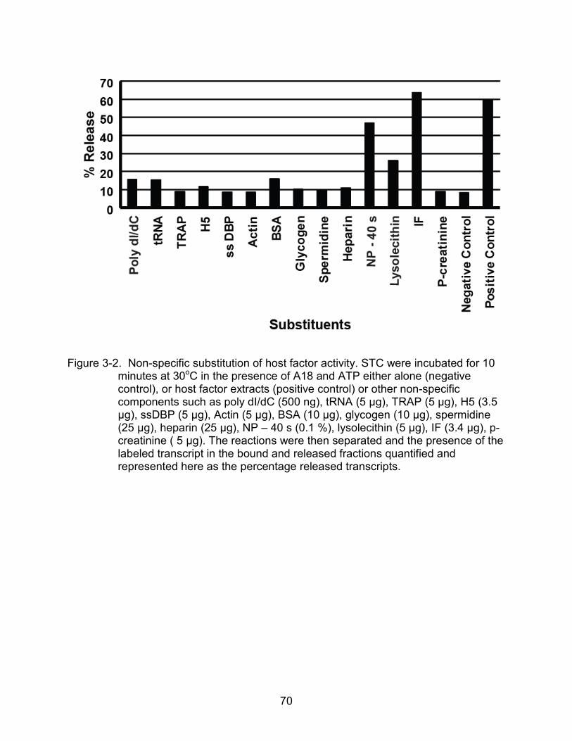
70
Figure 3-2. Non-specific substitution of host factor activity. STC were incubated for 10 minutes at 30oC in the presence of A18 and ATP either alone (negative control), or host factor extracts (positive control) or other non-specific components such as poly dI/dC (500 ng), tRNA (5 μg), TRAP (5 μg), H5 (3.5 μg), ssDBP (5 μg), Actin (5 μg), BSA (10 μg), glycogen (10 μg), spermidine (25 μg), heparin (25 μg), NP – 40 s (0.1 %), lysolecithin (5 μg), IF (3.4 μg), p-creatinine ( 5 μg). The reactions were then separated and the presence of the labeled transcript in the bound and released fractions quantified and represented here as the percentage released transcripts.

71
Figure 3-3. Detergent stimulation of A18 mediated termination. The termination potential of STCs was assayed by incubating the complexes under the four test reaction conditions with no added detergents as a control. Similar reactions were carried out in the presence of a detergent. The results are represented as the percentage of the total transcripts that were released into the supernatant.

72
Figure 3-4. Heat stability of the host factor activity. Host factor extracts were heat treated to different degrees for increasing lengths of time as indicated. The heat treated extracts were assayed for their activity in the termination assay by incubating STCs in the presence of untreated or heat treated host factor extracts and A18 and ATP.

73
Figure 3-5. Host factor is a protein. A & B. Trypsin treatment of host factor extracts. Host extracts were treated with zero (M), 7.5 U, 15 U, 38 U or 75 U of trypsin and loss of activity was analyzed by the termination assay (A) and SDS PAGE (B). C & D. Micrococcal nuclease sensitivity of the host extracts. Host extracts were treated with 0 (M), 12, 24 or 48 µg/ml micrococcal nuclease and tested for loss of activity in both the termination assay (c) and agarose gel electrophoresis (D).
.

74
Figure 3-6. Host factor is a ubiquitous eukaryotic protein. HeLa cytoplasmic extracts (HCE), drosophila nuclear extracts (DNE), yeast extracts (YE) and bovine serum albumin (BSA) controls were titrated into termination reactions containing STC, A18 and ATP. The percentage of total transcripts released into the supernatant was quantified and is graphically represented.

75
CHAPTER 4
BIOCHEMICAL CHARACTERISTICS OF A18 MEDIATED TERMINATION
The isolation of the stalled ternary complex (STC) made it possible to investigate
the properties of the termination of vaccinia post-replicative ternary complex in the
presence of A18 independent of the elongation properties of the transcription ternary
complex. The biochemical characteristics of A18 mediated termination are detailed in
this chapter. An important pre-requisite for termination of ternary complexes is the ability
of ternary complexes to pause. The role of pausing of the ternary complex during A18
mediated termination is also investigated in this chapter.
Specific Aim 3: Properties of A18 Mediated Termination of Stalled Ternary Complexes
The optimal conditions for A18 mediated termination were determined by
terminating the STC in the presence of A18 and HF under different reaction conditions.
These studies helped assess not only the reaction optima for A18 mediated termination,
such as salt and divalent metal ion requirements, but also the enzyme kinetics and
energy requirements for the process of termination.
Kinetics of Termination of STC
The kinetics of the termination reaction was examined. The concentration
dependence of A18 mediated termination was determined by incubating the STCs with
increasing concentrations of purified histidine tagged A18 in the presence of host factor
and ATP. The reactions when separated into bound and released fractions and
analyzed for the release of mRNA transcripts show a linear increase in termination with
increasing concentrations of A18. Release of 50 % of the transcripts was achieved at 20
ng of the purified protein and saturation levels of release under 60 % were achieved
76
with 100 ng of the protein (Fig. 4-1A). Higher concentrations of A18 when tested
mediated no further release of transcripts (data not shown).
The time course of A18 mediated termination was determined by assessing the
percentage of nascent mRNA transcripts released from a STC when such a complex
was incubated at 30oC in the presence of A18, host factor and ATP, for varying lengths
of time. The results when quantified show that the rate of termination increases linearly
with time and reaches maximum by 10 minutes, beyond which length of time no further
increase in termination is noted (Fig. 4-1B). These data indicate that termination of the
STC occurs in a concentration and time dependent manner.
Salt Optima for Termination
Ternary complexes in the various systems, prokaryotic, eukaryotic and even early
vaccinia early complexes have been shown to have distinct salt sensitivities. The salt
sensitivity of the ternary complex also varies along with the stage of transcription.
Termination of vaccinia early polymerase has been shown to be highly salt sensitive,
with a marked inhibition of termination seen in the presence of as much as 50 mM NaCl.
Transcription initiation, elongation and hence termination under our reaction conditions
were carried out at 80 mM KoAc. In order to determine the salt optima for and sensitivity
of the process of termination of STC generated from vaccinia intermediate gene specific
promoters, we titrated in varying concentrations of the different salts during A18
mediated termination reactions. Briefly, STC were washed in transcription buffer in the
absence of salt. The complexes were then terminated in the presence of A18, host
factor and ATP in buffers containing varying concentrations of salt. A18 and the host
factor were added prior to the indicated concentrations of salt.

77
Minimal levels of termination (30 %) over the background (10 %) seen regularly in
the absence of any factors is seen, indicating that termination of STC occurs
independent of salt (Fig. 4-2). Increase in termination is seen with increasing salt
concentrations with optima between 100 – 150 mM salt, beyond which concentration we
see a gradual inhibition of termination. Concentrations over 200 mM salt could inhibit
interaction of A18 and host factor with the STC or each other, thereby inhibiting
termination. Of the different salts tests, the STC is most stable in reactions containing
KoAc, as seen from the autoradiogram in figure 4-2. In the presence of KoAc,
destabilization of the ternary complex, indicated by the presence of lower molecular
weight transcripts is seen only in concentrations of 0.5 M salt. In the presence of the
other salts, lower molecular weight transcripts indicating unstable complexes appear at
salt concentrations as low as 200 – 250 mM salt. At higher salt concentrations (0.5 M)
of these salts, an increase in the terminated products was observed which can be
attributed to the destabilization and dissociation of the complex in the presence of high
salt.
Divalent Metal Ion Optima for Termination
The divalent metal ion optima for termination was tested by incubating STCs that
were washed and resuspended in transcription buffer lacking magnesium chloride, in
reactions containing 100 ng A18, 8.5 μg HF and increasing concentrations of
magnesium chloride between 0 – 20 mM. These magnesium titrations were carried out
under different ATP concentrations, 0.2 mM, 1 mM and 5 mM. Termination was
quantified as the percentage of transcripts released into the supernatant for each
reaction and represented graphically (Fig. 4-3). A18 mediated termination of vaccinia
intermediate ternary complexes is dependent on magnesium ion. This is not surprising

78
given that A18 belongs to the superfamily II helicases whose true substrate is the
Mg:ATP complex. The reaction is optimal at 5 mM magnesium beyond which
concentration the reaction is inhibited possibly due to the formation of dimagnesium-
ATP complexes. The percentage of terminated products increases in an ATP
dependent manner.
Energy Requirement for Termination
A requirement for hydrolysable ATP during termination was initially identified;
however the nature the energy requirement for A18 mediated termination could not be
examined since the process of termination was coupled to transcription elongation.
Isolation of STC and hence the uncoupling to the two processes allowed us to study the
energy requirement for A18 mediated termination. STCs were terminated in the
presence of A18 and HF and 1 mM concentrations of different NTPs and dNTP (Fig. 4-
4A). The results show that in the absence of ATP or the presence of a non-hydrolysable
ATP analog, AMP-PNP, termination is inhibited. Termination is mediated in the
presence of ATP and dATP but none of the other NTP/dNTP. The energy requirement
for termination mimics the energy requirement described for A18 in an in vitro ATPase
assay, indicating the significance of the enzyme’s ATPase function during termination.
Based on the energy requirement of the enzyme, we determined the optimal ATP
concentrations for the termination process. STC were allowed to termination in
presence of A18 and host factor under increasing concentrations of ATP. The reactions
were incubated at 30oC for 2, 5 or 10 minutes. The saturation curves derived for these
reactions indicate that for our reaction conditions the ATP maxima is achieved at 0.5
mM ATP (Fig. 4-4B). Based on the initial velocity curves of the reaction we were able to

79
determine an approximate Km for the enzyme in the termination process of 200 µM
ATP.
Specific Aim 4: A18 Mediated Termination of Paused Complexes
Transcriptional pausing, which allows for a temporary halt of the ternary complex
during the process of elongation, forms a key intermediate between elongation and
termination. Not all paused complexes terminate, since paused complexes are capable
of subsequent re-elongation. However, in the various termination mechanisms
examined, some of which were described in the introduction to this thesis, we see that
pausing of the ternary complex is necessary during termination. In our lab the role of
A18 has been examined both during elongation and on artificially stalled ternary
complexes. The experiments described in the following section of the thesis examine
the role of A18 in termination with respect to ternary complexes that are capable of
pausing.
In order to examine A18 mediated termination in the context of paused complexes,
we utilized a previously discussed template system. A schematic representation of the
transcription scenarios with this system is presented in figure 4-5. The bead bound
pG8GU template, as described in the materials and methods, contains the vaccinia
intermediate gene G8R promoter, followed sequentially by a 37 nucleotide G-less
cassette, a 41 nucleotide T-less cassette, a 9 T region and 169 nucleotide non-specific
downstream template (Fig. 4-5). Transcription through the G-less cassette in the
presence of radiolabeled nucleotide and 3’oMeGTP generates a pulse labeled transcript
(P) stalled at the end of the 37 nucleotide cassette. These complexes are washed under
high stringency conditions and allowed to resume elongation under different nucleotide
conditions to derive different end products. When the ternary complex is elongated with

80
1 mM NTPs in the absence of UTP, the complex transcribes through the T-less cassette
and pauses at the 9 T site, giving rise to transcripts referred to as T9 transcripts. Upon
elongation in the presence of a full complement of NTPs, the ternary complex
transcribes to the end of the template to generate a full length (FL) transcript.
Elongation in the presence of 1 mM NTPs and limiting concentrations of UTP generates
a mixed population of T9 and FL transcripts.
Termination of Paused Elongation Complexes
We investigated the effect of A18 mediated termination on paused complexes by
adding A18 and HF to elongation complexes under increasing concentrations of UTP.
The reactions were then separated into beadbound and released fractions and analyzed
on a 10 % urea polyacrylamide gel (Fig. 4-6). Heterogeneity of the pulse labeled
transcripts can be attributed to the 5’ poly A heads that are representative of vaccinia
post-replicative transcripts. Elongation in the absence of UTP generated T9 transcripts
that fractionated with the bound fractions in the absence of A18 and in the released
fractions in the presence of A18. Elongation in the presence of saturating (1 mM) UTP
generated run-off FL transcripts that were split between the bound and released
fractions in the absence of A18 and were completely released in the presence of A18.
Intermediate concentrations of UTP generated a mixed population of T9 and FL
transcripts that were also released in the presence of A18. Interestingly, under these
elongation conditions, discrete intermediate size transcripts between 150 – 200 bases
on the ladder were apparent. These transcripts appear only in the presence of A18 and
are released into the supernatant. A similar but diminished effect is noted in the
presence of 1 mM UTP indicated by the appearance of a single intermediate sized
transcript in the released lane in the presence of A18. The results imply that the

81
presence of both A18 and the HF during elongation reveals the presence of cryptic
pause sites of the ternary complex by mediating termination at these sites. Analysis of
the template sequence downstream from the 9 T site revealed several stretches of T-
rich regions which could act as pause sites in the presence of limiting concentrations of
UTP.
Early Time Course of Elongation
The results from the experiment in Figure 4-6 indicate that cryptic pause sites are
revealed in the presence of A18 and HF, but not in the presence of HF alone. The
absence of these paused polymerase complexes when A18 is absent could be
attributed to the extended incubation time (20 minutes) to which reactions are
subjected. Polymerases have been shown to form transient pauses, lasting between 1-6
seconds, during the normal course of elongation. The ternary complexes used in our
assays were generated from infected cell extracts and then stripped under high
stringency wash conditions. We wanted to confirm the ability of the polymerase in our
reactions to pause during elongation by examining the early time points during
elongation. Pulse-labeled complexes (P) were allowed to elongate under limiting (40
μM) UTP conditions for 110 seconds. Time points were collected every ten seconds and
analyzed on a 10 % urea polyacrylamide gel (Fig. 4-7). The results indicate the
appearance of the T9 transcript within 10 seconds of elongation, increasing in intensity
with time. The T9 transcripts form the major pause product and most of the transcripts
still remain as pulse labeled transcript. This is due to short span of the elongation time
and indicates only the early events during elongation. Even within this time span it is
clear from the appearance of the gradient of increasing transcript lengths with

82
increasing time that the ternary complexes that are being generated and investigated
under these conditions form transient pauses during elongation.
A18-specific Termination of Paused Complexes
A18 mediated termination has shown a dependence on host factor(s) in the
context of the stalled ternary complex. In this section we investigate the host factor
dependence of A18 mediated termination of paused complexes. Pulse labeled
complexes were generated as described in the preceding sections and elongated in the
presence of limiting (40 μM) UTP. Elongation was carried out under increasing
concentrations of A18, both in the presence and absence of the host factor extracts
(Fig. 4-8). Elongation in the absence of A18 and the host factor under these conditions
results in the formation of a mixed population of T9 and FL transcripts in the bound
fractions. The presence of the host factor by itself shows similar results. An overall
decrease in transcripts seen in the presence of the host factor could be due to
nucleases present in the extracts. Addition of A18 to these reactions results in the
appearance of the intermediate length paused complexes. Increasing concentrations of
A18 mediates a decrease in the amount of FL and higher molecular weight paused
complexes, indicating that A18 negatively affects transcription. Importantly, even in the
absence of the host factor A18 seems to mediate termination in a population of the
paused complexes as evidenced by the presence of transcripts in both the bound and
released lanes in the absence of the host factor. However, complete termination of the
paused complexes does require the presence of the host factor.

83
Figure 4-1. Kinetics of A18 mediated termination. A) STCs were incubated in the presence of 8.5 μg HF and 1 mM ATP with increasing concentrations of A18. B) STCs were incubated with 100 ng A18, 8.5 μg HF and 1 mM ATP a1 30oC for increasing lengths of time as indicated. Termination of ternary complex in both these experiments is represented by the percentage of total transcripts released into the supernatant is represented.

84
Figure 4-2. Salt optima for A18 mediated termination. STCs were terminated under different salt conditions, separated as bound and released fractions and run on a 6 % denaturing urea polyacrylamide gel. Each row of the autoradiogram represents a different salt assayed in the termination reaction. In each gel, the lanes below represent the bound fractions and the lanes below represent the released fractions. The results from the autoradiogram are also represented graphically in the panel below.

85
Figure 4-3. Metal ion optima for termination. STCs were terminated under increasing concentrations of magnesium ion, in the presence of A18 and HF. Termination reactions were carried out under different ATP conditions. Part A represents the titration between 0 – 20 mM magnesium chloride. Part B is a view of the finer concentrations of the titration curves.

86
Figure 4-4. Energy requirement for termination. A) SECs were terminated in the presence of A18 and HF. The reactions were separated as bound and released and termination represented as the percentage of released transcripts. The NTP or dNTP used as the energy source for each individual reaction is as indicated. B) SECs incubated with A18 and HF were titrated into reactions containing increasing concentrations of ATP. Successful termination at 2, 5 or 10 minutes was assessed by quantifying the percentage of total transcripts that were released into the supernatant and is represented graphically.

87
Figure 4-5. Transcription of paused complexes. The beadbound (brown circle) pG8GU template has a vaccinia G8R promoter (yellow box), followed sequentially by a 37 nt G-less cassette (blue box), a 41 nt T-less cassette (green box), a 9 T site (red box) and downstream non-specific DNA (grey box). Transcription through the G-less cassette with radiolabeled nucleotide and in the absence of GTP generates complexes with a pulse labeled transcript (P). Elongation of these ternary complexes in the absence of UTP pauses the complexes at the 9T site generating T9 transcripts. Elongation in the presence of saturating UTP generates complexes with full length (FL) transcripts. Elongation under limiting UTP conditions generates mixed populations of complexes.

88
Figure 4-6. Termination of paused complexes. Transcription of beadbound pG8GU with radiolabeled nucleotide and 3’oMeGTP generated pulse labeled transcripts (P), which were washed and elongated under increasing concentrations of UTP, either in the presence of HF alone or HF and A18 as indicated above each autoradiogram. The reactions were separated into bound and released fractions and run on a 10 % urea polyacrylamide gel. T9 indicates transcripts formed from complexes paused at the 9 T site and FL indicates read-through full length transcripts.

89
Figure 4-7. Early time course of elongation. Pulse labeled complexes (P) that had been washed under high stringency conditions and allowed to elongate in the presence of 1mM ATP, 1 mM CTP, 1 mM GTP and 40 μM UTP at 30oC for 110 seconds. Time points were collected every 10 seconds and analyzed on a 10 % urea polyacrylamide gel. (T9 – 9 T paused transcript; FL – full length transcript; L – ladder)

90
Figure 4-8. A18 specific termination. Pulse labeled complexes (P) were generated and elongated with 1mM ATP, 1 mM CTP, 1 mM GTP and 40 μM UTP, increasing concentrations of A18, either in the presence or the absence of HF as indicated above. The reactions were separated into bound and released fractions and run on a 10 % urea polyacrylamide gel. (T9 – 9 T paused transcript; FL – full length transcript; L – ladder)

91
CHAPTER 5 DISCUSSION
Transcription of vaccinia intermediate and late classes of genes is carried out in
the host cytoplasm. The generation of intermediate promoter specific transcription
complexes in vitro by incubating virus infected cell extracts with beadbound templates is
an important tool to study the process of viral post-replicative transcription. In this study
we have shown that stable forms of the ternary complex can be isolated by stripping the
intermediate transcription complexes in high stringency conditions containing 0.1 %
sarkosyl in the presence of 1 M salt. We have shown that these complexes are
functionally active, capable of elongation and termination. These active complexes can
be used to probe the roles of the both viral and host proteins in the regulation of
vaccinia intermediate and presumably late transcription. In our study, we have
manipulated these complexes to study the process of A18 mediated termination of
vaccinia intermediate genes.
Host Factor Requirement for A18 Mediated Termination
In contrast to early gene expression, vaccinia post-replicative gene expression is
regulated by cellular factors due to the cytoplasmic site of gene expression. As
described in the introduction, host factors have been shown to play a role in both
intermediate and late transcription initiation. Host factors presumably regulate the
process of transcription elongation. It has been shown previously that the process of
post-replicative termination also has a host factor requirement. Although the cellular
component that aids termination has proved resistant to our attempts at purification, we
have in our study shown that the host factor(s) is a protein(s) component of the host
extracts, due to the trypsin sensitivity and heat sensitivity of the active components. The
92
active component is a specific yet ubiquitously present eukaryotic protein or protein
complex. Our experience with the purification and identification of the host factor
suggests that the factor could be part of a dynamic complex involved in the regulation of
transcription. Purification of such a multi-subunit complex to homogeneity would prove
difficult through conventional chromatography. Eukaryotic transcription is regulated by
several such multi-subunit complexes which could be involved in regulation of viral
transcription. Vaccinia post-replicative ternary complex could, like its eukaryotic
counterpart, be present as a holoenzyme consisting of the viral core subunits and
additional host subunits. The host factor could be a component of the viral holoenzyme
that gets detached in vitro and requires to be added back in to make the polymerase
termination efficient. The presence of the protein in prokaryotic extracts has been
difficult to determine due to the presence of nucleases.
Properties of A18 Mediated Termination of STC
Termination assays involving STC have demonstrated that while both A18 and the
HF are required to terminate the stalled ternary complex, neither factor is required to be
part of the elongating polymerase in order to mediate termination. Vaccinia stalled
intermediate ternary complexes are most stable in potassium salt. Termination of these
complexes is insensitive to the absence of salt, but has definite salt optima at 100 mM
salt, conditions which have been shown to be inhibitory to the termination of vaccinia
early ternary complexes. Inhibition of termination at higher salts where the STC is still
stable hints at the disruption of protein-nucleic acid or protein-protein interactions that
may be essential for A18 mediated termination. The termination assay has a divalent
metal dependence with optima at 5 mM, higher concentrations than which are inhibitory
to termination. The energy requirement profile of A18 in the termination assay mimics
93
the energy requirement profile of the purified enzyme in an in vitro ATPase assay,
capable of hydrolyzing only ATP and dATP. These data suggest that the enzyme
utilizes its ATPase and translocase potential to bind the template DNA and translocate
along the template to function as remodeling the ternary complex. ATPase titrations of
the termination assay provide an approximate Km for the enzyme at substrate
concentrations of 200 μM ATP, which is a more physiologically relevant value when
compared to 2.4 mM derived for the enzyme in an in vitro ATPase assay. This indicates
that the function of the enzyme in the context of the ternary complex is energetically
more favorable.
A18 Mediated Termination and Transcriptional Pausing
We investigated the relationship between A18 mediated termination and
transcriptional pausing by introducing A18 to actively elongating ternary complexes,
capable of pausing at an artificial pause site introduced into the template. In the
presence of the HF, A18 terminates the paused complexes. A18 revealed the presence
of natural or intrinsic pause sites on the template by terminating the ternary complexes
at these sites, under limiting nucleotide conditions. At saturating concentrations of
nucleotides, when the ternary complex has an increased rate of elongation and a
decreased propensity to pause, we observe a reduction in the termination of intrinsically
paused complexes. This proves that pausing is a key requirement for A18-mediated
termination. In the context of the actively elongating ternary complex capable of
pausing, the presence of A18 by itself negatively affects the transcription potential of the
elongating complex as evidenced by a decrease in the amount of full length transcripts
and the increase in the amount of paused complexes with increasing concentrations of
A18 in bound fractions. Moreover, A18 alone seems to terminate a subset of the above

94
paused complexes independent of the host factor. This is direct contrast to the STC
where termination is solely dependent on the presence of both A18 and HF. The
vaccinia genome is AT rich with several T-rich regions that could serve as cryptic pause
sites from where elongating polymerases can by terminated by A18 leading to an
overall heterogeneity of the 3’ ends in vivo.
Model for A18 Mediated Termination
A comparison of the different mechanisms of termination in both prokaryotic and
eukaryotic systems shows two common features that are associated with transcription
termination. In factor dependent termination mechanisms the factors are either RNA or
DNA helicases that have translocase activity. The other common feature involves the
state of the RNA polymerase. Pausing or stalling of elongating RNA polymerases is a
key intermediate between active elongation and complete termination. In bacteria,
termination is mediated by rho, a hexameric helicase, capable of dissociating paused
elongation complexes utilizing an RNA-dependent ATPase activity to translocate along
the nascent mRNA transcript. The other factor dependent mechanism involves mfd, a
DNA helicase that translocates along the DNA template, capable of terminating a
transcription complex stalled at an area containing a DNA lesion in an energy
dependent manner. In addition to these factor-mediated mechanisms of termination,
there also exists a factor independent mechanism, intrinsic termination, where a stable
RNA hairpin loop structure formed when the polymerase pauses, further destabilizes
and terminates the complex. In eukaryotic system, although the mechanism for
termination is not clearly understood, the importance of pausing in termination and the
helicase nature of the transcription factors involved in termination has been well
documented. The following paragraphs contain a description of these common features.
95
Pausing is a key regulatory mechanism employed by elongating RNA
polymerases. Transcriptional complexes move in an asynchronous or discontinuous
manner along a DNA template. At some sequence specific regions of the template the
polymerase is capable of transient pausing. These pauses are not considered a normal
state of the elongating polymerase but an “off-pathway” state of the polymerase,
because for any given pause sequence in the DNA not all polymerases undergo
pausing; a subset of the polymerase population pauses at the site while the rest of them
continue their normal elongation. Bacterial RNA polymerases have been shown in vitro
to be capable of pausing ubiquitously once every 100-200 base pairs of the DNA
template for as short as 1-6 seconds. Ubiquitous pauses can be further stabilized or
prolonged by additional mechanisms that include i) backtracking of the RNA
polymerase, leading to a displacement and extrusion of the 3’ end of the RNA transcript
from the active site within the ternary complex, ii) protein interactions with the paused
polymerase, especially in the ssDNA region of the non-template strand, iii) stable RNA
hairpin loop structures, iv) nature of the downstream DNA template (Landick, 2006). In
their role as regulatory components of transcription, paused polymerases can either be
acted upon by positive elongation factors and resume productive elongation or back-
track excessively or decay into arrested complexes that are associated with the
template DNA but are incapable of elongation on their own or be terminated by
appropriate termination factors.
Helicases play a major role in many cellular processes including transcription.
Historically helicases were defined as nucleic acid dependent ATPases that couple
energy from NTP hydrolysis to unwinding duplex nucleic acid structures. Based on the

96
sequence similarities among these proteins, common motifs were identified and used to
classify new members of the family. However not all helicases are functionally capable
of unwinding double stranded nucleic acid. Currently, all helicases are considered motor
proteins capable of translocating along nucleic acids in an energy dependent manner. A
subset of these proteins is capable of unwinding duplex DNA or RNA molecules.
Another function associated with translocases/helicases is dismantling or remodeling
nucleoprotein complexes, such as nucleosomes, splicesosomes, transcription
complexes (Singleton et al., 2007).
A18 shares these features that are a hallmark of different mechanisms of
termination. Previous studies have identified A18 as a DNA helicase capable in vitro of
only a weak helicase activity activated by a ssDNA, dsDNA or DNA:RNA dependent
ATPase activity. We postulate that A18, by virtue of its helicase motifs, functions to
translocate along nucleic acid and disrupt or dissociate the ternary complex. In this
dissertation we have shown the relevance of pausing during A18 mediated termination.
The pause sites revealed during elongation in the presence of A18 indicates that A18
possibly stabilizes the RNAP at certain pause sites and ultimately causes its
termination.
Our overall hypothesis based on these data is that A18 binds and stabilizes
ubiquitously paused polymerases and utilizes ATPase derived energy to dissociate the
polymerase. Since the ATPase activity is required for termination and is activated by
either ssDNA, dsDNA or DNA:RNA hybrids, in the context of a paused polymerase
there are three possible regions for A18 to interact with the ternary complex, A1, A2 or
A3 (Fig. 5-1). In binding to position A1, which is the downstream DNA template, A18

97
could act as a roadblock protein to stabilize the paused polymerase and by virtue of its
translocase function exert an opposing force on the ternary complex and mediate its
disruption or dissociation. In binding to position A2, which is the exposed ssDNA region
of the non-template strand of the ternary complex, A18 could stabilize the ubiquitously
paused polymerase, one of the mechanisms suggested to stabilize pauses (Landick,
2006). Termination by this mechanism could be possible in a manner similar to early
vaccinia polymerases, where NPHI binds one of the polymerase subunits and couples
ssDNA dependent ATPase energy to mediate termination (Christen et al., 2008;
Piacente et al., 2008). This would involve in addition to A18’s 3’-5’ translocase activity
and ssDNA dependent ATPase activity, a possible interaction of A18 with the vaccinia
RNA polymerase, no such interaction has been identified to date. The third possibility
involves the binding of A18 to position A3, which is the upstream DNA sequence.
Termination in this scenario could be mediated by A18’s binding directly to the RNA
polymerase and stabilizing the pause. DNA and polymerase bound A18 could
dissociate the ternary complex by translocating in the direction against the direction of
normal polymerase movement. This mechanism would share similarities with mfd
mediated termination, in that mfd also interacts with both the RNA polymerase and the
upstream DNA. Mfd terminates only stalled ternary complexes incapable of elongation
by inducing a forward translocation of the polymerase and ultimately dissociating the
polymerase. However, in our scenario, A18 can mediate termination of elongation
competent polymerases by stabilizing their pausing. This would indicate that A18 upon
binding the upstream DNA and RNA polymerase would have to dissociate the complex
by translocating away from the polymerase rather than towards it as in the case of mfd.
98
There are two possible roles for the host factor during termination. In the first
scenario, the host protein or protein complex would interact with A18 and enhance or
activate its termination functions, which would be 1) the ability to bind DNA and catalyze
ATP hydrolysis, 2) ATPase catalyzed translocase activity, 3) interaction with the RNA
polymerase. Our data with paused polymerases suggest that A18 can mediate
termination of a subset of paused complexes independent of the host factor, indicating
that A18 does not require the host factor to activate its termination functions. A second
possible role for the host factor involves interaction and subsequent enhancement of the
ternary complex by the host protein or protein complex making the ternary complex
sensitive to A18 mediated termination. The RNA polymerase ternary complex can exist
in multiple conformational states that are dictated by the nature of its current
interactions. In our studies we examined termination from two sets of ternary
complexes; a stalled ternary complex which was completely resistant to A18 mediated
termination in the absence of the host factor and an actively elongating ternary complex
which was partially sensitive to A18 mediated termination in the absence of the host
factor. These data indicate that the active state of the polymerase plays a distinguishing
role between the two scenarios. The possible role of the host factor could be inducing a
termination sensitive state of the ternary complex. Moreover, under normal
circumstances during viral replication, the host protein could be a RNA polymerase
associated factor that was separated from the complex during in vitro manipulations.
Role of A18 in Viral Post-replicative Transcription Regulation
Post-replicative transcription termination in vaccinia virus lacks a distinct cis-acting
termination signal and results in the formation of transcripts of variable lengths with
ragged 3’ ends. In A18 defective mutants, infections result in normal early transcripts

99
but much longer than normal intermediate and late transcripts proving its importance in
transcription termination. Our model for A18 mediated termination can be applied to the
viral infection scenario where ubiquitous pauses of the elongating RNA polymerase are
stabilized by the presence of A18 leading to the dissociation of the complex. The AT-
rich (60 %) vaccinia genome serves as an ideal template to induce ubiquitous pausing
of an elongating RNA polymerase. Termination of these polymerases would result in
transcripts with heterogeneous 3’ ends. Owing to the cytoplasmic site of gene
expression the vaccinia post-replicative ternary complex is possibly regulated by both
viral and host factors. With the exception of the host factor requirement during A18
mediated termination in vitro, no other host requirement during elongation and
termination has been described. The role of viral transcription regulators like G2, J3 –
positive elongation factors and a multifunctional factor, H5 were identified during genetic
characterization of temperature sensitive mutants. Evidence from in vitro interaction
studies have indicated at a possible interaction of A18 with these factors. Although A18
does not need to interact with an elongating polymerase in order to terminate it, in the
context of the viral infection, the post-replicative elongation complex is probably
regulated by both positive regulators like J3 and G2 and negative regulators like A18
and the host factor, which bias the ternary complex either into productive elongation or
termination.
In addition, in some post-replicative scenarios that are exceptions to the rule, H5
has been shown to mediate cleavage of transcripts resulting in homogeneous
transcripts, indicating at a possibly more complex process of transcription regulation,
similar to those seen in eukaryotic systems. Transcription termination in eukaryotic
100
system involves transcription past the poly A signal which leads to the recruitment of 3’
end processing factors. Among the 3’ end processing factors are the multisubunit
cleavage and polyadenylation specificity factors (CPSF) and cleavage stimulation
factors (CstF) that cleave the nascent mRNA to generate two RNA molecules, a capped
mRNA that is subsequently polyadenylated and a polymerase associated RNA with an
uncapped 5’end. This uncapped molecule serves as a substrate for cellular
exonucleases that function in tandem with other factors to mediate termination of the
still elongating polymerase. The role of the vaccinia cleavage factor, H5, in the
formation of a few homogeneous post-replicative transcripts implies that the process of
transcription termination in vaccinia post-replicative genes might involve a mechanism
similar to eukaryotic co-transcriptional cleavage, where nascent mRNA transcripts are
generated by the cleavage of RNA at cryptic cleavage sites, leaving behind a
functionally elongating polymerase with an uncapped 5’ end that is terminated by A18.
Future Directions
A18 was identified during phenotypic studies of temperature sensitive mutants. A
role for the purified protein was described by a series of biochemical experiments,
ultimately leading to investigations of its properties in the context of a transcription
complex. A detailed understanding of the protein’s mechanistic role during termination
and macroscopic function in viral transcription regulation can be achieved through
multipronged studies involving the protein.
The properties of A18 mediated termination of a stalled ternary complex were
investigated in this dissertation. In the context of actively elongating polymerases under
limiting nucleotide conditions, we have shown that A18 is capable of i) negatively
affecting elongation, 2) stabilizing pauses and 3) terminating a subset of the

101
polymerases on its own. It will be interesting to analyze the properties of these three
functions, with respect to their salt sensitivity and hydrolysable energy requirement to
identify conditions to distinguish the three functions.
The ATPase activity associated with the protein was shown to be activated in the
presence of ssDNA, dsDNA and DNA:RNA hybrids. The levels of activation of A18’s
ATPase function in an in vitro assay were seen to vary depending on the nature of the
oligomer used. In addition A18 has ATP independent DNA binding property. It would
prove useful to investigate the nature of A18’s binding to nucleic acids in order to
determine if the enzyme has a sequence preference or specificity. Eukaryotic pol I
termination mechanism involves a two factor mechanism, where one of the factors
binds sequence specifically to downstream DNA regions to serve as a roadblock to the
polymerase complex. The nature of A18’s nucleic acid binding can be investigated
either by performing filter binding assays or non-denaturing gel analysis of A18 bound
labeled nucleic acids.
A18 is classified as belonging to the superfamily II helicase family of proteins and
has a weak DNA helicase activity in vitro. Our hypothesis suggests that A18 functions
as a translocase protein that ultimately mediates nucleoprotein remodeling or disruption.
The translocase function of A18 can be tested by performing the triple helix forming
oligonucleotide (TFO) assay (Whitehouse et al., 2003; Smith et al., 2007). Pyrimidine-
rich TFOs functions as a sequence specific dsDNA binding ligand. TFOs recognize the
complementary purine containing dsDNA and bind the major groove of dsDNA utilizing
Hoogsten hydrogen bonds to form T-AT and C-GC triplets. These triple helices form
only in low pH conditions and not under neutral pH. By incubating a radiolabeled TFO

102
with the target DNA at low pH triplexes are formed. The TFO can be dissociated from
the dsDNA in the presence of a translocase. Performing translocase assays under pH
conditions where re-binding of the TFO to the dsDNA is inhibited, we can identify the
ability of A18 to function as a translocase.
The role of A18 in the context of the viral transcription regulators is an important
area of investigation. This would involve transcription in the presence of purified
vaccinia polymerase. While purified vaccinia polymerase has been used to study early
vaccinia termination, post-replicative termination studies have been carried using
extract generated ternary complexes. In post-replicative vaccinia transcription, purified
polymerases have been re-constituted in vitro to study transcription initiation.
Characterization of this system to study elongation and termination would make it
possible to perform mechanistic studies involving transcription regulation. Using such a
re-constituted system, not only can we investigate the effect of individual transcription
factors but also the effect these factors have on each other and in combination on the
ternary complex.
A reconstituted post-replicative transcription system using purified polymerases
and the minimal required factors to initiate transcription in vitro can also be used to
investigate the structural details and the stability of the transcription complex, as has
been described for early vaccinia polymerases (Hagler & Shuman, 1992a; Hagler &
Shuman, 1992b; Hagler & Shuman, 1992c). Structural studies by DNA footprinting
analyses will help define parameters of the post-replicative ternary complexes such as
the length of the ternary complex, distance between the active site and the edges of the
ternary complex and length of the DNA-RNA hybrid. Such a well-defined ternary

103
complex will be suited to investigate the mechanistic details of A18 mediated
termination. Using this system we will be able to define the region of A18’s interaction
with the ternary complex. A mutant version of the EcoR1 enzyme (Gln111) that can bind
the recognition site but is catalytically inactive has been used in studies with mfd to
define the template region required for mfd binding during termination (Park et al.,
2002). This involves using templates where the elongating polymerase is stalled at
specific positions either downstream or upstream from the EcoR1 binding sites. Mutant
EcoR1 is allowed to bind templates with the stalled polymerases prior to A18 mediated
termination. Termination will be abrogated in those constructs where the EcoR1 mutant
is bound to A18 binding regions, thus identifying A18’s site of interaction with the ternary
complex.
This experimental setup will also help address a fundamental mechanistic aspect
of A18 mediated termination. Mfd and rho mediated transcription termination has been
shown to occur by a mechanism of forward translocation where the RNA polymerase
terminates when it is forced into forward translocation in the absence of RNA synthesis
(Park et al., 2002; Park & Roberts, 2006). A similar mechanism was proposed for
intrinsic terminators as well (Yarnell & Roberts, 1999). In recent years an alternate
allosteric model which does not require forward translocation to mediate termination has
been demonstrated for intrinsic terminators as well as rho dependent terminators
(Epshtein et al., 2007; Epshtein et al, 2010). Given the basic structural conservation
among the different RNA polymerases, A18 mediated termination would also be
mediated by either of these two processes. Evidence from the Gollnick lab suggests
that vaccinia early polymerase termination might involve forward translocation. Of the

104
three models for A18 mediated termination, binding of A18 to positions A1 and A3 (Fig.
5-1) and subsequent dissociation of the ternary would inhibit forward translocation. The
dependence of A18 mediated termination on forward translocation can be determined
by comparing A18’s ability to terminate under two different reaction scenarios. In one
case the ternary complex would be stalled at a region directly upstream from the mutant
EcoR1 protein allowing no space for the forward translocation of the polymerase. In
another setup the ternary complex would be stalled a few bases upstream from the
mutant EcoR1, allowing for forward translocation of the polymerase. Preferential
termination of one of these complexes over the other would help us gain an insight into
the mechanistic aspects of A18 mediated termination.
A different experimental approach to investigate the properties and function of A18
involves deletion analysis of the purified protein to test its in vitro functions. Most
superfamily II helicase proteins have, in addition to the helicase motifs, accessory
domains that confers specificity for each protein. These accessory domains can posses
catalytic function or be involved in protein interaction. A18 is a 56 KDa protein belonging
to the superfamily II helicases consisting of 493 amino acids. As seen in figure 5-2, the
helicase motifs occupy the central regions of the protein, leaving behind
uncharacterized 100 amino acid regions on both the amino and carboxyl terminal ends
which could function as accessory domains. We possess a number of assays to test for
different aspects of the enzyme’s function; ATPase assays, nucleic acid binding assay,
stabilization of paused complexes, host factor dependent and independent termination.
Deletion constructs of the protein can be expressed, and tested in these assays to
identify if these regions are required for normal ATPase function or additional nucleic

105
acid binding, possible host factor interaction. These deletion mutants can also be tested
for their ability to bind other viral transcription factors.
In conclusion, the work described in this dissertation has refined our
understanding of the vaccinia termination factor; in that it has identified pausing to be a
key mechanistic requirement for A18 mediated termination. It has also described a
possible role for the protein in stabilizing ubiquitously paused polymerases. We have
been able to show for the first time host factor independent termination by A18, leading
us to a possible hypothesis that the active conformation state of the polymerase is a
requirement for A18 mediated termination. Moreover, we have shown that the host
factor is a cellular protein or protein complex and suggest that it functions to modulate
the ternary complex to make it termination sensitive. Further investigation of the
mechanistic aspects of vaccinia post-replicative transcription termination and A18’s role
in terminating the ternary complex would further the understanding of the process of
eukaryotic termination.

106
Figure 5-1. Model for A18 mediated termination. An ubiquitously paused ternary elongation complex, represented here by the RNA polymerase in grey, DNA template in blue and RNA transcript in orange, are stably paused and ultimately terminated by the action of A18, shown here as the purple circles. A18 has three possible binding sites. A1, downstream DNA template region. A2, ssDNA in the ternary bubble. A3, upstream DNA template region. Grey arrow indicates direction of RNA polymerase transcription. Green arrows indicate direction of A18 translocation.

107
Figure 5-2. Schematic representation of the A18 protein. The protein A18 is depicted as the grey bar containing in purple the different helicase signature motifs that classify it as a superfamily 2 helicase. (N-amino terminus; C-carboxyl terminus)

108
LIST OF REFERENCES
Adhya, S., Gottesman, M., 1978. Control of transcription termination. Annu. Rev. Biochem. 47, 967-996.
Ahn, B.Y., Gershon, P.D., Moss, B., 1994. RNA polymerase-associated protein Rap94 confers promoter specificity for initiating transcription of vaccinia virus early stage genes. J. Biol. Chem. 269, 7552-7557.
Ahn, B.Y., Moss, B., 1992. RNA polymerase-associated transcription specificity factor encoded by vaccinia virus. Proc. Natl. Acad. Sci. U.S.A. 89, 3536-3540.
Allison, L.A., Moyle, M., Shales, M., Ingles, C.J., 1985. Extensive homology among the largest subunits of eukaryotic and prokaryotic RNA polymerases. Cell 42, 599-610.
Baldick Jr., C.J., Cassetti, M.C., Harris, N., Moss, B., 1994. Ordered assembly of a functional preinitiation transcription complex, containing vaccinia virus early transcription factor and RNA polymerase, on an immobilized template. J. Virol. 68, 6052-6056.
Baldick Jr., C.J., Keck, J.G., Moss, B., 1992. Mutational analysis of the core, spacer, and initiator regions of vaccinia virus intermediate-class promoters. J. Virol. 66, 4710-4719.
Baroudy, B.M., Moss, B., 1980. Purification and characterization of a DNA-dependent RNA polymerase from vaccinia virions. J. Biol. Chem. 255, 4372-4380.
Bayliss, C.D., Condit, R.C., 1995. The vaccinia virus A18R gene product is a DNA-dependent ATPase. J. Biol. Chem. 270, 1550-1556.
Beck, L.L., Smith, T.G., Hoover, T.R., 2007. Look, no hands! Unconventional transcriptional activators in bacteria. Trends Microbiol. 15, 530-537.
Bengali, Z., Townsley, A.C., Moss, B., 2009. Vaccinia virus strain differences in cell attachment and entry. Virology 389, 132-140.
Black, E.P., Condit, R.C., 1996. Phenotypic characterization of mutants in vaccinia virus gene G2R, a putative transcription elongation factor. J. Virol. 70, 47-54.
Black, E.P., Moussatche, N., Condit, R.C., 1998. Characterization of the interactions among vaccinia virus transcription factors G2R, A18R, and H5R. Virology 245, 313-322.
Boeger, H., Griesenbeck, J., Strattan, J.S., Kornberg, R.D., 2003. Nucleosomes unfold completely at a transcriptionally active promoter. Mol. Cell 11, 1587-1598.
Borukhov, S., Lee, J., Laptenko, O., 2005. Bacterial transcription elongation factors: new insights into molecular mechanism of action. Mol. Microbiol. 55, 1315-1324.

109
Borukhov, S., Nudler, E., 2008. RNA polymerase: the vehicle of transcription. Trends Microbiol. 16, 126-134.
Brakel, C., Kates, J.R., 1974a. Poly(A) polymerase from vaccinia virus-infected cells. I. Partial purification and characterization. J. Virol. 14, 715-723.
Brakel, C., Kates, J.R., 1974b. Poly(A) polymerase from vaccinia virus-infected cells. II. Product and primer characterization. J. Virol. 14, 724-732.
Brennan, C.A., Dombroski, A.J., Platt, T., 1987. Transcription termination factor rho is an RNA-DNA helicase. Cell 48, 945-952.
Browning, D.F., Busby, S.J., 2004. The regulation of bacterial transcription initiation. Nat. Rev. Microbiol. 2, 57-65.
Broyles, S.S., 1991. A role for ATP hydrolysis in vaccinia virus early gene transcription. Dissociation of the early transcription factor-promoter complex. J. Biol. Chem. 266, 15545-15548.
Broyles, S.S., 2003. Vaccinia virus transcription. J. Gen. Virol. 84, 2293-2303.
Broyles, S.S., Fesler, B.S., 1990. Vaccinia virus gene encoding a component of the viral early transcription factor. J. Virol. 64, 1523-1529.
Broyles, S.S., Li, J., Moss, B., 1991. Promoter DNA contacts made by the vaccinia virus early transcription factor. J. Biol. Chem. 266, 15539-15544.
Broyles, S.S., Moss, B., 1986. Homology between RNA polymerases of poxviruses, prokaryotes, and eukaryotes: nucleotide sequence and transcriptional analysis of vaccinia virus genes encoding 147-kDa and 22-kDa subunits. Proc. Natl. Acad. Sci. U.S.A. 83, 3141-3145.
Broyles, S.S., Moss, B., 1988. DNA-dependent ATPase activity associated with vaccinia virus early transcription factor. J. Biol. Chem. 263, 10761-10765.
Broyles, S.S., Yuen, L., Shuman, S., Moss, B., 1988. Purification of a factor required for transcription of vaccinia virus early genes. J. Biol. Chem. 263, 10754-10760.
Buratowski, S., 2003. The CTD code. Nat. Struct. Mol. Biol. 10, 679-680.
Burgess, R.R., 1969. Separation and characterization of the subunits of ribonucleic acid polymerase. J. Biol. Chem. 244, 6168-6176.
Cairns, B.R., 2009. The logic of chromatin architecture and remodelling at promoters. Nature 461, 193-198.
110
Carter, G.C., Law, M., Hollinshead, M., Smith, G.L., 2005. Entry of the vaccinia virus intracellular mature virion and its interactions with glycosaminoglycans. J. Gen. Virol. 86, 1279-1290.
Cassetti, M.A., Moss, B., 1996. Interaction of the 82-kDa subunit of the vaccinia virus early transcription factor heterodimer with the promoter core sequence directs downstream DNA binding of the 70-kDa subunit. Proc. Natl. Acad. Sci. U.S.A. 93, 7540-7545.
Chen, C.Y., Chang, C.C., Yen, C.F., Chiu, M.T.K., Chang, W.H., 2009. Mapping RNA exit channel on transcribing RNA polymerase II by FRET analysis. Proc. Natl. Acad. Sci. U.S.A. 106, 127-132.
Christen, L.A., Piacente, S., Mohamed, M.R., Niles, E.G., 2008. Vaccinia virus early gene transcription termination factors VTF and Rap94 interact with the U9 termination motif in the nascent RNA in a transcription ternary complex. Virology 376, 225-235.
Cochran, M.A., Puckett, C., Moss, B., 1985. In vitro mutagenesis of the promoter region for a vaccinia virus gene: evidence for tandem early and late regulatory signals. J. Virol. 54, 30-37.
Cohrs, R.J., Condit, R.C., Pacha, R.F., Thompson, C.L., Sharma, O.K., 1989. Modulation of ppp(A2'p)nA-dependent RNase by a temperature-sensitive mutant of vaccinia virus. J. Virol. 63, 948-951.
Conaway, J.W., Shilatifard, A., Dvir, A., Conaway, R.C., 2000. Control of elongation by RNA polymerase II. Trends Biochem. Sci. 25, 375-380.
Condit, R.C., Lewis, J.I., Quinn, M., Christen, L.M., Niles, E.G., 1996a. Use of lysolecithin-permeabilized infected-cell extracts to investigate the in vitro biochemical phenotypes of poxvirus ts mutations altered in viral transcription activity. Virology 218, 169-180.
Condit, R.C., Motyczka, A., 1981. Isolation and preliminary characterization of temperature-sensitive mutants of vaccinia virus. Virology 113, 224-241.
Condit, R.C., Moussatche, N., Traktman, P., 2006. In a nutshell: structure and assembly of the vaccinia virion. Adv. Virus Res. 66, 31-124.
Condit, R.C., Niles, E.G., 2002. Regulation of viral transcription elongation and termination during vaccinia virus infection. Biochim. Biophys. Acta 1577, 325-336.
Condit, R.C., Xiang, Y., Lewis, J.I., 1996b. Mutation of vaccinia virus gene G2R causes suppression of gene A18R ts mutants: implications for control of transcription. Virology 220, 10-19.

111
Cong, P., Shuman, S., 1992. Methyltransferase and subunit association domains of vaccinia virus mRNA capping enzyme. J. Biol. Chem. 267, 16424-16429.
Cong, P., Shuman, S., 1995. Mutational analysis of mRNA capping enzyme identifies amino acids involved in GTP binding, enzyme-guanylate formation, and GMP transfer to RNA. Mol. Cell. Biol. 15, 6222-6231.
Core, L.J., Lis, J.T., 2008. Transcription regulation through promoter-proximal pausing of RNA polymerase II. Science 319, 1791-1792.
Costanzo, F., Fiume, L., La, P.M., Mannini-Palenzona, A., Novello, F., Stirpe, F., 1970. Ribonucleic acid polymerase induced by vaccinia virus: lack of inhibition by rifampicin and alpha-amanitin. J. Virol. 5, 266-269.
Coupar, B.E.H., Boyle, D.B., Both, G.W., 1987. Effect of in vitro mutations in a vaccinia virus early promoter region monitored by herpes simplex virus thymidine kinase expression in recombinant vaccinia virus. J. Gen. Virol. 68, 2299-2309.
Cramer, P., Armache, K.J., Baumli, S., Benkert, S., Brueckner, F., Buchen, C., Damsma, G.E., Dengl, S., Geiger, S.R., Jasiak, A.J., Jawhari, A., Jennebach, S., Kamenski, T., Kettenberger, H., Kuhn, C.D., Lehmann, E., Leike, K., Sydow, J.F., Vannini, A., 2008. Structure of eukaryotic RNA polymerases. Annu. Rev. Biophys. 37, 337-352.
Cramer, P., Bushnell, D.A., Kornberg, R.D., 2001. Structural basis of transcription: RNA polymerase II at 2.8 angstrom resolution. Science 292, 1863-1876.
Cresawn, S.G., Prins, C., Latner, D.R., Condit, R.C., 2007. Mapping and phenotypic analysis of spontaneous isatin-beta-thiosemicarbazone resistant mutants of vaccinia virus. Virology 363, 319-332.
Cyrklaff, M., Risco, C., Fernandez, J.J., Jimenez, M.V., Estaban, M., Baumeister, W., Carrascosa, J.L., 2005. Cryo-electron tomography of vaccinia virus. Proc. Natl. Acad. Sci. U.S.A. 102, 2772-2777.
D'Costa, S.M., Antczak, J.B., Pickup, D.J., Condit, R.C., 2004. Post-transcription cleavage generates the 3' end of F17R transcripts in vaccinia virus. Virology 319, 1-11.
D'Costa, S.M., Bainbridge, T.W., Condit, R.C., 2008. Purification and properties of the vaccinia virus mRNA processing factor. J. Biol. Chem. 283, 5267-5275.
D'Costa, S.M., Bainbridge, T.W., Kato, S.E., Prins, C., Kelley, K., Condit, R.C., 2010. Vaccinia H5 is a multifunctional protein involved in viral DNA replication, postreplicative gene transcription, and virion morphogenesis. Virology 401, 49-60.
Darst, S.A., Edwards, A.M., Kubalek, E.W., Kornberg, R.D., 1991. Three-dimensional structure of yeast RNA polymerase II at 16 + resolution. Cell 66, 121-128.

112
Darst, S.A., Ribi, H.O., Pierce, D.W., Kornberg, R.D., 1988. Two-dimensional crystals of Escherichia coli RNA polymerase holoenzyme on positively charged lipid layers. J. Mol. Biol. 203, 269-273.
Darzacq, X., Shav-Tal, Y., de Turris, V., Brody, Y., Shenoy, S. M., Phair, R. D., Singer, R. H., 2007. In vivo dynamics of RNA polymerase II transcription. Nat. Struct. Mol. Biol. 14, 796-806.
Davison, A.J., Moss, B., 1989a. Structure of vaccinia virus early promoters. J. Mol. Biol. 210, 749-769.
Davison, A.J., Moss, B., 1989b. Structure of vaccinia virus late promoters. J. Mol. Biol. 210, 771-784.
Deng, L., Shuman, S., 1997. Elongation properties of vaccinia virus RNA polymerase: pausing, slippage, 3’ end addition, and termination site choice. Biochemistry 36, 15892-15899.
Deng, L., Shuman, S., 1998. Vaccinia NPH-I, a DExH-box ATPase, is the energy coupling factor for mRNA transcription termination. Genes Dev. 12, 538-546.
Dolan, J.W., Marshall, N.F., Richardson, J.P., 1990. Transcription termination factor rho has three distinct structural domains. J. Biol. Chem. 265, 5747-5754.
Dvir, A., Conaway, J.W., Conaway, R.C., 2001. Mechanism of transcription initiation and promoter escape by RNA polymerase II. Curr. Opin. Genet. Dev. 11, 209-214.
Ebright, R.H., Busby, S., 1995. The Escherichia coli RNA polymerase [alpha] subunit: structure and function. Curr. Opin. Genet. Dev. 5, 197-203.
Edwards, A.M., Darst, S.A., Feaver, W.J., Thompson, N.E., Burgess, R.R., Kornberg, R.D., 1990. Purification and lipid-layer crystallization of yeast RNA polymerase II. Proc. Natl. Acad. Sci. U.S.A. 87, 2122-2126.
Edwards, A.M., Kane, C.M., Young, R.A., Kornberg, R.D., 1991. Two dissociable subunits of yeast RNA polymerase II stimulate the initiation of transcription at a promoter in vitro. J. Biol. Chem. 266, 71-75.
Ensinger, M.J., Martin, S.A., Paoletti, E., Moss, B., 1975. Modification of the 5'-terminus of mRNA by soluble guanylyl and methyl transferases from vaccinia virus. Proc. Natl. Acad. Sci. U.S.A. 72, 2525-2529.
Epshtein, V., Cardinale, C.J., Ruckenstein, A.E., Borukhov, S., Nudler, E., 2007. An allosteric path to transcription termination. Mol. Cell 28, 991-1001.
Epshtein, V., Dutta, D., Wade, J., Nudler, E., 2010. An allosteric mechanism of Rho-dependent transcription termination. Nature 463, 245-249.

113
Farnham, P.J., Platt, T., 1981. Rho-independent termination: dyad symmetry in DNA causes RNA polymerase to pause during transcription in vitro. Nucleic Acids Res. 9, 563-577.
Finkel, J.S., Chinchilla, K., Ursic, D., Culbertson, M.R., 2010. Sen1p performs two genetically separate functions in transcription and processing of U5 small nuclear RNA in Saccharomyces cerevisiae. Genetics 184, 107-118.
Fu, J., Gnatt, A.L., Bushnell, D.A., Jensen, G.J., Thompson, N.E., Burgess, R.R., David, P.R., Kornberg, R.D., 1999. Yeast RNA polymerase II at 5 + resolution. Cell 98, 799-810.
Fuda, N.J., Ardehali, M.B., Lis, J.T., 2009. Defining mechanisms that regulate RNA polymerase II transcription in vivo. Nature 461, 186-192.
Geiselmann, J., Yager, T.D., Gill, S.C., Calmettes, P., von Hippel, P.H., 1992. Physical properties of the Escherichia coli transcription termination factor rho. 1. Association states and geometry of the rho hexamer. Biochemistry 31, 111-121.
Gershon, P.D., Ahn, B.Y., Garfield, M., Moss, B., 1991. Poly(A) polymerase and a dissociable polyadenylation stimulatory factor encoded by vaccinia virus. Cell 66, 1269-1278.
Gershon, P.D., Moss, B., 1990. Early transcription factor subunits are encoded by vaccinia virus late genes. Proc. Natl. Acad. Sci. U.S.A 87, 4401-4405.
Gershon, P.D., Moss, B., 1992. Transition from rapid processive to slow nonprocessive polyadenylation by vaccinia virus poly(A) polymerase catalytic subunit is regulated by the net length of the poly(A) tail. Genes Dev. 6, 1575-1586.
Gershon, P.D., Moss, B., 1993. Stimulation of poly(A) tail elongation by the VP39 subunit of the vaccinia virus-encoded poly(A) polymerase. J. Biol. Chem. 268, 2203-2210.
Ghosh, P., Ishihama, A., Chatterji, D., 2001. Escherichia coli RNA polymerase subunit omega and its N-terminal domain bind full-length β' to facilitate incorporation into the α2β subassembly. Eur. J. Biochem. 268, 4621-4627.
Glover-Cutter, K., Kim, S., Espinosa, J., Bentley, D.L., 2008. RNA polymerase II pauses and associates with pre-mRNA processing factors at both ends of genes. Nat. Struct. Mol. Biol. 15, 71-78.
Gnatt, A.L., Cramer, P., Fu, J., Bushnell, D.A., Kornberg, R.D., 2001. Structural basis of transcription: an RNA polymerase II elongation complex at 3.3 A resolution. Science 292, 1876-1882.
Hagler, J., Shuman, S., 1992a. A freeze-frame view of eukaryotic transcription during elongation and capping of nascent mRNA. Science 255, 983-986.
114
Hagler, J., Shuman, S., 1992b. Stability of ternary transcription complexes of vaccinia virus RNA polymerase at promoter-proximal positions. J. Biol. Chem. 267, 7644-7654.
Hagler, J., Shuman, S., 1992c. Structural analysis of ternary complexes of vaccinia RNA polymerase. Proc. Natl. Acad. Sci. U.S.A. 89, 10099-10103.
Hagler, J., Shuman, S., 1992d. Ternary complex formation by vaccinia virus RNA polymerase at an early viral promoter: analysis by native gel electrophoresis. J. Virol. 66, 2982-2989.
Hagler, J., Shuman, S., 1993. Nascent RNA cleavage by purified ternary complexes of vaccinia RNA polymerase. J. Biol. Chem. 268, 2166-2173.
Hahn, S., 2004. Structure and mechanism of the RNA polymerase II transcription machinery. Nat. Struct. Mol. Biol. 11, 394-403.
Han, M., Kim, U.J., Kayne, P., Grunstein, M., 1988. Depletion of histone H4 and nucleosomes activates the PHO5 gene in Saccharomyces cerevisiae. EMBO J. 7, 2221-2228.
Hanggi, M., Bannwarth, W., Stunnenberg, H.G., 1986. Conserved TAAAT motif in vaccinia virus late promoters: overlapping TATA box and site of transcription initiation. EMBO J. 5, 1071-1076.
Haugen, S.P., Ross, W., Gourse, R.L., 2008. Advances in bacterial promoter recognition and its control by factors that do not bind DNA. Nat. Rev. Microbiol. 6, 507-519.
Hawley, D.K., McClure, W.R., 1983. Compilation and analysis of Escherichia coli promoter DNA sequences. Nucleic Acids Res. 11, 2237-2255.
Heuser, J., 2005. Deep-etch EM reveals that the early poxvirus envelope is a single membrane bilayer stabilized by a geodetic “honeycomb” surface coat. J. Cell Biol. 169, 269-283.
Higman, M.A., Christen, L.A., Niles, E.G., 1994. The mRNA (guanine-7-)methyltransferase domain of the vaccinia virus mRNA capping enzyme. Expression in Escherichia coli and structural and kinetic comparison to the intact capping enzyme. J. Biol Chem. 269, 14974-14981.
Hirschmann, P., Vos, J.C., Stunnenberg, H.G., 1990. Mutational analysis of a vaccinia virus intermediate promoter in vivo and in vitro. J. Virol. 64, 6063-6069.
Holmes, W.M., Platt, T., Rosenberg, M., 1983. Termination of transcription in E. coli. Cell 32, 1029-1032.

115
Howard, B.H., de Crombrugghe, B., 1976. ATPase activity required for termination of transcription by the Escherichia coli protein factor rho. J. Biol. Chem. 251, 2520-2524.
Hsu, L.M., Vo, N.V., Kane, C.M., Chamberlin, M.J., 2003. In vitro studies of transcript initiation by Escherichia coli RNA polymerase. 1. RNA chain initiation, abortive initiation, and promoter escape at three bacteriophage promoters. Biochemistry 42, 3777-3786.
Jasiak, A.J., Hartmann, H., Karakasili, E., Kalocsay, M., Flatley, A., Kremmer, E., Strasser, K., Martin, D.E., Soding, J., Cramer, P., 2008. Genome-associated RNA polymerase II includes the dissociable Rpb4/7 subcomplex. J. Biol. Chem. 283, 26423-26427.
Jin, D.J., Burgess, R.R., Richardson, J.P., Gross, C.A., 1992. Termination efficiency at rho-dependent terminators depends on kinetic coupling between RNA polymerase and rho. Proc. Natl. Acad. Sci. U.S.A. 89, 1453-1457.
Jones, E.V., Puckett, C., Moss, B., 1987. DNA-dependent RNA polymerase subunits encoded within the vaccinia virus genome. J. Virol. 61, 1765-1771.
Juven-Gershon, T., Hsu, J.Y., Theisen, J.W., Kadonaga, J.T., 2008. The RNA polymerase II core promoter -- the gateway to transcription. Curr. Opin. Cell Biol. 20, 253-259.
Juven-Gershon, T., Kadonaga, J.T., 2010. Regulation of gene expression via the core promoter and the basal transcriptional machinery. Dev. Biol. 339, 225-229.
Kates, J., Beeson, J., 1970. Ribonucleic acid synthesis in vaccinia virus. II. Synthesis of polyriboadenylic acid. J. Mol. Biol. 50, 19-33.
Kates, J.R., McAuslan, B.R., 1967. Poxvirus DNA-dependent RNA polymerase. Proc. Natl. Acad. Sci. U.S.A. 58, 134-141.
Katsafanas, G.C., Moss, B., 2004. Vaccinia virus intermediate stage transcription is complemented by Ras-GTPase-activating protein SH3 domain-binding protein (G3BP) and cytoplasmic activation/proliferation-associated protein (p137) individually or as a heterodimer. J. Biol. Chem. 279, 52210-52217.
Kawauchi, J., Mischo, H., Braglia, P., Rondon, A., Proudfoot, N.J., 2008. Budding yeast RNA polymerases I and II employ parallel mechanisms of transcriptional termination. Genes Dev. 22, 1082-1092.
Keck, J.G., Baldick, C.J., Moss, B., 1990. Role of DNA replication in vaccinia virus gene expression: A naked template is required for transcription of three late trans-activator genes. Cell 61, 801-809.

116
Kelleher, R.J., Flanagan, P.M., Kornberg, R.D., 1990. A novel mediator between activator proteins and the RNA polymerase II transcription apparatus. Cell 61, 1209-1215.
Kettenberger, H., Armache, K.J., Cramer, P., 2004. Complete RNA Polymerase II Elongation Complex Structure and Its Interactions with NTP and TFIIS. Mol. Cell 16, 955-965.
Kim, M., Krogan, N.J., Vasiljeva, L., Rando, O.J., Nedea, E., Greenblatt, J.F., Buratowski, S., 2004. The yeast Rat1 exonuclease promotes transcription termination by RNA polymerase II. Nature 432, 517-522.
Kim, Y.J., Bjorklund, S., Li, Y., Sayre, M.H., Kornberg, R.D., 1994. A multiprotein mediator of transcriptional activation and its interaction with the C-terminal repeat domain of RNA polymerase II. Cell 77, 599-608.
Knutson, B.A., Liu, X., Oh, J., Broyles, S.S., 2006. Vaccinia virus intermediate and late promoter elements are targeted by the TATA-binding protein. J. Virol. 80, 6784-6793.
Knutson, B.A., Oh, J., Broyles, S.S., 2009. Downregulation of vaccinia virus intermediate and late promoters by host transcription factor YY1. J. Gen. Virol. 90, 1592-1599.
Kornberg, R.D., 2005. Mediator and the mechanism of transcriptional activation. Trends Biochem. Sci. 30, 235-239.
Kornberg, R.D., 2007. The molecular basis of eukaryotic transcription. Proc. Natl. Acad. Sci. U.S.A. 104, 12955-12961.
Lackner, C.A., Condit, R.C., 2000. Vaccinia virus gene A18R DNA helicase is a transcript release factor. J. Biol. Chem. 275, 1485-1494.
Landick, R., 2006. The regulatory roles and mechanism of transcriptional pausing. Biochem. Soc. Trans. 34, 1062-1066.
Larkin, R.M., Guilfoyle, T.J., 1997. Reconstitution of yeast and arabidopsis RNA polymerase α-like subunit heterodimers. J. Biol. Chem. 272, 12824-12830.
Latner, D.R., Xiang, Y., Lewis, J.I., Condit, J., Condit, R.C., 2000. The vaccinia virus bifunctional gene J3 (nucleoside-2'-O-)-methyltransferase and poly(A) polymerase stimulatory factor is implicated as a positive transcription elongation factor by two genetic approaches. Virology 269, 345-355.
Lee-Chen, G.J., Bourgeois, N., Davidson, K., Condit, R.C., Niles, E.G., 1988. Structure of the transcription initiation and termination sequences of seven early genes in the vaccinia virus HindIII D fragment. Virology 163, 64-79.
117
Lee-Chen, G.J., Niles, E.G., 1988. Transcription and translation mapping of the 13 genes in the vaccinia virus Hindlll D fragment. Virology 163, 52-63.
Li, J., Broyles, S.S., 1993. The DNA-dependent ATPase activity of vaccinia virus early gene transcription factor is essential for its transcription activation function. J. Biol. Chem. 268, 20016-20021.
Li, J., Pennington, M.J., Broyles, S.S., 1994. Temperature-sensitive mutations in the gene encoding the small subunit of the vaccinia virus early transcription factor impair promoter binding, transcription activation, and packaging of multiple virion components. J. Virol. 68, 2605-2614.
Lisser, S., Margalit, H., 1993. Compilation of E. coli mRNA promoter sequences. Nucleic Acids Res. 21, 1507-1516.
Lowery, C., Richardson, J.P., 1977a. Characterization of the nucleoside triphosphate phosphohydrolase (ATPase) activity of RNA synthesi termination factor p. I. Enzymatic properties and effects of inhibitors. J. Biol. Chem. 252, 1375-1380.
Lowery, C., Richardson, J.P., 1977b. Characterization of the nucleoside triphosphate phosphohydrolase (ATPase) activity of RNA synthesis termination factor p. II. Influence of synthetic RNA homopolymers and random copolymers on the reaction. J. Biol. Chem. 252, 1381-1385.
Lowery-Goldhammer, C., Richardson, J.P., 1974. An RNA-dependent nucleoside triphosphate phosphohydrolase (ATPase) associated with Rho termination factor. Proc. Natl. Acad. Sci. U.S.A. 71, 2003-2007.
Luo, Y., Mao, X., Deng, L., Cong, P., Shuman, S., 1995. The D1 and D12 subunits are both essential for the transcription termination factor activity of vaccinia virus capping enzyme. J. Virol. 69, 3852-3856.
Mahr, A., Roberts, B.E., 1984. Arrangement of late RNAs transcribed from a 7.1-kilobase EcoRI vaccinia virus DNA fragment. J. Virol. 49, 510-520.
Malik, S., Roeder, R.G., 2005. Dynamic regulation of pol II transcription by the mammalian Mediator complex. Trends Biochem. Sci. 30, 256-263.
Martin, S.A., Paoletti, E., Moss, B., 1975. Purification of mRNA guanylyltransferase and mRNA (guanine-7-) methyltransferase from vaccinia virions. J. Biol. Chem. 250, 9322-9329.
Mathew, R., Chatterji, D., 2006. The evolving story of the omega subunit of bacterial RNA polymerase. Trends Microbiol. 14, 450-455.
Mercer, J., Helenius, A., 2008. Vaccinia virus uses macropinocytosis and apoptotic mimicry to enter host cells. Science 320, 531-535.

118
Minakhin, L., Bhagat, S., Brunning, A., Campbell, E. A., Darst, S.A., Ebright, R.H., Severinov, K., 2001. Bacterial RNA polymerase subunit ω and eukaryotic RNA polymerase subunit RPB6 are sequence, structural, and functional homologs and promote RNA polymerase assembly. Proc. Natl. Acad. Sci. U.S.A. 98, 892-897.
Mohamed, M.R., Latner, D.R., Condit, R.C., Niles, E.G., 2001. Interaction between the J3R subunit of vaccinia virus poly(A) polymerase and the H4L subunit of the viral RNA polymerase. Virology 280, 143-152.
Mohamed, M.R., Niles, E.G., 2000. Interaction between nucleoside triphosphate phosphohydrolase I and the H4L subunit of the viral RNA polymerase is required for vaccinia virus early gene transcript release. J. Biol. Chem. 275, 25798-25804.
Moss, B., Rosenblum, E.N., Gershowitz, A., 1975. Characterization of a polyriboadenylate polymerase from vaccinia virions. J. Biol. Chem. 250, 4722-4729.
Mukherjee, K., Chatterji, D., 1997. Studies on the omega subunit of Escherichia coli RNA polymerase--its role in the recovery of denatured enzyme activity. Eur. J. Biochem. 247, 884-889.
Mulligan, M.E., Hawley, D.K., Entriken, R., McClure, W.R., 1984. Escherichia coli promoter sequences predict in vitro RNA polymerase selectivity. Nucleic Acids Res. 12, 789-800.
Munyon, W., Paoletti, E., Grace Jr., J.T., 1967. RNA polymerase activity in purified infectious vaccinia virus. Proc. Natl. Acad. Sci. U.S.A. 58, 2280-2287.
Nevins, J.R., Joklik, W.K., 1977a. Isolation and partial characterization of the poly(A) polymerases from HeLa cells infected with vaccinia virus. J. Biol. Chem. 252, 6939-6947.
Nevins, J.R., Joklik, W.K., 1977b. Isolation and properties of the vaccinia virus DNA-dependent RNA polymerase. J. Biol. Chem. 252, 6930-6938.
Osbourn, A.E., Field, B., 2009. Operons. Cell. Mol. Life Sci. 66, 3755-3775.
Pacha, R.F., Condit, R.C., 1985. Characterization of a temperature-sensitive mutant of vaccinia virus reveals a novel function that prevents virus-induced breakdown of RNA. J. Virol. 56, 395-403.
Park, J.S., Marr, M.T., Roberts, J.W., 2002. E. coli Transcription repair coupling factor (Mfd protein) rescues arrested complexes by promoting forward translocation. Cell 109, 757-767.
Park, J.S., Roberts, J.W., 2006. Role of DNA bubble rewinding in enzymatic transcription termination. Proc. Natl. Acad. Sci. U.S.A. 103, 4870-4875.
119
Patel, D.D., Pickup, D.J., 1989. The second-largest subunit of the poxvirus RNA polymerase is similar to the corresponding subunits of procaryotic and eucaryotic RNA polymerases. J. Virol. 63, 1076-1086.
Piacente, S., Christen, L., Dickerman, B., Mohamed, M.R., Niles, E.G., 2008. Determinants of vaccinia virus early gene transcription termination. Virology 376, 211-224.
Price, D.H., 2008. Poised polymerases: on your mark...get set...go! Mol. Cell 30, 7-10.
Prins, C., Cresawn, S.G., Condit, R.C., 2004. An isatin-beta-thiosemicarbazone-resistant vaccinia virus containing a mutation in the second largest subunit of the viral RNA polymerase is defective in transcription elongation. J. Biol. Chem. 279, 44858-44871.
Proudfoot, N.J., 1989. How RNA polymerase II terminates transcription in higher eukaryotes. Trends Biochem. Sci. 14, 105-110.
Richard, P., Manley, J.L., 2009. Transcription termination by nuclear RNA polymerases. Genes Dev. 23, 1247-1269.
Richardson, J.P., 1970. Rho factor function in T4 RNA transcription. Cold Spring Harb. Symp. Quant. Biol. 35, 127-133.
Richardson, L.V., Richardson, J.P., 1996. Rho-dependent termination of transcription is governed primarily by the upstream Rho utilization (rut) sequences of a terminator. J. Biol. Chem. 271, 21597-21603.
Roberts, J., Park, J.S., 2004. Mfd, the bacterial transcription repair coupling factor: translocation, repair and termination. Curr. Opin. Microbiol. 7, 120-125.
Roberts, J.W., 1969. Termination factor for RNA synthesis. Nature 224, 1168-1174.
Rondon, A.G., Mischo, H.E., Kawauchi, J., Proudfoot, N.J., 2009. Fail-safe transcriptional termination for protein-coding genes in S. cerevisiae. Mol. Cell 36, 88-98.
Rosales, R., Harris, N., Ahn, B.Y., Moss, B., 1994. Purification and identification of a vaccinia virus-encoded intermediate stage promoter-specific transcription factor that has homology to eukaryotic transcription factor SII (TFIIS) and an additional role as a viral RNA polymerase subunit. J. Biol. Chem. 269, 14260-14267.
Rosel, J.L., Earl, P.L., Weir, J.P., Moss, B., 1986. Conserved TAAATG sequence at the transcriptional and translational initiation sites of vaccinia virus late genes deduced by structural and functional analysis of the HindIII H genome fragment. J. Virol. 60, 436-449.
120
Rosonina, E., Kaneko, S., Manley, J.L., 2006. Terminating the transcript: breaking up is hard to do. Genes Dev. 20, 1050-1056.
Ross, W., Gourse, R.L., 2009. Analysis of RNA polymerase-promoter complex formation. Methods 47, 13-24.
Sanz, P., Moss, B., 1999. Identification of a transcription factor, encoded by two vaccinia virus early genes, that regulates the intermediate stage of viral gene expression. Proc. Natl. Acad. Sci. U.S.A. 96, 2692-2697.
Sawadogo, M., Roeder, R.G., 1985. Factors involved in specific transcription by human RNA polymerase II: analysis by a rapid and quantitative in vitro assay. Proc. Natl. Acad. Sci. U.S.A. 82, 4394-4398.
Sayre, M.H., Tschochner, H., Kornberg, R.D., 1992. Reconstitution of transcription with five purified initiation factors and RNA polymerase II from Saccharomyces cerevisiae. J. Biol. Chem. 267, 23376-23382.
Scaife, J., Beckwith, J.R., 1966. Mutational alteration of the maximal level of Lac operon expression. Cold Spring Harb. Symp. Quant. Biol. 31, 403-408.
Schmidt, M.C., Chamberlin, M.J., 1984. Binding of rho factor to Escherichia coli RNA polymerase mediated by nusA protein. J. Biol. Chem. 259, 15000-15002.
Schwartz, A., Margeat, E., Rahmouni, A.R., Boudvillain, M., 2007. Transcription termination factor rho can displace streptavidin from biotinylated RNA. J. Biol. Chem. 282, 31469-31476.
Selby, C.P., Sancar, A., 1993. Molecular mechanism of transcription-repair coupling. Science 260, 53-58.
Shultzaberger, R.K., Chen, Z., Lewis, K.A., Schneider, T.D., 2007. Anatomy of Escherichia coli sigma70 promoters. Nucleic Acids Res. 35, 771-788.
Shuman, S., Broyles, S.S., Moss, B., 1987. Purification and characterization of a transcription termination factor from vaccinia virions. J. Biol. Chem. 262, 12372-12380.
Shuman, S., Morham, S.G., 1990. Domain structure of vaccinia virus mRNA capping enzyme. Activity of the Mr 95,000 subunit expressed in Escherichia coli. J. Biol. Chem. 265, 11967-11972.
Shuman, S., Moss, B., 1988. Vaccinia virus poly(A) polymerase. Specificity for nucleotides and nucleotide analogs. J. Biol. Chem. 263, 8405-8412.
Shuman, S., Moss, B., 1989. Bromouridine triphosphate inhibits transcription termination and mRNA release by vaccinia virions. J. Biol. Chem. 264, 21356-21360.

121
Sigurdsson, S., Dirac-Svejstrup, A.B., Svejstrup, J.Q., 2010. Evidence that transcript cleavage is essential for RNA polymerase II transcription and cell viability. Mol. Cell 38, 202-210.
Simpson, D.A., Condit, R.C., 1994. The vaccinia virus A18R protein plays a role in viral transcription during both the early and the late phases of infection. J. Virol. 68, 3642-3649.
Simpson, D.A., Condit, R.C., 1995. Vaccinia virus gene A18R encodes an essential DNA helicase. J. Virol. 69, 6131-6139.
Sims, R.J., Belotserkovskaya, R., Reinberg, D., 2004. Elongation by RNA polymerase II: the short and long of it. Genes Dev. 18, 2437-2468.
Singleton, M.R., Dillingham, M.S., Wigley, D.B., 2007. Helicases and nucleic acid translocases. Annu. Rev. Biochem. 76, 23-50.
Smith, A.J., Szczelkun, M.D., Savery, N.J., 2007. Controlling the motor activity of a transcription-repair coupling factor: autoinhibition and role of RNA polymerase. Nuc. Acid Res. 35, 1802-1811.
Spencer, E., Shuman, S., Hurwitz, J., 1980. Purification and properties of vaccinia virus DNA-dependent RNA polymerase. J. Biol. Chem. 255, 5388-5395.
Svejstrup, J.Q., Vichi, P., Egly, J.M., 1996. The multiple roles of transcription/repair factor TFIIH. Trends Biochem. Sci. 21, 346-350.
Sweetser, D., Nonet, M., Young, R.A., 1987. Prokaryotic and eukaryotic RNA polymerases have homologous core subunits. Proc. Natl. Acad. Sci. U.S.A. 84, 1192-1196.
Townsley, A.C., Weisberg, A.S., Wagenaar, T.R., Moss, B., 2006. Vaccinia virus entry into cells via a low-pH-dependent endosomal pathway. J. Virol. 80, 8899-8908.
Venkataraman, K., Brown, K.M., Gilmartin, G.M., 2005. Analysis of a noncanonical poly(A) site reveals a tripartite mechanism for vertebrate poly(A) site recognition. Genes Dev. 19, 1315-1327.
Venkatesan, S., Baroudy, B.M., Moss, B., 1981. Distinctive nucleotide sequences adjacent to multiple initiation and termination sites of an early vaccinia virus gene. Cell 25, 805-813.
Venkatesan, S., Gershowitz, A., Moss, B., 1982. Complete Nucleotide Sequences of Two Adjacent Early Vaccinia Virus Genes Located Within the Inverted Terminal Repetition. J. Virol. 44, 637-646.

122
Vos, J.C., Sasker, M., Stunnenberg, H.G., 1991a. Promoter melting by a stage-specific vaccinia virus transcription factor is independent of the presence of RNA polymerase. Cell 65, 105-113.
Vos, J.C., Sasker, M., Stunnenberg, H.G., 1991b. Vaccinia virus capping enzyme is a transcription initiation factor. EMBO J. 10, 2553-2558.
Wei, R.R., Richardson, J.P., 2001. Identification of an RNA-binding Site in the ATP binding domain of Escherichia coli Rho by H2O2/Fe-EDTA cleavage protection studies. J. Biol. Chem. 276, 28380-28387.
West, S., Proudfoot, N.J., Dye, M.J., 2008. Molecular dissection of mammalian RNA polymerase II transcriptional termination. Mol. Cell 29, 600-610.
Westover, K.D., Bushnell, D.A., Kornberg, R.D., 2004. Structural basis of transcription: separation of RNA from DNA by RNA polymerase II. Science 303, 1014-1016.
Whitbeck, J.C., Foo, C.H., Ponce de Leon, M., Eisenberg, R.J., Cohen, G.H., 2009. Vaccinia virus exhibits cell-type-dependent entry characteristics. Virology 385, 383-391.
Whitehouse, I., Stockdale, C., Flaus, A., Szczelkun, M.D., Owen-Hughes, T., 2003. Evidence for DNA translocation by ISWI Chromatin remodeling enzyme. 23Mol. Cell. Biol. 23, 1935-1945.
Wright, C.F., Oswald, B.W., Dellis, S., 2001. Vaccinia virus late transcription is activated in vitro by cellular heterogeneous nuclear ribonucleoproteins. J. Biol. Chem. 276, 40680-40686.
Xiang, Y., Simpson, D.A., Spiegel, J., Zhou, A., Silverman, R.H., Condit, R.C., 1998. The vaccinia virus A18R DNA helicase is a postreplicative negative transcription elongation factor. J. Virol. 72, 7012-7023.
Yamaguchi, Y., Takagi, T., Wada, T., Yano, K., Furuya, A., Sugimoto, S., Hasegawa, J., Handa, H., 1999. NELF, a multisubunit complex containing RD, cooperates with DSIF to repress RNA polymerase II elongation. Cell 97, 41-51.
Xie, Z., Price, D., 1997. Drosophila factor 2, an RNA polymerase II transcript release factor, has DNA-dependent ATPase activity. J. Biol. Chem. 272, 31902-31907.
Yang, C., Bolotin, E., Jiang, T., Sladek, F.M., Martinez, E., 2007. Prevalence of the initiator over the TATA box in human and yeast genes and identification of DNA motifs enriched in human TATA-less core promoters. Gene 389, 52-65.
Yang, Z., Moss, B., 2009. Interaction of the vaccinia virus RNA polymerase-associated 94-kilodalton protein with the early transcription factor. J. Virol. 83, 12018-12026.

123
Yarnell, W.S., Roberts, J.W., 1999. Mechanism of intrinsic transcription termination and anti-termination. Science 284, 611-615.
Yu, L., Shuman, S., 1996. Mutational analysis of the RNA triphosphatase component of vaccinia virus mRNA capping enzyme. J. Virol. 70, 6162-6168.
Yuen, L., Moss, B., 1987. Oligonucleotide sequence signaling transcriptional termination of vaccinia virus early genes. Proc. Natl. Acad. Sci. U.S.A. 84, 6417-6421.
Zhang, G., Campbell, E.A., Minakhin, L., Richter, C., Severinov, K., Darst, S.A., 1999. Crystal structure of Thermus aquaticus core RNA polymerase at 3.3 + resolution. Cell 98, 811-824.
Zhang, Z., Gilmour, D.S., 2006. Pcf11 is a termination factor in drosophila that dismantles the elongation complex by bridging the CTD of RNA polymerase II to the nascent transcript. Mol. Cell 21, 65-74.

124
BIOGRAPHICAL SKETCH
Aparna Manoharan was born in Madras, India in the year 1982. She enrolled for
undergraduate studies in 1999 at the Birla Institute of Technology and Science (BITS),
Pilani, India where she graduated in 2003 with a master’s (Hons.) in biological sciences.
She was admitted into the Interdisciplinary Program in Biomedical Sciences at the
University of Florida in 2003. She joined the Condit lab in the summer of 2004 where
she started work on her dissertation. In the summer of 2010 she receives her Ph.D. in
biomedical sciences.