role of angiotensin-converting enzyme in myeloid cell
TRANSCRIPT

INVITED REVIEW LETTER Open Access
Role of angiotensin-converting enzyme inmyeloid cell immune responsesDuo-Yao Cao1, Suguru Saito1, Luciana C. Veiras1, Derick Okwan-Duodu1,2, Ellen A. Bernstein1, Jorge F. Giani1,2,Kenneth E. Bernstein1,2 and Zakir Khan1,2*
* Correspondence: [email protected] of BiomedicalSciences, Cedars-Sinai MedicalCenter, Los Angeles, CA 90048, USA2Department of Pathology andLaboratory Medicine, Cedars-SinaiMedical Center, Davis Res. Bldg.,Rm. 2014, 8700 Beverly Blvd, LosAngeles, CA 90048, USA
Abstract
Angiotensin-converting enzyme (ACE), a dicarboxypeptidase, plays a major role inthe regulation of blood pressure by cleaving angiotensin I into angiotensin II (Ang II),a potent vasoconstrictor. Because of its wide substrate specificity and tissue distribution,ACE affects many diverse biological processes. In inflammatory diseases, includinggranuloma, atherosclerosis, chronic kidney disease and bacterial infection, ACE expressiongets upregulated in immune cells, especially in myeloid cells. With increasing evidencesconnecting ACE functions to the pathogenesis of these acquired diseases, it is suggestedthat ACE plays a vital role in immune functions. Recent studies with mouse models ofbacterial infection and tumor suggest that ACE plays an important role in the immuneresponses of myeloid cells. Inhibition of ACE suppresses neutrophil immune response tobacterial infection. In contrast, ACE overexpression in myeloid cells strongly inducedbacterial and tumor resistance in mice. A detailed biochemical understanding of howACE activates myeloid cells and which ACE peptide(s) (substrate or product) mediatethese effects could lead to the development of novel therapies for boosting immunityagainst a variety of stimuli, including bacterial infection and tumor.
Keywords: Angiotensin-converting enzyme, Myeloid cells, Immune response, Neutrophils,Macrophages, Dendritic cells, Hematopoiesis, Methicillin-resistant Staphylococcus aureus(MRSA), Melanoma, MHC class I antigen presentation
This article was specially invited by the editors and represents work by leading
researchers.
IntroductionThe renin-angiotensin system (RAS) is a major regulator for blood pressure, fluid and
electrolyte balance, in which sequential action of two enzymes, renin and ACE produce a
bioactive peptide angiotensin II (Ang II) [1, 2]. It is well established that RAS, via the Ang
II AT1 receptor, plays a crucial role in cardiovascular and renal functions by regulating
blood pressure, electrolyte and volume homeostasis [2, 3]. This traditional concept of the
RAS as a circulating endocrine system has been evolved enormously with time and many
new RAS-regulatory components including peptide molecules, enzymes and G protein-
© The Author(s). 2020 Open Access This article is licensed under a Creative Commons Attribution 4.0 International License, whichpermits use, sharing, adaptation, distribution and reproduction in any medium or format, as long as you give appropriate credit tothe original author(s) and the source, provide a link to the Creative Commons licence, and indicate if changes were made. Theimages or other third party material in this article are included in the article's Creative Commons licence, unless indicated otherwisein a credit line to the material. If material is not included in the article's Creative Commons licence and your intended use is notpermitted by statutory regulation or exceeds the permitted use, you will need to obtain permission directly from the copyrightholder. To view a copy of this licence, visit http://creativecommons.org/licenses/by/4.0/.
Cellular & MolecularBiology Letters
Cao et al. Cellular & Molecular Biology Letters (2020) 25:31 https://doi.org/10.1186/s11658-020-00225-w

coupled receptor have been identified [4–7]. The development of ACE inhibitors (ACEi,
eg. enalapril, captopril, lisinopril, ramipril) or AT1 (Angiotensin II receptor type 1)
blockers (ARBs, eg. losartan) not only revolutionized the treatment strategies to treat
hypertension, but also provided a tool for the discovery of many unknown functions of
ACE components in different physiological and pathological mechanisms.
Increasing number of studies suggest that, in different tissues, a local RAS may operate ei-
ther systemically or entirely independent to the circulating counterpart, and it may act as a
whole or in part to meet the specific needs of the individual tissues via autocrine and/or para-
crine pathways [8, 9]. For example, in bone marrow (BM), it affects critical steps of blood cell
production, such as hematopoietic niche [10], myelopoiesis [11], and the development of
other cellular lineages including lymphocytic [12]. There are also enough studies suggesting
local operation of RAS in other organs including cardiac, vascular and renal tissues [13–17].
Renin enzyme is highly specific and extremely limited in its tissue expression, while
ACE is relatively nonspecific and widely expressed in different tissues [5]. Other than
Ang I, ACE can cleave variety of substrates including bradykinin, substance P, tetrapep-
tide N-acetyl-seryl-aspartyl-lysyl-proline (AcSDKP), enkephalins, neurotensin and
others. Because of this wide tissue distribution and substrate specificity, ACE may affect
many diverse biological functions, including renal development, fertility, hematopoiesis
and immunity [5, 18, 19]. This review aims to discuss new biological functions of ACE
in different aspects of the immune response.
ACE and immunoinflammatory diseases
Inflammation plays a critical role in immune activation. Many studies have found ACE
to be a potent pro-inflammatory modulator [20] that plays a role in the recruitment of
inflammatory cells into tissues by regulating chemokines and adhesion molecules [21].
ACE not only functions as a cell membrane ectopeptidase, but it can be secreted into
extracellular milieu by activated myelomonocytic or other lineage cells, and thus, it act
as both local and systemic regulator of peptides [22]. The association of ACE with im-
munoinflammatory diseases has been well established. First report published in 1975
that showed higher serum ACE in patients with sarcoidosis. Now, it is known that cir-
culating ACE is elevated in many other granulomatous diseases, such as Gaucher’s dis-
ease and tuberculosis [23, 24]. In sarcoidal granulomas the increased ACE activity is
predominately contributed by epithelioid cells and macrophages of the granuloma, and
a higher serum level of ACE is observed in a majority of patients [25]. In tuberculosis
granuloma, increased ACE is mostly contributed by alveolar macrophages [26].
Based on CD14 and CD16 expression, monocytes can be divided into two subpopula-
tions Mo1 and Mo2. Percentage of Mo2 increased in many inflammatory diseases in-
cluding chronic kidney disease (CDKs), which expresses higher level of ACE along with
CD14 and CD16 [17, 27]. These monocytes are proinflammatory in nature and associ-
ated with increased atherosclerosis and cardiovascular mortality in hemodialysis
patients [28]. It is now believed that all RAS components express in immune cells. For
example, monocytes and macrophages express renin, ACE, Ang I and Ang II, AT1 and
AT2 receptors [29]. In nervous system also, all RAS components are locally synthesized
by different cell types, including astrocytes, microglia and neutrons [30, 31], and ACE
expression is increased in many neurological autoimmune diseases including
Cao et al. Cellular & Molecular Biology Letters (2020) 25:31 Page 2 of 12

encephalitis and multiple sclerosis [18, 32]. There is no clear understanding why ACE
expression increases in these immunoinflammatory conditions. One possibility is that
ACE activity increases the activation of immune cells in response to immune challenge.
ACE in hematopoiesis and myeloproliferation
BM is a complex and highly organized system that produces all circulating cells. How-
ever, hematopoiesis is tightly regulated by a variety of factors including enzymes, recep-
tors, hormones, cytokines, growth factors and bioactive peptides. The early clues about
the RAS effects on hematopoiesis came from the reduced hematocrit of patients, who
were treating with the ACEi enalapril [33]. Studies further suggested that enalapril
caused erythrocytosis in patients with renal transplantation by reducing the hematocrit
level [34, 35]. In rare patients, ACEi treatment also causes anemia due to its effects on
immune system [36]. In clinical studies, people find that ACEi suppress RBC produc-
tion and decrease white blood cells In clinical studies [37, 38]. These finding clearly
hypothesize a possible role of the RAS in hematopoiesis. Over time, all the known com-
ponents of the RAS, such as angiotensinogen, renin, ACE, AT1a, AT2 (Angiotensin II
receptor type 2), Mas (G protein-coupled receptor) and ACE2 (Angiotensin converting
enzyme 2), have been identified in the BM, and it is now believed that local RAS oper-
ates in the BM [8, 39]. These RAS components, particularly ACE-mediated peptides,
affect several critical steps of hematopoietic cell development in physiological and
pathological conditions [8, 9, 39].
The existence of ACE in primitive lympho-hematopoietic cells, embryonic and fetal
tissues suggests that ACE not only affects erythropoietic progenitors, but might have ef-
fects on neoplastic tissues and primitive pluripotential hematopoietic stem cell popula-
tions [40, 41]. The ACE substrate Ac-SDKP has been extensively studied in the
regulation of stem cell proliferation. ACE inhibition by enalapril increases the level of
Ac-SDKP in both plasma and BM [42, 43], which in turn prevents hematopoietic stem
cell proliferation [44]. In the BM, stromal cells produce a large amount of Ac-SDKP
[43], and the increased ACE expression in stromal cells significantly decreases the Ac-
SDKP level in the BM microenvironment. Rousseau-Plasse et al. (1996) demonstrated
that ACE plays an important role in the recruitment of primitive stem cells into S-
phase by hydrolyzing Ac-SDKP [45]. Comte and colleagues then observed significant
changes in the circulatory hematopoietic progenitors in healthy volunteers following
administration of enalapril. It increased plasma and urinary Ac-SDKP in association
with an increased number of mixed colony-forming unit (mixed-CFU) hematopoietic
progenitors, and in contrast, reduced the number of granulocyte-monocytic colony-
forming unit (CFU-GM) and Burst-forming unit-erythroid (BFU-E) hematopoietic pro-
genitors in the circulation [46]. Ang II is another important peptide of ACE implicated
in hematopoietic cell development. AT1 receptor is expressed by CD34+ hematopoietic
progenitors and stromal cells in human tissues [47]. By binding to AT1a receptor on
CD34+ hematopoietic stem cells, Ang II increases proliferation of these cells [47]. Fur-
ther study validated that ACE inhibition by captopril caused myelosuppression by inhi-
biting stem cell and progenitor cell proliferation rather than depleting the BM cells
[48]. These studies suggest that ACE has a regulatory role in hematopoiesis.
Cao et al. Cellular & Molecular Biology Letters (2020) 25:31 Page 3 of 12

There is now abundant evidence that ACE affect myelopoiesis. These data come from
genetically modified mice with either ACE knockout (KO) or selective overexpression
of ACE in myeloid cells. Absence of ACE suppresses differentiation of myelomonocytic
precursors in ACE KO mice [11]. These mice showed a higher percentage of immature
myeloid precursors including myeloblasts, myelocytes, and metamyelocytes as com-
pared to WT mice. In contrast, a significant reduction of mature neutrophils was found
in ACE-KO BM as compared to WT BM [11]. A dramatic reduction in nucleated
erythroid precursors was found in the BM of ACE KO mice leading to anemia as com-
pared to WT mice. ACE inhibition also increases early BM progenitors, such as LSK
cells (a fraction enriched for hematopoietic stem cells) in ACE KO mice BM [11]. In
addition, increased extramedullary hematopoiesis was found in the spleen that caused
an expansion of immature myeloid cells in ACE KO mice as compared to WT [11].
Similar myelopoiesis abnormalities were observed by pharmacologic inhibition of ACE
in mice. In conclusion, these data suggest that ACE knockout enhanced myeloprolifera-
tion with reduced differentiation that ultimately increased the number of immature
myelomonocytic lineage cells in mice (Fig. 1).
ACE and myeloid cell immune response
Myeloid cells are a heterogenous population derived from a common myeloid progeni-
tor that belongs to the innate immune system. As a first line of defense, myeloid cells
(neutrophils, macrophages, and dendritic cells) are recruited to the inflammatory site
and elicit a variety of immune responses to eliminate potential threats from outside
and inside. Also, macrophages and dendritic cells (DCs) function as classical antigen
presenting cells (APCs), therefore, these cells are crucial for the initiation of an adaptive
Fig. 1 ACE upregulation enhances myeloid cell immune responses. In physiological conditions, ACE expressionincreased during the differentiation and functional maturation of myeloid-derived cells. Upon immune challenge, theexpression of ACE further increased in activated myeloid cells facilitated the optimal immune responses of these cells.Upregulation of ACE in myeloid cells (eg. NeuACE neutrophils and ACE10/10 macrophages) strongly enhancedimmune responses of these cells, beyond the normal capacity of WT cells. In neutrophils, ACE upregulation inducedoxidative bactericidal response, which is due the upregulation of NADPH oxidase activity. In macrophages, ACEupregulation enhanced M1 activation of macrophages due to the increased activation of NF-kB, STAT1 and TNFα,which in turn gives a strong anti-bacterial and anti-tumor phenotype. In APCs (DCs and macrophages), ACE trims thepeptide repertoire before they are bound to MHC class I complex and displayed by cells, which activates T cell –adaptive immune response and humoral immune response
Cao et al. Cellular & Molecular Biology Letters (2020) 25:31 Page 4 of 12

immune response. There is now strong evidence that ACE facilitates the immune acti-
vation of myeloid cells, as discussed below (Fig. 1).
Neutrophil immune response
Neutrophils, also called polymorphonuclear leucocytes, are an essential component of
the innate immune response which plays a major role during acute inflammation [49].
In blood, mature neutrophils are derived from the BM with the goal to locate and kill
harmful invading pathogens [50]. In a lipopolysaccharide (LPS)-induced acute lung in-
flammation mouse model, when mice were pre-treated with the ACEi enalapril, it re-
duced pulmonary recruitment of neutrophils following LPS treatment [51]. This may
demonstrate that ACE could assist in neutrophil recruitment during pulmonary
inflammation.
As protein expression markedly changes in activated neutrophils, we evaluated the
expression of ACE in neutrophils following methicillin-resistant Staphylococcus aureus
(MRSA) challenge. ACE expression upregulated in neutrophils upon activation with
MRSA [52]. To study whether ACE expression is associated with immune functions of
neutrophils, ACE KO and WT mice were subcutaneously challenged with MRSA and
then mice were sacrificed day 3 post-infection. ACE knockout significantly reduced
bacterial resistance in mice as manifested by larger lesion size (Fig. 2) and higher tissue
bacterial burden in ACE KO mice as compared to WT mice. To directly access the ef-
fect of ACE on neutrophil activity, intracellular bacterial killing by purified ACE KO
and WT neutrophils were determined. The intracellular killing of bacteria was signifi-
cantly higher by WT neutrophils as compared to ACE KO neutrophils [52].
Because the lack of ACE expression is linked with neutrophil immune suppression,
we also investigated the phenotype of animals overexpressing ACE in neutrophils. To
study this, a transgenic mouse line called NeuACE mice was generated by using a c-
fms-ACE construct [52]. These mice overexpress ACE (~ 10 fold) in neutrophils. In
contrast to ACE KO, NeuACE mice show enhanced resistance to bacterial infections as
compared to WT mice [52]. To investigate the role of Ang II, mice were pre-treated
Fig. 2 ACE affects anti-bacterial defense. In mice, ACE knockout suppressed bacterial resistance. Representativeimages showing MRSA skin lesion at day 4 post-bacterial subcutaneous injection (1 × 10⁷ CFU/mouse flank)[Cao D-Y et al. Unpublished data]
Cao et al. Cellular & Molecular Biology Letters (2020) 25:31 Page 5 of 12
with either the losartan (ARB) or ramipril (ACEi) for several days before infection.
Ramipril treatment eliminated any differences between NeuACE and WT mice. In con-
trast, losartan had no major effect [52]. This indicates that AngII-AT1R does not medi-
ate ACE effect on neutrophil antibacterial activity. By using renin inhibitor and Ang II
infusion in mice, we confirmed that no Ang peptide mediates neutrophil-ACE effects.
Further, the phenotype of ACE overexpressing NeuACE neutrophils appears independ-
ent of AcSDKP, bradykinin-B2K receptor and substance P-NK1 receptor [52]. Some-
times, treatment with ACEi caused angioedema (a mucosal immunopathological
condition) in patients due to inhibition of kinin degradation and consequent inhibition
of kinin-B1KR signaling pathway [53, 54]. However, the role of B1KR in neutrophil im-
mune response needs to be investigated.
The production of reactive oxygen species (ROS) is very important for neutrophil
antimicrobial activity. We found that ACE directly affects ROS production in neutro-
phils. Upon immune challenge, ACE KO neutrophils produce less ROS, while NeuACE
neutrophils produce more ROS as compared to WT neutrophils (Fig. 1). Further, our
study demonstrated that ACE increases neutrophil antibacterial activity by enhancing
NADPH oxidase enzyme activation, an effect that is independent of the angiotensin II
AT1 receptor [52].
ACEi are generally considered safe and used by millions of patients for the treatment
of hypertension and cardiovascular diseases. As ACE plays an important role in neutro-
phil anti-bacterial activity, any neutrophilic immune suppression by ACEi may increase
risk of infection in vulnerable patients, such as patients with weak immunity. Indeed,
some clinical studies have found an association between the uses of ACEi and increased
risk of infection including sepsis and urinary tract infection [55–57]. Such infections
were not noted with an ARB [57]. Therefore, our findings caution in the use of ACEi
under conditions where patients are vulnerable to infections. The uses of ACEi and risk
of infection needs to be further investigated.
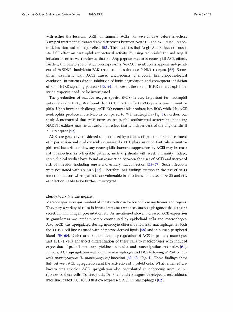
Macrophages immune response
Macrophages as major residential innate cells can be found in many tissues and organs.
They play a variety of roles in innate immune responses, such as phagocytosis, cytokine
secretion, and antigen presentation etc. As mentioned above, increased ACE expression
in granulomas was predominately contributed by epithelioid cells and macrophages.
Also, ACE was upregulated during monocyte differentiation into macrophages in both
the THP-1 cell line cultured with adipocyte-derived lipids [58] and in human peripheral
blood [59, 60]. Under uremic conditions, up-regulation of ACE in primary monocytes
and THP-1 cells enhanced differentiation of these cells to macrophages with induced
expression of proinflammatory cytokines, adhesion and transmigration molecules [61].
In mice, ACE upregulation was found in macrophages and DCs following MRSA or Lis-
teria monocytogenes (L. monocytogenes) infection [62, 63] (Fig. 1). These findings show
link between ACE upregulation and the activation of myeloid cells. What remained un-
known was whether ACE upregulation also contributed in enhancing immune re-
sponses of these cells. To study this, Dr. Shen and colleagues developed a recombinant
mice line, called ACE10/10 that overexpressed ACE in macrophages [62].
Cao et al. Cellular & Molecular Biology Letters (2020) 25:31 Page 6 of 12
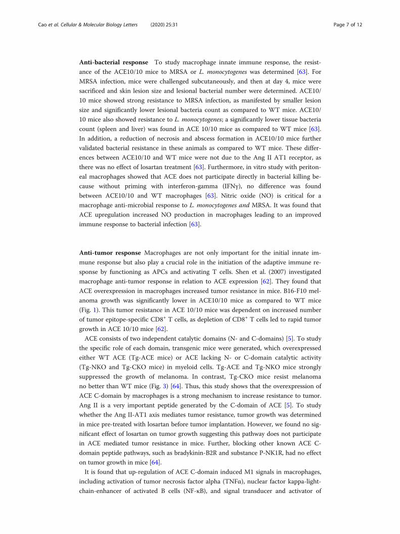
Anti-bacterial response To study macrophage innate immune response, the resist-
ance of the ACE10/10 mice to MRSA or L. monocytogenes was determined [63]. For
MRSA infection, mice were challenged subcutaneously, and then at day 4, mice were
sacrificed and skin lesion size and lesional bacterial number were determined. ACE10/
10 mice showed strong resistance to MRSA infection, as manifested by smaller lesion
size and significantly lower lesional bacteria count as compared to WT mice. ACE10/
10 mice also showed resistance to L. monocytogenes; a significantly lower tissue bacteria
count (spleen and liver) was found in ACE 10/10 mice as compared to WT mice [63].
In addition, a reduction of necrosis and abscess formation in ACE10/10 mice further
validated bacterial resistance in these animals as compared to WT mice. These differ-
ences between ACE10/10 and WT mice were not due to the Ang II AT1 receptor, as
there was no effect of losartan treatment [63]. Furthermore, in vitro study with periton-
eal macrophages showed that ACE does not participate directly in bacterial killing be-
cause without priming with interferon-gamma (IFNγ), no difference was found
between ACE10/10 and WT macrophages [63]. Nitric oxide (NO) is critical for a
macrophage anti-microbial response to L. monocytogenes and MRSA. It was found that
ACE upregulation increased NO production in macrophages leading to an improved
immune response to bacterial infection [63].
Anti-tumor response Macrophages are not only important for the initial innate im-
mune response but also play a crucial role in the initiation of the adaptive immune re-
sponse by functioning as APCs and activating T cells. Shen et al. (2007) investigated
macrophage anti-tumor response in relation to ACE expression [62]. They found that
ACE overexpression in macrophages increased tumor resistance in mice. B16-F10 mel-
anoma growth was significantly lower in ACE10/10 mice as compared to WT mice
(Fig. 1). This tumor resistance in ACE 10/10 mice was dependent on increased number
of tumor epitope-specific CD8+ T cells, as depletion of CD8+ T cells led to rapid tumor
growth in ACE 10/10 mice [62].
ACE consists of two independent catalytic domains (N- and C-domains) [5]. To study
the specific role of each domain, transgenic mice were generated, which overexpressed
either WT ACE (Tg-ACE mice) or ACE lacking N- or C-domain catalytic activity
(Tg-NKO and Tg-CKO mice) in myeloid cells. Tg-ACE and Tg-NKO mice strongly
suppressed the growth of melanoma. In contrast, Tg-CKO mice resist melanoma
no better than WT mice (Fig. 3) [64]. Thus, this study shows that the overexpression of
ACE C-domain by macrophages is a strong mechanism to increase resistance to tumor.
Ang II is a very important peptide generated by the C-domain of ACE [5]. To study
whether the Ang II-AT1 axis mediates tumor resistance, tumor growth was determined
in mice pre-treated with losartan before tumor implantation. However, we found no sig-
nificant effect of losartan on tumor growth suggesting this pathway does not participate
in ACE mediated tumor resistance in mice. Further, blocking other known ACE C-
domain peptide pathways, such as bradykinin-B2R and substance P-NK1R, had no effect
on tumor growth in mice [64].
It is found that up-regulation of ACE C-domain induced M1 signals in macrophages,
including activation of tumor necrosis factor alpha (TNFα), nuclear factor kappa-light-
chain-enhancer of activated B cells (NF-κB), and signal transducer and activator of
Cao et al. Cellular & Molecular Biology Letters (2020) 25:31 Page 7 of 12

transcription 1 (STAT1), and in contrast inhibition M2 signals including STAT3
and STAT6 was found. This appears to reprogram these cells towards a more clas-
sically activated M1 phenotype that is responsible for the enhanced tumor resistance
(Fig. 1) [64].
Metabolic effects Metabolic reprogramming is closely associated with the polarization
of macrophages [65]. For example, LPS dependent inflammation by M1 macrophages
mostly relies on glycolysis and fatty acid biosynthesis. In contrast, tissue repair activity
by M2 macrophages switches their metabolism to fatty acid oxidation and oxidative
phosphorylation [65]. To investigate the molecular basis of how ACE affects cells
phenotypically, metabolism of ACE overexpressing myeloid cells (ACE10/10 and
NeuACE) was determined. It was found that ACE up-regulation significantly increased
ATP, Krebs cycle intermediates and electron transport chain proteins (NDUFB8,
ATP5A, and ATP5β) in ACE10/10 macrophages and NeuACE neutrophils as compared
to WT cells [66]. This appears to underpin some of the phenotypic differences between
these cells and myeloid cells expressing WT levels of ACE, such as superoxide produc-
tion and phagocytic bacterial killing. Consistent with anti-tumor response, the C-
domain of ACE predominately enhanced ATP production in these cells. Again, there
was no effect of Ang II on myeloid cell ATP production [66].
Fig. 3 Melanoma tumor growth. In mice, overexpression of ACE C-domain enhanced macrophage anti-tumor activity. Representative images showing tumor growth at day 14 after intradermal injection of B16-F10 melanoma cells (106 cells/mouse) [Cao D-Y et al. Unpublished data]
Cao et al. Cellular & Molecular Biology Letters (2020) 25:31 Page 8 of 12

Dendritic cells and adaptive immune response
ACE is also expressed by dendritic cells (DCs) [67, 68]. Ang II and bradykinin have
been implicated in DCs maturation and Th1 cell development in a mouse model of
Trypanosoma cruzi infection [69]. ACE expression increased during the differentiation
of DCs and was further increased when these cells were activated with the pro-
inflammatory cytokine IFN-γ [70]. Similarly, when mice were challenged with listeria,
ACE expression was up in splenic macrophages and DCs [70]. Major histocompatibility
complex (MHC) class I proteins play a critical role in the activation of CD8+ T cell
adaptive immune responses against intracellular pathogens and perhaps also against tu-
mors. APCs, such as DCs and macrophages phagocytize cellular debris and present
MHC class I bound antigens for CD8+ T cells. Mice studies suggest that ACE can trim
the peptide repertoire displayed on the surface of APCs as part of the MHC class I
complex (Fig. 1) [70, 71].
In addition to myeloid cells, ACE also plays a role in the activation of lymphoid cells.
Hoch et al. (2009) found a direct autocrine effect of ACE and Ang II on T cell function,
including activation, expression of tissue-homing markers, and production of cytokines
[72], probably due to superoxide production by T cell NADPH oxidase. In a rodent
model of cerebral malaria, following activation by infection, T cells show a significant
increase of CD69 expression, while this reduced to normal when mice were treated
with losartan and captopril [73]. In a clinical study of pulmonary sarcoidosis, the per-
centage of lymphocytes and the CD4/CD8 ratio in bronchoalveolar lavage fluid (BALF)
is coordinated with ACE activity [74]. Further, a connection between Th1 cell cytokines
(IL-12 and IL-18) and ACE activity was determined in BALF [75]. ACE and Ang II have
also been implicated in encephalitis [17]. All these findings suggest an active participa-
tion of ACE in adaptive immune activation. Unfortunately, the critical ACE function
and catalytic substrates are still obscure and need more investigation.
ConclusionsACE is one of the most vital and well-studied peptidases in the RAS. However, in re-
cent years, ACE functions are found to coordinate with immune responses. ACE is up-
regulated in many immunological diseases, such as granuloma. Similarly, upregulation
of ACE was reported in myeloid cells following immune challenges. By comparing ACE
KO, WT and ACE overexpressing neutrophils and macrophages, our studies have dem-
onstrated that ACE not only plays a physiological role in myeloid cell immune re-
sponse, but if overexpressed, ACE further enhances immune responses against a variety
of stimuli, such as bacterial infection and tumor, which is beyond the normal ability of
WT cells. However, whether ACE overexpression has similar effects on human myeloid
cells needs to be investigated. In both tumor and infection studies with transgenic mice
overexpressing only an active C-domain or an active N-domain, we found that the C-
domain of ACE is important for increasing the immune responses of myeloid cells.
However, none of the known ACE C-domain peptides, such as all angiotensin peptides,
bradykinin and Substance p were found to mediate these ACE effects on neutrophils
and macrophages. Identification of ACE peptide(s) (substrate or product) that elicit an
increased immune response may hold great promise for therapeutic manipulation to
boost the immune response against a variety of stimuli, including infections and
tumors.
Cao et al. Cellular & Molecular Biology Letters (2020) 25:31 Page 9 of 12

AbbreviationsACE: Angiotensin-converting enzyme; RAS: Renin-Angiotensin System; Ang II: Angiotensin II; ACEi: ACE inhibitors;ARBs: AT1 blockers; BM: Bone Marrow; AcSDKP: Tetrapeptide N-Acetyl-Seryl-Aspartyl-Lysyl-Proline; WT: Wild type; ACEKO: ACE Knockout; NeuACE: Neutrophil ACE overexpression; Tg-ACE: Transgenic ACE; Tg-CKO: Transgenic ACE Cdomain knockout; Tg-NKO: Transgenic ACE N domain knockout; APCs: Antigen Presenting Cells; DCs: Dendritic Cells;LPS: Lipopolysaccharide; MRSA: Methicillin-Resistant Staphylococcus aureus; L. monocytogenes: Listeria monocytogenes;IFNγ: Interferon-gamma; NO: Nitric oxide; TNFα: Tumor Necrosis Factor alpha; NF-κB: Nuclear Factor Kappa-light-chain-enhancer of activated B cells; STAT: Signal Transducer and Activator of Transcription; MHC: Major HistocompatibilityComplex; BALF: Bronchoalveolar Lavage Fluid
AcknowledgementsThe authors thank Brian Taylor of Cedars-Sinai Medical Center for providing administrative helps during our studies.
Availability of data and materialNot Applicable.
Authors’ contributionsAll authors substantially contributed to the discussion of the content. S.S., L.C.V. E.A.B., and Z.K. researched data for thearticle. D.-Y.C., D. O.-D., J.F.G., K.E.B. and Z.K. drafted and edited the manuscript before submission. L.C.V. and Z.K.revised the manuscript. The author(s) read and approved the final manuscript.
FundingThis work was supported by American Heart Association (AHA) Grants 19CDA34760010 (Z.K.), 17GRNT33661206 (K.E.B.),16SDG30130015 (J.F.G.), and the National Institutes of Health Grants P01HL129941 (K.E.B.), R01AI143599 (K.E.B.),R01HL142672 (J.F.G.).
Ethics approval and consent to participateAll animal experiment protocols were approved by the Cedars-Sinai Institutional Animal Care and Usage Committee(IACUC No. 4978).
Consent for publicationNot Applicable.
Competing interestsNo conflicts of interest, financial or otherwise, are declared by the authors.
Received: 20 January 2020 Accepted: 5 May 2020
References1. Peart WS. Renin-angiotensin system. N Engl J Med. 1975;292(6):302–6.2. Nishimura H. Renin-angiotensin system in vertebrates: phylogenetic view of structure and function. Anat Sci Int. 2017;
92(2):215–47.3. Chappell MC. Biochemical evaluation of the renin-angiotensin system: the good, bad, and absolute? Am J Physiol Heart
Circ Physiol. 2016;310(2):H137–52.4. Jones ES, Vinh A, McCarthy CA, Gaspari TA, Widdop RE. AT2 receptors: functional relevance in cardiovascular disease.
Pharmacol Ther. 2008;120(3):292–316.5. Bernstein KE, Ong FS, Blackwell WL, Shah KH, Giani JF, Gonzalez-Villalobos RA, et al. A modern understanding of the
traditional and nontraditional biological functions of angiotensin-converting enzyme. Pharmacol Rev. 2013;65(1):1–46.6. Bernstein KE, Shen XZ, Gonzalez-Villalobos RA, Billet S, Okwan-Duodu D, Ong FS, et al. Different in vivo functions of the
two catalytic domains of angiotensin-converting enzyme (ACE). Curr Opin Pharmacol. 2011;11(2):105–11.7. Savoia C, Burger D, Nishigaki N, Montezano A, Touyz RM. Angiotensin II and the vascular phenotype in hypertension.
Expert Rev Mol Med. 2011;13:e11.8. Haznedaroglu IC, Ozturk MA. Towards the understanding of the local hematopoietic bone marrow renin-angiotensin
system. Int J Biochem Cell Biol. 2003;35(6):867–80.9. Haznedaroglu IC, Beyazit Y. Pathobiological aspects of the local bone marrow renin-angiotensin system: a review.
J Renin-Angiotensin-Aldosterone Syst. 2010;11(4):205–13.10. Hubert C, Savary K, Gasc JM, Corvol P. The hematopoietic system: a new niche for the renin-angiotensin system. Nat
Clin Pract Cardiovasc Med. 2006;3(2):80–5.11. Lin C, Datta V, Okwan-Duodu D, Chen X, Fuchs S, Alsabeh R, et al. Angiotensin-converting enzyme is required for
normal myelopoiesis. Faseb J. 2011;25(4):1145–55.12. Haznedaroglu IC, Beyazit Y. Local bone marrow renin-angiotensin system in primitive, definitive and neoplastic
haematopoiesis. Clin Sci (Lond). 2013;124(5):307–23.13. Danser AH, Saris JJ, Schuijt MP, van Kats JP. Is there a local renin-angiotensin system in the heart? Cardiovasc Res. 1999;
44(2):252–65.14. Dostal DE, Baker KM. The cardiac renin-angiotensin system: conceptual, or a regulator of cardiac function? Cardiovasc
Res. 1999;85(7):643–50.15. Holtz J, Goetz RM. Vascular renin-angiotensin-system, endothelial function and atherosclerosis? Basic Res Cardiol. 1994;
89:71–86.16. Paul M, Stock P, Langheinrich M, Liefeldt L, Schonfelder G, Bohm M. Role of the cardiac renin-angiotensin system in
human heart failure. Adv Exp Med Biol. 1995;377:279–83.
Cao et al. Cellular & Molecular Biology Letters (2020) 25:31 Page 10 of 12

17. Valdivielso JM, Rodriguez-Puyol D, Pascual J, Barrios C, Bermudez-Lopez M, Sanchez-Nino MD, et al. Atherosclerosis inchronic kidney disease: more, less, or just different? Arterioscler Thromb Vasc Biol. 2019;39(10):1938–66.
18. Platten M, Youssef S, Hur EM, Ho PP, Han MH, Lanz TV, et al. Blocking angiotensin-converting enzyme inducespotent regulatory T cells and modulates TH1- and TH17-mediated autoimmunity. Proc Natl Acad Sci U S A. 2009;106(35):14948–53.
19. Jurewicz M, McDermott DH, Sechler JM, Tinckam K, Takakura A, Carpenter CB, et al. Human T and natural killer cellspossess a functional renin-angiotensin system: further mechanisms of angiotensin II-induced inflammation. J Am SocNephrol. 2007;18(4):1093–102.
20. Song GG, Kim JH, Lee YH. Associations between the angiotensin-converting enzyme insertion/deletion polymorphismand susceptibility to sarcoidosis: a meta-analysis. J Renin-Angiotensin-Aldosterone Syst. 2015;16(1):219–26.
21. Ruiz-Ortega M, Bustos C, Hernandez-Presa MA, Lorenzo O, Plaza JJ, Egido J. Angiotensin II participates in mononuclearcell recruitment in experimental immune complex nephritis through nuclear factor-kappa B activation and monocytechemoattractant protein-1 synthesis. J Immunol. 1998;161(1):430–9.
22. Rameshwar P, Ganea D, Gascon P. In vitro stimulatory effect of substance P on hematopoiesis. Blood. 1993;81(2):391–8.23. Brice EA, Friedlander W, Bateman ED, Kirsch RE. Serum angiotensin-converting enzyme activity, concentration, and
specific activity in granulomatous interstitial lung disease, tuberculosis, and COPD. Chest. 1995;107(3):706–10.24. Baudin B. New aspects on angiotensin-converting enzyme: from gene to disease. Clin Chem Lab Med. 2002;40(3):256–65.25. Iannuzzi MC, Rybicki BA, Teirstein AS. Sarcoidosis. N Engl J Med. 2007;357(21):2153–65.26. Stanton LA, Fenhalls G, Lucas A, Gough P, Greaves DR, Mahoney JA, et al. Immunophenotyping of macrophages in
human pulmonary tuberculosis and sarcoidosis. Int J Exp Pathol. 2003;84(6):289–304.27. Ulrich C, Seibert E, Heine GH, Fliser D, Girndt M. Monocyte angiotensin converting enzyme expression may be associated
with atherosclerosis rather than arteriosclerosis in hemodialysis patients. Clin J Am Soc Nephrol. 2011;6(3):505–11.28. Ulrich C, Heine GH, Garcia P, Reichart B, Georg T, Krause M, et al. Increased expression of monocytic angiotensin-
converting enzyme in dialysis patients with cardiovascular disease. Nephrol Dial Transplant. 2006;21(6):1596–602.29. Okamura A, Rakugi H, Ohishi M, Yanagitani Y, Takiuchi S, Moriguchi K, et al. Upregulation of renin-angiotensin system
during differentiation of monocytes to macrophages. J Hypertens. 1999;17(4):537–45.30. Labandeira-Garcia JL, Rodriguez-Perez AI, Garrido-Gil P, Rodriguez-Pallares J, Lanciego JL, Guerra MJ. Brain renin-
angiotensin system and microglial polarization: implications for aging and Neurodegeneration. Front Aging Neurosci.2017;9:129.
31. de Kloet AD, Liu M, Rodriguez V, Krause EG, Sumners C. Role of neurons and glia in the CNS actions of the renin-angiotensin system in cardiovascular control. Am J Physiol Regul Integr Comp Physiol. 2015;309(5):R444–58.
32. Constantinescu CS, Goodman DB, Grossman RI, Mannon LJ, Cohen JA. Serum angiotensin-converting enzyme inmultiple sclerosis. Arch Neurol. 1997;54(8):1012–5.
33. Griffing GT, Melby JC. Enalapril (MK-421) and the white cell count and haematocrit. Lancet. 1982;1(8285):1361.34. Vlahakos DV, Canzanello VJ, Madaio MP, Madias NE. Enalapril-associated anemia in renal transplant recipients treated for
hypertension. Am J Kidney Dis. 1991;17(2):199–205.35. Gould AB, Goodman SA. Effect of an angiotensin-converting enzyme inhibitor on blood pressure and erythropoiesis in
rats. Eur J Pharmacol. 1990;181(3):225–34.36. Hirakata H, Onoyama K, Iseki K, Kumagai H, Fujimi S, Omae T. Worsening of anemia induced by long-term use of
captopril in hemodialysis patients. Am J Nephrol. 1984;4(6):355–60.37. Sica DS. Pharmacotherapy in congestive heart failure: ACE inhibitors and anemia in congestive heart failure. Congest
Heart Fail. 2000;6(6):330–2.38. Hashmi HR, Jabbour R, Schreiber Z, Khaja M. Benazepril-induced Agranulocytosis: a case report and review of the
literature. Am J Case Rep. 2016;17:425–8.39. Strawn WB, Richmond RS, Ann Tallant E, Gallagher PE, Ferrario CM. Renin-angiotensin system expression in rat bone
marrow haematopoietic and stromal cells. Br J Haematol. 2004;126(1):120–6.40. Jokubaitis VJ, Sinka L, Driessen R, Whitty G, Haylock DN, Bertoncello I, et al. Angiotensin-converting enzyme (CD143)
marks hematopoietic stem cells in human embryonic, fetal, and adult hematopoietic tissues. Blood. 2008;111(8):4055–63.41. Zambidis ET, Park TS, Yu W, Tam A, Levine M, Yuan X, et al. Expression of angiotensin-converting enzyme (CD143) identifies
and regulates primitive hemangioblasts derived from human pluripotent stem cells. Blood. 2008;112(9):3601–14.42. Azizi M, Rousseau A, Ezan E, Guyene TT, Michelet S, Grognet JM, et al. Acute angiotensin-converting enzyme inhibition
increases the plasma level of the natural stem cell regulator N-acetyl-seryl-aspartyl-lysyl-proline. J Clin Invest. 1996;97(3):839–44.
43. Li J, Volkov L, Comte L, Herve P, Praloran V, Charbord P. Production and consumption of the tetrapeptide AcSDKP, anegative regulator of hematopoietic stem cells, by hematopoietic microenvironmental cells. Exp Hematol. 1997;25(2):140–6.
44. Robinson S, Lenfant M, Wdzieczak-Bakala J, Melville J, Riches A. The mechanism of action of the tetrapeptide acetyl-N-Ser-asp-Lys-pro (AcSDKP) in the control of haematopoietic stem cell proliferation. Cell Prolif. 1992;25(6):623–32.
45. Rousseau-Plasse A, Lenfant M, Potier P. Catabolism of the hemoregulatory peptide N-acetyl-Ser-asp-Lys-pro: a newinsight into the physiological role of the angiotensin-I-converting enzyme N-active site. Bioorg Med Chem. 1996;4(7):1113–9.
46. Comte L, Lorgeot V, Volkov L, Allegraud A, Aldigier JC, Praloran V. Effects of the angiotensin-converting enzymeinhibitor enalapril on blood haematopoietic progenitors and acetyl-N-Ser-asp-Lys-pro concentrations. Eur J Clin Investig.1997;27(9):788–90.
47. Rodgers KE, Xiong S, Steer R. diZerega GS. Effect of angiotensin II on hematopoietic progenitor cell proliferation. StemCells. 2000;18(4):287–94.
48. Chisi JE, Wdzieczak-Bakala J, Thierry J, Briscoe CV, Riches AC. Captopril inhibits the proliferation of hematopoietic stemand progenitor cells in murine long-term bone marrow cultures. Stem Cells. 1999;17(6):339–44.
49. Kolaczkowska E, Kubes P. Neutrophil recruitment and function in health and inflammation. Nat Rev Immunol. 2013;13(3):159–75.
50. Amulic B, Cazalet C, Hayes GL, Metzler KD, Zychlinsky A. Neutrophil function: from mechanisms to disease. Annu RevImmunol. 2012;30:459–89.
Cao et al. Cellular & Molecular Biology Letters (2020) 25:31 Page 11 of 12

51. Arndt PG, Young SK, Poch KR, Nick JA, Falk S, Schrier RW, et al. Systemic inhibition of the angiotensin-convertingenzyme limits lipopolysaccharide-induced lung neutrophil recruitment through both bradykinin and angiotensin II-regulated pathways. J Immunol. 2006;177(10):7233–41.
52. Khan Z, Shen XZ, Bernstein EA, Giani JF, Eriguchi M, Zhao TV, et al. Angiotensin-converting enzyme enhances theoxidative response and bactericidal activity of neutrophils. Blood. 2017;130(3):328–39.
53. Molinaro G, Cugno M, Perez M, Lepage Y, Gervais N, Agostoni A, et al. Angiotensin-converting enzyme inhibitor-associated angioedema is characterized by a slower degradation of des-arginine (9)-bradykinin. J Pharmacol Exp Ther.2002;303(1):232–7.
54. Prat A, Biernacki K, Saroli T, Orav JE, Guttmann CR, Weiner HL, et al. Kinin B1 receptor expression on multiple sclerosismononuclear cells: correlation with magnetic resonance imaging T2-weighted lesion volume and clinical disability. ArchNeurol. 2005;62(5):795–800.
55. Pouwels KB, Visser ST, Hak E. Effect of pravastatin and fosinopril on recurrent urinary tract infections. J AntimicrobChemother. 2013;68(3):708–14.
56. Pouwels KB, Bos JH, Hak E. ACE inhibitors and urinary tract infections. Epidemiology. 2014;25(3):466–7.57. Dial S, Nessim SJ, Kezouh A, Benisty J, Suissa S. Antihypertensive agents acting on the renin-angiotensin system and the
risk of sepsis. Br J Clin Pharmacol. 2014;78(5):1151–8.58. Kohlstedt K, Trouvain C, Namgaladze D, Fleming I. Adipocyte-derived lipids increase angiotensin-converting enzyme
(ACE) expression and modulate macrophage phenotype. Basic Res Cardiol. 2011;106(2):205–15.59. Diet F, Pratt RE, Berry GJ, Momose N, Gibbons GH, Dzau VJ. Increased accumulation of tissue ACE in human
atherosclerotic coronary artery disease. Circulation. 1996;94(11):2756–67.60. Saijonmaa O, Nyman T, Fyhrquist F. Atorvastatin inhibits angiotensin-converting enzyme induction in differentiating
human macrophages. Am J Physiol Heart Circ Physiol. 2007;292(4):H1917–21.61. Trojanowicz B, Ulrich C, Seibert E, Fiedler R, Girndt M. Uremic conditions drive human monocytes to pro-atherogenic
differentiation via an angiotensin-dependent mechanism. PLoS One. 2014;9(7):e102137.62. Shen XZ, Li P, Weiss D, Fuchs S, Xiao HD, Adams JA, et al. Mice with enhanced macrophage angiotensin-converting
enzyme are resistant to melanoma. Am J Pathol. 2007;170(6):2122–34.63. Okwan-Duodu D, Datta V, Shen XZ, Goodridge HS, Bernstein EA, Fuchs S, et al. Angiotensin-converting enzyme
overexpression in mouse myelomonocytic cells augments resistance to Listeria and methicillin-resistant Staphylococcusaureus. J Biol Chem. 2010;285(50):39051–60.
64. Khan Z, Cao DY, Giani JF, Bernstein EA, Veiras LC, Fuchs S, et al. Overexpression of the C-domain of angiotensin-converting enzyme reduces melanoma growth by stimulating M1 macrophage polarization. J Biol Chem. 2019;294(12):4368–80.
65. Galvan-Pena S, O'Neill LA. Metabolic reprograming in macrophage polarization. Front Immunol. 2014;5:420.66. Cao DY, Spivia WR, Veiras LC, Khan Z, Peng Z, Jones AE, et al. ACE over expression in myeloid cells increases oxidative
metabolism and cellular ATP. J Biol Chem. 2020;295(5):1369–84.67. Danilov SM, Sadovnikova E, Scharenborg N, Balyasnikova IV, Svinareva DA, Semikina EL, et al. Angiotensin-converting
enzyme (CD143) is abundantly expressed by dendritic cells and discriminates human monocyte-derived dendritic cellsfrom acute myeloid leukemia-derived dendritic cells. Exp Hematol. 2003;31(12):1301–9.
68. Eisenlohr LC, Bacik I, Bennink JR, Bernstein K, Yewdell JW. Expression of a membrane protease enhances presentation ofendogenous antigens to MHC class I-restricted T lymphocytes. Cell. 1992;71(6):963–72.
69. Scharfstein J, Monteiro AC, Schmitz V, Svensjo E. Angiotensin-converting enzyme limits inflammation elicited byTrypanosoma cruzi cysteine proteases: a peripheral mechanism regulating adaptive immunity via the innate kininpathway. Biol Chem. 2008;389(8):1015–24.
70. Shen XZ, Billet S, Lin C, Okwan-Duodu D, Chen X, Lukacher AE, et al. The carboxypeptidase ACE shapes the MHC class Ipeptide repertoire. Nat Immunol. 2011;12(11):1078–85.
71. Shen XZ, Lukacher AE, Billet S, Williams IR, Bernstein KE. Expression of angiotensin-converting enzyme changes majorhistocompatibility complex class I peptide presentation by modifying C termini of peptide precursors. J Biol Chem.2008;283(15):9957–65.
72. Hoch NE, Guzik TJ, Chen W, Deans T, Maalouf SA, Gratze P, et al. Regulation of T-cell function by endogenouslyproduced angiotensin II. Am J Physiol Regul Integr Comp Physiol. 2009;296(2):R208–16.
73. Silva-Filho JL, Souza MC, Ferreira-Dasilva CT, Silva LS, Costa MF, Padua TA, et al. Angiotensin II is a new componentinvolved in splenic T lymphocyte responses during Plasmodium berghei ANKA infection. PLoS One. 2013;8(4):e62999.
74. De Smet D, Martens GA, Berghe BV, Meysman M, Heylen O, Gorus FK, et al. Use of likelihood ratios improvesinterpretation of laboratory testing for pulmonary sarcoidosis. Am J Clin Pathol. 2010;134(6):939–47.
75. Mroz RM, Korniluk M, Stasiak-Barmuta A, Chyczewska E. Upregulation of Th1 cytokine profile in bronchoalveolar lavagefluid of patients with hypersensitivity pneumonitis. J Physiol Pharmacol. 2008;59:499–505.
Publisher’s NoteSpringer Nature remains neutral with regard to jurisdictional claims in published maps and institutional affiliations.
Cao et al. Cellular & Molecular Biology Letters (2020) 25:31 Page 12 of 12