role of autophagy in granulocyte-colony stimulating factor
TRANSCRIPT

D I A B E T E S & M E T A B O L I S M J O U R N A L
This is an Open Access article distributed under the terms of the Creative Commons Attribution Non-Commercial License (https://creativecommons.org/licenses/by-nc/4.0/) which permits unrestricted non-commercial use, distribution, and reproduction in any medium, provided the original work is properly cited.
Copyright © 2021 Korean Diabetes Association https://e-dmj.org
Role of Autophagy in Granulocyte-Colony Stimulating Factor Induced Anti-Apoptotic Effects in Diabetic CardiomyopathyGuang-Yin Shen1,2,*, Jeong-Hun Shin1,*, Yi-Sun Song3,*, Hyun-Woo Joo3, In-Hwa Park3, Jin-Hee Seong3, Na-Kyoung Shin3, A-Hyeon Lee3, Young Jong Cho4, Yonggu Lee1, Young-Hyo Lim1, Hyuck Kim5, Kyung-Soo Kim1,3
1Division of Cardiology, Department of Internal Medicine, Hanyang University College of Medicine, Seoul, Korea,2Department of Cardiology, Jilin University Jilin Central Hospital, Jilin, China,3Graguate School of Biomedical Science and Engineering, Hanyang University, Seoul, 4Laboratory Medicine, Kangwon National University School of Medicine, Chuncheon, 5Department of Thoracic Surgery, Hanyang University College of Medicine, Seoul, Korea
Background: We previously, reported that granulocyte-colony stimulating factor (G-CSF) reduces cardiomyocyte apoptosis in diabetic cardiomyopathy. However, the underlying mechanisms are not yet fully understood. Therefore, we investigated whether the mechanisms underlying of the anti-apoptotic effects of G-CSF were associated with autophagy using a rat model of diabetic cardiomyopathy.Methods: Diabetic cardiomyopathy was induced in rats through a high-fat diet combined with low-dose streptozotocin and the rats were then treated with G-CSF for 5 days. Rat H9c2 cardiac cells were cultured under high glucose conditions as an in vitro model of diabetic cardiomyopathy. The extent of apoptosis and protein levels related to autophagy (Beclin-1, microtubule-binding protein light chain 3 [LC3]-II/LC3-I ratio, and P62) were determined for both models. Autophagy determination was performed using an Autophagy Detection kit.Results: G-CSF significantly reduced cardiomyocyte apoptosis in the diabetic myocardium in vivo and led to an increase in Be-clin-1 level and the LC3-II/LC3-I ratio, and decreased P62 level. Similarly, G-CSF suppressed apoptosis, increased Beclin-1 level and LC3-II/LC3-I ratio, and decreased P62 level in high glucose-induced H9c2 cardiac cells in vitro. These effects of G-CSF were abrogated by 3-methyladenine, an autophagy inhibitor. In addition, G-CSF significantly increased autophagic flux in vitro.Conclusion: Our results suggest that the anti-apoptotic effect of G-CSF might be significantly associated with the up-regulation of autophagy in diabetic cardiomyopathy.
Keywords: Apoptosis; Autophagy; Diabetic cardiomyopathies; Granulocyte colony-stimulating factor; Myocytes, cardiac
Corresponding author: Kyung-Soo Kim https://orcid.org/0000-0002-0891-1023 Division of Cardiology, Department of Internal Medicine, Hanyang University College of Medicine, 222-1 Wangsimni-ro, Seongdong-gu, Seoul 04763, Korea E-mail: [email protected]
*Guang-Yin Shen, Jeong-Hun Shin, and Yi-Sun Song contributed equally to this study as first authors.
Received: Feb. 28, 2020; Accepted: Sep. 27, 2020
INTRODUCTION
Diabetes mellitus increases the risk of developing diabetic car-diomyopathy, a specific cardiomyopathy first described by Rubler et al. [1]. Diabetic cardiomyopathy leads to heart failure in patients with diabetes that is independent of hypertension and coronary artery disease, and is characterized by diastolic
dysfunction, ventricular hypertrophy, myocardial fibrosis, and cardiomyocyte apoptosis [1,2]. Although the mechanisms are not fully understood, overwhelming evidence indicates that cardiomyocyte apoptosis plays an important role in the devel-opment of diabetic cardiomyopathy [3,4].
Autophagy is a self-degradative process that removes protein aggregates and damaged organelles, and is important for bal-
Original ArticleBasic Research
https://doi.org/10.4093/dmj.2020.0049pISSN 2233-6079 · eISSN 2233-6087
Diabetes Metab J 2021;45:594-605

G-CSF upregulates autophagy in diabetic cardiomyopathy
595Diabetes Metab J 2021;45:594-605 https://e-dmj.org
ancing the sources of energy in development as well as in re-sponse to nutrient stress [5]. The majority of studies have indi-cated that autophagy is the most important regulatory target of cell survival, and it plays an important role in the development and prognosis of heart disease [6,7]. Zou and Xie [8] demon-strated that diabetes induces cardiomyocyte apoptosis and suppresses cardiac autophagy in diabetic mice. Moreover, re-cent studies have demonstrated, in experimental and clinical settings, that cardiomyocyte apoptosis is correlated with the inhibition of autophagy induced by hyperglycemia [9,10].
Granulocyte-colony stimulating factor (G-CSF) is a growth factor that mediates the proliferation, differentiation, and sur-vival of hematopoietic progenitor cells as well as the mobiliza-tion of bone marrow cells [11]. Recent studies indicated that G-CSF improves cardiac function both after myocardial in-farction and in dilated cardiomyopathy [12,13]. We previously reported that G-CSF treatment of diabetic rats improved car-diac diastolic dysfunction and attenuated cardiomyocyte apop-tosis [14,15]. Therefore, in this study, we explored whether the mechanisms underlying the anti-apoptotic effects of G-CSF were associated with autophagy using a rat model of diabetic cardiomyopathy.
METHODS
AnimalsMale Sprague-Dawley rats (Koatech, Pyeongtaek, Korea), aged
7 weeks and weighing 210 to 230 g, were used in this study. A combination of a high-fat diet (HFD, 60.3% of total calories come from fat, D12492; Research Diets Inc., New Brunswick, NJ, USA) and a low-dose of streptozotocin (STZ; Sigma-Al-drich, St. Louis, MO, USA) was effectively used to induce a rat model of diabetic cardiomyopathy [16]. The rats were main-tained in a specific pathogen-free facility at the Hanyang Uni-versity Medical School Animal Experiment Center under con-trolled conditions: temperature, 23°C±2°C; humidity, 55%± 5%; and with an alternating 12-hour light/dark cycle. The ex-periments were performed in compliance with the animal re-search: reporting of in vivo experiments (ARRIVE) guidelines on animal research [17], and the research protocol was ap-proved by the Hanyang University Institutional Animal Care and Use Committee (HY-IACUC-16-0107).
In vivo experimental design and drug treatmentThe experimental design is outlined in Fig. 1. Seven-week-old rats were randomly assigned to one of two dietary regimens, either normal chow diet (n=6) or HFD (n=16) for an initial period of 7 weeks. After 6 weeks (at 13 weeks of age), the HFD group received a single intraperitoneal injection of STZ 30 mg/kg in 0.1 mmol/L citrate buffer, and the normal chow group received an injection of an equivalent volume of citrate buffer vehicle. One week later (at 14 weeks of age), fasting blood glu-cose (FBG) was measured, and rats with blood glucose level ≥200 mg/dL (11.1 mmol/L) were considered to have diabetes
Fig. 1. Scheme of the animal experiment. Diabetes was induced in rats by feeding for 7 weeks with a high-fat diet and low-dose streptozotocin (30 mg/kg) injection. Rat were then randomized for treatment with granulocyte-colony stimulating factor (G-CSF) or saline administrated intraperitoneally, for 5 days. Body weight, biochemical analysis, and echocardiography were performed both pre- and post-treatment. At 18 weeks of age, all rats were euthanized for histology and protein analysis. SD, Sprague-Dawley.
SD rat(n=6)
Saline treatment(n=6)
Normal chow diet(n=6)
SD rat(n=16)
Saline treatment(n=8)
G-CSF treatment(n=8)
At 14 weeks of ageAt 7 weeks of age At 18 weeks of age
Analysis
High fat diet & Low dose streptozotocin(n=16)
Shen GY, et al.
596 Diabetes Metab J 2021;45:594-605 https://e-dmj.org
mellitus [18]. At 15 weeks of age, the diabetic group rats were randomly divided into two subgroups: diabetic rats treated with saline (n=8) and diabetic rats treated with G-CSF (n=8). Rats in the G-CSF treatment group were injected intraperito-neally with recombinant human G-CSF (200 μg/kg/day, Leu-costim; Dong-A Pharmacological, Seoul, Korea) for 5 days. Rats in the normal chow group and the diabetic group treated with saline were injected intraperitoneally with an equivalent volume of saline for 5 days. At 18 weeks of age, all rats were eu-thanized for laboratory analysis.
Body weight and biochemical analysisBody weight, FBG, total cholesterol (TC), triglyceride (TG), and fasting insulin levels were measured. Blood samples were collected from tail veins after 8 hours of fasting. Serum glucose, TC, and TG were measured using an Olympus AU400 auto analyzer (Olympus GmbH, Hamburg, Germany). Fasting in-sulin was measured using an EZRMI 13K kit (Millipore, St. Charles, Mo, USA) according to the manufacturer’s instruc-tions. Insulin resistance was estimated by the homeostasis model assessment of insulin resistance (HOMA-IR), using the following formula: HOMA-IR=fasting insulin (μU/mL)× fasting plasma glucose (mmol/L)/22.5 [19].
EchocardiographyEchocardiography was performed pre- and post-treatment. The rats were anesthetized by intramuscular injection of a mixture of zoletil 50 (30 mg/kg; Virbac SA, Carros, France) and rompun (10 mg/kg; Bayer Korea, Seoul, Korea) [20]. Serial echocardio-graphic examinations (VIVID E9; GE Healthcare with a 12 probe, Chicago, IL, USA) were performed by a single sonogra-pher, with the rats in the left lateral decubitus position; the left side of the chest was shaved in order to obtain a clear image. The measurements included left ventricular ejection fraction (LVEF), early peak velocity of the early diastolic filling wave (E), and early mitral annulus velocity during the diastolic phase (E´) [14]. All measurements were based on the mean of five consec-utive cardiac cycles; mean values were used in analyses.
Detection of myocardial apoptosis by TUNEL assayApoptotic cells in the myocardium were detected by the termi-nal deoxynucleotidyl transferase (TDT)–mediated dUTP–bio-tin nick end–labeling (TUNEL) assay in paraffin sections using an In situ Cell Death Detection kit (Roche, Mannheim, Ger-many). The stained sections were photographed using a light
microscope (Leica DM 4000B; Leica Microsystems, Wetzlar, Germany). Five regions from each digitized image were select-ed at random, and the numbers of healthy and TUNEL-posi-tive (apoptotic) nuclei were quantified. The apoptotic index was calculated as the number of TUNEL-positive nuclei/total number of nuclei [15]. All data were evaluated by an indepen-dent blinded investigator.
Cell cultureH9c2 cardiac cells (ATCC, Manassas, VA, USA) were cultured in Dulbecco’s modified Eagle’s medium (DMEM; Life Tech-nologies, New York, NY, USA) containing 5.5 mM glucose, 1% fetal bovine serum (Life Technologies), and 1% penicillin and streptomycin (Life Technologies) at 37°C in a humidified incu-bator containing 5% CO2 [21]. When cells reached 60% con-fluence they were divided into six treatment groups: (1) incu-bation with DMEM containing 5.5 mM glucose (normal); (2) incubation with DMEM containing 45 mM glucose (high glu-cose [HG]); (3) incubation with HG DMEM supplemented with 3 μg/mL G-CSF; (4) incubation with HG DMEM supple-mented with 3 μg/mL G-CSF and 5 mM 3-methyladenine (3-MA; an autophagy inhibitor; Sigma-Aldrich, St. Louis, MO, USA); (5) incubation with HG DMEM supplemented with 5 mM 3-MA; (6) incubation with HG DMEM supplemented with 50 nM rapamycin (Sigma-Aldrich), an autophagy activa-tor. After 36 hours, cells were harvested for flow cytometry and Western blot analysis of autophagy-related proteins. All tests were repeated at least three times.
Western blot analysis The halves of the hearts were homogenized, and total protein was extracted using protein lysis buffer (Pro-prep; iNtRON, Seongnam, Korea). Cardiac tissue samples containing 60 μg to-tal protein and H9c2 cell extracts containing 10 μg total pro-teins were boiled for 10 minutes and loaded onto sodium do-decyl sulfate-polyacrylamide gel electrophoresis (SDS-PAGE) gels (8% stacking and 10%, 15% separating gels). Separated pro-teins were transferred to nitrocellulose membranes (NC, 0.45 μm pore size; Bio-Rad, Hercules, CA, USA) or Iimmobilon-P transfer membrane (PVDF, 0.45 μm pore size; Millipore, Biller-ica, MD, USA). After blocking in 5% bovine serum albumin so-lution (Sigma-Aldrich) or 5% skim milk solution (BD Biosci-ences, San Diego, CA, USA) for 60 minutes, the membranes were incubated with primary antibody overnight at 4°C. The primary antibodies used are specified in Supplementary Table 1.

G-CSF upregulates autophagy in diabetic cardiomyopathy
597Diabetes Metab J 2021;45:594-605 https://e-dmj.org
Blots were incubated with horseradish peroxidase-conjugated anti-rabbit antibody (1:2,000; Jackson Immunoresearch, West Grove, IA, USA) or anti-mouse antibody (1:2,000; Jackson Im-munoresearch) for 1 hour at room temperature. Glyceralde-hyde-3-phosphate dehydrogenase was used as a protein loading control. Positive protein bands were visualized using an ECL kit (GenDEPOT, Barker, NY, USA), and results were quantified with an image analyzer (Image lab 3.0; Bio-Rad).
Flow cytometryH9c2 cardiac cells were cultured in 6-well plates and incubated with the appropriate drugs. Cells were resuspended in 500 μL of 1×binding buffer with 2 μL of Annexin V fluorescein iso-thiocyanate (FITC) and 2 μL of propidium iodide (FITC An-nexin V Apoptosis Detection Kit I; BD Biosciences), and incu-bated at room temperature for 15 minutes in the dark. Cells stained with Annexin V-FITC and/or propidium iodide were analyzed by flow cytometry [22]. Three independent experi-ments were conducted for each condition investigated, with 1×10⁴ cells analyzed per experiment.
Autophagic flux detection assayAutophagy determination was performed using an Autophagy Detection kit (ab139484; Abcam, Cambridge, UK) according to the manufacturer’s protocol [23-25]. Autophagy Assay Kit ab139484 measures autophagic vacuoles and monitors au-tophagic flux in live cells using a dye that selectively labels au-tophagic vacuoles. The green dye accumulates in autophagy vacuoles based on the pH present in the vacuole. Moreover, it is pH clamed for pre-autophagosomes, autophagosomes, and autophagolysosomes. The quantity of stained vesicles reflects the degree of autophagy in the cell population. For flow cytom-etry, after treatment, cells underwent trypsinization and were pelleted. Cells were centrifuged at 125 rcf for 7 minutes to pel-let the cells, then washed with 1x Assay buffer, and thereafter incubated with 250 μL of the diluted green stain solution for 30 minutes at 37°C in the dark. For fluorescence microscopy, cells were on 24-well plates on coverslips. After treatment, the medium was removed, and cells washed with 1x Assay buffer, following incubation with 100 μL microscopy dual detection reagent for 30 minutes at 37°C in the dark.
Statistical analysesSPSS version 22.0 software (IBM Co., Armonk, NY, USA) was used for statistical analyses. All data are expressed as mean±
standard deviation, except for histological and echocardiology data, which are expressed as mean±standard error. Data were analyzed using one-way analysis of variance (ANOVA) analy-sis (for multiple comparisons), and post hoc multiple compari-sons were made with Tukey’s test (equal variances assumed) or Dunnett’s T3 test (equal variances not assumed). P values less than 0.05 were considered significant.
RESULTS
Body weight and biochemical analysis At the end of the experiment, there were no significant differ-ences in body weight between the diabetic and normal rats. The diabetic rats showed significantly higher FBG, TC, and TG levels compared with normal rats. The diabetic rats treated with saline also showed significantly higher HOMA-IR levels than normal rats, but there was no significant difference in the HOMA-IR level between the diabetic rats treated with G-CSF and normal rats (Supplementary Table 2). These results con-firmed the successful development of a diabetic rat model us-ing a combination of HFD and low-dose STZ.
Effect of G-CSF on cardiac diastolic dysfunctionEchocardiography was performed to assess cardiac function, pre- and post-treatment. At pre-treatment, LVEF was pre-served, but the E’ velocity was significantly lower and the E/E’ ratio was significantly higher in diabetic rats than in normal rats, suggesting that the diabetic rats developed diastolic dys-function. At post-treatment, echocardiography revealed that the E’ velocity was significantly higher and the E/E’ ratio was significantly lower in diabetic rats treated with G-CSF than in diabetic rats treated with saline, whereas LVEF and E velocity did not differ significantly between groups. In addition, there was a significant increase in E’ velocity (2.53±0.51 cm/sec vs. 4.00±0.55 cm/sec, P<0.05) and decrease in E/E’ ratio (27.35± 5.01 vs. 17.19±2.13, P<0.05) in diabetic rats treated with G-CSF compared with pre-treatment (Supplementary Table 3). G-CSF significantly reduced the extent of fibrosis in the myo-cardium in diabetic rats, as observed in our previous study (Supplementary Fig. 1) [14,17]. Taken together, these results demonstrated that G-CSF has an ameliorative effect on dia-stolic dysfunction in a rat model of diabetic cardiomyopathy.
Effect of G-CSF on cardiomyocyte apoptosis in cardiac tissue The apoptotic index was significantly lower in diabetic rats

Shen GY, et al.
598 Diabetes Metab J 2021;45:594-605 https://e-dmj.org
treated with G-CSF than in diabetic rats treated with saline (25.12%±4.24% vs. 34.51%±3.93%, P<0.05). However, there was no significant difference in the apoptotic index between diabetic rats treated with G-CSF and normal rats (Fig. 2A and B). To understand the molecular basis of increased apoptosis in the myocardium of diabetic rats, we measured the level of anti-apoptotic protein, B-cell lymphoma 2 (Bcl-2). The Bcl-2 protein level was significantly higher in diabetic rats treated with G-CSF than in diabetic rats treated with saline (82.86%± 14.76% vs. 52.99%±19.58%, P<0.05) (Fig. 2C and D). These
results suggested that G-CSF has an anti-apoptotic effect on the diabetic myocardium.
Effect of G-CSF on autophagy in cardiac tissue To clarify the effect of G-CSF on autophagy, we measured the cardiac Beclin-1 level, microtubule-binding protein light chain 3 (LC3)-II/LC3-I ratio, and P62 level, which are used as molec-ular markers of autophagy. Beclin-1 level was significantly higher in diabetic rats treated with G-CSF than in diabetic rats treated with saline (134.55%±25.46% vs. 70.08%±21.84%,
Fig. 2. Effect of granulocyte-colony stimulating factor (G-CSF) on myocardial apoptosis in the diabetic myocardium. (A) Repre-sentative images of terminal deoxynucleotidyl transferase (TDT)–mediated dUTP–biotin nick end–labeling (TUNEL) assay staining of myocardium for each group 4 weeks after treatment (magnification ×400). Apoptotic nuclei are stained brown and non-apoptotic nuclei are stained blue on TUNEL assay staining. (B) Quantitative analysis of apoptotic cells in the myocardium of each group. (C) Level of B-cell lymphoma 2 (Bcl-2) protein in cardiac tissue was detected by Western blotting. Glyceraldehyde-3-phosphate dehydrogenase (GAPDH) was used as a loading control. (D) Quantitative Western blot analysis of Bcl-2. The expres-sion level was normalized by comparison with GAPDH expression. Protein levels are expressed as mean±standard deviation. Histology data are expressed as mean±standard error. aP<0.05 vs. normal group, bP<0.05 vs. saline group (n=6–8 per group).
50
40
30
20
10
0
200
150
100
50
0
Apop
totic
inde
x (%
)Bc
l-2/G
APD
H
Normal
Normal
Saline
Saline
G-CSF
G-CSFA
B
Bcl-2
GAPDH
Normal Saline G-CSF C
D
a
a
b
b

G-CSF upregulates autophagy in diabetic cardiomyopathy
599Diabetes Metab J 2021;45:594-605 https://e-dmj.org
P<0.05) (Fig. 3A and B). Moreover, the LC3-II/LC3-I ratio was also significantly higher (134.57±26.21 vs. 64.48±11.59, P<0.05) (Fig. 3A and C), whereas the P62 level was signifi-cantly lower (110.97%±13.85% vs. 169.56%±18.14%, P<0.05) (Fig. 3A and D), in diabetic rats treated with G-CSF than in di-abetic rats treated with saline. These results suggest that the an-ti-apoptotic effect of G-CSF is significantly associated with up-regulation of autophagy in the diabetic myocardium.
Effect of G-CSF on apoptosis in H9c2 cardiac cells To clarify the effect of G-CSF on HG-induced apoptosis in H9c2 cardiac cells, we cultured H9c2 cardiac cells in HG me-dia and measured the apoptosis rate by flow cytometry. HG media significantly increased the apoptosis rate of H9c2 cardi-ac cells compared with low glucose media (29.50%±3.90% vs. 16.50%±2.30%, P<0.05) (Fig. 4). Treatment with G-CSF sig-nificantly decreased the HG-induced apoptosis rate to 17.7% (Fig. 4). This effect was reversed to 33.1% by the autophagy in-hibitor 3-MA (Fig. 4).
Effect of G-CSF on up-regulation of autophagic flux in H9c2 cardiac cellsTo investigate the effect of G-CSF on autophagy, we measured the Beclin-1 protein level, the LC3-II/LC3-I ratio, and P62 lev-el. Treatment with G-CSF significantly increased Beclin-1 level (75.00%±5.37% vs. 50.41%±7.86%, P<0.05) and the LC3-II/LC3-I ratio (119.36%±14.37% vs. 75.07%±5.41%, P<0.05) in H9c2 cardiac cells cultured in HG media (Fig. 5A-C); these in-creases were reduced upon treatment with 3-MA (36.99%± 3.06% vs. 75.00%±5.37%, P<0.05; and 51.01%±7.41% vs. 119.36%±14.37%, P<0.05) (Fig. 5A-C). The levels of P62 in H9c2 cardiac cells was significantly lower in HG media supple-mented with G-CSF alone (68.57%±5.31% vs. 134.46%± 19.55%, P<0.05) (Fig. 5A and D), but was increased by 3-MA (68.57%±5.31% vs. 144.35%±5.40%, P<0.05) (Fig. 5A and D).
Furthermore, the apoptosis rate increased to 37.6% in H9c2 cardiac cells cultured in HG media supplemented with 3-MA, but decreased to 18.1% in cells cultured in HG media supple-mented with rapamycin (Supplementary Fig. 2); this observa-
Fig. 3. Effect of granulocyte-colony stimulating factor (G-CSF) on autophagy in the diabetic myocardium. (A) Representative im-ages showing the levels of autophagy-related proteins Beclin-1, the microtubule-binding protein light chain 3 (LC3)-II/LC3-I ra-tio, and P62 in the diabetic myocardium measured by Western blot at 18 weeks of age (4 weeks after G-CSF and saline treatment). Glyceraldehyde-3-phosphate dehydrogenase (GAPDH) was used as a loading control. (B, C, D) Quantitative Western blot analy-sis of Beclin-1, the LC3-II/LC3-I ratio, and P62. Protein levels were normalized by comparison with GAPDH expression. All data are expressed as mean±standard deviation. aP<0.05 vs. normal group, bP<0.05 vs. saline group (n=6–8 per group).
200
150
100
50
0
200
150
100
50
0
200
150
100
50
0
Becli
n-1/
GA
PDH
P62/
GA
PDH
LC3-
II/L
C3-I
Normal
NormalNormal
Saline
SalineSaline
G-CSF
G-CSFG-CSF
Beclin-1
LC3-ILC3-II
P62
GAPDH
Normal Saline G-CSF A B
DC
b
b
a
b

Shen GY, et al.
600 Diabetes Metab J 2021;45:594-605 https://e-dmj.org
Fig. 5. Effect of granulocyte-colony stimulating factor (G-CSF) on autophagy in high glucose-induced H9c2 cardiac cells. (A) Rep-resentative images of levels of autophagy-related proteins Beclin-1, the microtubule-binding protein light chain 3 (LC3)-II/LC3-I ratio, and P62 in H9c2 cardiac cells measured by Western blot. (B, C, D) Quantitative Western blot analysis of Beclin-1, the LC3-II/LC3-I ratio, and P62. Protein levels were normalized by comparison with glyceraldehyde-3-phosphate dehydrogenase (GAPDH) expression. GAPDH was used as a loading control. All data are expressed as mean±standard deviation. 3-MA, 3-methyladenine. aP<0.05 vs. H9c2 cardiac cells cultured in low glucose media, bP<0.05 vs. H9c2 cardiac cells cultured in high glucose media, cP<0.05 vs. H9c2 cardiac cells cultured in high glucose media containing G-CSF (n=5 per group).
Beclin-1
LC3-ILC3-II
P62
GAPDH
Fig. 4. Effect of granulocyte-colony stimulating factor (G-CSF) on apoptosis in high glucose-induced H9c2 cardiac cells. (A) Dot plots displaying the stages of apoptotic death of H9c2 cardiac cells: Annexin−/PI− (Q3), viable cells; Annexin+/PI− (Q4), cells under-going apoptosis; Annexin+/PI+ (Q2), cells in end-stage apoptosis or that are already dead; Annexin−/PI+ (Q1), cells that are in ne-crosis. (a) H9c2 cardiac cells cultured in low glucose media; (b) H9c2 cardiac cells cultured in high glucose media; (c) H9c2 cardiac cells cultured in high glucose media containing G-CSF (3 μg/mL); (d) H9c2 cardiac cells cultured in high glucose media containing G-CSF (3 μg/mL) and 3-methyladenine (3-MA; 5 mM). (B) Quantitative analysis of apoptotic cells (Q2+Q4). All data are expressed as mean±standard deviation. FITC, fluorescein isothiocyanate. aP<0.05 vs. H9c2 cardiac cells cultured in low glucose media, bP<0.05 vs. H9c2 cardiac cells cultured in high glucose media, cP<0.05 vs. H9c2 cardiac cells cultured in high glucose media contain-ing G-CSF (n=5 per group).
50
40
30
20
10
0
200
150
100
50
0
200
150
100
50
0
200
150
100
50
0
Apop
totic
inde
x (%
)Be
clin-
1/G
APD
HP6
2/G
APD
H
LC3-
II/L
C3-I
−
−−
−−
−
−−
−−
−
−−
−−
High glucose
High glucoseHigh glucose
High glucoseHigh glucose
G-CSF
G-CSFG-CSF
G-CSFG-CSF
3-MA
3-MA3-MA
3-MA3-MA
+
++
++
−
−−
−−
−
−−
−−
+
++
++
+
++
++
−
−−
−−
+
++
++
+
++
++
+
++
++
a
a
a
a
b
a,b
a,b
a,b
b,c
a,b,c
a,b,c
a,b,c
A B
BA
DC
Prop
idiu
m io
dide
Annexin V-FITC
G-CSF upregulates autophagy in diabetic cardiomyopathy
601Diabetes Metab J 2021;45:594-605 https://e-dmj.org
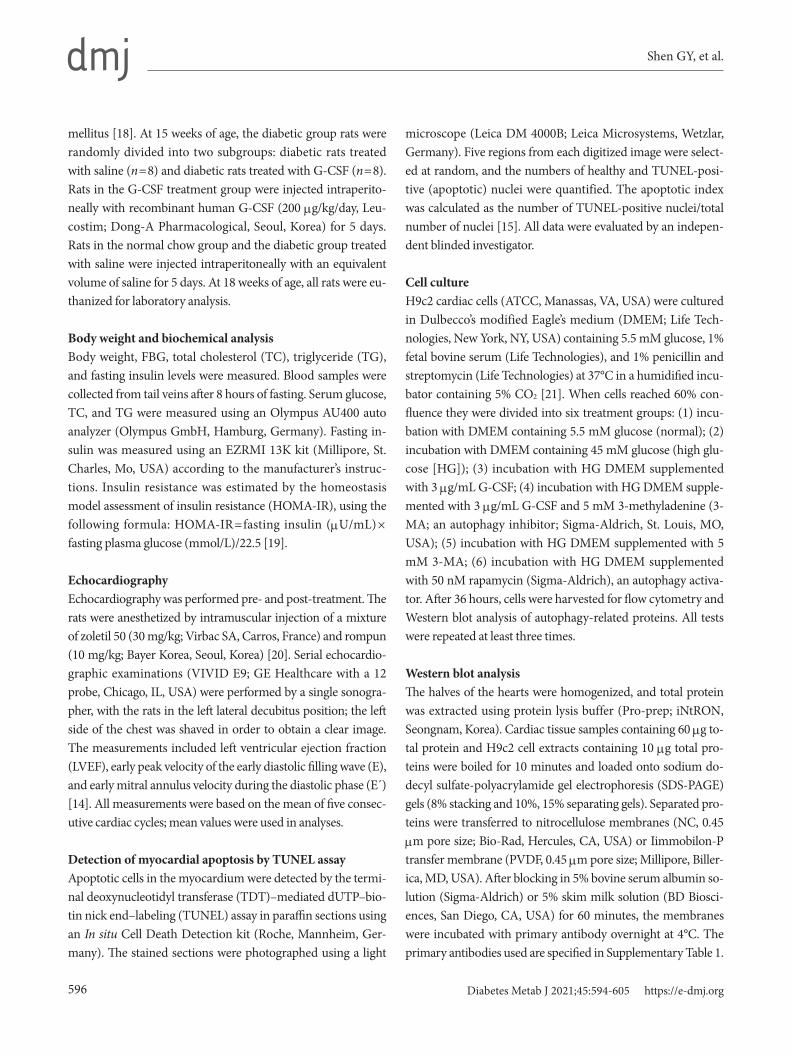
tion was consistent with the reduction in apoptosis in H9c2 cardiac cells induced by G-CSF under HG condition. In addi-tion, the effect of G-CSF on autophagic flux in H9c2 cardiac cells was investigated using an Autophagy Detection kit. Using flow cytometry, we confirmed that treatment with G-CSF sig-nificantly increased autophagic flux (11.68% ±1.92% vs. 9.40%±0.45%, P<0.05) in H9c2 cardiac cells cultured in HG
media (Fig. 6A and B). Additionally, using fluorescence mi-croscopy, we showed that treatment with G-CSF increased the fluorescent signal, thus indicating enhanced autophagic flux in H9c2 cardiac cells cultured in HG media (Fig. 6C).
Taken together, these results also suggest that the anti-apop-totic effect of G-CSF in H9c2 cardiac cells under diabetic con-ditions is linked to the up-regulation of autophagy.
Fig. 6. Effect of granulocyte-colony stimulating factor (G-CSF) on autophagic flux in high glucose-induced H9c2 cardiac cells. Au-tophagic flux evaluated high glucose-induced H9c2 cardiac cells by flow cytometry and fluorescence microscopy. Representative histogram (A) and bar graph (B), where fluorescence increase of autophagy green indicates autophagic flux increase. (C) Autopha-gic flux evaluated by fluorescence microscopy (magnification ×400), photos are representative of four to five independent experi-ments. Arrows indicate autophagic vesicles. The inserts in (C) show higher magnification. Rapamycin was used as a positive control of autophagy. All data are expressed as mean±standard deviation. Normal, normal condition group. aP<0.05 vs. H9c2 cardiac cells cultured in normal condition, bP<0.05 vs. H9c2 cardiac cells cultured in high glucose media containing G-CSF (n=4–5 per group).
20
16
12
8
4
0Au
toph
agy g
reen
(%)
Cou
nt
Normal High glucose G-CSF B
C
A
a,b
a150
100
50
0103102101 104 105
Autophagy green
NormalHigh glucoseG-CSFRapamycin

Shen GY, et al.
602 Diabetes Metab J 2021;45:594-605 https://e-dmj.org
DISCUSSION
The present study demonstrated that G-CSF reduced cardio-myocyte apoptosis and up-regulated autophagy in the diabetic myocardium. In addition, the anti-apoptotic effect of G-CSF in H9c2 cardiac cells under diabetic conditions was counteracted by treatment with an autophagy inhibitor. Moreover, we con-firmed that G-CSF increased autophagic flux in vitro. These data indicated that the anti-apoptotic effects of G-CSF in a rat model of diabetic cardiomyopathy may be mediated by up-regulation of autophagy.
Cardiomyocyte apoptosis is an important mechanism in the development of diabetic cardiomyopathy, which is closely as-sociated with cardiac dysfunction, hypertrophy, and fibrosis [26,27]. In addition, several studies revealed that the reduction of cardiomyocyte apoptosis prevents diabetic cardiomyopathy both in animal models and in vitro experiments [28,29]. In previous experimental studies, G-CSF treatment reduced car-diomyocyte apoptosis by modulating apoptosis-related pro-teins [15]; however, the mechanisms underlying this anti-apoptotic effect of G-CSF in diabetic cardiomyopathy remain unclear.
Autophagy is an important mechanism for cell survival, as it maintains the quality of proteins and organelles [30]. Studies have suggested that down-regulation of autophagy and the re-sulting accumulation of abnormal proteins and organelles, leads to apoptosis and cardiac dysfunction in various cardiac diseases, such as ischemic heart disease, cardiac hypertrophy, and heart failure [31,32]. Sishi et al. [33] and Wang et al. [34] demonstrated that up-regulation of autophagy prevented car-diomyocyte apoptosis in doxorubicin-induced cardiomyopa-thy and hypertensive heart disease, respectively. Moreover, overwhelming evidence indicates that cardiomyocyte apopto-sis, which plays an important role in the development of dia-betic cardiomyopathy, is induced by the impairment of au-tophagy [35-37].
The major finding in this study was that G-CSF increased Beclin-1level and the LC3-II/LC3-I ratio and decreased P62 level in the diabetic myocardium. Beclin-1 is an essential in-ductor of autophagy activity that binds to class III phosphati-dylinositol 3-kinase to form a kinase complex in mammals [38]. LC3 is the major regulatory protein that promotes the in-duction of the autophagosome membrane [39]. When autoph-agy is initiated, LC3-I is conjugated to phosphatidylethanol-amine to form LC3-II, which is required for the formation of
the autophagosome [40]. P62 is another major factor that tar-gets specific cargo for autophagy; P62 accumulates when au-tophagy is inhibited, and its levels decrease when autophagy is induced [41]. Consistent with our data, Zhao et al. [42] report-ed that heme oxygenase-1 up-regulated the expression of Be-clin-1 and LC3-II in diabetic mice and suggesting that heme oxygenase-1 prevents diabetic cardiomyopathy by up-regula-tion autophagy. Moreover, activation of AMP-activated protein kinase was shown to protect cardiac structure and function by up-regulation of Beclin-1 and LC3-II expression, suggesting that increasing cardiac autophagy protect would protect cardiac structure and function in the diabetic myocardium [43]. In this study we showed that G-CSF up-regulated cardiac autophagy, as indicated by the increase in Beclin-1 level and LC3-II/LC3-I ratio and decrease in P62 level in the diabetic myocardium.
In this study, to confirm the effect of G-CSF on HG-induced apoptosis in cardiac cells, we cultured H9c2 cardiac cells with HG media, to create a diabetic cardiomyopathy model [44], and measured the rate of apoptosis using these cells. H9c2 car-diac cells are a commercially available myogenic cell line de-rived from embryonic rat ventricular tissue [45], which show cardiac-specific characteristics, such as morphological, bio-chemical, and electrophysiological characterization [46]. H9c2 cardiac cells offer a unique in vitro model to study the meta-bolic activity of the heart [47]. Moreover, we used 3-MA (an autophagy inhibitor) and rapamycin (an autophagy inducer) to further confirm that apoptosis of H9c2 cardiac cells was re-lated to autophagy under diabetic conditions. We found that G-CSF reduced apoptosis of H9c2 cardiac cells, concurrent with the up-regulation of autophagy; these effects were abro-gated by 3-MA. We also confirmed that inhibition of autopha-gy by 3-MA increased H9c2 cardiac cell apoptosis under dia-betic conditions, whereas up-regulation of autophagy by ra-pamycin reduced H9c2 cardiac cell apoptosis, under diabetic conditions. Jia et al. [48] previously showed that safflower ex-tract reduced apoptosis of H9c2 cardiac cells treated with an-giotensin II, by increasing LC3-II expression. They also report-ed that the anti-apoptotic effect of safflower was reversed by 3-MA and that rapamycin reduced apoptosis, suggesting that safflower inhibits apoptosis via the up-regulation of autophagy. Gao et al. [49] similarly reported that 3-MA abrogated the an-ti-apoptotic effect of fasudil, suggesting that fasudil protects H9c2 cardiac cells from apoptosis via increasing Beclin-1 level and LC3-II/LC3-I ratio and decreasing P62 level under diabet-ic conditions. Guo et al. [50] also demonstrated that G-CSF
G-CSF upregulates autophagy in diabetic cardiomyopathy
603Diabetes Metab J 2021;45:594-605 https://e-dmj.org
promoted autophagy and reduced neural tissue damage after spinal cord injury in mice. Inhibition of autophagy by 3-MA partially blocked the neuroprotective effect induced by G-CSF, suggesting that G-CSF reduced neural tissue damage through up-regulation of autophagy. Moreover, we confirmed that G-CSF reduced the up-regulated autophagic flux under diabetic conditions. Considering our data and previous studies, we sug-gest that the anti-apoptotic effect of G-CSF is potentially medi-ated by the up-regulation of autophagy in H9c2 cardiac cells, under diabetic condition.
This study does, however, have several limitations. First, we demonstrated the effects of 3-MA and rapamycin in in vitro diabetic condition experiments, but we did not confirm the systemic effects of 3-MA and rapamycin in diabetic rats. In ad-dition, we did not perform genetic knockdown or gain-of-au-tophagy methods that are necessary to determine the func-tional role of autophagy in the anti-apoptotic effects of G-CSF and to rule out the potential nonspecific effects of 3-MA and rapamycin that are unrelated to autophagy. Second, we were unable to investigate the down-stream signaling of autophagy-related proteins such as Beclin-1, LC3, and P62. Additional studies are required to further elucidate the detailed mecha-nisms regarding effect of G-CSF on apoptosis linked to up-reg-ulation of autophagy. Third, we cannot rule out the possibility that the anti-apoptotic effect of G-CSF is associated with any other previously postulated mechanism such as the action of G-CSF directly or through the G-CSF receptor-mediated sig-naling pathway, a systemic effect, mobilization or homing of bone marrow stem cells, or other paracrine effects; such as fi-brosis, vascularization, oxidative stress, or endoplasmic reticu-lum stress. Further studies regarding the precise mechanism of the anti-apoptotic effect of G-CSF are also worth exploring.
In conclusion, the results of our study indicate that the anti-apoptotic effect of G-CSF may be significantly associated with the up-regulation of autophagy in diabetic cardiomyopathy. These findings suggest that G-CSF could potentially be used a novel therapeutic drug for the treatment of patients with dia-betic cardiomyopathy.
SUPPLEMENTARY MATERIALS
Supplementary materials related to this article can be found online at https://doi.org/10.4093/dmj.2020.0049.
CONFLICTS OF INTEREST
No potential conflict of interest relevant to this article was re-ported.
AUTHOR CONTRIBUTIONS
Conception or design: G.Y.S., J.H.S., Y.S.S.Acquisition, analysis, or interpretation of data: G.Y.S., Y.S.S., H.W.J., I.H.P., J.H.S., N.K.S., A.H.L., Y.J.C.Drafting the work or revising: Y.L., Y.H.L., H.K.Final approval of the manuscript: K.S.K.
ORCID
Guang-Yin Shen https://orcid.org/0000-0001-5442-6881Jeong-Hun Shin https://orcid.org/0000-0002-6718-9763Yi-Sun Song https://orcid.org/0000-0001-9797-7325Kyung-Soo Kim https://orcid.org/0000-0002-0891-1023
FUNDING
None
ACKNOWLEDGMENTS
This research was supported by the Basic Science Research Pro-gram through the National Research Foundation of Korea (NRF), funded by the Ministry of Education (2016R1D1A1-B03931479).
REFERENCES
1. Rubler S, Dlugash J, Yuceoglu YZ, Kumral T, Branwood AW, Grishman A. New type of cardiomyopathy associated with dia-betic glomerulosclerosis. Am J Cardiol 1972;30:595-602.
2. Acar E, Ural D, Bildirici U, Sahin T, Yilmaz I. Diabetic cardio-myopathy. Anadolu Kardiyol Derg 2011;11:732-7.
3. Cai L, Li W, Wang G, Guo L, Jiang Y, Kang YJ. Hyperglycemia-induced apoptosis in mouse myocardium: mitochondrial cyto-chrome C-mediated caspase-3 activation pathway. Diabetes 2002;51:1938-48.
4. Frustaci A, Kajstura J, Chimenti C, Jakoniuk I, Leri A, Maseri A, et al. Myocardial cell death in human diabetes. Circ Res 2000;87:1123-32.

Shen GY, et al.
604 Diabetes Metab J 2021;45:594-605 https://e-dmj.org
5. Glick D, Barth S, Macleod KF. Autophagy: cellular and molec-ular mechanisms. J Pathol 2010;221:3-12.
6. Levine B, Kroemer G. Autophagy in the pathogenesis of dis-ease. Cell 2008;132:27-42.
7. Orogo AM, Gustafsson AB. Therapeutic targeting of autopha-gy: potential and concerns in treating cardiovascular disease. Circ Res 2015;116:489-503.
8. Zou MH, Xie Z. Regulation of interplay between autophagy and apoptosis in the diabetic heart: new role of AMPK. Au-tophagy 2013;9:624-5.
9. Hsu HC, Chen CY, Lee BC, Chen MF. High-fat diet induces cardiomyocyte apoptosis via the inhibition of autophagy. Eur J Nutr 2016;55:2245-54.
10. Mellor KM, Reichelt ME, Delbridge LM. Autophagy anomalies in the diabetic myocardium. Autophagy 2011;7:1263-7.
11. Demetri GD, Griffin JD. Granulocyte colony-stimulating fac-tor and its receptor. Blood 1991;78:2791-808.
12. Deindl E, Zaruba MM, Brunner S, Huber B, Mehl U, Assmann G, et al. G-CSF administration after myocardial infarction in mice attenuates late ischemic cardiomyopathy by enhanced ar-teriogenesis. FASEB J 2006;20:956-8.
13. Huttmann A, Duhrsen U, Stypmann J, Noppeney R, Nuckel H, Neumann T, et al. Granulocyte colony-stimulating factor-in-duced blood stem cell mobilisation in patients with chronic heart failure: feasibility, safety and effects on exercise tolerance and cardiac function. Basic Res Cardiol 2006;101:78-86.
14. Lim YH, Joe JH, Jang KS, Song YS, So BI, Fang CH, et al. Ef-fects of granulocyte-colony stimulating factor (G-CSF) on dia-betic cardiomyopathy in Otsuka Long-Evans Tokushima fatty rats. Cardiovasc Diabetol 2011;10:92.
15. Shin JH, Lim YH, Song YS, So BI, Park JY, Fang CH, et al. Granulocyte-colony stimulating factor reduces cardiomyocyte apoptosis and ameliorates diastolic dysfunction in Otsuka Long-Evans Tokushima Fatty rats. Cardiovasc Drugs Ther 2014;28:211-20.
16. Srinivasan K, Viswanad B, Asrat L, Kaul CL, Ramarao P. Com-bination of high-fat diet-fed and low-dose streptozotocin-treated rat: a model for type 2 diabetes and pharmacological screening. Pharmacol Res 2005;52:313-20.
17. Kilkenny C, Browne WJ, Cuthill IC, Emerson M, Altman DG. Improving bioscience research reporting: the ARRIVE guide-lines for reporting animal research. PLoS Biol 2010;8:e1000412.
18. Ti Y, Xie GL, Wang ZH, Bi XL, Ding WY, Wang J, et al. TRB3 gene silencing alleviates diabetic cardiomyopathy in a type 2 diabetic rat model. Diabetes 2011;60:2963-74.
19. Song YS, Fang CH, So BI, Park JY, Lee Y, Shin JH, et al. Time course of the development of nonalcoholic Fatty liver disease in the Otsuka long-evans Tokushima Fatty rat. Gastroenterol Res Pract 2013;2013:342648.
20. Song YS, Joo HW, Park IH, Shen GY, Lee Y, Shin JH, et al. Transplanted human amniotic epithelial cells secrete paracrine proangiogenic cytokines in rat model of myocardial infarction. Cell Transplant 2015;24:2055-64.
21. Liu L, Ding WY, Zhao J, Wang ZH, Zhong M, Zhang W, et al. Activin receptor-like kinase 7 mediates high glucose-induced H9c2 cardiomyoblast apoptosis through activation of Smad2/3. Int J Biochem Cell Biol 2013;45:2027-35.
22. Song YS, Joo HW, Park IH, Shen GY, Lee Y, Shin JH, et al. Bone marrow mesenchymal stem cell-derived vascular endothelial growth factor attenuates cardiac apoptosis via regulation of cardiac miRNA-23a and miRNA-92a in a rat model of myocar-dial infarction. PLoS One 2017;12:e0179972.
23. Sharif T, Martell E, Dai C, Kennedy BE, Murphy P, Clements DR, et al. Autophagic homeostasis is required for the pluripo-tency of cancer stem cells. Autophagy 2017;13:264-84.
24. Almasi S, Kennedy BE, El-Aghil M, Sterea AM, Gujar S, Parti-da-Sanchez S, et al. TRPM2 channel-mediated regulation of autophagy maintains mitochondrial function and promotes gastric cancer cell survival via the JNK-signaling pathway. J Biol Chem 2018;293:3637-50.
25. Perez-Arizti JA, Ventura-Gallegos JL, Galvan Juarez RE, Ra-mos-Godinez MDP, Colin-Val Z, Lopez-Marure R. Titanium dioxide nanoparticles promote oxidative stress, autophagy and reduce NLRP3 in primary rat astrocytes. Chem Biol Interact 2020;317:108966.
26. Fang ZY, Prins JB, Marwick TH. Diabetic cardiomyopathy: ev-idence, mechanisms, and therapeutic implications. Endocr Rev 2004;25:543-67.
27. Huynh K, Bernardo BC, McMullen JR, Ritchie RH. Diabetic cardiomyopathy: mechanisms and new treatment strategies targeting antioxidant signaling pathways. Pharmacol Ther 2014;142:375-415.
28. Fiordaliso F, Li B, Latini R, Sonnenblick EH, Anversa P, Leri A, et al. Myocyte death in streptozotocin-induced diabetes in rats in angiotensin II-dependent. Lab Invest 2000;80:513-27.
29. Li K, Cui YC, Zhang H, Liu XP, Zhang D, Wu AL, et al. Gluta-mine reduces the apoptosis of H9C2 cells treated with high-glucose and reperfusion through an oxidation-related mecha-nism. PLoS One 2015;10:e0132402.
30. Lavandero S, Chiong M, Rothermel BA, Hill JA. Autophagy in
G-CSF upregulates autophagy in diabetic cardiomyopathy
605Diabetes Metab J 2021;45:594-605 https://e-dmj.org
cardiovascular biology. J Clin Invest 2015;125:55-64. 31. Yan L, Vatner DE, Kim SJ, Ge H, Masurekar M, Massover WH,
et al. Autophagy in chronically ischemic myocardium. Proc Natl Acad Sci U S A 2005;102:13807-12.
32. Zhu H, Rothermel BA, Hill JA. Autophagy in load-induced heart disease. Methods Enzymol 2009;453:343-63.
33. Sishi BJ, Loos B, van Rooyen J, Engelbrecht AM. Autophagy upregulation promotes survival and attenuates doxorubicin-induced cardiotoxicity. Biochem Pharmacol 2013;85:124-34.
34. Wang ZV, Rothermel BA, Hill JA. Autophagy in hypertensive heart disease. J Biol Chem 2010;285:8509-14.
35. Cai L, Kang YJ. Cell death and diabetic cardiomyopathy. Car-diovasc Toxicol 2003;3:219-28.
36. Xu X, Hua Y, Nair S, Zhang Y, Ren J. Akt2 knockout preserves cardiac function in high-fat diet-induced obesity by rescuing cardiac autophagosome maturation. J Mol Cell Biol 2013;5:61-3.
37. Xu X, Kobayashi S, Chen K, Timm D, Volden P, Huang Y, et al. Diminished autophagy limits cardiac injury in mouse models of type 1 diabetes. J Biol Chem 2013;288:18077-92.
38. Kang R, Zeh HJ, Lotze MT, Tang D. The Beclin 1 network regu-lates autophagy and apoptosis. Cell Death Differ 2011;18:571-80.
39. Kobayashi S, Liang Q. Autophagy and mitophagy in diabetic cardiomyopathy. Biochim Biophys Acta 2015;1852:252-61.
40. Cherra SJ 3rd, Kulich SM, Uechi G, Balasubramani M, Mount-zouris J, Day BW, et al. Regulation of the autophagy protein LC3 by phosphorylation. J Cell Biol 2010 Aug;190:533-9.
41. Bjorkoy G, Lamark T, Pankiv S, Overvatn A, Brech A, Johan-sen T. Monitoring autophagic degradation of p62/SQSTM1. Methods Enzymol 2009;452:181-97.
42. Zhao Y, Zhang L, Qiao Y, Zhou X, Wu G, Wang L, et al. Heme oxygenase-1 prevents cardiac dysfunction in streptozotocin-diabetic mice by reducing inflammation, oxidative stress, apoptosis and enhancing autophagy. PLoS One 2013;8:e75927.
43. Xie Z, Lau K, Eby B, Lozano P, He C, Pennington B, et al. Im-provement of cardiac functions by chronic metformin treat-ment is associated with enhanced cardiac autophagy in diabet-ic OVE26 mice. Diabetes 2011;60:1770-8.
44. Song H, Zandstra PW, Radisic M. Engineered heart tissue model of diabetic myocardium. Tissue Eng Part A 2011;17: 1869-78.
45. Kimes BW, Brandt BL. Properties of a clonal muscle cell line from rat heart. Exp Cell Res 1976;98:367-81.
46. Hescheler J, Meyer R, Plant S, Krautwurst D, Rosenthal W, Schultz G. Morphological, biochemical, and electrophysiologi-cal characterization of a clonal cell (H9c2) line from rat heart. Circ Res 1991;69:1476-86.
47. Watkins SJ, Borthwick GM, Arthur HM. The H9C2 cell line and primary neonatal cardiomyocyte cells show similar hyper-trophic responses in vitro. In Vitro Cell Dev Biol Anim 2011; 47:125-31.
48. Jia Z, Liu Y, Su H, Li M, Zhang M, Zhu Y, et al. Safflower extract inhibiting apoptosis by inducing autophagy in myocardium derived H9C2 cell. Int J Clin Exp Med 2015;8:20254-62.
49. Gao H, Hou F, Dong R, Wang Z, Zhao C, Tang W, et al. Rho-Kinase inhibitor fasudil suppresses high glucose-induced H9c2 cell apoptosis through activation of autophagy. Cardiovasc Ther 2016;34:352-9.
50. Guo Y, Liu S, Zhang X, Wang L, Gao J, Han A, et al. G-CSF promotes autophagy and reduces neural tissue damage after spinal cord injury in mice. Lab Invest 2015;95:1439-49.