role of endothelial shear stress in the natural history of ... · in the natural history of...
TRANSCRIPT

Adialts
FMNHMNFcHCIF
2
Journal of the American College of Cardiology Vol. 49, No. 25, 2007© 2007 by the American College of Cardiology Foundation ISSN 0735-1097/07/$32.00P
STATE-OF-THE-ART PAPER
Role of Endothelial Shear Stressin the Natural History of CoronaryAtherosclerosis and Vascular RemodelingMolecular, Cellular, and Vascular Behavior
Yiannis S. Chatzizisis, MD, MSC,*† Ahmet Umit Coskun, PHD,‡ Michael Jonas, MD,†Elazer R. Edelman, MD, PHD, FACC,*† Charles L. Feldman, SCD,* Peter H. Stone, MD, FACC*
Boston and Cambridge, Massachusetts
Although the entire coronary tree is exposed to the atherogenic effect of the systemic risk factors, atheroscle-rotic lesions form at specific arterial regions, where low and oscillatory endothelial shear stress (ESS) occur. LowESS modulates endothelial gene expression through complex mechanoreception and mechanotransduction pro-cesses, inducing an atherogenic endothelial phenotype and formation of an early atherosclerotic plaque. Eachearly plaque exhibits an individual natural history of progression, regression, or stabilization, which is dependentnot only on the formation and progression of atherosclerosis but also on the vascular remodeling response. Al-though the pathophysiologic mechanisms involved in the remodeling of the atherosclerotic wall are incompletelyunderstood, the dynamic interplay between local hemodynamic milieu, low ESS in particular, and the biology ofthe wall is likely to be important. In this review, we explore the molecular, cellular, and vascular processes sup-porting the role of low ESS in the natural history of coronary atherosclerosis and vascular remodeling and indi-cate likely mechanisms concerning the different natural history trajectories of individual coronary lesions. Ath-erosclerotic plaques associated with excessive expansive remodeling evolve to high-risk plaques, because lowESS conditions persist, thereby promoting continued local lipid accumulation, inflammation, oxidative stress,matrix breakdown, and eventually further plaque progression and excessive expansive remodeling. An enhancedunderstanding of the pathobiologic processes responsible for atherosclerosis and vascular remodeling mightallow for early identification of a high-risk coronary plaque and thereby provide a rationale for innovative diag-nostic and/or therapeutic strategies for the management of coronary patients and prevention of acute coronarysyndromes. (J Am Coll Cardiol 2007;49:2379–93) © 2007 by the American College of Cardiology Foundation
ublished by Elsevier Inc. doi:10.1016/j.jacc.2007.02.059
sbtfacbm
oenailaEma
therosclerosis is a chronic, inflammatory, fibroproliferativeisease primarily of large- and medium-sized conduit arter-
es (1,2). Although the entire vasculature is exposed to thetherogenic effects of the systemic risk factors (e.g., hyper-ipidemia, cigarette smoking, hypertension, diabetes melli-us, chronic infections, and genetic predisposition), athero-clerotic lesions form at specific regions of the arterial tree,
rom the *Cardiovascular Division, Brigham and Women’s Hospital, Harvardedical School, Boston, Massachusetts; ‡Mechanical and Industrial Engineering,ortheastern University, Boston, Massachusetts; and †Harvard-MIT Division ofealth Sciences and Technology, Massachusetts Institute of Technology, Cambridge,assachusetts. Dr. Stone receives research grants from Boston Scientific Co.,ovartis Pharmaceutical Co., and the National Institutes of Health (NIH). Dr.eldman receives research grants from Boston Scientific Co. and Novartis Pharma-eutical Co. Dr. Edelman is receiving a research grant from the National Institutes ofealth (R01 HL 49039). This work was supported by grants from Boston Scientifico., Novartis Pharmaceutical Co., the NIH (R01 HL 49039), the Center for
nnovative and Minimally Invasive Therapy (CIMIT), and the Hellenic Harvardoundation.
mManuscript received January 2, 2007; revised manuscript received February 22,
007, accepted February 26, 2007.
uch as in the vicinity of branch points, the outer wall ofifurcations, and the inner wall of curvatures, where dis-urbed flow occurs (3). Local factors, such as hemodynamicorces, play a major role in the regional localization oftherosclerosis (4–7). These local hemodynamic forces in-lude flow-generated endothelial shear stress (ESS) andlood pressure-derived tensile stress, with ESS playing theost fundamental role in atherosclerosis.The first evidence implicating ESS in the localization
f atherosclerosis was described over 40 years ago by Carot al. (8). Later, sophisticated computational fluid dy-amic simulations in autopsy-based models of coronaryrteries (9), carotid bifurcations (10), and distal abdom-nal aortas (11) showed that areas with low ESS corre-ated to the localization of atherosclerosis found atutopsy. Further support of the atherogenic role of lowSS was also derived from in vivo experiments in animalodels (12–14). In vivo investigations in humans, usingcombination of intravascular ultrasound (IVUS) or
agnetic resonance imaging and computational fluid
oiuermndmn
D
Etoa1pt�
tlo
T
Continued on next page
2380 Chatzizisis et al. JACC Vol. 49, No. 25, 2007Shear Stress, Atherosclerosis, and Remodeling June 26, 2007:2379–93
dynamics confirmed the mecha-nistic role of low ESS in thedevelopment and progression ofatherosclerosis (15–17). Morerecent molecular and cellularstudies have begun to clarify thedetailed pathways by which lowESS leads to atherosclerosis aswell as the development of thincap fibroatheromas, presumed orsuspected “vulnerable plaques,”responsible for acute coronarysyndromes (18–22).
In addition to the processes ofatherosclerosis development andprogression, the local remodelingcharacteristics of the arterial wallin response to plaque growthconstitute crucial determinantsof the natural history and clinicalmanifestations of an individualatherosclerotic lesion. Local fac-tors undoubtedly play an impor-tant role in the nature of theremodeling response as well(5,15,22–25).
The purposes of this revieware to explore the molecular,cellular, and vascular biologicprocesses supporting the role oflow ESS in the natural history
f coronary atherosclerosis and vascular remodeling andndicate likely mechanisms concerning the different nat-ral history trajectories of individual coronary lesions. Annhanced understanding of the pathobiologic processesesponsible for atherosclerosis and vascular remodelingight allow for early identification of a high-risk coro-
ary plaque and thereby provide a rationale for innovativeiagnostic and/or therapeutic strategies for the manage-ent of coronary patients and prevention of acute coro-
ary syndromes.
efinitions of ESS and Blood Flow Patterns
ndothelial shear stress is the tangential stress derived fromhe friction of the flowing blood on the endothelial surfacef the arterial wall and is expressed in units of force / unitrea (N/m2 or Pascal [Pa] or dyne/cm2; 1 N/m2 � 1 Pa �0 dyne/cm2) (26,27) (Table 1). Endothelial shear stress isroportional to the product of the blood viscosity (�) andhe spatial gradient of blood velocity at the wall (ESS �
� dv/dy) (Fig. 1).The nature of fluid flow through a tube is dependent on
he velocity of flow and the presence of geometric irregu-arities or obstructions. Fluid flow might be either laminar
Abbreviationsand Acronyms
EC � endothelial cell
ECM � extracellular matrix
eNOS � endothelial nitricoxide synthase
ESS � endothelial shearstress
IEL � internal elasticlamina
IL � interleukin
LDL � low-densitylipoprotein cholesterol
MAPK � mitogen-activatedprotein kinase
MMP � matrixmetalloproteinase
NF-�B � nuclear factor-kappa B
NO � nitric oxide
ROS � reactive oxygenspecies
SREBP � sterol regulatoryelements binding protein
TCFA � thin capfibroatheroma
TF � transcription factor
VSMC � vascular smoothmuscle cell
r turbulent (26,28) (Fig. 2). Laminar flow refers to a
erminology of Arterial Hemodynamics
Table 1 Terminology of Arterial Hemodynamics
Term Definition
Endothelial shear stress (ESS) The tangential force derived by the frictionof the flowing blood on the endothelialsurface. It is the product of the shear rateat the wall and the blood viscosity (�).
Shear rate The spatial gradient of blood velocity, whichdescribes how fast the blood velocityincreases from areas at the arterial walltoward areas at the center of the lumen(i.e., dv/dy, where dv is change in flowvelocity unit and dy is change in unit ofradial distance from the wall).Physiologically, the shear rate decreasesat the center of the lumen and graduallyincreases toward the wall.
Blood viscosity A principal property of blood related to itsinternal friction that causes blood toresist flow. Hematocrit is the majordeterminant of blood viscosity.
Newtonian blood behavior Constant blood viscosity independent ofshear rate. In large-sized arteries (e.g.,aorta) blood behaves largely in aNewtonian fashion.
Non-Newtonian blood behavior Non-constant blood viscosity inverselyrelated to shear rate. Blood has non-Newtonian properties, especially in veins,small-sized arteries, and in themicrocirculation.
Laminar flow Smooth, streamlined blood flow whereviscous forces prevail against inertialforces.
Undisturbed laminar blood flow Smooth streamlined flow characterized byconcentric layers of blood moving inparallel along the course of the artery.The highest velocity is found at the centreof the lumen, whereas the lowest velocityoccurs along the wall. Uniform laminarblood flow primarily occurs in relativelystraight arterial segments.
Disturbed laminar blood flow Disturbed laminar flow characterized byreversed flow (i.e., flow separation,recirculation, and reattachment toforward flow). Disturbed laminar bloodflow occurs in arterial segments withgeometric irregularities (e.g., curvatures,branches, bifurcations), or upstream anddownstream of stenoses.
Turbulent blood flow Flow in which the blood velocity at any givenpoint varies continuously over time, eventhough the overall flow is steady. Inturbulent flow the inertial forces are moresignificant than viscous forces. Turbulentblood flow rarely occurs but has beendescribed in human aorta at peaksystole, during heavy exercise in much ofthe central arterial system, distal tosevere stenoses (�75%), and inaneurysms.
Reynolds number (Re) The ratio of blood inertial forces to viscousforces. For a given geometry, whether theflow will be laminar or turbulent isdetermined by its Reynolds number. Forlow Re values blood flow is laminar,whereas for high Re values (typically,above 2,000) blood flow is turbulent.

st(wr(pflflRwt
itc
ru1pmo
C
2381JACC Vol. 49, No. 25, 2007 Chatzizisis et al.June 26, 2007:2379–93 Shear Stress, Atherosclerosis, and Remodeling
treamlined flow and can be further divided into undis-urbed laminar flow, characterized by smooth streamlinesFig. 2A), and disturbed laminar flow, characterized by areasith reversed flow (i.e., flow separation, recirculation, and
eattachment to forward flow) or circumferential swirling26,29) (Fig. 2B). In turbulent flow the velocity at any givenoint varies continuously over time, even though the overallow is steady (Fig. 2C). For a given geometry, whether theow will be laminar or turbulent is determined by itseynolds number (Re); for low Re values, flow is laminar,hereas for high Re values (typically, above 2,000), flow is
urbulent (7,26,30).The pulsatile (unsteady) nature of the arterial blood flow
n combination with the complex geometric configuration ofhe coronaries determines the ESS patterns, which areharacterized by direction and magnitude (10,31,32). In
Figure 1 Definition of ESS
Endothelial shear stress (ESS) is proportional to the product of the bloodviscosity (�) and the spatial gradient of blood velocity at the wall (dv/dy).
ontinued
Table 1 Continued
Term Definition
Steady blood flow Blood flow in which velocity does not varywith time. This type of flow does notoccur in vivo; however, it has been largelyused in computational fluid dynamicstudies.
Pulsatile (unsteady) blood flow Blood flow with periodically changingvelocity during the cardiac cycle.
Steady ESS ESS that does not vary with time (i.e.,constant direction and magnitude).
Pulsatile ESS Unidirectional ESS with a magnitude varying,typically, within a range of 15 to 70dyne/cm2 over the cardiac cycle, yieldinga positive time-average.
Low ESS Unidirectional ESS with a periodically varyingmagnitude over the cardiac cycle, yieldinga significantly low time-average (�10 to12 dyne/cm2).
Oscillatory ESS Bidirectional ESS with a periodically varyingmagnitude over the cardiac cycle, yielding avery low time-average, usually close to 0.
ESS spatial gradient ESS variations over short distances. HighESS spatial gradients occur primarily ingeometrically irregular arterial regions.
elatively straight arterial segments, ESS is pulsatile andnidirectional with a magnitude that varies within a range of5 to 70 dyne/cm2 over the cardiac cycle and yields aositive time-average (4–6) (Fig. 3). In contrast, in geo-etrically irregular regions, where disturbed laminar flow
ccurs, pulsatile flow generates low and/or oscillatory ESS.
Figure 2 Characteristics of Flow Patterns
Schematic figure illustrating the characteristics of flow patterns. (A) Undis-turbed laminar flow is a smooth streamlined flow chacterized by concentric lay-ers of blood moving in parallel along the course of the artery; (B) disturbedlaminar flow is characterized by reversed flow (i.e., flow separation, recircula-tion, and reattachment to forward flow); (C) in turbulent flow the blood velocityat any given point varies continuously over time, even though the overall flow issteady. Adapted from Munson et al. (28). Re � Reynolds number.
Figure 3 Definition and Example ofPulsatile, Low, and Oscillatory ESS
Definition of pulsatile, low, and oscillatoryendothelial shear stress (ESS). Adapted from Ku et al. (10).

Lpr�acsi(isupb(eehs
atal
nctm(ac
ESM
Ebb(np(cc(o
2382 Chatzizisis et al. JACC Vol. 49, No. 25, 2007Shear Stress, Atherosclerosis, and Remodeling June 26, 2007:2379–93
ow ESS refers to ESS that is unidirectional at any givenoint but has a periodically fluctuating magnitude thatesults in a significantly low time-average (approximately10 to 12 dyne/cm2) (4,6,15) (Fig. 3). However, the
bsolute threshold effect of low ESS is likely dependent ononcomitant conditions, such as systemic factors, or inter-pecies differences (33). Low ESS typically occurs at thenner areas of curvatures as well as upstream of stenoses34). Oscillatory ESS is characterized by significant changesn both direction (bidirectional) and magnitude betweenystole and diastole, resulting in a very low time-average,sually close to 0 (4–6) (Fig. 3). Oscillatory ESS occursrimarily downstream of stenoses, at the lateral walls ofifurcations, and in the vicinity of branch points6,10,34,35). Beside the temporal oscillations, ESS experi-nces significant spatial oscillations over short distances,specially in geometrically irregular regions, resulting inigh spatial gradients, which are also involved in athero-clerosis (14,35–37).
Although low ESS and oscillatory ESS are closelyssociated with atherogenesis, the relative importance ofhese different ESS patterns is unclear. In a mouse carotidrtery in vivo model, both low ESS and oscillatory ESS
Figure 4 Endothelial Mechanotransduction of ESS
Local endothelial shear stress (ESS) is sensed by luminal endothelial mechanoreckinase receptors (TKRs), nicotinamide adenine dinucleotide phosphate (NADPH) oxproteoglycans. Also, ESS signals are transmitted through the cytoskeleton to the bcomplex consisting of platelet endothelial cell adhesion molecule-1 (PECAM-1) andvated integrins phosphorylate and activate a multiple complex of non-receptor tyroguanine nucleotide exchange factors (Sos, C3G), thereby activating Ras family GTPtriggers various parallel downstream cascades of serine kinases; each of these kivating mitogen-activated protein kinases (MAPKs). Besides integrin-mediated mechtiated by luminal or junctional mechanoreceptors. These pathways include the proprotein kinase C (PKC), activation of Rho family small GTPases (which mediate theECs), release of endothelial nitric oxide synthase (eNOS) and other signaling moleUltimately, all of these signaling pathways lead to phosphorylation of several trans(AP-1). These TF proteins bind positive or negative shear stress responsive elemeexpression, thereby modulating cellular function and morphology.
ed to atherosclerotic plaque formation, but only low, c
onoscillatory ESS was associated with inflammatoryhanges and proclivity to rupture (21). Different vascularerritories (e.g., femoral, carotid, and coronary arteries)ight also respond differently to various ESS stimuli
33). The magnitude of local low ESS is criticallyssociated with the severity of atherosclerotic plaqueharacteristics (19).
SS Mechanoreception,ignal Transduction, andechanosensitive Gene Expression
ndothelial cell (EC) surfaces (luminal, junctional, andasal) are equipped with numerous mechanoreceptors capa-le of detecting and responding to ESS stimuli (38–40)Fig. 4). After activation of mechanoreceptors, a complexetwork of several intracellular pathways is triggered, arocess known as mechanotransduction (38,39,41– 45)Fig. 4). These pathways are activated simultaneously andross-talk with each other; the great majority of themonverge into the mitogen-activated protein kinaseMAPKs) cascade at various levels, suggesting the key rolef MAPKs in ESS mechanotransduction (39). Cytoskeleton
, such as ion channels (K�, Ca2�, Na�, Cl�), G-proteins, caveolae, tyrosineand xanthine oxidase (XO), plasma membrane lipid bilayer, and heparan sulfater junctional endothelial surface, where certain integrins or a mechanosensoryare activated, respectively, and initiate a downstream signaling cascade. Acti-nases (FAK, c-Src, Shc, paxillin, and p130CAS), adaptor proteins (Grb2, Crk), andctive Ras plays a pivotal role in intracellular transduction of ESS signals as itphosphorylates and hence activates the next one downstream, ultimately acti-nsduction, ESS activates a number of other downstream signaling pathways ini-of reactive oxygen species (ROS) from NADPH oxidase and XO, activation of
deling cytoskeleton resulting in temporary or permanent structural changes offrom caveolae, and activation of phosphoinositide-3 kinase (PI3K)-Akt cascade.n factors (TFs), such as nuclear factor-kappa � (NF-�B) and activator protein-1REs) at promoters of mechanosensitive genes inducing or suppressing their
eptorsidaseasal oFlk-1
sine kiase. A
nasesanotra
ductionremo
culescriptio
nts (SS
onstitutes a central mediator in ESS signaling by providing

assj(snmttnrssrtpt
R
Lava(metstaEmto
IeitaL(cemgfhmivcsi
atrpHrlafmbclt
2383JACC Vol. 49, No. 25, 2007 Chatzizisis et al.June 26, 2007:2379–93 Shear Stress, Atherosclerosis, and Remodeling
scaffold for the formation or translocation of variousignaling molecules, serving as a bond between the luminalurface, where ESS is imposed, and several luminal, basal, orunctional formations, where the signaling pathways initiate45) (Fig. 5). These pathways lead to phosphorylation ofeveral transcription factors (TFs), which bind positive oregative shear stress responsive elements (SSREs) at pro-oters of mechanosensitive genes, inducing or suppressing
heir expression and, ultimately, modulating cellular func-ion and morphology (4,6,46–48). In arterial regions withon-disturbed flow, where ESS varies within a physiologicange, the ECs express various atheroprotective genes anduppress several pro-atherogenic ones, leading eventually totability and quiescence in that region (6,48). In contrast, inegions with low and disturbed flow where low ESS occurs,he atheroprotective genes are suppressed, whereas thero-atherogenic genes are upregulated, thereby promotinghe atherosclerotic process (6,48).
ole of Low ESS in Atherosclerosis
ow ESS attenuates nitric oxide (NO)-dependenttheroprotection. Nitric oxide, a key component of normalascular tone, also possesses strong anti-inflammatory, anti-poptotic, anti-mitogenic, and anti-thrombotic properties49) (Table 2). Physiologic pulsatile ESS constitutes theost potent stimulus for continuous NO production by the
ndothelium, an effect that is regulated at either transcrip-ional level through upregulation of endothelial nitric oxideynthase (eNOS) gene expression (50) or at post-ranscriptional level by eNOS protein phosphorylation andctivation (51). In arterial regions with disturbed flow, lowSS reduces the bioavailability of NO by decreasing eNOSessenger ribonucleic acid (mRNA) and protein expression,
hereby exposing the endothelium to the atherogenic effect
Figure 5 Role of Cytoskeleton in ESS Mechanotransduction
The endothelial cytoskeleton transmits the shear forces to the focal adhesions loccade starts. The shear forces can also be transmitted to mechanoreceptors at theESS � endothelial shear stress.
f local and systemic risk factors (12,13,49,52,53) (Fig. 6). (
n addition, low ESS downregulates prostacyclin, anotherndothelial vasodilatory substance (4,53), while upregulat-ng endothelin-1 (ET-1) (4,52,53), a potent vasoconstric-ive and mitogenic molecule, thereby precipitatingtherosclerosis.ow ESS promotes low-density lipoprotein cholesterol
LDL) uptake, synthesis, and permeability. Low ESSauses a sustained endothelial activation of sterol regulatorylements binding proteins (SREBPs), a family of endoplas-ic reticulum-bound TFs that upregulate the expression of
enes encoding LDL receptor, cholesterol synthase, andatty acid synthase (54) (Fig. 6). In the context of systemicyperlipidemia, this effect results in an increased engage-ent and synthesis of LDL by the ECs, ultimately promot-
ng the subendothelial accumulation of LDL (55). Acti-ated SREBPs also appear to induce interleukin (IL)-8 and,oncomitantly, monocyte accumulation into the intima,uggesting an additional role of these TFs in the localnflammatory processes (56).
In addition to active SREBPs-dependent LDL uptakend synthesis, disturbed flow increases the permeability ofhe endothelial surface to LDL (14,38,57) (Fig. 6). Theegulation of cell cycle and survival by shear forces mightlay an important role in increasing LDL permeability.ighly mitotic and apoptotic activity of ECs was found in
egions susceptible to atherosclerosis, where low and oscil-atory ESS occur (58,59). The accentuated ECs mitosis andpoptosis as well as the conformational changes of ECsrom fusiform to polygonal shape associated with low ESSight be responsible for the widening of the junctions
etween ECs (4,55,60). These small “gaps” between ECs, inombination with flow stagnation and the subsequent pro-ongation of the residence time of circulating LDL, facili-ate the infiltration of LDL underneath the endothelium
t the basal endothelial surface, where a downstream intracellular signaling cas-cell junctions, luminal surface, and nucleus (N). Adapted from Davies et al. (45).
ated acell–
6,29).

Lawooeae[bdmadrf[LcmscpocsAibroN
tgv
EF
Continued on next column
C
AmoIcsgTm
2384 Chatzizisis et al. JACC Vol. 49, No. 25, 2007Shear Stress, Atherosclerosis, and Remodeling June 26, 2007:2379–93
ow ESS promotes oxidative stress. Once LDL particlesre engulfed in the subendothelial layer, they are associatedith intimal proteoglycans, become entrapped, and undergoxidative modification (1,2). Low ESS promotes productionf reactive oxygen species (ROS) into the intima and,ventually, oxidation of LDL, by enhancing gene expressionnd post-transcriptional activity of the major oxidativenzymes (nicotinamide adenine dinucleotide phosphateNADPH] oxidase and xanthine oxidase) at EC mem-ranes (49,61,62) (Fig. 6). Low ESS appears also toownregulate the intracellular ROS scavengers, such asanganese superoxide dismutase and glutathione, further
ugmenting local oxidative stress (47,63). Generated ROSegrade NO and its co-factors (e.g., tetrahydrobiopterin),educing the bioavailability of atheroprotective NO andurther enhancing the production of ROS (e.g., superoxideO2
�] or peroxynitrite [ONOO�]) (49).ow ESS promotes inflammation. The recruitment ofirculating inflammatory cells (monocytes, T-lymphocytes,ast cells, eosinophils, dendritic cells) into the intima to
cavenge oxidized LDL constitutes a major pathogeneticomponent in the atherosclerotic process (64). Low ESSlays a key role in the localized attachment and infiltrationf these cells into the arterial wall through activation ofertain TFs, notably nuclear factor-kappa � (NF-�B), andubsequent translocation to the nucleus (36,65–67) (Fig. 6).ctivation of NF-�B is further promoted by low shear-
nduced oxidative stress (61). In addition, a negative feed-ack mechanism occurs between NF-�B and NO, in thateduced eNOS expression and subsequent NO productionccurring in low ESS regions increases the activity ofF-�B (67).Various endothelial genes are upregulated downstream
o low shear-induced NF-�B activation. These includeenes that encode several adhesion molecules, such as
ontinued
Table 2 Continued
Effect of Low ESS
Plaque calcification
BMP-4 Upregulated
Plaque thrombogenecity
eNOS/NO Downregulated
Prostacyclin Downregulated
Thrombomodulin No effect (38,104)
t-PA Downregulated (38,105)
Blood stagnation— accumulation ofblood thrombogenic factorsclose to the wall
Increased (6,29)
CE � angiotensin-converting enzyme; bFGF � basic fibroblast growth factor; BMP � boneorphogenic protein; EC � endothelial cell; eNOS/NO � endothelial nitric oxide synthase/nitric
xide; ESS � endothelial shear stress; ET � endothelin; ICAM � intercellular adhesion molecule;FN � interferon; IL � interleukin; LDL � low-density lipoprotein cholesterol; MCP � monocytehemoattractant protein; MMP � matrix metalloproteinase; Mn SOD � manganese-dependantuperoxide dismutase; NADPH � nicotinamide adenine dinucleotide phosphate; PAI � plasmino-en activator inhibitor; PDGF � platelet derived growth factor; TGF � transforming growth factor;NF � tumor necrosis factor; t-PA � tissue plasminogen activator; VCAM � vascular cell adhesionolecule; VEGF � vascular endothelial growth factor; VSMC � vascular smooth muscle cell.
ndothelial Genes and Vascularunctions Regulated by Low ESS in Atherosclerosis
Table 2 Endothelial Genes and VascularFunctions Regulated by Low ESS in Atherosclerosis
Effect of Low ESS
Impaired flow-dependent vasodilation
Vasodilators
eNOS/NO Downregulated (4,12,13,52,53)
Prostacyclin Downregulated (4,53)
Vasoconstrictors
ET-1 Upregulated (4,52,53)
Subendothelial accumulation of LDL
Endothelial LDL uptake and synthesis Increased (54)
Endothelial LDL permeability Increased (14,57)
Blood stagnation—accumulation ofLDL close to the wall
Increased (6,29)
ECs proliferation and apoptosis Increased (58,59)
Oxidative stress
Oxidative enzymes
NADPH oxidase Upregulated (61)
Xanthine oxidase Upregulated (62)
Antioxidative enzymes
Mn SOD Downregulated (47)
Glutathione Downregulated (63)
Inflammation
Chemoattractants (MCP-1) Upregulated (46,47,66)
Adhesion molecules (VCAM-1,ICAM-1, E-selectin)
Upregulated (21,46,47,65-68)
Cytokines (TNF-�, IL-1, IFN-�) Upregulated (46,47)
BMP-4 Upregulated (7,101)
Leukocyte pseudopod projection Increased (69)
Blood stagnation—accumulation ofmonocytes close to the wall
Increased (6,29,68)
VSMCs migration, differentiation, andproliferation
Growth promoters
PDGF-A, PDGF-B Upregulated (4,70)
ET-1 Upregulated
VEGF Upregulated (21,71)
bFGF Unclear (4,73)
ACE Unclear (4,38)
Angiotensin II Unclear (38,73)
Growth inhibitors
eNOS/NO Downregulated
TGF-� Downregulated (4,74,75)
PAI-1 Downregulated (22,72)
Regulation of extracellular matrixcontent and composition
Increased matrix degradation
MMP-2, MMP-9 Upregulated (21,22,80-82)
Cathepsin L Upregulated (93)
Reduced matrix synthesis
IFN-� Upregulated
VSMCs apoptosis Increased
eNOS/NO Downregulated
TGF-� Downregulated
Neovascularization
VEGF Upregulated
Other angiogenic factors(e.g., angiopoietin-2)
Upregulated (46)
ascular cell adhesion molecule (VCAM)-1; intercellular

atpaamregiaflm(jbmueopLmpV(vL
cfe(tfaciaEuVatdptiicc(Lafia
2385JACC Vol. 49, No. 25, 2007 Chatzizisis et al.June 26, 2007:2379–93 Shear Stress, Atherosclerosis, and Remodeling
dhesion molecule (ICAM)-1 and E-selectin; chemoat-ractant chemokines, such as monocyte chemoattractantrotein (MCP)-1; and pro-inflammatory cytokines, suchs tumor necrosis factor (TNF)-�, interleukin (IL)-1,nd interferon (IFN)-� (21,46,47,65– 68). Adhesionolecules are expressed on EC surface and mediate the
olling and adhesion of circulating leukocytes on thendothelial surface, whereas MCP-1 promotes transmi-ration of leukocytes, particularly monocytes, into thentima. The intimal infiltration of inflammatory cellsppears also to be mechanistically facilitated by bloodow stagnation and endothelial junction widening, pri-arily occurring in areas with disturbed flow (6,29,68)
Fig. 6). Low ESS might also provoke pseudopod pro-ection through mechanotransduction processes andinding of leukocytes on the endothelium (69). Onceonocytes infiltrate underneath the endothelium they
ndergo structural and functional alterations and differ-ntiate to macrophages, which sustain the inflammation,xidative stress, and dynamic matrix remodeling, therebyromoting atherosclerosis progression (1,2,64).ow ESS promotes vascular smooth muscle cell (VSMC)igration, differentiation, and proliferation. Low ESS
romotes endothelial gene and protein expression of potentSMC mitogens, such as platelet-derived growth factor
PDGF)-A and -B isoforms (4,70), ET-1 (4,52,53), andascular endothelial growth factor (VEGF) (21,71) (Fig. 6).
Figure 6 Role of Low ESS in Atherosclerosis
In arterial regions with disturbed laminar flow, low endothelial shear stress (ESS) sthereby promoting atherogenesis, atherosclerotic plaque formation and progressio� intercellular adhesion molecule; IFN � interferon; IL � interleukin; LDL � low-dematrix metalloproteinase; NO � nitric oxide; PDGF � platelet-derived growth factortransforming growth factor; TNF � tumor necrosis factor; t-PA � tissue plasminogegrowth factor; VSMC � vascular smooth muscle cell; other abbreviations as in Fig
ow ESS-induced formation of ROS and pro-inflammatory c
ytokines also promote the expression of these growthactors (1). Also, low and disturbed flow decreases thendothelial expression of plasminogen activator inhibitorPAI)-1, an inhibitor of VSMC migration (22,72). Al-hough the effect of low ESS on basic fibroblast growthactor (bFGF), angiotensin converting enzyme (ACE), andngiotensin II is yet unclear (4,38,73), potent suppressors ofell growth and migration, such as NO (52) and transform-ng growth factor (TGF)-� (4,74,75) are downregulated inreas with low and disturbed flow (Fig. 6). Ultimately, lowSS-mediated over-expression of growth promoters andnder-expression of growth inhibitors by the ECs stimulateSMCs to migrate from media to intima through a region-
lly disrupted internal elastic lamina (IEL) (76,77). Withinhe intima VSMCs acquire a “synthetic” phenotype, pro-ucing collagen and other extracellular matrix (ECM)roteins, and proliferate (1). Over time, VSMCs along withhe fibroblasts create a fibrous cap around the lipid coresolating the thrombogenic lipid material from the circulat-ng platelets. The fibrous cap along with the lipid coreonstitute the so-called early atherosclerotic plaque (fibrousap atheroma; American Heart Association type IV lesion)78,79).ow ESS promotes ECM degradation in vascular wall
nd plaque fibrous cap. The ECM of vascular wall andbrous cap is composed of a complex mixture of collagennd elastin fibers within a ground substance of proteogly-
he endothelial function and structure toward an atherosclerotic phenotype,vascular remodeling. BMP � bone morphogenic protein; ET � endothelin; ICAM
lipoprotein cholesterol; MCP � monocyte chemoattractant protein; MMP �
P � sterol regulatory elements binding protein; TF � transcription factor; TGF �
vator; VCAM � vascular cell adhesion molecule; VEGF � vascular endothelial
hifts tn, andnsity; SREBn acti
ure 4.
ans and glycosaminoglycans. In vitro and in vivo animal

eg(gap(rmt(Eotttce
tpbKap(bNlLpdIat(abttpatEdPNtbmmeVat
a(Pmsu(apeocfp(Liealaut(db(iit
RA
TpsrsaTcd(eEeMtbm(vil
2386 Chatzizisis et al. JACC Vol. 49, No. 25, 2007Shear Stress, Atherosclerosis, and Remodeling June 26, 2007:2379–93
xperiments have demonstrated that low ESS upregulatesene expression and activity of matrix metalloproteinasesMMPs), particularly MMP-2 (or gelatinase-A) and �9 (orelatinase-B) (21,22,80–82), which are the major proteasesssociated with ECM degradation in the atheroscleroticlaques (83– 86) (Fig. 6). Pro-inflammatory cytokinesTNF-�, IL-1, IFN-�) comprise the major stimuli for theelease of MMPs from their key cellular sources (ECs,acrophages, VSMCs, T-lymphocytes, and mast cells) via
he MAPKs pathway and subsequent activation of TFse.g., NF-�B and activator protein [AP]-1) (86–88). LowSS increases MMPs expression by ECs through activationf these TFs. Moreover, low ESS enhances the accumula-ion of macrophages and VSMCs within the plaque, wherehe upregulated pro-inflammatory cytokines stimulate themo secrete MMPs. Reactive oxygen species, which areentral effectors in low ESS signaling, also enhance thexpression and activity of MMPs (86).
Whereas MMPs are the matrix degrading endopeptidaseshat have been most extensively investigated, several otherroteases have been shown to play a key role in matrixreakdown, including cysteine proteases (e.g., cathepsins S,, L) (89–92), serine proteases (e.g., tissue plasminogen
ctivator [t-PA], urokinase-plasminogen activator [u-PA],lasmin) (87), and mast cells-derived chymase and tryptase64,87). Low ESS upregulates the expression of cathepsin Ly ECs, macrophages, and VSMCs, probably through anF-�B–dependent and cytokine-dependent pathway simi-
ar to that of MMPs (93) (Fig. 6).ow ESS attenuates ECM synthesis in vascular wall andlaque fibrous cap. In addition to intensive ECM degra-ation, low and disturbed flow attenuates ECM synthesis.nterferon-�, a pro-inflammatory cytokine derived by thectivated T-lymphocytes in response to low ESS, consti-utes a potent inhibitor of collagen synthesis by VSMCs94,95) and simultaneously promotes Fas-related VSMCpoptosis (96). Vascular smooth muscle cell apoptosis cane also induced by low shear-generated oxidative stresshrough activation of Fas signaling pathways (97). Next toheir role in VSMC turnover, TGF-� and NO constituteotent inducers of collagen synthesis by VSMCs as well asnti-inflammatory molecules (49,74). Downregulated endo-helial expression of TGF-� and eNOS genes due to lowSS might contribute to increased inflammation and re-uced matrix synthesis (98) (Fig. 6).otential role of low ESS in plaque neovascularization.eovascularization (angiogenesis) constitutes a key factor in
he progression and vulnerability of atherosclerotic plaquesy supplying them with lipoproteins, inflammatory cells,atrix proteases, and ROS (99). Low ESS indirectly pro-otes intimal neovascularization by inducing intimal thick-
ning and thus ischemia, upregulating the expression ofEGF (21,71) and other angiogenic factors (e.g.,
ngiopoietin-2) (46), enhancing local inflammation, oxida-
ive stress, and expression of matrix degrading enzymes and lccentuating EC and VSMC migration and proliferation100) (Fig. 6).otential role of low ESS in plaque calcification. Boneorphogenic protein (BMP)-4, a member of the TGF-�
uperfamily of cytokines (74), has recently been shown to bepregulated in ECs exposed to low and oscillatory ESS7,101) (Fig. 6). The BMP-4 stimulates the expression andctivity of NADPH oxidase, thereby leading to ROSroduction, NF-�B activation, pro-inflammatory cytokinexpression, and subsequent increased monocyte adhesivityf ECs. In addition, BMP-4 participates in plaque calcifi-ation, suggesting a potential role of low ESS in theormation of spotty deposits of calcium at the base of thelaque, close to the IEL, surrounded by inflammatory cells19,102,103).ow ESS increases plaque thrombogenecity. Low ESS
ncreases plaque thrombogenecity by downregulating thexpression of eNOS and prostacyclin, well known for theirnti-thrombotic properties (52,53) (Fig. 6). Furthermore,ow ESS exerts no effect on thrombomodulin, a majornticoagulant of endothelial surface, which is physiologicallypregulated by laminar flow (38,104), whereas it decreaseshe expression of t-PA, thereby promoting thrombosis38,105) (Fig. 6). Blood stagnation occurring at areas withisturbed flow might also facilitate the accumulation oflood thrombogenic factors (e.g., platelets) close to the wall29). All these thrombogenic actions might be detrimentaln the setting of an acute fibrous cap disruption, contribut-ng to abrupt thrombus formation and, therefore, manifes-ation of an acute coronary syndrome.
ole of Low ESS intherosclerotic Wall Remodeling
he nature and clinical significance of an atheroscleroticlaque is dependent not only on the formation and progres-ion of atherosclerosis but also on the vascular remodelingesponse to that atherosclerosis (106,107). A controlled andelf-limited physiologic process of matrix protein synthesisnd breakdown maintains the integrity of the arterial wall.he key mediators of this balance are the matrix-producing
ells, primarily VSMCs and fibroblasts, and the matrix-egrading proteases, primarily MMPs and cathepsins86,91). The function of VSMCs is regulated by a dynamicquilibrium between growth-promoting (e.g., PDGF,T-1, VEGF, angiotensin II) and growth-inhibiting mol-
cules (e.g., TGF-�, NO, IFN-�). Similarly, the activity ofMPs and cathepsins is regulated by a balance between
heir synthesis and post-transcriptional activation and inhi-ition by their inhibitors (e.g., tissue inhibitors of matrixetalloproteinases [TIMPs], cystatin C for cathepsins)
86,91). Although the pathophysiologic mechanisms in-olved in the remodeling of the atherosclerotic wall arencompletely understood, the dynamic interplay betweenocal hemodynamic milieu and the biology of the wall is
ikely to be important (23,86).
Eiabeatlrawseaa
trptlsbedsetc
2387JACC Vol. 49, No. 25, 2007 Chatzizisis et al.June 26, 2007:2379–93 Shear Stress, Atherosclerosis, and Remodeling
xpansive remodeling. Expansive (or outward) remodel-ng, the process of arterial enlargement in response to localtherosclerotic plaque formation or hemodynamic distur-ance, was initially described in primates (108). Glagovt al. (109) was the first to demonstrate in human coronaryrteries that the presence of atherosclerotic plaque withinhe arterial wall leads to vessel enlargement so that theumen remains preserved (i.e., compensatory expansiveemodeling). More recent studies indicated that althoughpproximately 60% of atherosclerotic coronary arteriesith minor luminal stenosis exhibit compensatory expan-
ive remodeling, approximately 20% exhibit excessivexpansive remodeling, such that both vessel and lumenre actually larger than the neighboring, non-involved
Figure 7 Proposed Natural History of Coronary Atherosclerosis
The initiating process of atherosclerosis in an atherosclerosis-prone host is a lowatheroma, which might be diffuse. The vascular response to that early fibroatheromthere is local compensatory expansive remodeling, then the local ESS is normalizelesion evolves to a quiescent plaque with limited inflammation. However, in the prundergo excessive expansive remodeling. In this context the local low ESS environpetuating vicious cycle is established among local low ESS, excessive expansive rfibroatheroma. The stenotic plaques might either evolve with a phenotype promotinend-stage of scarring in the setting of prior inflamed thin cap fibroatheroma througundergo local erosion or develop calcified nodules and lead to local thrombus formthe figure are based on intravascular ultrasound studies (23,110,111).
reas (23,110,111) (Fig. 7). (
Although in normal arteries, low ESS elicits an adap-ive response of the arterial wall leading to constrictiveemodeling and, consequently, an increase in ESS tohysiologic levels, in atherosclerotic arteries the responseo low ESS is very complex (15,21,23,24,112). Low ESSeads to the development of focal plaque and, in theetting of a continued low ESS environment, the walleneath the plaque becomes inflamed and acquires thenzymatic products that shift the ECM balance towardegradation. Within such an environment IEL undergoesevere fragmentation, and the atherosclerotic processxtends into the media degrading the collagen and elas-in fibers, thereby promoting arterial expansion and ac-ommodation of the enlarging plaque (20 –22,86,113)
elial shear stress (ESS) environment, leading to the formation of an early fibro-ly determines the nature of the subsequent natural history of that plaque. Ifhemodynamic stimulus for further plaque progression is resolved, and the earlyof certain local, systemic, and genetic factors, the local vascular wall might
persists, promoting further plaque progression and vessel expansion. A self-per-ling, and plaque inflammation, transforming the early fibroatheroma to a thin capproliferation consistently throughout their natural history course or represent antitive microruptures and healing. Also, the stenotic plaques might infrequentlyand manifestation of an acute coronary syndrome. The percentages reported in
endotha like
d, theesencementemodeg fibro
h repeation
Fig. 7).
tcteIsIipeeSgilCcms(hsrtatpafiit
RioQ
Talamsbcvlpaol7lic
ncbFwrlwb(
maifleDbeetimppfiemwudwpEcDmnnadtbtaawtmbeHia
2388 Chatzizisis et al. JACC Vol. 49, No. 25, 2007Shear Stress, Atherosclerosis, and Remodeling June 26, 2007:2379–93
It is currently unknown what factors determine whetherhe expansive remodeling response to atherosclerosis be-omes either compensatory or excessive. Recent observa-ions, however, indicate that low ESS leads to excessivexpansive remodeling and, furthermore, that the severity ofEL degradation and excessive expansive remodeling isignificantly associated with the magnitude of low ESS (20).n the setting of very low ESS, local lipid accumulation,nflammation, and oxidative stress are enhanced, therebyromoting intensive ECM degradation, culminating inxcessive vascular wall expansion, which perpetuates orxacerbates the local low ESS environment (19,20) (Fig. 7).ystemic factors (e.g., magnitude of hyperlipidemia, hyper-lycemia, hypertension) and genetic factors might alsonterplay with the low ESS microenvironment and modu-ate the excessive expansion of the arterial wall (19,20).
onstrictive remodeling. When fibroproliferative pro-esses predominate against inflammation and subsequentatrix breakdown, the atherosclerotic wall undergoes con-
trictive or inward remodeling, leading to luminal narrowing114). Approximately 20% of even minimally diseaseduman coronary arteries exhibit constrictive remodeling,uggesting that vascular constriction might occur as a directesponse to plaque growth (23,110,115,116) (Fig. 7). His-opathology data showed that constrictive remodeling mightlso occur as a stage in the evolution of high-risk plaqueshrough processes of wound healing in response to repetitivelaque microruptures (117,118) (Fig. 7). Studies indicatelso that lipid-lowering treatment with statins enhancesbroproliferative processes leading to constrictive remodel-
ng (119). Low ESS does not appear to play a direct role inhe pathobiology of constrictive remodeling.
ole of Low ESSn the Differential Developmentf Early Fibroatheroma Into High-Risk,uiescent, or Stenotic Atherosclerotic Plaque
he classification of individual atherosclerotic lesions has beenn issue of substantial and ongoing debate concerning histo-ogic characteristics and functional correlates. The first system-tic classification was reported by Stary et al. (78) and laterodified by Virmani et al. (79). In this review we use a
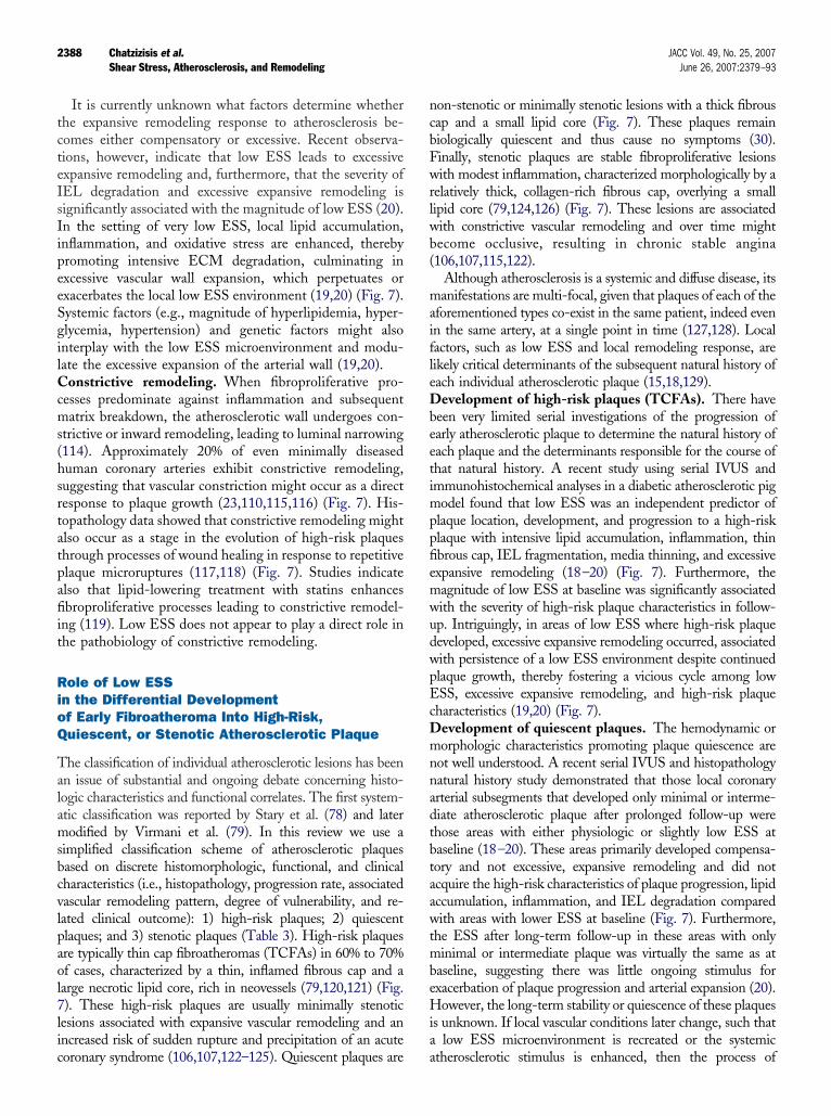
implified classification scheme of atherosclerotic plaquesased on discrete histomorphologic, functional, and clinicalharacteristics (i.e., histopathology, progression rate, associatedascular remodeling pattern, degree of vulnerability, and re-ated clinical outcome): 1) high-risk plaques; 2) quiescentlaques; and 3) stenotic plaques (Table 3). High-risk plaquesre typically thin cap fibroatheromas (TCFAs) in 60% to 70%f cases, characterized by a thin, inflamed fibrous cap and aarge necrotic lipid core, rich in neovessels (79,120,121) (Fig.). These high-risk plaques are usually minimally stenoticesions associated with expansive vascular remodeling and anncreased risk of sudden rupture and precipitation of an acute
oronary syndrome (106,107,122–125). Quiescent plaques are aon-stenotic or minimally stenotic lesions with a thick fibrousap and a small lipid core (Fig. 7). These plaques remainiologically quiescent and thus cause no symptoms (30).inally, stenotic plaques are stable fibroproliferative lesionsith modest inflammation, characterized morphologically by a
elatively thick, collagen-rich fibrous cap, overlying a smallipid core (79,124,126) (Fig. 7). These lesions are associatedith constrictive vascular remodeling and over time mightecome occlusive, resulting in chronic stable angina106,107,115,122).
Although atherosclerosis is a systemic and diffuse disease, itsanifestations are multi-focal, given that plaques of each of the
forementioned types co-exist in the same patient, indeed evenn the same artery, at a single point in time (127,128). Localactors, such as low ESS and local remodeling response, areikely critical determinants of the subsequent natural history ofach individual atherosclerotic plaque (15,18,129).
evelopment of high-risk plaques (TCFAs). There haveeen very limited serial investigations of the progression ofarly atherosclerotic plaque to determine the natural history ofach plaque and the determinants responsible for the course ofhat natural history. A recent study using serial IVUS andmmunohistochemical analyses in a diabetic atherosclerotic pig
odel found that low ESS was an independent predictor oflaque location, development, and progression to a high-risklaque with intensive lipid accumulation, inflammation, thinbrous cap, IEL fragmentation, media thinning, and excessivexpansive remodeling (18–20) (Fig. 7). Furthermore, theagnitude of low ESS at baseline was significantly associatedith the severity of high-risk plaque characteristics in follow-p. Intriguingly, in areas of low ESS where high-risk plaqueeveloped, excessive expansive remodeling occurred, associatedith persistence of a low ESS environment despite continuedlaque growth, thereby fostering a vicious cycle among lowSS, excessive expansive remodeling, and high-risk plaque
haracteristics (19,20) (Fig. 7).evelopment of quiescent plaques. The hemodynamic ororphologic characteristics promoting plaque quiescence are
ot well understood. A recent serial IVUS and histopathologyatural history study demonstrated that those local coronaryrterial subsegments that developed only minimal or interme-iate atherosclerotic plaque after prolonged follow-up werehose areas with either physiologic or slightly low ESS ataseline (18–20). These areas primarily developed compensa-ory and not excessive, expansive remodeling and did notcquire the high-risk characteristics of plaque progression, lipidccumulation, inflammation, and IEL degradation comparedith areas with lower ESS at baseline (Fig. 7). Furthermore,
he ESS after long-term follow-up in these areas with onlyinimal or intermediate plaque was virtually the same as at
aseline, suggesting there was little ongoing stimulus forxacerbation of plaque progression and arterial expansion (20).owever, the long-term stability or quiescence of these plaques
s unknown. If local vascular conditions later change, such thatlow ESS microenvironment is recreated or the systemic
therosclerotic stimulus is enhanced, then the process of

peDstrrTaTfidatd
vf(aEmi
C
BarocfsaTuIarcpaoam
icse
fpmIotApppmaahlpobt
rmI3fraupeainhpnCl
C
2389JACC Vol. 49, No. 25, 2007 Chatzizisis et al.June 26, 2007:2379–93 Shear Stress, Atherosclerosis, and Remodeling
rogressive atherosclerosis, inflammation, and vascular remod-ling might again re-emerge.
evelopment of stenotic (fibrous) plaques. Stenotic le-ions either evolve with a phenotype promoting fibroprolifera-ion consistently throughout its natural history course (115) orepresent an end-stage of scarring in the setting of priorepetitive microruptures of an inflamed TCFA (117) (Fig. 7).he local hemodynamic or morphologic factors responsible for
n early plaque to evolve into a fibrous plaque are unknown.he magnitude of local ESS stimuli for cellular proliferation/brosis versus inflammation might play a decisive role inetermining whether the balance between ECM degradationnd synthesis favors less inflammation and more ECM syn-hesis (i.e., development of stenotic plaque) or favors ECMegradation (i.e., development of TCFA) (130) (Fig. 7).Stenotic plaques infrequently undergo local erosion or de-
elop calcified nodules, which might lead to local thrombusormation and manifestation of an acute coronary syndrome20% to 40% of cases) (79). Low ESS does not appear to playrole in the pathophysiology of plaque erosion. However, highSS, which occurs at the neck of highly stenotic plaques,ight be responsible for the local endothelial erosion and
nduction of acute coronary thrombosis (31) (Fig. 7).
linical Implications of Assessing ESS
ecause ruptured TCFAs account for the great majority of thecute coronary syndromes, early understanding of the degree ofisk associated with an individual plaque and the identificationf plaques at risk to evolve to TCFAs is anticipated to haveonsiderable clinical impact. Although systemic therapy is theoundation to reduce risk in patients with coronary disease,ystemic strategies alone might be insufficient to adequatelyddress the high-risk patient or the high-risk coronary lesion.he PROVE-IT–TIMI-22 (Pravastatin or Atorvastatin Eval-ation and Infection Therapy–Thrombolysis in Myocardialnfarction-22) trial, for example, indicated that although veryggressive systemic therapy (i.e., atorvastatin 80 mg q.d.)educed the primary end point of death or a major cardiovas-ular event by 16% compared with standard statin therapy (i.e.,ravastatin 40 mg q.d.) after a mean follow up of 24 months,primary end point event nevertheless still occurred in 22.4%f the intensive treatment group (131). Clearly an incrementalpproach beyond systemic therapy would be of value in the
lassification Scheme for the Natural History of Early Atherosclero
Table 3 Classification Scheme for the Natural History of Early A
Plaque Trajectory Histopathology Progression Rate
Quiescent plaque Small lipid coreThick fibrous cap
Minimal C
Stenotic plaque Small lipid coreVery thick fibrouscap
Gradual C
High-risk plaque Large lipid coreThin andinflamed fibrouscap
Increased E
anagement of these high-risk patients. A focused strategy of d
dentification of an early stage of a high-risk lesion might beomplementary to systemic therapy by enabling a highlyelective local intervention to avert a future acute coronaryvent (103).
Several in vivo technologies for the assessment of theunctional and morphologic characteristics of a particularlaque now exist, including multislice computed tomography,agnetic resonance imaging, position emission tomography,
VUS-based virtual histology and palpography, thermography,ptical coherence tomography, near-infrared spectroscopy, in-ravascular magnetic resonance imaging, and angioscopy (103).lthough these modalities might be useful to characterize aarticular plaque, they might be insufficient to optimallyredict future risk, because they provide a snapshot of thelaque at only a single point in time. Thus, these modalities areost useful to identify only the ends of the spectrum betweenstenotic plaque and a high-risk plaque, but they cannot
ddress the stimuli responsible for the subsequent naturalistory of that plaque. Incorporation of an in vivo assessment of
ocal ESS stimuli and local remodeling behavior of a particularlaque might substantially enhance the prognostic significancef these imaging modalities, because one can then have insightoth into the existing nature and the future natural history ofhat plaque (15,23,25).
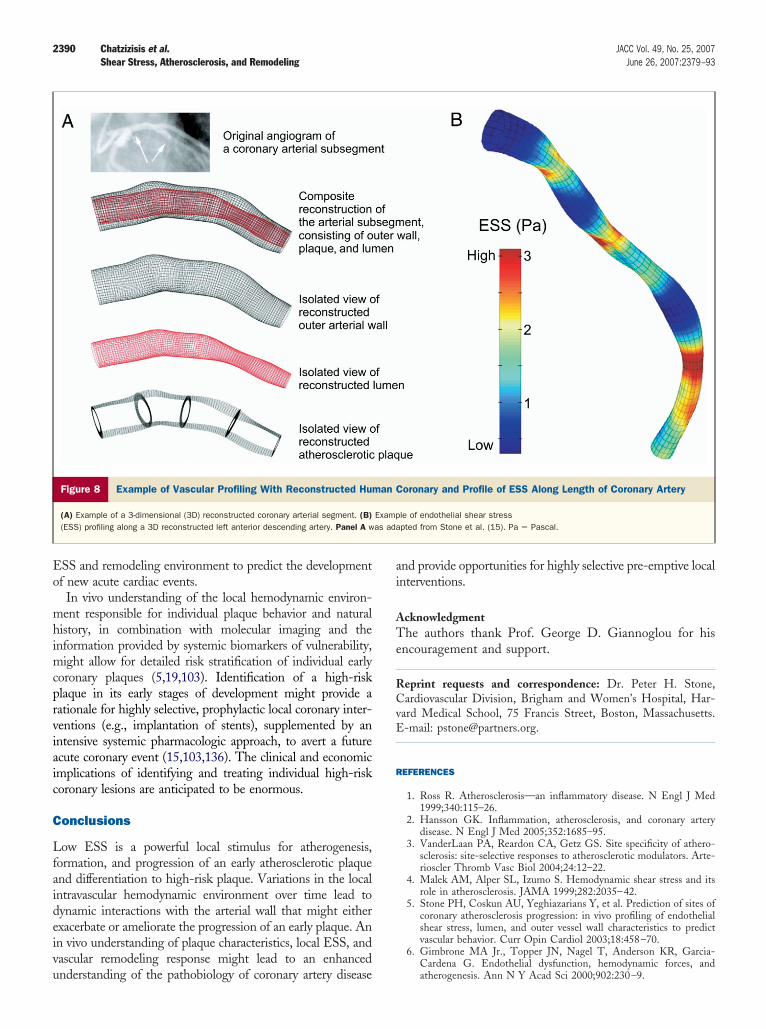
The most comprehensive technique for investigating theelationship between ESS and vascular pathobiology is aethodology known as “vascular profiling,” which uses routine
VUS and coronary angiography to create an accurate-dimensional representation of the coronary artery, and thisorms the basis of identifying both local ESS and vascularemodeling behavior (5,15) (Fig. 8). Vascular profiling isccurate (132–134) and highly reproducible (135) and can besed to track changes in lumen, wall thickness, and ESS ineriods as short as 6 to 9 months in human (15,23) or inxperimental animals (18,19). Future technologies might beble to non-invasively assess local ESS and remodeling behav-or with multi-slice computed tomography, magnetic reso-ance imaging, or other imaging approaches (17). A naturalistory clinical study of atherosclerotic plaques using vascularrofiling techniques in patients with coronary artery disease isow underway (PREDICTION [Prediction of Progression oforonary Artery Disease and Clinical Outcome Using Vascu-
ar Profiling of Shear Stress and Wall Morphology] trial) to
aques (Early Fibroatheromas)
osclerotic Plaques (Early Fibroatheromas)
ular Remodeling Proclivity to Rupture Clinical Manifestation
nsatory expansiveodeling
Low Asymptomatic
ctive remodeling Low Stable angina
ve expansiveodeling
High Acute coronary syndrome
tic Pl
ther
Vasc
omperem
onstri
xcessirem
etermine the incremental value of characterizing the local
Eo
mhimcprviaic
C
Lfaideivu
ai
ATe
RCvE
R
2390 Chatzizisis et al. JACC Vol. 49, No. 25, 2007Shear Stress, Atherosclerosis, and Remodeling June 26, 2007:2379–93
SS and remodeling environment to predict the developmentf new acute cardiac events.
In vivo understanding of the local hemodynamic environ-ent responsible for individual plaque behavior and natural
istory, in combination with molecular imaging and thenformation provided by systemic biomarkers of vulnerability,
ight allow for detailed risk stratification of individual earlyoronary plaques (5,19,103). Identification of a high-risklaque in its early stages of development might provide aationale for highly selective, prophylactic local coronary inter-entions (e.g., implantation of stents), supplemented by anntensive systemic pharmacologic approach, to avert a futurecute coronary event (15,103,136). The clinical and economicmplications of identifying and treating individual high-riskoronary lesions are anticipated to be enormous.
onclusions
ow ESS is a powerful local stimulus for atherogenesis,ormation, and progression of an early atherosclerotic plaquend differentiation to high-risk plaque. Variations in the localntravascular hemodynamic environment over time lead toynamic interactions with the arterial wall that might eitherxacerbate or ameliorate the progression of an early plaque. Ann vivo understanding of plaque characteristics, local ESS, andascular remodeling response might lead to an enhanced
Figure 8 Example of Vascular Profiling With Reconstructed Hum
(A) Example of a 3-dimensional (3D) reconstructed coronary arterial segment. (B)(ESS) profiling along a 3D reconstructed left anterior descending artery. Panel A w
nderstanding of the pathobiology of coronary artery disease
nd provide opportunities for highly selective pre-emptive localnterventions.
cknowledgmenthe authors thank Prof. George D. Giannoglou for his
ncouragement and support.
eprint requests and correspondence: Dr. Peter H. Stone,ardiovascular Division, Brigham and Women’s Hospital, Har-
ard Medical School, 75 Francis Street, Boston, Massachusetts.-mail: [email protected].
EFERENCES
1. Ross R. Atherosclerosis—an inflammatory disease. N Engl J Med1999;340:115–26.
2. Hansson GK. Inflammation, atherosclerosis, and coronary arterydisease. N Engl J Med 2005;352:1685–95.
3. VanderLaan PA, Reardon CA, Getz GS. Site specificity of athero-sclerosis: site-selective responses to atherosclerotic modulators. Arte-rioscler Thromb Vasc Biol 2004;24:12–22.
4. Malek AM, Alper SL, Izumo S. Hemodynamic shear stress and itsrole in atherosclerosis. JAMA 1999;282:2035–42.
5. Stone PH, Coskun AU, Yeghiazarians Y, et al. Prediction of sites ofcoronary atherosclerosis progression: in vivo profiling of endothelialshear stress, lumen, and outer vessel wall characteristics to predictvascular behavior. Curr Opin Cardiol 2003;18:458–70.
6. Gimbrone MA Jr., Topper JN, Nagel T, Anderson KR, Garcia-
oronary and Profile of ESS Along Length of Coronary Artery
le of endothelial shear stresspted from Stone et al. (15). Pa � Pascal.
an C
Exampas ada
Cardena G. Endothelial dysfunction, hemodynamic forces, andatherogenesis. Ann N Y Acad Sci 2000;902:230–9.

2391JACC Vol. 49, No. 25, 2007 Chatzizisis et al.June 26, 2007:2379–93 Shear Stress, Atherosclerosis, and Remodeling
7. Cunningham KS, Gotlieb AI. The role of shear stress in thepathogenesis of atherosclerosis. Lab Invest 2005;85:9–23.
8. Caro CG, Fitz-Gerald JM, Schroter RC. Arterial wall shear anddistribution of early atheroma in man. Nature 1969;223:1159–60.
9. Asakura T, Karino T. Flow patterns and spatial distribution ofatherosclerotic lesions in human coronary arteries. Circ Res 1990;66:1045–66.
10. Ku DN, Giddens DP, Zarins CK, Glagov S. Pulsatile flow andatherosclerosis in the human carotid bifurcation. Positive correlationbetween plaque location and low oscillating shear stress. Arterioscle-rosis 1985;5:293–302.
11. Moore JE Jr., Xu C, Glagov S, Zarins CK, Ku DN. Fluid wall shearstress measurements in a model of the human abdominal aorta:oscillatory behavior and relationship to atherosclerosis. Atheroscle-rosis 1994;110:225–40.
12. Gambillara V, Chambaz C, Montorzi G, Roy S, Stergiopulos N,Silacci P. Plaque-prone hemodynamics impair endothelial function inpig carotid arteries. Am J Physiol Heart Circ Physiol 2006;290:H2320–8.
13. Cheng C, van Haperen R, de Waard M, et al. Shear stress affects theintracellular distribution of eNOS: direct demonstration by a novel invivo technique. Blood 2005;106:3691–8.
14. Buchanan JR Jr., Kleinstreuer C, Truskey GA, Lei M. Relationbetween non-uniform hemodynamics and sites of altered permeabil-ity and lesion growth at the rabbit aorto-celiac junction. Atheroscle-rosis 1999;143:27–40.
15. Stone PH, Coskun AU, Kinlay S, et al. Effect of endothelial shearstress on the progression of coronary artery disease, vascular remod-eling, and in-stent restenosis in humans: in vivo 6-month follow-upstudy. Circulation 2003;108:438–44.
16. Wentzel JJ, Krams R, Schuurbiers JC, et al. Relationship betweenneointimal thickness and shear stress after Wallstent implantation inhuman coronary arteries. Circulation 2001;103:1740–5.
17. Wentzel JJ, Corti R, Fayad ZA, et al. Does shear stress modulateboth plaque progression and regression in the thoracic aorta? Humanstudy using serial magnetic resonance imaging. J Am Coll Cardiol2005;45:846–54.
18. Chatzizisis YS, Jonas M, Coskun AU, et al. Low endothelial shearstress (ESS) is responsible for the heterogeneity and severity ofcoronary atherosclerotic plaques: an in-vivo IVUS natural historystudy (abstr). Circulation 2006;114:II23.
19. Chatzizisis YS, Jonas M, Coskun AU, et al. Low endothelial shearstress (ESS) predicts the development of high-risk coronary athero-sclerotic plaques: a correlative IVUS and histopathology naturalhistory study (abstr). J Am Coll Cardiol 2007;49 Suppl A:334A.
20. Chatzizisis YS, Jonas M, Coskun AU, et al. Low endothelial shearstress (ESS) leads to expansive remodeling of atherosclerotic coronarysubsegments: an in-vivo followup IVUS study (abstr). J Am CollCardiol 2007;49 Suppl:335A.
21. Cheng C, Tempel D, van Haperen R, et al. Atherosclerotic lesionsize and vulnerability are determined by patterns of fluid shear stress.Circulation 2006;113:2744–53.
22. Gambillara V, Montorzi G, Haziza-Pigeon C, Stergiopulos N,Silacci P. Arterial wall response to ex vivo exposure to oscillatoryshear stress. J Vasc Res 2005;42:535–44.
23. Stone PH, Coskun AU, Kinlay S, et al. Regions of low endothelialshear stress are sites where coronary plaque progress and vascularremodeling occurs in humans: an in-vivo serial study. Eur Heart J2007:28:705–10.
24. Wentzel JJ, Janssen E, Vos J, et al. Extension of increased athero-sclerotic wall thickness into high shear stress regions is associatedwith loss of compensatory remodeling. Circulation 2003;108:17–23.
25. Wentzel JJ, Kloet J, Andhyiswara I, et al. Shear-stress and wall-stressregulation of vascular remodeling after balloon angioplasty: effect ofmatrix metalloproteinase inhibition. Circulation 2001;104:91–6.
26. Nichols WW, O’Rourke MF. McDonald’s Blood Flow in Arteries:Theoretical, Experimental and Clincal Principles. 5th edition.London: A Hodder Arnold Publication, 2005.
27. Slager CJ, Wentzel JJ, Gijsen FJ, et al. The role of shear stress in thegeneration of rupture-prone vulnerable plaques. Nat Clin PractCardiovasc Med 2005;2:401–7.
28. Munson BR, Young DF, Okiishi TH. Fundamentals of Fluid
Mechanics. Canada: John Wiley & Sons, 1990.29. Feldman CL, Ilegbusi OJ, Hu Z, Nesto R, Waxman S, Stone PH.Determination of in vivo velocity and endothelial shear stress patternswith phasic flow in human coronary arteries: a methodology topredict progression of coronary atherosclerosis. Am Heart J 2002;143:931–9.
30. MacIsaac AI, Thomas JD, Topol EJ. Toward the quiescent coronaryplaque. J Am Coll Cardiol 1993;22:1228–41.
31. Feldman CL, Stone PH. Intravascular hemodynamic factors respon-sible for progression of coronary atherosclerosis and development ofvulnerable plaque. Curr Opin Cardiol 2000;15:430–40.
32. Papaioannou TG, Karatzis EN, Vavuranakis M, Lekakis JP, StefanadisC. Assessment of vascular wall shear stress and implications for athero-sclerotic disease. Int J Cardiol 2006;113:12–8.
33. Cheng C, Helderman F, Tempel D, et al. Large variations inabsolute wall shear stress levels within one species and betweenspecies. Atherosclerosis 2006 Dec 11;[e-pub ahead of print].
34. Ku D. Blood flow in arteries. Annu Rev Fluid Mech 1997;79:399–434.
35. Soulis JV, Giannoglou GD, Chatzizisis YS, et al. Spatial and phasicoscillation of non-Newtonian wall shear stress in human left coronaryartery bifurcation: an insight to atherogenesis. Coron Artery Dis2006;17:351–8.
36. Nagel T, Resnick N, Dewey CF Jr., Gimbrone MA Jr. Vascularendothelial cells respond to spatial gradients in fluid shear stress byenhanced activation of transcription factors. Arterioscler ThrombVasc Biol 1999;19:1825–34.
37. Giannoglou GD, Soulis JV, Farmakis TM, Farmakis DM, LouridasGE. Haemodynamic factors and the important role of local low staticpressure in coronary wall thickening. Int J Cardiol 2002;86:27–40.
38. Traub O, Berk BC. Laminar shear stress: mechanisms by whichendothelial cells transduce an atheroprotective force. ArteriosclerThromb Vasc Biol 1998;18:677–85.
39. Li YS, Haga JH, Chien S. Molecular basis of the effects of shearstress on vascular endothelial cells. J Biomech 2005;38:1949–71.
40. Lehoux S, Castier Y, Tedgui A. Molecular mechanisms of thevascular responses to haemodynamic forces. J Intern Med 2006;259:381–92.
41. Tzima E, Irani-Tehrani M, Kiosses WB, et al. A mechanosensorycomplex that mediates the endothelial cell response to fluid shearstress. Nature 2005;437:426–31.
42. Tzima E, del Pozo MA, Shattil SJ, Chien S, Schwartz MA.Activation of integrins in endothelial cells by fluid shear stressmediates Rho-dependent cytoskeletal alignment. Embo J 2001;20:4639–47.
43. Shyy JY, Chien S. Role of integrins in endothelial mechanosensing ofshear stress. Circ Res 2002;91:769–75.
44. Lehoux S, Tedgui A. Signal transduction of mechanical stresses inthe vascular wall. Hypertension 1998;32:338–45.
45. Davies PF, Barbee KA, Volin MV, et al. Spatial relationships in earlysignaling events of flow-mediated endothelial mechanotransduction.Annu Rev Physiol 1997;59:527–49.
46. Dai G, Kaazempur-Mofrad MR, Natarajan S, et al. Distinct endo-thelial phenotypes evoked by arterial waveforms derived fromatherosclerosis-susceptible and -resistant regions of human vascula-ture. Proc Natl Acad Sci U S A 2004;101:14871–6.
47. Brooks AR, Lelkes PI, Rubanyi GM. Gene expression profiling ofhuman aortic endothelial cells exposed to disturbed flow and steadylaminar flow. Physiol Genomics 2002;9:27–41.
48. Resnick N, Yahav H, Shay-Salit A, et al. Fluid shear stress and thevascular endothelium: for better and for worse. Prog Biophys MolBiol 2003;81:177–99.
49. Harrison DG, Widder J, Grumbach I, Chen W, Weber M, SearlesC. Endothelial mechanotransduction, nitric oxide and vascular in-flammation. J Intern Med 2006;259:351–63.
50. Lam CF, Peterson TE, Richardson DM, et al. Increased blood flowcauses coordinated upregulation of arterial eNOS and biosynthesis oftetrahydrobiopterin. Am J Physiol Heart Circ Physiol 2006;290:H786–93.
51. Go YM, Boo YC, Park H, et al. Protein kinase B/Akt activates c-JunNH(2)-terminal kinase by increasing NO production in response toshear stress. J Appl Physiol 2001;91:1574–81.
52. Ziegler T, Bouzourene K, Harrison VJ, Brunner HR, Hayoz D.
Influence of oscillatory and unidirectional flow environments on the
2392 Chatzizisis et al. JACC Vol. 49, No. 25, 2007Shear Stress, Atherosclerosis, and Remodeling June 26, 2007:2379–93
expression of endothelin and nitric oxide synthase in culturedendothelial cells. Arterioscler Thromb Vasc Biol 1998;18:686–92.
53. Qiu Y, Tarbell JM. Interaction between wall shear stress andcircumferential strain affects endothelial cell biochemical production.J Vasc Res 2000;37:147–57.
54. Liu Y, Chen BP, Lu M, et al. Shear stress activation of SREBP1 inendothelial cells is mediated by integrins. Arterioscler Thromb VascBiol 2002;22:76–81.
55. Chien S. Molecular and mechanical bases of focal lipid accumulationin arterial wall. Prog Biophys Mol Biol 2003;83:131–51.
56. Yeh M, Cole AL, Choi J, et al. Role for sterol regulatory element-binding protein in activation of endothelial cells by phospholipidoxidation products. Circ Res 2004;95:780–8.
57. Himburg HA, Grzybowski DM, Hazel AL, LaMack JA, Li XM,Friedman MH. Spatial comparison between wall shear stress mea-sures and porcine arterial endothelial permeability. Am J PhysiolHeart Circ Physiol 2004;286:H1916–22.
58. White CR, Haidekker M, Bao X, Frangos JA. Temporal gradients inshear, but not spatial gradients, stimulate endothelial cell prolifera-tion. Circulation 2001;103:2508–13.
59. Tricot O, Mallat Z, Heymes C, Belmin J, Leseche G, Tedgui A.Relation between endothelial cell apoptosis and blood flow directionin human atherosclerotic plaques. Circulation 2000;101:2450–3.
60. Chen YL, Jan KM, Lin HS, Chien S. Ultrastructural studies onmacromolecular permeability in relation to endothelial cell turnover.Atherosclerosis 1995;118:89–104.
61. Hwang J, Ing MH, Salazar A, et al. Pulsatile versus oscillatory shearstress regulates NADPH oxidase subunit expression: implication fornative LDL oxidation. Circ Res 2003;93:1225–32.
62. McNally JS, Davis ME, Giddens DP, et al. Role of xanthineoxidoreductase and NAD(P)H oxidase in endothelial superoxideproduction in response to oscillatory shear stress. Am J Physiol HeartCirc Physiol 2003;285:H2290–7.
63. Mueller CF, Widder JD, McNally JS, McCann L, Jones DP,Harrison DG. The role of the multidrug resistance protein-1 inmodulation of endothelial cell oxidative stress. Circ Res 2005;97:637–44.
64. Libby P. Inflammation in atherosclerosis. Nature 2002;420:868–74.65. Orr AW, Sanders JM, Bevard M, Coleman E, Sarembock IJ,
Schwartz MA. The subendothelial extracellular matrix modulatesNF-kappaB activation by flow: a potential role in atherosclerosis.J Cell Biol 2005;169:191–202.
66. Collins T, Cybulsky MI. NF-kappaB: pivotal mediator or innocentbystander in atherogenesis? J Clin Invest 2001;107:255–64.
67. Mohan S, Hamuro M, Sorescu GP, et al. IkappaBalpha-dependentregulation of low-shear flow-induced NF-kappa B activity: role ofnitric oxide. Am J Physiol Cell Physiol 2003;284:C1039–47.
68. Hsiai TK, Cho SK, Wong PK, et al. Monocyte recruitment toendothelial cells in response to oscillatory shear stress. FASEB J2003;17:1648–57.
69. Moazzam F, DeLano FA, Zweifach BW, Schmid-Schonbein GW.The leukocyte response to fluid stress. Proc Natl Acad Sci U S A1997;94:5338–43.
70. Palumbo R, Gaetano C, Antonini A, et al. Different effects of highand low shear stress on platelet-derived growth factor isoform releaseby endothelial cells: consequences for smooth muscle cell migration.Arterioscler Thromb Vasc Biol 2002;22:405–11.
71. Conklin BS, Zhong DS, Zhao W, Lin PH, Chen C. Shear stressregulates occludin and VEGF expression in porcine arterial endothe-lial cells. J Surg Res 2002;102:13–21.
72. Redmond EM, Cullen JP, Cahill PA, et al. Endothelial cells inhibitflow-induced smooth muscle cell migration: role of plasminogenactivator inhibitor-1. Circulation 2001;103:597–603.
73. Passerini AG, Milsted A, Rittgers SE. Shear stress magnitude anddirectionality modulate growth factor gene expression in precondi-tioned vascular endothelial cells. J Vasc Surg 2003;37:182–90.
74. Grainger DJ. Transforming growth factor beta and atherosclerosis: sofar, so good for the protective cytokine hypothesis. ArteriosclerThromb Vasc Biol 2004;24:399–404.
75. Borkowski P, Robinson MJ, Kusiak JW, Borkowski A, Brathwaite C,Mergner WJ. Studies on TGF-beta 1 gene expression in the intimaof the human aorta in regions with high and low probability of
developing atherosclerotic lesions. Mod Pathol 1995;8:478–82.76. Jones GT, Jiang F, McCormick SP, Dusting GJ. Elastic laminadefects are an early feature of aortic lesions in the apolipoprotein Eknockout mouse. J Vasc Res 2005;42:237–46.
77. Sukhova GK, Wang B, Libby P, et al. Cystatin C deficiency increaseselastic lamina degradation and aortic dilatation in apolipoproteinE-null mice. Circ Res 2005;96:368–75.
78. Stary HC, Chandler AB, Dinsmore RE, et al. A definition ofadvanced types of atherosclerotic lesions and a histological classifica-tion of atherosclerosis. A report from the Committee on VascularLesions of the Council on Arteriosclerosis, American Heart Associ-ation. Circulation 1995;92:1355–74.
79. Virmani R, Kolodgie FD, Burke AP, Farb A, Schwartz SM. Lessonsfrom sudden coronary death: a comprehensive morphological classi-fication scheme for atherosclerotic lesions. Arterioscler Thromb VascBiol 2000;20:1262–75.
80. Magid R, Murphy TJ, Galis ZS. Expression of matrixmetalloproteinase-9 in endothelial cells is differentially regulated byshear stress. Role of c-Myc. J Biol Chem 2003;278:32994–9.
81. Godin D, Ivan E, Johnson C, Magid R, Galis ZS. Remodeling ofcarotid artery is associated with increased expression of matrixmetalloproteinases in mouse blood flow cessation model. Circulation2000;102:2861–6.
82. Bassiouny HS, Song RH, Hong XF, Singh A, Kocharyan H, GlagovS. Flow regulation of 72-kD collagenase IV (MMP-2) after experi-mental arterial injury. Circulation 1998;98:157–63.
83. Choudhary S, Higgins CL, Chen IY, et al. Quantitation andlocalization of matrix metalloproteinases and their inhibitors inhuman carotid endarterectomy tissues. Arterioscler Thromb VascBiol 2006;26:2351–8.
84. de Nooijer R, Verkleij CJ, von der Thusen JH, et al. Lesionaloverexpression of matrix metalloproteinase-9 promotes intraplaquehemorrhage in advanced lesions but not at earlier stages of athero-genesis. Arterioscler Thromb Vasc Biol 2006;26:340–6.
85. Galis ZS, Sukhova GK, Lark MW, Libby P. Increased expression ofmatrix metalloproteinases and matrix degrading activity in vulnerableregions of human atherosclerotic plaques. J Clin Invest 1994;94:2493–503.
86. Galis ZS, Khatri JJ. Matrix metalloproteinases in vascular remodelingand atherogenesis: the good, the bad, and the ugly. Circ Res 2002;90:251–62.
87. Dollery CM, Libby P. Atherosclerosis and proteinase activation.Cardiovasc Res 2006;69:625–35.
88. Chase AJ, Bond M, Crook MF, Newby AC. Role of nuclearfactor-kappa B activation in metalloproteinase-1, -3, and -9 secretionby human macrophages in vitro and rabbit foam cells produced invivo. Arterioscler Thromb Vasc Biol 2002;22:765–71.
89. Lutgens E, Lutgens SP, Faber BC, et al. Disruption of the cathepsinK gene reduces atherosclerosis progression and induces plaque fibro-sis but accelerates macrophage foam cell formation. Circulation2006;113:98–107.
90. Rodgers KJ, Watkins DJ, Miller AL, et al. Destabilizing role ofcathepsin S in murine atherosclerotic plaques. Arterioscler ThrombVasc Biol 2006;26:851–6.
91. Liu J, Sukhova GK, Sun JS, Xu WH, Libby P, Shi GP. Lysosomalcysteine proteases in atherosclerosis. Arterioscler Thromb Vasc Biol2004;24:1359–66.
92. Pasterkamp G, Galis ZS, de Kleijn DP. Expansive arterial remodel-ing: location, location, location. Arterioscler Thromb Vasc Biol2004;24:650–7.
93. Platt MO, Ankeny RF, Jo H. Laminar shear stress inhibits cathepsinL activity in endothelial cells. Arterioscler Thromb Vasc Biol 2006;26:1784–90.
94. Xu Y, Wang L, Buttice G, Sengupta PK, Smith BD. Interferongamma repression of collagen (COL1A2) transcription is mediatedby the RFX5 complex. J Biol Chem 2003;278:49134–44.
95. Tousoulis D, Antoniades C, Koumallos N, Stefanadis C. Pro-inflammatory cytokines in acute coronary syndromes: from bench tobedside. Cytokine Growth Factor Rev 2006;17:225–33.
96. Rosner D, Stoneman V, Littlewood T, et al. Interferon-gammainduces Fas trafficking and sensitization to apoptosis in vascularsmooth muscle cells via a PI3K- and Akt-dependent mechanism.
Am J Pathol 2006;168:2054–63.
1
1
1
1
1
1
1
1
1
1
1
1
1
1
1
1
1
1
1
1
1
1
1
1
1
1
1
1
1
1
1
1
1
1
1
1
1
2393JACC Vol. 49, No. 25, 2007 Chatzizisis et al.June 26, 2007:2379–93 Shear Stress, Atherosclerosis, and Remodeling
97. Um HD, Orenstein JM, Wahl SM. Fas mediates apoptosis in humanmonocytes by a reactive oxygen intermediate dependent pathway.J Immunol 1996;156:3469–77.
98. Lutgens E, Gijbels M, Smook M, et al. Transforming growthfactor-beta mediates balance between inflammation and fibrosisduring plaque progression. Arterioscler Thromb Vasc Biol 2002;22:975–82.
99. Moreno PR, Purushothaman KR, Fuster V, et al. Plaque neovascu-larization is increased in ruptured atherosclerotic lesions of humanaorta: implications for plaque vulnerability. Circulation 2004;110:2032–8.
00. Urbich C, Dernbach E, Reissner A, Vasa M, Zeiher AM, DimmelerS. Shear stress-induced endothelial cell migration involves integrinsignaling via the fibronectin receptor subunits alpha(5) and beta(1).Arterioscler Thromb Vasc Biol 2002;22:69–75.
01. Sorescu GP, Song H, Tressel SL, et al. Bone morphogenic protein 4produced in endothelial cells by oscillatory shear stress inducesmonocyte adhesion by stimulating reactive oxygen species productionfrom a nox1-based NADPH oxidase. Circ Res 2004;95:773–9.
02. Shao JS, Cai J, Towler DA. Molecular mechanisms of vascularcalcification: lessons learned from the aorta. Arterioscler ThrombVasc Biol 2006;26:1423–30.
03. Waxman S, Ishibashi F, Muller JE. Detection and treatment ofvulnerable plaques and vulnerable patients: novel approaches toprevention of coronary events. Circulation 2006;114:2390–411.
04. Malek AM, Jackman R, Rosenberg RD, Izumo S. Endothelialexpression of thrombomodulin is reversibly regulated by fluid shearstress. Circ Res 1994;74:852–60.
05. Papadaki M, Ruef J, Nguyen KT, et al. Differential regulation ofprotease activated receptor-1 and tissue plasminogen activator expres-sion by shear stress in vascular smooth muscle cells. Circ Res1998;83:1027–34.
06. Nakamura M, Nishikawa H, Mukai S, et al. Impact of coronaryartery remodeling on clinical presentation of coronary artery disease:an intravascular ultrasound study. J Am Coll Cardiol 2001;37:63–9.
07. Schoenhagen P, Ziada KM, Kapadia SR, Crowe TD, Nissen SE,Tuzcu EM. Extent and direction of arterial remodeling in stableversus unstable coronary syndromes: an intravascular ultrasoundstudy. Circulation 2000;101:598–603.
08. Armstrong ML, Heistad DD, Marcus ML, Megan MB, Piegors DJ.Structural and hemodynamic response of peripheral arteries ofmacaque monkeys to atherogenic diet. Arteriosclerosis 1985;5:336–46.
09. Glagov S, Weisenberg E, Zarins CK, Stankunavicius R, Kolettis GJ.Compensatory enlargement of human atherosclerotic coronary arter-ies. N Engl J Med 1987;316:1371–5.
10. Feldman CL, Coskun AU, Yeghiazarians Y, et al. Remodelingcharacteristics of minimally diseased coronary arteries are consistentalong the length of the artery. Am J Cardiol 2006;97:13–6.
11. Sipahi I, Tuzcu EM, Schoenhagen P, et al. Paradoxical increase inlumen size during progression of coronary atherosclerosis: observa-tions from the REVERSAL trial. Atherosclerosis 2006;189:229–35.
12. Korshunov VA, Berk BC. Strain-dependent vascular remodeling: the“Glagov phenomenon” is genetically determined. Circulation 2004;110:220–6.
13. Bentzon JF, Pasterkamp G, Falk E. Expansive remodeling is aresponse of the plaque-related vessel wall in aortic roots of apoE-deficient mice: an experiment of nature. Arterioscler Thromb VascBiol 2003;23:257–62.
14. Mintz GS, Kent KM, Pichard AD, Satler LF, Popma JJ, Leon MB.Contribution of inadequate arterial remodeling to the development offocal coronary artery stenoses. An intravascular ultrasound study.Circulation 1997;95:1791–8.
15. Hirose M, Kobayashi Y, Mintz GS, et al. Correlation of coronaryarterial remodeling determined by intravascular ultrasound withangiographic diameter reduction of 20% to 60%. Am J Cardiol2003;92:141–5.
16. Taylor AJ, Burke AP, Farb A, et al. Arterial remodeling in the leftcoronary system: the role of high-density lipoprotein cholesterol.
J Am Coll Cardiol 1999;34:760–7.17. Burke AP, Kolodgie FD, Farb A, et al. Healed plaque ruptures andsudden coronary death: evidence that subclinical rupture has a role inplaque progression. Circulation 2001;103:934–40.
18. Ward MR, Pasterkamp G, Yeung AC, Borst C. Arterial remodeling.Mechanisms and clinical implications. Circulation 2000;102:1186–91.
19. Schoenhagen P, Tuzcu EM, Apperson-Hansen C, et al. Determi-nants of arterial wall remodeling during lipid-lowering therapy: serialintravascular ultrasound observations from the Reversal of Athero-sclerosis with Aggressive Lipid Lowering Therapy (REVERSAL)trial. Circulation 2006;113:2826–34.
20. Schaar JA, Muller JE, Falk E, et al. Terminology for high-risk andvulnerable coronary artery plaques. Report of a meeting on thevulnerable plaque, June 17 and 18, 2003, Santorini, Greece. EurHeart J 2004;25:1077–82.
21. Naghavi M, Libby P, Falk E, et al. From vulnerable plaque tovulnerable patient: a call for new definitions and risk assessmentstrategies: part I. Circulation 2003;108:1664–72.
22. Rodriguez-Granillo GA, Serruys PW, Garcia-Garcia HM, et al.Coronary artery remodelling is related to plaque composition. Heart2006;92:388–91.
23. Kotani J, Mintz GS, Castagna MT, et al. Intravascular ultrasoundanalysis of infarct-related and non-infarct-related arteries in patientswho presented with an acute myocardial infarction. Circulation2003;107:2889–93.
24. Varnava AM, Mills PG, Davies MJ. Relationship between coronaryartery remodeling and plaque vulnerability. Circulation 2002;105:939–43.
25. Ivan E, Khatri JJ, Johnson C, et al. Expansive arterial remodeling isassociated with increased neointimal macrophage foam cell content:the murine model of macrophage-rich carotid artery lesions. Circu-lation 2002;105:2686–91.
26. Pasterkamp G, Schoneveld AH, van der Wal AC, et al. Relation ofarterial geometry to luminal narrowing and histologic markers forplaque vulnerability: the remodeling paradox. J Am Coll Cardiol1998;32:655–62.
27. Cutlip DE, Chhabra AG, Baim DS, et al. Beyond restenosis:five-year clinical outcomes from second-generation coronary stenttrials. Circulation 2004;110:1226–30.
28. Casscells W, Naghavi M, Willerson JT. Vulnerable atheroscleroticplaque: a multifocal disease. Circulation 2003;107:2072–5.
29. Rodriguez-Granillo GA, Garcia-Garcia HM, Wentzel J, et al.Plaque composition and its relationship with acknowledged shearstress patterns in coronary arteries. J Am Coll Cardiol 2006;47:884–5.
30. Silver AE, Vita JA. Shear-stress-mediated arterial remodeling inatherosclerosis: too much of a good thing? Circulation 2006;113:2787–9.
31. Cannon CP, Braunwald E, McCabe CH, et al. Intensive versusmoderate lipid lowering with statins after acute coronary syndromes.N Engl J Med 2004;350:1495–504.
32. Slager CJ, Wentzel JJ, Schuurbiers JC, et al. True 3-dimensionalreconstruction of coronary arteries in patients by fusion of angiogra-phy and IVUS (ANGUS) and its quantitative validation. Circulation2000;102:511–6.
33. Giannoglou GD, Chatzizisis YS, Sianos G, et al. In-vivo validationof spatially correct three-dimensional reconstruction of human cor-onary arteries by integrating intravascular ultrasound and biplaneangiography. Coron Artery Dis 2006;17:533–43.
34. Chatzizisis YS, Giannoglou GD, Matakos A, et al. In-vivo accuracyof geometrically correct three-dimensional reconstruction of humancoronary arteries: is it influenced by certain parameters? Coron ArteryDis 2006;17:545–51.
35. Coskun AU, Yeghiazarians Y, Kinlay S, et al. Reproducibility ofcoronary lumen, plaque, and vessel wall reconstruction and ofendothelial shear stress measurements in vivo in humans. CatheterCardiovasc Interv 2003;60:67–78.
36. Muller JE, Tawakol A, Kathiresan S, Narula J. New opportunities foridentification and reduction of coronary risk: treatment of vulnerable
patients, arteries, and plaques. J Am Coll Cardiol 2006;47:C2–6.