role of inflammatory cytokines in insulin resistance among...
TRANSCRIPT

American Journal of Internal Medicine 2016; 4(5): 79-84
http://www.sciencepublishinggroup.com/j/ajim
doi: 10.11648/j.ajim.20160405.11
ISSN: 2330-4316 (Print); ISSN: 2330-4324 (Online)
Role of Inflammatory Cytokines in Insulin Resistance Among Chronic Hepatitis C Patients
Nouman Mohammed Elgarem1, Mohammad Ahmad Elghobary
1, Rasha Hamed El Sherif
2,
Maha Assem Hussien1, Yousra Hamed Mourad
1
1Departments of Internal Medicine, Faculty of Medicine, Cairo University, Cairo, Egypt 2Clinical and Chemical Pathology, Faculty of Medicine, Cairo University, Cairo, Egypt
Email address: [email protected] (N. M. Elgarem), [email protected] (M. A. Elghobary), [email protected] (M. A. Hussien),
[email protected] (R. H. E. Sherif), [email protected] (Y. H. Mourad)
To cite this article: Nouman Mohammed Elgarem, Mohammad Ahmad Elghobary, Rasha Hamed El Sherif, Maha Assem Hussien. Yousra Hamed Mourad. Role
of Inflammatory Cytokines in Insulin Resistance Among Chronic Hepatitis C Patients. American Journal of Internal Medicine.
Vol. 4, No. 5, 2016, pp. 79-84. doi: 10.11648/j.ajim.20160405.11
Received: July 18, 2016; Accepted: August 2, 2016; Published: August 31, 2016
Abstract: Insulin resistance plays an essential role in the pathogenesis of diabetes associated with HCV. High levels of
inflammatory cytokines have been found in HCV-infected patients. The aim of the study was to investigate the association of
HCV infection with impaired glucose metabolism and to highlight the role of inflammatory cytokines as an initial mechanism
involved in insulin resistance development in HCV infection. It included 3 groups of patients: Group I: 50 HCV patients with
DM. Group II: 50 HCV patients without DM and Group III: 25 patients with DM alone as control subjects. Insulin resistance
was evaluated using the (HOMA- IR) index. We measured the levels of fasting insulin, CRP and two of the inflammatory
cytokines of the innate immunity (TNF-α and IL-6 by ELISA). It was found that insulin resistance, CRP and IL6 in group (I)
were significantly higher when compared to group (II) and (III) with P-value < 0.001. CRP in group (II) was significantly
higher when compared to group (III) with P-value < 0.05. Serum level of (TNF-α) in group (I) was significantly higher when
compared to group (II) with P-value < 0.05 and group (III) with P-value < 0.001 as well as in group (II) it was significantly
higher when compared to group (III) with P-value < 0.001. CRP, IL-6 and TNF-α had statistically significant positive direct
correlation to insulin resistance. In conclusion there was a strong relationship between inflammatory cytokines and the
occurrence of insulin resistance in chronic HCV patients with type 2 diabetes mellitus.
Keywords: Diabetes, Inflammatory Cytokines, Insulin Resistance and Chronic HCV Infection
1. Introduction
"Diabetes and hepatitis C virus (HCV) infection are severe
health problems in the developing countries. [1] Many
studies have suggested that the prevalence of diabetes
mellitus is high among patients with chronic hepatitis C with
or without coexisting cirrhosis [2], [3] Insulin resistance
which is induced by HCV seems to play a vital role in the
pathogenesis of HCV- induced glucose intolerance [1]."
"Chronic inflammation plays a role in the development of
insulin resistance through enhancing inflammatory cytokines.
HCV establishes chronic infection through cellular targets
involved in innate immunity [4] Cytokines of the innate
immune response mainly IL-6 and tumor necrosis factor
(TNF)-α stimulate production of acute-phase proteins by the
liver [3] In type 2 diabetes, there is an ongoing cytokine-
mediated acute-phase response and this is closely involved in
the pathogenesis of the disease [4]."
The aim of our study was to investigate the association of
HCV infection with impaired glucose metabolism and to
highlight the role of inflammatory cytokines as an initial
mechanism involved in insulin resistance development in
HCV infection.
2. Materials and Methods
2.1. Patients
The study included 100 patients with chronic liver disease

80 Nouman Mohammed Elgarem et al.: Role of Inflammatory Cytokines in Insulin Resistance
Among Chronic Hepatitis C Patients
with Grade A score according to the modified Child-Turcotte-
Pugh score. They were recruited from the inpatient ward and
the outpatient clinic of the Internal Medicine Hospital at Cairo
University during the period from June 2013 to March 2014.
The patients were divided into three groups. Group A included
50 chronic HCV patients with Type 2 DM, Group B included
50 chronic HCV patients without DM and Group C included
25 patients with type 2 DM with matched; age, sex and disease
duration as control subjects.
2.2. Exclusion Criteria
Patients with other concomitant hepatic viral infections
(HBV), CLD stage B & C according to modified Child-
Turcotte-Pugh score, autoimmune hepatitis, hepatorenal
syndrome, hepatic focal lesions, other coexisting medical
illness and obese patients (BMI >30). All groups were
carefully matched by age, sex, BMI, negative family history
of type 2 diabetes, fasting blood glucose and severity of liver
disease which was assessed according to Modified Child
Pugh's classification.
2.3. Ethical Aspects
Research protocols were approved by the medical ethics
committee of Kasralainy Medical School, Cairo University.
All participants provided a written informed consent after the
research protocols were explained to them. Informed consent
was obtained from all the study participants and their
approval was taken by signature.
2.4. Procedures
All participants underwent a complete screening panel,
including medical history, clinical examination and
measurement of body mass index.
Specimen collection: Blood samples were collected from
each patient after 6-8 hours of overnight fasting using a sterile
plastic syringe to withdraw venous blood by a single puncture
technique of the most accessible vein. Samples were dispensed
into 3 sterile tubes: The first tube contained K-EDTA solution
(1.2 mg/ml) as an anticoagulant for complete blood picture.
The second one contained sodium citrate solution (3.2 mg/ml)
as an anticoagulant for prothrombin time assay. The third tube
was without an anticoagulant (clotted sample) for biochemical
investigations, serum TNF-α and IL-6. Serum was prepared by
centrifugation for 10 minutes at 3000 rpm at room
temperature, aliquoted and stored after labeling at -20°C until
analysis.
Biochemical profile included the following: serum
electrolytes (Na, K), liver enzymes (ALT, AST), serum
albumin, total proteins, serum bilirubin (total, direct),
alkaline phosphatase (ALP) and fasting glucose. Biochemical
tests were formed using Synchron CX5 auto-analyzer,
Beckman Instrumentation, California, USA. Complete blood
count (CBC) with stress on total leucocytic count was
performed using Sysmex KX-21N, USA. Prothrombin time
(PT) and partial thromboplastin time (PTT) were performed
on the Sysmex CA 1500, Dade Behring, Germany. All
patients were screened for HCV Ab by ELISA, HBsAg,
HBcAb, Bilharsial Ab.
Fasting insulin level was determined using active Insulin
Enzyme-Linked Immunosorbent Assay (ELISA) DSL-10-
1600 Diagnostic Systems laboratories, Inc. Corporate.
Normal values of fasting Insulin 6-35 (µu/ml). Insulin
resistance was determined by the homeostasis model
assessment (HOMA-IR) according to the formula:
HOMA-IR = fasting glucose (mmol / liter) x fasting insulin
(milliU / liter)
22.5 (glucose in mmol/l) or 403 (glucose in mg/ml)
A HOMA score close to 1 indicates normal insulin sensitivity.
Insulin resistance is associated with high HOMA scores.
Inflammatory cytokines: High sensitivity C-reactive
protein was determined using ELISA Kit (Diagnostic
Biochem. Candada Inc.). Serum tumor necrosis factor α
(TNF-α), Interleukin 6 (IL 6) were determined using a
sandwich (ELISA) kit (Koma Biotech Inc., Korea).
Abdominal ultrasound was done with special stress on
liver texture and size, splenomegaly, portal vein diameter and
amount of ascites.
Statistical analysis Data was analyzed through statistical
package of social science software program, version 18
(SPSS). Statistical Package for the Social Science; SPSS Inc.,
Chicago, Illinois, USA. Data were summarized using mean
and SD for quantitative variables and frequency and
percentage for qualitative variables. A comparison between
groups was performed using the one-way analysis of variance
test for quantitative variables with the post-hoc Tukey’s test for
pairwise comparisons and the χ2-test for qualitative variables.
The Spearman correlation coefficient was calculated to test the
association between quantitative variables. P values equal to or
less than 0.05 were considered statistically significant.
3. Results
There was no significant difference in age and BMI
between the 3 groups.
Table 1. Showing the demographic data of the patients. Parameter Group I (mean±SD) Group II (mean±SD) Group III (mean±SD) ANOVA P-value
Age (years) 47.700±12.1404 44.867±13.763 48.867±9.881 0.42
BMI 26.792±4.974 26.083±6.474 25.719±9.036 0.835
Our results showed that CRP, TNF-α, IL6, FBG, Fasting
insulin, Insulin Resistance and Insulin Secretion were
statistically significant different between the three groups with
p value <0.001 except for the TNF-α its p value was <0.05.
Using the post-hoc Tukey's test for pairwise comparisons,
CRP, TNF-α, IL6, FBG, Fasting insulin, Insulin Resistance

American Journal of Internal Medicine 2016; 4(5): 79-84 81
and Insulin Secretion were statistically significant higher in
diabetic patients with HCV infection in comparison with
non-diabetic patients with HCV infection and control group
with p value <0.001 except for the TNF-α in diabetic patients
with HCV infection in comparison with non-diabetic patients
with HCV infection, its p value was <0.05.
CRP and Insulin Resistance were statistically significant
higher in non-diabetic patients with HCV infection in
comparison with control group with p value <0.05, also TNF-
α, FBG, and Insulin Secretion were statistically significant
higher in non-diabetic patients with HCV infection in
comparison with control group with p value <0.001, however
IL6 and Fasting insulin were higher in non-diabetic patients
with HCV infection in comparison with control group with
no statistical difference.
CRP, TNF-α, and IL6 had statistically significant positive
direct correlation with insulin resistance with p value <0.001,
< 0.001 and <0.004 respectively.
Table 2. Showing Mean and Standard deviation of the main laboratory parameters among the three groups included in our study.
parameter Group (I) n = 50 Group (II) n = 50 Group (III) n = 25 ANOVA
Mean ± S.D Mean ± S.D Mean ± S.D P value
CRP mg/L 9.44 ± 2.165 A 7.70 ± 2.726 B 5.82 ± 1.316 C <0.001
TNF-α (pg/ml) 626.12 ± 238.738 A 618 ± 302.805 B 422.76 ± 243.190 C <0.05
IL6 pg/m 215.63 ± 62.004 A 167.62 ±47.719 B 173.72 ± 57.427 B <0.001
FBG mg/dl 168.3 ± 52.234 A 89.95 ± 11.898 B 198.28 ± 30.245 C <0.001
Fasting insulin (µLu/ml) 20.850 ± 5.092 A 11.357± 2.302 B 6.846 ± 1.088 B <0.001
Insulin Resistance 3.020 ± 0.719 A 1.457 ± 0.287 B 1.064 ± 0.160 C <0.001
Insulin Secretion 36.552 ± 18.855 A 97.476 ± 25.847 B 72.93± 26.17 C <0.001
Values are expressed as mean ± SD; ANOVA, analysis of variance. The post-hoc Tukey’s test: groups having different letters are statistically significantly
different at a P value of 0.05; *The P value is statistically significant at P < 0.05 and highly significant with P < 0.001.
Figure 1. Represents CRP mean and S.D. level among the three groups.
Figure 2. Represents mean and SD of interleukin 6 level among the 3 groups.

82 Nouman Mohammed Elgarem et al.: Role of Inflammatory Cytokines in Insulin Resistance
Among Chronic Hepatitis C Patients
Figure 3. Representing mean and S.D. of the TNF-α (pg/ml) in the three groups.
Table 3. Showing Pearson correlation coefficient between insulin resistance and CRP, TNF -α -and IL-6.
Parameter CRP TNF-α IL-6
Insulin resistance
r 0.592 0.581 0.404
p-value <0.001 <0.001 <0.004
r, Spearman correlation coefficient; *The P value is statistically significant.
Figure 4. Showing correlation between Insulin resistance and CRP mg/L.
Represents statistically significant positive correlation
between C-reactive protein and insulin resistance.
Figure 5. Showing Correlation between IL6 pg/ml and insulin resistance.
Represents statistically significant positive correlation
coefficient between insulin resistance and interleukin 6 (r =
0.581, P- value 0.001).
Figure 6. Showing correlation between TNF-α (pg/ml) and insulin
resistance.
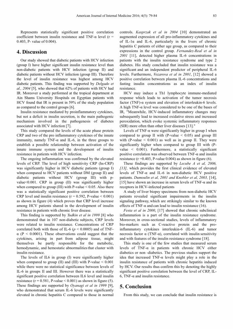
American Journal of Internal Medicine 2016; 4(5): 79-84 83
Represents statistically significant positive correlation
coefficient between insulin resistance and TNF-α level (r =
0.403, P- value of 0.004).
4. Discussion
Our study showed that diabetic patients with HCV infection
(group I) have higher significant insulin resistance level than
non-diabetic patients with HCV infection (group II) and
diabetic patients without HCV infection (group III). Therefore
the level of insulin resistance was highest among HCV
diabetic patients. This finding was supported by Delgado et
al., 2004 [5], who showed that 62% of patients with HCV had
IR. Moreover a study performed at the tropical department at
Ain Shams University Hospitals on Egyptian patients with
HCV found that IR is present in 59% of the study population
as compared to the control groups [6].
Insulin resistance mediated by proinflammatory cytokines,
but not a deficit in insulin secretion, is the main pathogenic
mechanism involved in the pathogenesis of diabetes
associated with HCV infection [7].
This study compared the levels of the acute phase protein
CRP and two of the pro inflammatory cytokines of the innate
immunity, namely TNF-α and IL-6, in the three groups to
establish a possible relationship between activation of the
innate immune system and the development of insulin
resistance in patients with HCV infection.
The ongoing inflammation was confirmed by the elevated
levels of CRP. The level of high sensitivity CRP (hs-CRP)
was significantly higher in HCV diabetic patients (group I)
when compared to HCV patients without DM (group II) and
diabetic patients without HCV (group III) with p-
value<0.001. CRP in group (II) was significantly higher
when compared to group (III) with P-value < 0.05. Also there
was a statistically significant positive correlation between
CRP level and insulin resistance (r = 0.592, P-value < 0.001)
as shown in figure (4) which proves that CRP level increase
among HCV patients shared in the development of insulin
resistance in patients with chronic liver disease.
This finding is supported by Yudkin et al in 1999 [8] who
demonstrated that in 107 non-diabetic subjects, CRP levels
were related to insulin resistance. Concentrations of CRP
correlated both with those of IL-6 (p < 0.0005) and of TNF-
α (P < 0.0001). These observations could suggest that the
cytokines, arising in part
from adipose tissue, might
themselves be partly responsible
for the metabolic,
hemodynamic, and hemostatic abnormalities that cluster with
insulin resistance.
The levels of IL6 in group (I) were significantly higher
when compared to group (II) and (III) with P-value < 0.001
while there were no statistical significance between levels of
IL-6 in groups II and III. However there was a statistically
significant positive correlation between IL6 level and insulin
resistance (r = 0.581, P-value < 0.001) as shown in figure (5).
These findings are supported by Oyanagi et al in 1999 [9],
who demonstrated that serum IL-6 levels were significantly
elevated in chronic hepatitis C compared to those in normal
controls. Kasprzak et al in 2004 [10] demonstrated an
augmented expression of all pro-inflammatory cytokines and
of IL-1α and IL-6, particularly in the livers of chronic
hepatitis C patients of either age group, as compared to their
expressions in the control group. Fernandez-Real et al in
2001 [11], detected higher plasma IL-6 concentrations in
patients with the insulin resistance syndrome and type 2
diabetes. His study concluded that insulin resistance was a
significant and an independent predictor of peripheral IL-6
levels. Furthermore, Vozarova et al in 2001, [12] showed a
positive correlation between plasma IL-6 concentrations and
fasting insulin concentrations as an index of insulin
resistance.
HCV may induce a Th1 lymphocyte immune-mediated
response which leads to activation of the tumor necrosis
factor (TNF)-α system and elevation of interleukin-6 levels.
A high TNF-α level was considered to be one of the bases of
IR, "Meanwhile, HCV-induced inflammatory changes may
subsequently lead to increased oxidative stress and increased
peroxidation, which evoke systemic inflammatory responses
(SIR) more often than other liver diseases [13]."
Levels of TNF-α were significantly higher in group I when
compared to group II with (P-value < 0.05) and group III
with (P-value < 0.001) as well as in group II. They were
significantly higher when compared to group III with (P-
value < 0.001). Furthermore, a statistically significant
positive correlation was observed between TNF-α and insulin
resistance (r =0.403, P-value 0.004) as shown in figure (6).
These findings are supported by Lecube A et al, 2006,
[13], which provides the first clinical evidence of elevated
levels of TNF-α and IL-6 in non-diabetic HCV positive
patients. Dumoulin et al, 2001 and Knobler et al, 2003, [14],
[15] have shown an increase in serum levels of TNF-α and its
receptors in HCV-infected patients
A study of liver biopsy specimens from non-diabetic HCV
patients
revealed significant impairments in the insulin
signaling pathway, which are strikingly similar to the known
effects of TNF-α and can lead to insulin resistance (16).
Festa et al in 2000, [17] showed that chronic subclinical
inflammation is a part of the insulin resistance syndrome.
Moreover, in cross-sectional studies, levels of inflammatory
bio-markers
such as C-reactive protein and the pro-
inflammatory cytokines interleukin-6 (IL-6) and tumor
necrosis factor α (TNF-α), correlated with insulin sensitivity
and with features of the insulin resistance syndrome [18]. This study is one of the few studies that measured serum
levels of TNF-α in patients with chronic HCV either
diabetics or non -diabetics. The previous studies support the
idea that increased TNF-α levels might play a role in the
insulin resistance of patients with chronic hepatitis induced
by HCV. Our results thus confirm this by denoting the highly
significant positive correlation between the level of CRP, IL-
6, TNF-α and insulin resistance.
5. Conclusion
From this study, we can conclude that insulin resistance is

84 Nouman Mohammed Elgarem et al.: Role of Inflammatory Cytokines in Insulin Resistance
Among Chronic Hepatitis C Patients
more correlated to HCV diabetic patients than HCV non-
diabetic patients and that there is a highly significant positive
correlation between level of CRP, IL-6, TNF-α and insulin
resistance. Investigating the association between HCV and
DM is highly important. It represents a major public health
problem that probably affects hundreds of thousands of
patients. Many more HCV-infected patients may have
impaired glucose tolerance before becoming overtly diabetic.
DM may also adversely affect the course of chronic hepatitis
C and be associated with increased liver steatosis and
fibrosis, and recent evidence suggests that steatosis and
diabetes may also significantly enhance the risk of
hepatocellular cancer. These patients may be less responsive
to therapy and may show an increased prevalence of
hepatocellular cancer. Finally, elucidating the risk factors and
associated features of the HCV-diabetes association may
shed a light on mechanisms of the disease. The HCV-DM
association may constitute an example in which an
exaggerated TNF response is deleterious to the host. Future
research should focus on reaffirming this conclusion and
defining effective interventions and strategies to block the
local TNF response.
Therefore, IR should be assessed in patients infected with
hepatits C virus, since IR is a risk factor for fibrosis
progression. Antiviral treatment of the patients at risk is
recommended as soon as possible, to selectively improve
treatment outcome in these patients and thus prevent fibrosis
progression while specific pharmaceutical treatment of
insulin resistance is not yet established. Towards the future,
HCV infection needs to be viewed not only as a liver disease
but also as a metabolic disease.
References
[1] Sher Zaman Safi, Humaira Shah, Gracie OngSiok Yan, Rajes Qvist (2015): Insulin resistance provides the connection between hepatitis C virus and diabetes. Hepat Mon; 15 (1): e23941.
[2] Hui JM, Sud A, Farrell GC, Bandara P, et al (2003): Insulin resistance is associated with chronic hepatitis C virus infection and fibrosis progression. Gastroenterology; 125: 1695-704.
[3] Gentilucci UV, Picardi A, and Pozzilli P (2006): Glucose abnormalities in patients with hepatitis C virus infection: epidemiology and pathogenesis: response to Lecube et al. Diabetes Care; 29 (11): 2558-2559.
[4] Simo R, Lecube A, Genesca, Esteban JR, and Hernandez C (2006): Sustained Virological Response Correlates With Reduction in the Incidence of Glucose Abnormalities in Patients with Chronic Hepatitis C Virus Infection. Diabetes Care; 29 (11): 2462-66.
[5] Delgado-Borrego A, Casson D, Schoenfeld D, Ma S, Adam T, Sergio JH et al (2004): Hepatitis C virus is independently associated with increased insulin resistance after liver transplantation. Transplantation; 77 (5): 703-10.
[6] Mahmoud HM (2005): Prevalence of insulin resistance in non-diabetic HCV patients. (Master’s thesis). Ain Shams University.
[7] Lecube A, Cristina Hernández, Joan Genescà and Rafael Simó (2006): Glucose Abnormalities in Patients with Hepatitis C Virus Infection. DiabetesCare; 29 (5): 1140-9 (a).
[8] Yudkin JS, Stehouwer CD, Emeis JJ, Coppack SW (1999): C-reactive protein in healthy subjects: associations with obesity, insulin resistance, and endothelial dysfunction: a potential role for cytokines originating from adipose tissue? Arterioscler Thromb Vasc Biol; 19: 972-8.
[9] Oyanagi Y, Takahashi T, Matsui S, Takahashi S, Boku S, Takahashi K et al (1999): Enhanced expression of interleukin-6 in chronic hepatitis C. Liver.; 19 (6): 464-72.
[10] Kasprzak A, Seidel J, Spachacz R, Biczysko W, Makowska A, Kaczmarek E et al. (2004): Intracellular expression of pro-inflammatory cytokines (IL-1, TNF-α, and IL-6) in chronic hepatitis C. Roczniki Akademii Medycznej w Biaymstoku; 49: 207-9.
[11] Fernandez-Real JM, Vayreda M, Richart C, Gutierrez C, Broch M, Vendrell J et al (2001): Circulating Interleukin 6 Levels, Blood Pressure, and Insulin Sensitivity in Apparently Healthy Men and Women. The Journal of Clinical Endocrinology & Metabolism; 86 (3): 1154-59.
[12] Vozarova B, Weyer C, Hanson K, Tataranni P, Bogardus C and Pratley RE (2001): Circulating Interleukin-6 in relation to Adiposity, Insulin Action, and Insulin Secretion. Obes Res.; 9: 414–17.
[13] Lecube A, Hernández C, Genescà J and Simó R. (2006) Proinflammatory cytokines, insulin resistance, and insulin secretion in chronic hepatitis C patients: a case-control study. Diabetes Care; 29: 1096-101 (b).
[14] Dumoulin FL, Wennrich U, Nischalke HD Leifeld L, Fischer HP, Sauerbruch T et al (2001): Intrahepatic mRNA levels of interferon gamma and tumor necrosis factor alpha and response to antiviral treatment of chronic hepatitis C. J Hum Virol; 4: 195-99.
[15] Knobler H, Zhornicky T, Sandler A, Haran N, Ashur Y and Schattner A (2003): TNF- induced insulin resistance may mediate the HCV-diabetes association. Am J Gastroenterol; 98: 2751–6.
[16] Aytug S, Reich D, Sapiro L E, Bernstein D and Begum N (2003): Impaired IRS-1/PI3-kinase signaling in patients with HCV: a mechanism for increased prevalence of type 2 diabetes. Hepatology; 38: 1384-92.
[17] Festa A, D'Agostino R Jr, Howard G, Mykkänen L, Russell P. Tracy RP, Haffner SM (2000): Chronic subclinical inflammation as part of the insulin resistance syndrome: the Insulin Resistance Atherosclerosis Study (IRAS). Circulation; 102: 42-7.
[18] Knobler H and Schattner A (2005): TNF-α chronic hepatitis C and diabetes: a novel triad QJM; 98 (1): 1-6.