role of ret protein-tyrosine kinase inhibitors in the ... · r. roskoski jr., a. sadeghi-nejad /...
TRANSCRIPT
I
RR
Ra
b
a
ARAA
CACLPSSV
KCKPPRT
C
CrnP
h1
Pharmacological Research 128 (2018) 1–17
Contents lists available at ScienceDirect
Pharmacological Research
journa l homepage: www.e lsev ier .com/ locate /yphrs
nvited Review
ole of RET protein-tyrosine kinase inhibitors in the treatmentET-driven thyroid and lung cancers
obert Roskoski Jr. a,∗, Abdollah Sadeghi-Nejad b
Blue Ridge Institute for Medical Research, 3754 Brevard Road, Suite 116, Box 19, Horse Shoe, NC 28742-8814, United StatesDepartment of Pediatrics, Tufts Medical Center, Tufts University School of Medicine, 800 Washington Street, Boston, MA 02111-1552, United States
r t i c l e i n f o
rticle history:eceived 21 December 2017ccepted 21 December 2017vailable online 25 December 2017
hemical compounds studied in this article:lectinib (PubMed CID: 49806720)abozantinib (PubMED CID: 25102847)envatinib (PubMED CID: 9823820)onatinib (PubMED CID: 24826799orafenib (PubMED CID: 216239)unitinib (PubMED CID: 5329102andetanib (PubMed CID: 3081361)
eywords:atalytic spine/E/D/Drotein kinase inhibitor classificationrotein kinase structureegulatory spineargeted cancer therapy
a b s t r a c t
RET is a transmembrane receptor protein-tyrosine kinase that is required for the development of thenervous system and several other tissues. The mechanism of activation of RET by its glial-cell derivedneurotrophic factor (GDNF) ligands differs from that of all other receptor protein-tyrosine kinases owingto the requirement for additional GDNF family receptor-� (GFR�) co-receptors (GFR�1/2/3/4). RET pointmutations have been reported in multiple endocrine neoplasia (MEN2A, MEN2B) and medullary thyroidcarcinoma. In contrast, RET fusion proteins have been reported in papillary thyroid and non-small celllung adenocarcinomas. More than a dozen fusion partners of RET have been described in papillary thyroidcarcinomas, most frequently CCDC6-RET and NCOA4-RET. RET-fusion proteins, commonly KIF5B-RET,have also been found in non-small cell lung cancer (NSCLC). Several drugs targeting RET have beenapproved by the FDA for the treatment of cancer: (i) cabozantinib and vandetanib for medullary thyroidcarcinomas and (ii) lenvatinib and sorafenib for differentiated thyroid cancers. In addition, alectinib andsunitinib are approved for the treatment of other neoplasms. Each of these drugs is a multikinase inhibitorthat has activity against RET. Previous X-ray studies indicated that vandetanib binds within the ATP-binding pocket and forms a hydrogen bond with A807 within the RET hinge and it makes hydrophobiccontact with L881 of the catalytic spine which occurs in the floor of the adenine-binding pocket. Ourmolecular modeling studies indicate that the other antagonists bind in a similar fashion. All of theseantagonists bind to the active conformation of RET and are therefore classified as type I inhibitors. Thedrugs also make variable contacts with other residues of the regulatory and catalytic spines. None of thesedrugs was designed to bind preferentially to RET and it is hypothesized that RET-specific antagonists
might produce even better clinical outcomes. Currently the number of new cases of neoplasms bearingRET mutations or RET-fusion proteins is estimated to be about 10,000 per year in the United States. Thisis about the same as the incidence of chronic myelogenous leukemia for which imatinib and secondand third generation BCR-Abl non-receptor protein-tyrosine kinase antagonists have proven clinicallyefficacious and which are commercially successful. These findings warrant the continued developmentof specific antagonists targeting RET-driven neoplasms.© 2017 Published by Elsevier Ltd.
ontents
1. Overview of RET protein-tyrosine kinase . . . . . . . . . . . . . . . . . . . . . . . . . . . . . . . . . .
2. Thyroid cancers . . . . . . . . . . . . . . . . . . . . . . . . . . . . . . . . . . . . . . . . . . . . . . . . . . . . . . . . . . . .
2.1. Classification of thyroid cancers . . . . . . . . . . . . . . . . . . . . . . . . . . . . . . . . . . . .
Abbreviations: ARTN, artemin; AS, activation segment; CD, cadherin-like domain; CDKTT, carboxyterminal tail; EGFR, epidermal growth factor receptor; FGFR, fibroblast growteceptor-�; GK, gatekeeper; GRL, Gly-rich loop; JM, juxtamembrane segment; KD, kinaseurturin; NSCLC, non-small cell lung cancer; PDGFR, platelet-derived growth factor receKC, protein kinase C; RS or R-spine, regulatory spine; Sh1, shell residue 1; TM, transmem∗ Corresponding author.
E-mail addresses: [email protected] (R. Roskoski Jr.), [email protected] (A.
ttps://doi.org/10.1016/j.phrs.2017.12.021043-6618/© 2017 Published by Elsevier Ltd.
. . . . . . . . . . . . . . . . . . . . . . . . . . . . . . . . . . . . . . . . . . . . . . . . . . . . . . . . . . . . . . . . . . . . . . . . . . . . . . 2. . . . . . . . . . . . . . . . . . . . . . . . . . . . . . . . . . . . . . . . . . . . . . . . . . . . . . . . . . . . . . . . . . . . . . . . . . . . . . 2
. . . . . . . . . . . . . . . . . . . . . . . . . . . . . . . . . . . . . . . . . . . . . . . . . . . . . . . . . . . . . . . . . . . . . . . . . . . . . . 3
, cyclin-dependent protein kinase; CL, catalytic loop; CS or C-spine, catalytic spine;h factor receptor; GDNF, glial-cell derived neurotrophic factor; GFR�, GDNF familye domain; KID, kinase insert domain; MEN, multiple endocrine neoplasia; NTRN,
ptor; PI-3K, phosphatidylinositol 3-kinase; PSPN, persephin; PKA, protein kinase A;brane segment; VEGFR, vascular endothelial growth factor receptor.
Sadeghi-Nejad).
2 R. Roskoski Jr., A. Sadeghi-Nejad / Pharmacological Research 128 (2018) 1–17
2.2. Treatment of thyroid cancers . . . . . . . . . . . . . . . . . . . . . . . . . . . . . . . . . . . . . . . . . . . . . . . . . . . . . . . . . . . . . . . . . . . . . . . . . . . . . . . . . . . . . . . . . . . . . . . . . . . . . . . . . . . . . . . . . . . . . 42.3. Driver mutations in thyroid cancers . . . . . . . . . . . . . . . . . . . . . . . . . . . . . . . . . . . . . . . . . . . . . . . . . . . . . . . . . . . . . . . . . . . . . . . . . . . . . . . . . . . . . . . . . . . . . . . . . . . . . . . . . . . . . 4
3. RET fusion proteins in lung cancers . . . . . . . . . . . . . . . . . . . . . . . . . . . . . . . . . . . . . . . . . . . . . . . . . . . . . . . . . . . . . . . . . . . . . . . . . . . . . . . . . . . . . . . . . . . . . . . . . . . . . . . . . . . . . . . . . . . . . . 54. Properties of the RET protein-tyrosine kinase domain . . . . . . . . . . . . . . . . . . . . . . . . . . . . . . . . . . . . . . . . . . . . . . . . . . . . . . . . . . . . . . . . . . . . . . . . . . . . . . . . . . . . . . . . . . . . . . . . . . 5
4.1. Primary, secondary, and tertiary structures of the human RET catalytic domain . . . . . . . . . . . . . . . . . . . . . . . . . . . . . . . . . . . . . . . . . . . . . . . . . . . . . . . . . . . . . . . 54.2. The hydrophobic spines of human RET, VEGFR2, Bruton tyrosine kinase, and murine PKA catalytic domains. . . . . . . . . . . . . . . . . . . . . . . . . . . . . . . . .8
5. Drugs approved and in clinical trials for the treatment of RET-driven thyroid and lung cancers . . . . . . . . . . . . . . . . . . . . . . . . . . . . . . . . . . . . . . . . . . . . . . . . . . . . . . 85.1. Clinical trial summary . . . . . . . . . . . . . . . . . . . . . . . . . . . . . . . . . . . . . . . . . . . . . . . . . . . . . . . . . . . . . . . . . . . . . . . . . . . . . . . . . . . . . . . . . . . . . . . . . . . . . . . . . . . . . . . . . . . . . . . . . . . . 85.2. Classification of protein kinase-drug complexes . . . . . . . . . . . . . . . . . . . . . . . . . . . . . . . . . . . . . . . . . . . . . . . . . . . . . . . . . . . . . . . . . . . . . . . . . . . . . . . . . . . . . . . . . . . . . . . . . 95.3. Structures of RET-drug complexes . . . . . . . . . . . . . . . . . . . . . . . . . . . . . . . . . . . . . . . . . . . . . . . . . . . . . . . . . . . . . . . . . . . . . . . . . . . . . . . . . . . . . . . . . . . . . . . . . . . . . . . . . . . . . . 10
6. RET point mutations . . . . . . . . . . . . . . . . . . . . . . . . . . . . . . . . . . . . . . . . . . . . . . . . . . . . . . . . . . . . . . . . . . . . . . . . . . . . . . . . . . . . . . . . . . . . . . . . . . . . . . . . . . . . . . . . . . . . . . . . . . . . . . . . . . . . 137. Clinical outcomes in response to RET inhibitors . . . . . . . . . . . . . . . . . . . . . . . . . . . . . . . . . . . . . . . . . . . . . . . . . . . . . . . . . . . . . . . . . . . . . . . . . . . . . . . . . . . . . . . . . . . . . . . . . . . . . . . 138. Epilogue . . . . . . . . . . . . . . . . . . . . . . . . . . . . . . . . . . . . . . . . . . . . . . . . . . . . . . . . . . . . . . . . . . . . . . . . . . . . . . . . . . . . . . . . . . . . . . . . . . . . . . . . . . . . . . . . . . . . . . . . . . . . . . . . . . . . . . . . . . . . . . . . . 15
Conflict of interest . . . . . . . . . . . . . . . . . . . . . . . . . . . . . . . . . . . . . . . . . . . . . . . . . . . . . . . . . . . . . . . . . . . . . . . . . . . . . . . . . . . . . . . . . . . . . . . . . . . . . . . . . . . . . . . . . . . . . . . . . . . . . . . . . . . . . . 15Acknowledgments . . . . . . . . . . . . . . . . . . . . . . . . . . . . . . . . . . . . . . . . . . . . . . . . . . . . . . . . . . . . . . . . . . . . . . . . . . . . . . . . . . . . . . . . . . . . . . . . . . . . . . . . . . . . . . . . . . . . . . . . . . . . . . . . . . . . . . 15
. . . . . .
1
isdwfcilnctctt(m(o
3dbutitedi[k
trrtlafdoaaa
References . . . . . . . . . . . . . . . . . . . . . . . . . . . . . . . . . . . . . . . . . . . . . . . . . . . . . . . . . . . .
. Overview of RET protein-tyrosine kinase
RET is a transmembrane receptor protein-tyrosine kinase thats required for the normal development of the brain, the peripheralympathetic and parasympathetic nervous systems, the neuroen-ocrine thyroid calcitonin producing C-cells, thyroid and lung asell as hematopoietic progenitors and other tissues [1]. This review
ocuses on the role of RET in the pathogenesis of thyroid and lungancers and a discussion of the small molecule drugs that target RETn the treatment of these disorders. We will focus on cabozantinib,envatinib, sorafenib, and vandetanib, which are RET and multiki-ase inhibitors that are FDA-approved for the treatment of thyroidancers. We will also consider (i) alectinib, an ALK/RET antagonisthat is approved for the treatment of ALK+ non-small cell lung can-er (NSCLC), (ii) ponatinib, a multikinase inhibitor approved for thereatment of Philadelphia chromosome positive acute lymphoblas-ic leukemia (ALL) and chronic myelogenous leukemia (CML), andiii) sunitinib, a multikinase inhibitor that is approved for the treat-
ent of renal cell carcinoma (RCC), gastrointestinal stromal tumorsGIST), and pancreatic neuroendocrine tumors (pNET) (www.brimr.rg/PKI/PKIs.htm).
As reported in 1985, Takahashi et al. transformed murine NIHT3 fibroblasts with sonicated human lymphoma DNA [2]. Theyetermined that the transforming sequence encompassed 34 kilo-ases and was made up of a rearrangement of two normal butnlinked DNA segments. Because this transforming sequence washe product of gene rearrangement during transfection, they namedt “REarranged during Transfection,” or RET. Ishizaka et al. mappedhe human RET gene to chromosome 10 (10q11.2) [3]. Durbeet al. subsequently identified RET as the receptor for the glial-cellerived neurotrophic factor (GDNF) [4]. Beside GDNF, this family
ncludes artemin (ARTN), neurturin (NTRN), and persephin (PSPN)1]. The nearest relatives of the RET receptor protein-tyrosineinase include FGFR1/2/3/4 and VEGFR1/2/3 [5].
The mechanism of activation of RET by its ligands differs fromhat of all other receptor protein-tyrosine kinases owing to theequirement for additional GDNF family receptor-� (GFR�) co-eceptors (GFR�1/2/3/4) [1]. These co-receptors are tethered tohe external plasma membrane by a glycosylphosphatidylinositolinkage and they form binary complexes with the extracellular RET-ctivating ligands that in turn bind to RET. Members of the GDNFamily exist as a homodimer linked by a disulfide bond. The GDNFimer interacts with two GFR� complexes to form a dimer (a dimer
f dimers) that interacts with two RET receptors to form a hex-meric complex that consists of two receptors, two co-receptors,nd a ligand dimer (Fig. 1). Although there may be some crossovermong the ligands and co-receptors, the chief ligand/co-receptor. . . . . . . . . . . . . . . . . . . . . . . . . . . . . . . . . . . . . . . . . . . . . . . . . . . . . . . . . . . . . . . . . . . . . . . . . . . . 15
complexes include GDNF/GFR�1, NTRN/GFR�2, ARTN/GFR�3, andPSPN/GFR�4 (Table 1) [1].
In the absence of ligand-mediated stimulation, RET existsin a dormant non-phosphorylated state. Following ligand-mediated stimulation, the RET receptor dimer undergoes trans-phosphorylation leading to increased activity. Unlike that ofmost other receptor protein-tyrosine kinases [6], the activatingphosphorylation sites occur outside of the activation segment[7–9]. There are 18 tyrosine residues within the RET intracel-lular domain (Table 2). Tyr900 and Tyr905 occur within theactivation segment, Y660 and Y697 occur within the JM seg-ment, and Y1029/1062/1090/1096 occur in the carboxyterminaltail. Plaza-Menacho et al. reported that Y697 and Y1062 representearly phosphorylation sites while Y900 and Y905 represent latephosphorylation sites [8,9]. These investigators reported that phos-phorylation of Y697 within the JM segment plays an important rolein enhancing RET activity.
Additional receptor phosphorylation creates docking sites for avariety of proteins that result in intracellular signaling by a plethoraof pathways [1,8]. For example, pY752 and pY928 are docking sitesthat lead to the activation of the JAK-STAT pathway and cell survival(Table 2). pY981 is a docking site that leads to the activation of Srcand the PI–3 K/AKT pathway and cell survival while pY1015 leads tothe docking of PLC� and the activation of PKC. pY1062 and pY1096are docking sites that lead to the activation of the Ras/MAP kinasepathway and cell proliferation along with the PI–3 K/AKT pathwayand cell survival as mediated by several docking proteins includingShc1 and Grb2 and Dok1/2/4/5/6. Moreover, the activation segmentpY905 is a docking site for Grb7/10 that contributes to the activationof the Ras/MAP kinase cell proliferation pathway [1].
RET pre-mRNA undergoes alternative splicing to yield threeprotein isoforms: RET9, RET44, and RET51 (Fig. 1) [1]. These dif-fer at the carboxyterminus with 9, 43, and 51 unique C-terminalamino acid residues following codon 1063. Although RET dif-fers in its mode of activation from other protein-tyrosine kinasesowing to the requirement for co-receptors, it contains a typicalextracellular domain, a transmembrane segment followed by anintracellular juxtamembrane segment, a kinase domain, and a car-boxyterminal tail (Fig. 2). The kinase domain contains an insert of15 residues. The extracellular domain contains four cadherin-likedomain (CD1/2/3/4) repeats of about 110 amino acid residues [13]and a cysteine-rich segment of about 150 amino acid residues justproximal to the transmembrane segment. RET is the only receptor
protein-tyrosine kinase with cadherin-like domains. An extracel-lular Ca2+-binding site occurs between CD2 and CD3.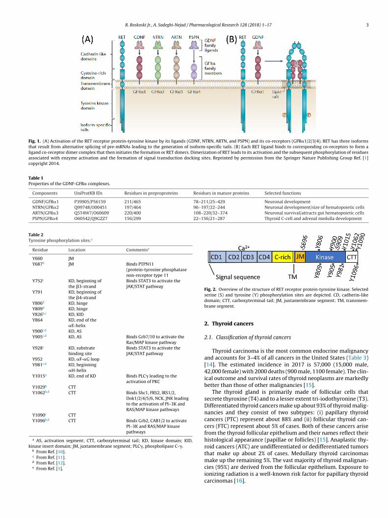
R. Roskoski Jr., A. Sadeghi-Nejad / Pharmacological Research 128 (2018) 1–17 3
Fig. 1. (A) Activation of the RET receptor protein-tyrosine kinase by its ligands (GDNF, NTRN, ARTN, and PSPN) and its co-receptors (GFR�1/2/3/4). RET has three isoformsthat result from alternative splicing of pre-mRNAs leading to the generation of isoform-specific tails. (B) Each RET ligand binds to corresponding co-receptors to form aligand co-receptor dimer complex that then initiates the formation or RET dimers. Dimerization of RET leads to its activation and the subsequent phosphorylation of residuesassociated with enzyme activation and the formation of signal transduction docking sites. Reprinted by permission from the Springer Nature Publishing Group Ref. [1]copyright 2014.
Table 1Properties of the GDNF-GFR� complexes.
Components UniProtKB IDs Residues in preproproteins Residues in mature proteins Selected functions
GDNF/GFR�1 P39905/P56159 211/465 78–211/25–429 Neuronal developmentNTRN/GFR�2 Q99748/O00451 197/464 96–197/22–244 Neuronal development/size of hematopoietic cellsARTN/GFR�3 Q5T4W7/O60609 220/400 108–220/32–374 Neuronal survival/attracts gut hematopoietic cellsPSPN/GFR�4 O60542/Q9GZZ7 156/299 22–156/21–287 Thyroid C-cell and adrenal medulla development
Table 2Tyrosine phosphorylation sites.a
Residue Location Commentse
Y660 JMY687b JM Binds PTPN11
(protein-tyrosine phosphatasenon-receptor type 11
Y752c KD, beginning ofthe �3-strand
Binds STAT3 to activate theJAK/STAT pathway
Y791 KD, beginning ofthe �4-strand
Y806d KD, hingeY809d KD, hingeY826b,c KD, KIDY864 KD, end of the
�E-helixY900c,d KD, ASY905c,d KD, AS Binds Grb7/10 to activate the
Ras/MAP kinase pathwayY928c KD, substrate
binding siteBinds STAT3 to activate theJAK/STAT pathway
Y952 KD, �F-�G loopY981c,d KD, beginning
�H-helixY1015a KD, end of KD Binds PLC� leading to the
activation of PKCY1029b CTTY1062b,d CTT Binds Shc1, FRS2, IRS1/2,
Dok1/2/4/5/6, NCK, JNK leadingto the activation of PI–3K andRAS/MAP kinase pathways
Y1090c CTTY1096b,d CTT Binds Grb2, CAB1/2 to activate
PI–3K and RAS/MAP kinasepathways
a AS, activation segment; CTT, carboxyterminal tail; KD, kinase domain; KID,kinase insert domain; JM, juxtamembrane segment; PLC�, phospholipase C-�.
b From Ref. [10].c From Ref. [11].d From Ref. [12].e From Ref. [1].
Fig. 2. Overview of the structure of RET receptor protein-tyrosine kinase. Selectedserine (S) and tyrosine (Y) phosphorylation sites are depicted. CD, cadherin-likedomain; CTT, carboxyterminal tail; JM, juxtamembrane segment; TM, transmem-brane segment.
2. Thyroid cancers
2.1. Classification of thyroid cancers
Thyroid carcinoma is the most common endocrine malignancyand accounts for 3–4% of all cancers in the United States (Table 3)[14]. The estimated incidence in 2017 is 57,000 (15,000 male,42,000 female) with 2000 deaths (900 male, 1100 female). The clin-ical outcome and survival rates of thyroid neoplasms are markedlybetter than those of other malignancies [15].
The thyroid gland is primarily made of follicular cells thatsecrete thyroxine (T4) and to a lesser extent tri-iodothyronine (T3).Differentiated thyroid cancers make up about 93% of thyroid malig-nancies and they consist of two subtypes: (i) papillary thyroidcancers (PTC) represent about 88% and (ii) follicular thyroid can-cers (FTC) represent about 5% of cases. Both of these cancers arisefrom the thyroid follicular epithelium and their names reflect theirhistological appearance (papillae or follicles) [15]. Anaplastic thy-roid cancers (ATC) are undifferentiated or dedifferentiated tumorsthat make up about 2% of cases. Medullary thyroid carcinomas
make up the remaining 5%. The vast majority of thyroid malignan-cies (95%) are derived from the follicular epithelium. Exposure toionizing radiation is a well-known risk factor for papillary thyroidcarcinomas [16].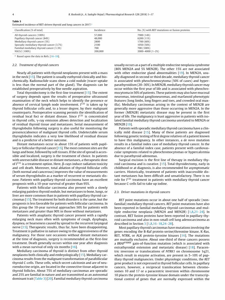
4 R. Roskoski Jr., A. Sadeghi-Nejad / Pharmacological Research 128 (2018) 1–17
Table 3Estimated incidence of RET-driven thyroid and lung cancers in 2017.a
Classification (% of total) Incidence No. (%) with RET mutations or fusion proteins
All thyroid cancers (100%) 57,000 7900 (14%)Papillary thyroid cancer (84%) 48,000 6200 (11%)Medullary thyroid cancer (5%) 2800 1700 (60%)Sporadic medullary thyroid cancer (3.7%) 2100 1050 (50%)
2
icie
oeatcriotpta
llswoa(oep
elcptm
ednTmatw
nctrtad
Familial medullary thyroid cancer (1.3%) 700
NSCLC (100%) 200,000
a Based upon the data in Refs. [16–19].
.2. Treatment of thyroid cancers
Nearly all patients with thyroid neoplasms present with a massn the neck [15]. The patient is usually euthyroid clinically and bio-hemically. Radionuclide scans show a cold nodule (tracer uptakes less than the normal part of the gland). The diagnosis can bestablished preoperatively by fine needle aspiration.
Total thyroidectomy is the first-line treatment [15]. The extentf surgery depends upon the results of preoperative ultrasoundxamination of the neck which helps to identify the presence orbsence of cervical lymph node involvement. I131 is taken up byhyroid follicular cells and, to a lesser degree, by their malignantounterparts. Postoperative scanning permits the identification ofesidual local foci or distant disease. Since I131 is concentratedn thyroid cells, �-ray emission allows detection and localizationf residual thyroid tissue and metastases. Serial measurement ofhyroglobulin following surgery is also useful for monitoring theresence/absence of malignant thyroid cells. Undetectable serumhyroglobulin indicates a very low likelihood of residual diseasend is a predictor of a better outcome.
Distant metastases occur in about 15% of patients with papil-ary or follicular thyroid cancer [15]. The most common sites are theungs and bone, followed by soft tissues. If the site of spread is acces-ible and localized, surgery is the treatment of choice. In patientsith unresectable disease or distant metastases, a therapeutic dose
f I131 is a treatment option. Here, �-rays induce radiation toxicitynd cell death. Moreover, total ablation of thyroid follicular cellsboth normal and cancerous) improves the value of measurementsf serum thyroglobulin as a marker of recurrent or metastatic dis-ase. Patients with papillary thyroid carcinoma have an excellentrognosis with a 10-year survival of greater than 95%.
Patients with follicular carcinoma also present with a slowlynlarging painless thyroid nodule, but metastases to bone, lungs, oriver are more common than in patients with papillary thyroid car-inomas [15]. The treatment for both disorders is the same, but therognosis is less favorable for patients with follicular carcinoma. Inhis group the 10-year survival approaches 50% for patients with
etastases and greater than 90% in those without metastases.Patients with anaplastic thyroid cancer present with a rapidly
nlarging neck mass often with symptoms of cough, dysphagia,yspnea, or hoarseness caused by spread to the recurrent laryngealerve [15]. Therapeutic results, thus far, have been disappointing.reatment is palliative in nature owing to the aggressiveness of thealignancy. For those rare occasions without distant metastases
t the time of diagnosis, surgery is recommended as the first-linereatment. Death generally occurs within one year after diagnosisith a mean survival of only six months [16].
Medullary carcinoma of thyroid is distinct from other thyroideoplasms both clinically and embryologically [15]. Medullary car-inoma results from the malignant transformation of parafollicular
hyroid C-cells. These cells, which secret calcitonin and are of neu-oendocrine origin, are located in the connective tissue adjacent tohyroid follicles. About 75% of medullary carcinomas are sporadicnd 25% are familial in nature and are transmitted as an autosomalominant trait (Table 3) [20]. Familial medullary thyroid carcinoma700 (100%)2000–4000 (1–2%)
usually occurs as a part of a multiple endocrine neoplasia syndrome(80% MEN2A and 5% MEN2B). The other 15% are not associatedwith other endocrine gland abnormalities [19]. In MEN2A, usu-ally diagnosed in second or third decade, medullary thyroid canceris associated with pheochromocytoma (50% of cases) and hyper-parathyroidism (20–30%). In MEN2B, medullary thyroid cancer mayoccur within the first year of life and is associated with pheochro-mocytoma in 50% of patients. These patients may also have mucosalneuromas, intestinal ganglioneuromas, and marfanoid phenotypicfeatures (long limbs, long fingers and toes, and crowded oral max-illa). Medullary carcinomas arising in the context of MEN2B aregenerally more aggressive than those occurring in MEN2A. In theformer (MEN2B) metastatic disease may be present in the firstyear of life. The malignancy is least aggressive in patients with iso-lated familial medullary thyroid carcinoma unrelated to MEN2A orMEN2B [19].
Patients with sporadic medullary thyroid carcinoma have a clin-ically mild disease [15]. Many of these patients are diagnosedfollowing genetic testing of first degree relatives of a patient knownto have this malignancy. In other instances, a de novo mutationresults in a familial index case of medullary thyroid cancer. In theabsence of a familial index case, patients present with cardiovas-cular symptoms related to pheochromocytomas or hypercalcemiadue to parathyroid adenomas.
Surgical excision is the first line of therapy in medullary thy-roid carcinoma and is curative. [15]. Total thyroidectomy, early inchildhood or at diagnosis, is also recommended for asymptomaticcarriers. Historically, treatment of patients with inaccessible dis-tant metastases has been difficult and unsatisfactory. There is norole for I131 treatment in patients with medullary thyroid cancerbecause C-cells fail to take up iodine.
2.3. Driver mutations in thyroid cancers
RET point mutations occur in about one half of sporadic (non-familial) medullary thyroid cancers. RET point mutations have alsobeen reported in familial medullary thyroid carcinomas and mul-tiple endocrine neoplasia (MEN2A and MEN2B) [1,8,21–24]. Incontrast, RET fusion proteins have been reported in papillary thy-roid carcinoma and also in non-small cell lung adenocarcinoma asdescribed in Section 3 [1,8,16–18,24–28].
Most papillary thyroid carcinomas have mutations involving thegenes encoding the B-Raf protein-serine/threonine kinase, K-Ras,RET, NTRK, or ALK protein-tyrosine kinases [16]. The mutationsare mutually exclusive. About one-third of these cancers possessa BRAFV600E gain-of-function mutation (which is associated withextrathyroidal extension and metastatic disease) [18]. Paracen-tric inversion or translocations of NTRK1 on chromosome 1q21,which result in enzyme activation, are present in 5–10% of pap-illary thyroid malignancies. Under physiologic conditions, the RET
gene product is not expressed in thyroid follicular cells. In papillarycancers, however, a reciprocal translocation between chromo-somes 10 and 17 or a paracentric inversion within chromosome10 places the protein-tyrosine kinase domain under the transcrip-tional control of genes that are normally expressed within the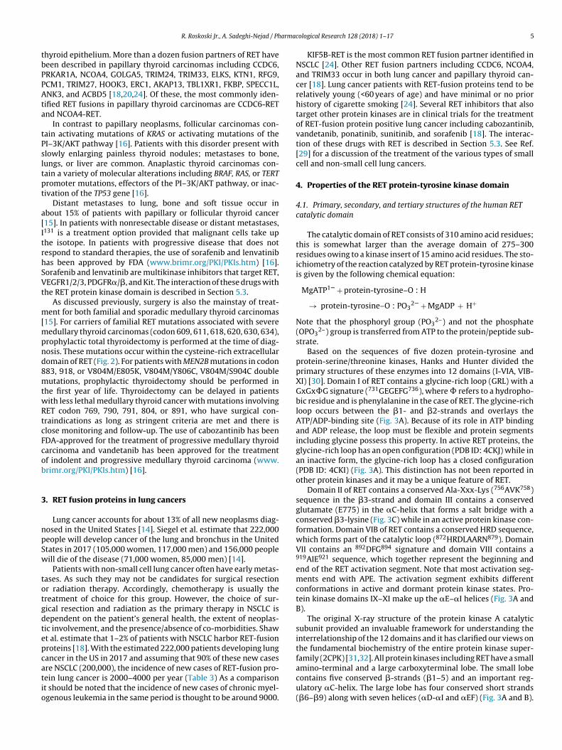
armac
tbPPAta
tPsltpt
a[ItrhSVt
m[mpnd8mtwRtcFcob
3
npSw
totgdtepcatio
R. Roskoski Jr., A. Sadeghi-Nejad / Ph
hyroid epithelium. More than a dozen fusion partners of RET haveeen described in papillary thyroid carcinomas including CCDC6,RKAR1A, NCOA4, GOLGA5, TRIM24, TRIM33, ELKS, KTN1, RFG9,CM1, TRIM27, HOOK3, ERC1, AKAP13, TBL1XR1, FKBP, SPECC1L,NK3, and ACBD5 [18,20,24]. Of these, the most commonly iden-
ified RET fusions in papillary thyroid carcinomas are CCDC6-RETnd NCOA4-RET.
In contrast to papillary neoplasms, follicular carcinomas con-ain activating mutations of KRAS or activating mutations of theI–3K/AKT pathway [16]. Patients with this disorder present withlowly enlarging painless thyroid nodules; metastases to bone,ungs, or liver are common. Anaplastic thyroid carcinomas con-ain a variety of molecular alterations including BRAF, RAS, or TERTromoter mutations, effectors of the PI–3K/AKT pathway, or inac-ivation of the TP53 gene [16].
Distant metastases to lung, bone and soft tissue occur inbout 15% of patients with papillary or follicular thyroid cancer15]. In patients with nonresectable disease or distant metastases,131 is a treatment option provided that malignant cells take uphe isotope. In patients with progressive disease that does notespond to standard therapies, the use of sorafenib and lenvatinibas been approved by FDA (www.brimr.org/PKI/PKIs.htm) [16].orafenib and lenvatinib are multikinase inhibitors that target RET,EGFR1/2/3, PDGFR�/�, and Kit. The interaction of these drugs with
he RET protein kinase domain is described in Section 5.3.As discussed previously, surgery is also the mainstay of treat-
ent for both familial and sporadic medullary thyroid carcinomas15]. For carriers of familial RET mutations associated with severe
edullary thyroid carcinomas (codon 609, 611, 618, 620, 630, 634),rophylactic total thyroidectomy is performed at the time of diag-osis. These mutations occur within the cysteine-rich extracellularomain of RET (Fig. 2). For patients with MEN2B mutations in codon83, 918, or V804M/E805K, V804M/Y806C, V804M/S904C doubleutations, prophylactic thyroidectomy should be performed in
he first year of life. Thyroidectomy can be delayed in patientsith less lethal medullary thyroid cancer with mutations involvingET codon 769, 790, 791, 804, or 891, who have surgical con-raindications as long as stringent criteria are met and there islose monitoring and follow-up. The use of cabozantinib has beenDA-approved for the treatment of progressive medullary thyroidarcinoma and vandetanib has been approved for the treatmentf indolent and progressive medullary thyroid carcinoma (www.rimr.org/PKI/PKIs.htm) [16].
. RET fusion proteins in lung cancers
Lung cancer accounts for about 13% of all new neoplasms diag-osed in the United States [14]. Siegel et al. estimate that 222,000eople will develop cancer of the lung and bronchus in the Unitedtates in 2017 (105,000 women, 117,000 men) and 156,000 peopleill die of the disease (71,000 women, 85,000 men) [14].
Patients with non-small cell lung cancer often have early metas-ases. As such they may not be candidates for surgical resectionr radiation therapy. Accordingly, chemotherapy is usually thereatment of choice for this group. However, the choice of sur-ical resection and radiation as the primary therapy in NSCLC isependent on the patient’s general health, the extent of neoplas-ic involvement, and the presence/absence of co-morbidities. Shawt al. estimate that 1–2% of patients with NSCLC harbor RET-fusionroteins [18]. With the estimated 222,000 patients developing lung
ancer in the US in 2017 and assuming that 90% of these new casesre NSCLC (200,000), the incidence of new cases of RET-fusion pro-ein lung cancer is 2000–4000 per year (Table 3) As a comparisont should be noted that the incidence of new cases of chronic myel-genous leukemia in the same period is thought to be around 9000.ological Research 128 (2018) 1–17 5
KIF5B-RET is the most common RET fusion partner identified inNSCLC [24]. Other RET fusion partners including CCDC6, NCOA4,and TRIM33 occur in both lung cancer and papillary thyroid can-cer [18]. Lung cancer patients with RET-fusion proteins tend to berelatively young (<60 years of age) and have minimal or no priorhistory of cigarette smoking [24]. Several RET inhibitors that alsotarget other protein kinases are in clinical trials for the treatmentof RET-fusion protein positive lung cancer including cabozantinib,vandetanib, ponatinib, sunitinib, and sorafenib [18]. The interac-tion of these drugs with RET is described in Section 5.3. See Ref.[29] for a discussion of the treatment of the various types of smallcell and non-small cell lung cancers.
4. Properties of the RET protein-tyrosine kinase domain
4.1. Primary, secondary, and tertiary structures of the human RETcatalytic domain
The catalytic domain of RET consists of 310 amino acid residues;this is somewhat larger than the average domain of 275–300residues owing to a kinase insert of 15 amino acid residues. The sto-ichiometry of the reaction catalyzed by RET protein-tyrosine kinaseis given by the following chemical equation:
MgATP1– + protein-tyrosine–O : H
→ protein-tyrosine–O : PO32– + MgADP + H+
Note that the phosphoryl group (PO32–) and not the phosphate
(OPO32–) group is transferred from ATP to the protein/peptide sub-
strate.Based on the sequences of five dozen protein-tyrosine and
protein-serine/threonine kinases, Hanks and Hunter divided theprimary structures of these enzymes into 12 domains (I-VIA, VIB-XI) [30]. Domain I of RET contains a glycine-rich loop (GRL) with aGxGx�G signature (731GEGEFG736), where � refers to a hydropho-bic residue and is phenylalanine in the case of RET. The glycine-richloop occurs between the �1- and �2-strands and overlays theATP/ADP-binding site (Fig. 3A). Because of its role in ATP bindingand ADP release, the loop must be flexible and protein segmentsincluding glycine possess this property. In active RET proteins, theglycine-rich loop has an open configuration (PDB ID: 4CKJ) while inan inactive form, the glycine-rich loop has a closed configuration(PDB ID: 4CKI) (Fig. 3A). This distinction has not been reported inother protein kinases and it may be a unique feature of RET.
Domain II of RET contains a conserved Ala-Xxx-Lys (756AVK758)sequence in the �3-strand and domain III contains a conservedglutamate (E775) in the �C-helix that forms a salt bridge with aconserved �3-lysine (Fig. 3C) while in an active protein kinase con-formation. Domain VIB of RET contains a conserved HRD sequence,which forms part of the catalytic loop (872HRDLAARN879). DomainVII contains an 892DFG894 signature and domain VIII contains a919AIE921 sequence, which together represent the beginning andend of the RET activation segment. Note that most activation seg-ments end with APE. The activation segment exhibits differentconformations in active and dormant protein kinase states. Pro-tein kinase domains IX–XI make up the �E–�I helices (Fig. 3A andB).
The original X-ray structure of the protein kinase A catalyticsubunit provided an invaluable framework for understanding theinterrelationship of the 12 domains and it has clarified our views onthe fundamental biochemistry of the entire protein kinase super-
family (2CPK) [31,32]. All protein kinases including RET have a smallamino-terminal and a large carboxyterminal lobe. The small lobecontains five conserved �-strands (�1–5) and an important reg-ulatory �C-helix. The large lobe has four conserved short strands(�6–�9) along with seven helices (�D-�I and �EF) (Fig. 3A and B).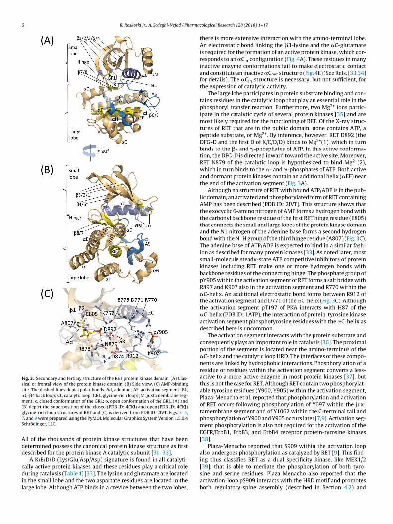
6 R. Roskoski Jr., A. Sadeghi-Nejad / Pharma
Fig. 3. Secondary and tertiary structure of the RET protein kinase domain. (A) Clas-sical or frontal view of the protein kinase domain. (B) Side view. (C) AMP-bindingsite. The dashed lines depict polar bonds. Ad, adenine; AS, activation segment; BL,�C-�4 back loop; CL, catalytic loop; GRL, glycine-rich loop; JM, juxtamembrane seg-ment; c, closed conformation of the GRL; o, open conformation of the GRL. (A) and(g7S
Add
cdil
B) depict the superposition of the closed (PDB ID: 4CKI) and open (PDB ID: 4CKJ)lycine-rich loop structures of RET and (C) is derived from PDB ID: 2IVT. Figs. 3–5,, and 9 were prepared using the PyMOL Molecular Graphics System Version 1.5.0.4chrödinger, LLC.
ll of the thousands of protein kinase structures that have beenetermined possess the canonical protein kinase structure as firstescribed for the protein kinase A catalytic subunit [31–33].
A K/E/D/D (Lys/Glu/Asp/Asp) signature is found in all catalyti-ally active protein kinases and these residues play a critical roleuring catalysis (Table 4) [33]. The lysine and glutamate are located
n the small lobe and the two aspartate residues are located in thearge lobe. Although ATP binds in a crevice between the two lobes,
cological Research 128 (2018) 1–17
there is more extensive interaction with the amino-terminal lobe.An electrostatic bond linking the �3-lysine and the �C-glutamateis required for the formation of an active protein kinase, which cor-responds to an �Cin configuration (Fig. 4A). These residues in manyinactive enzyme conformations fail to make electrostatic contactand constitute an inactive �Cout structure (Fig. 4E) (See Refs. [33,34]for details). The �Cin structure is necessary, but not sufficient, forthe expression of catalytic activity.
The large lobe participates in protein substrate binding and con-tains residues in the catalytic loop that play an essential role in thephosphoryl transfer reaction. Furthermore, two Mg2+ ions partic-ipate in the catalytic cycle of several protein kinases [35] and aremost likely required for the functioning of RET. Of the X-ray struc-tures of RET that are in the public domain, none contains ATP, apeptide substrate, or Mg2+. By inference, however, RET D892 (theDFG-D and the first D of K/E/D/D) binds to Mg2+(1), which in turnbinds to the �- and �-phosphates of ATP. In this active conforma-tion, the DFG-D is directed inward toward the active site. Moreover,RET N879 of the catalytic loop is hypothesized to bind Mg2+(2),which in turn binds to the �- and �-phosphates of ATP. Both activeand dormant protein kinases contain an additional helix (�EF) nearthe end of the activation segment (Fig. 3A).
Although no structure of RET with bound ATP/ADP is in the pub-lic domain, an activated and phosphorylated form of RET containingAMP has been described (PDB ID: 2IVT). This structure shows thatthe exocyclic 6-amino nitrogen of AMP forms a hydrogen bond withthe carbonyl backbone residue of the first RET hinge residue (E805)that connects the small and large lobes of the protein kinase domainand the N1 nitrogen of the adenine base forms a second hydrogenbond with the N–H group of the third hinge residue (A807) (Fig. 3C).The adenine base of ATP/ADP is expected to bind in a similar fash-ion as described for many protein kinases [33]. As noted later, mostsmall-molecule steady-state ATP competitive inhibitors of proteinkinases including RET make one or more hydrogen bonds withbackbone residues of the connecting hinge. The phosphate group ofpY905 within the activation segment of RET forms a salt bridge withR897 and K907 also in the activation segment and R770 within the�C-helix. An additional electrostatic bond forms between R912 ofthe activation segment and D771 of the �C-helix (Fig. 3C). Althoughthe activation segment pT197 of PKA interacts with H87 of the�C-helix (PDB ID: 1ATP), the interaction of protein-tyrosine kinaseactivation segment phosphotyrosine residues with the �C-helix asdescribed here is uncommon.
The activation segment interacts with the protein substrate andconsequently plays an important role in catalysis [36]. The proximalportion of the segment is located near the amino-terminus of the�C-helix and the catalytic loop HRD. The interfaces of these compo-nents are linked by hydrophobic interactions. Phosphorylation of aresidue or residues within the activation segment converts a less-active to a more-active enzyme in most protein kinases [37], butthis is not the case for RET. Although RET contain two phosphorylat-able tyrosine residues (Y900, Y905) within the activation segment,Plaza-Menacho et al. reported that phosphorylation and activationof RET occurs following phosphorylation of Y697 within the jux-tamembrane segment and of Y1062 within the C-terminal tail andphosphorylation of Y900 and Y905 occurs later [7,9]. Activation seg-ment phosphorylation is also not required for the activation of theEGFR/ErbB1, ErbB3, and ErbB4 receptor protein-tyrosine kinases[38].
Plaza-Menacho reported that S909 within the activation loopalso undergoes phosphorylation as catalyzed by RET [9]. This find-
ing thus classifies RET as a dual specificity kinase, like MEK1/2[39], that is able to mediate the phosphorylation of both tyro-sine and serine residues. Plaza-Menacho also reported that theactivation-loop pS909 interacts with the HRD motif and promotesboth regulatory-spine assembly (described in Section 4.2) and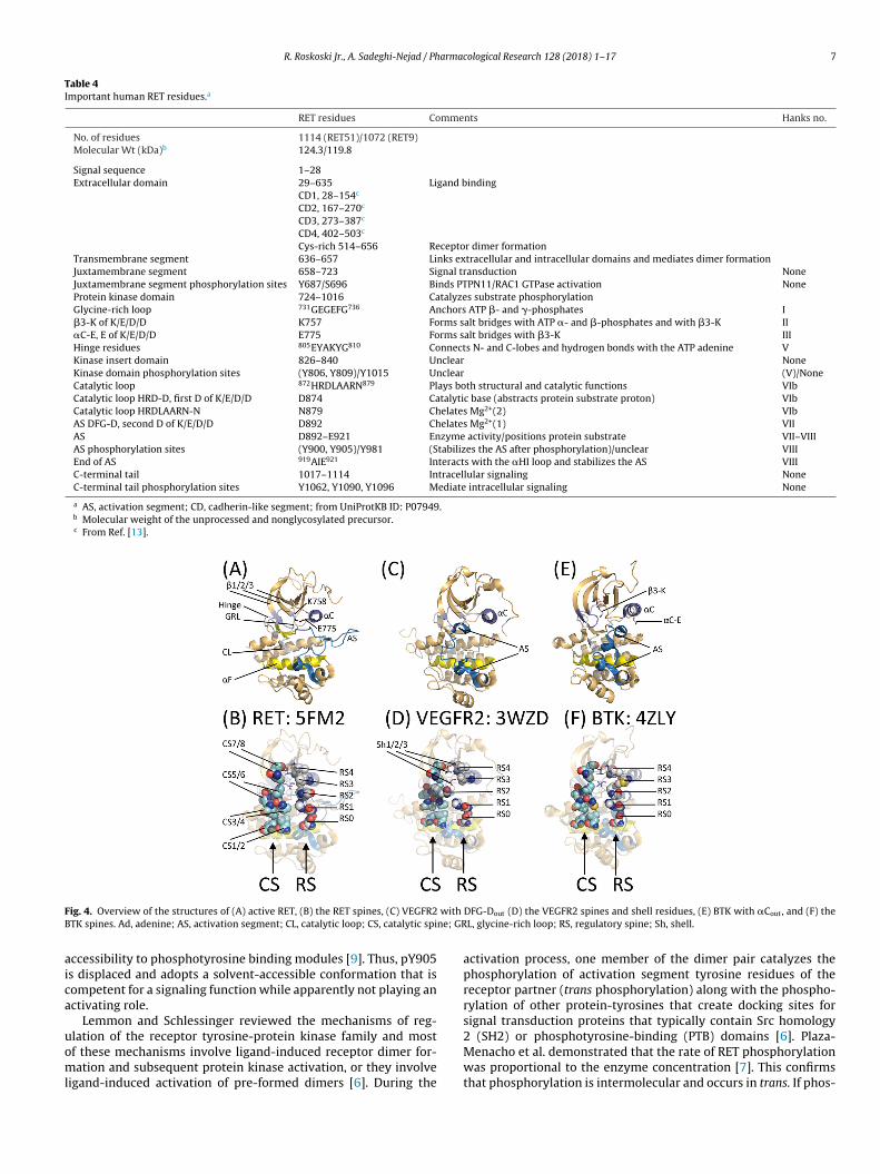
R. Roskoski Jr., A. Sadeghi-Nejad / Pharmacological Research 128 (2018) 1–17 7
Table 4Important human RET residues.a
RET residues Comments Hanks no.
No. of residues 1114 (RET51)/1072 (RET9)Molecular Wt (kDa)b 124.3/119.8
Signal sequence 1–28Extracellular domain 29–635 Ligand binding
CD1, 28–154c
CD2, 167–270c
CD3, 273–387c
CD4, 402–503c
Cys-rich 514–656 Receptor dimer formationTransmembrane segment 636–657 Links extracellular and intracellular domains and mediates dimer formationJuxtamembrane segment 658–723 Signal transduction NoneJuxtamembrane segment phosphorylation sites Y687/S696 Binds PTPN11/RAC1 GTPase activation NoneProtein kinase domain 724–1016 Catalyzes substrate phosphorylationGlycine-rich loop 731GEGEFG736 Anchors ATP �- and �-phosphates I�3-K of K/E/D/D K757 Forms salt bridges with ATP �- and �-phosphates and with �3-K II�C-E, E of K/E/D/D E775 Forms salt bridges with �3-K IIIHinge residues 805EYAKYG810 Connects N- and C-lobes and hydrogen bonds with the ATP adenine VKinase insert domain 826–840 Unclear NoneKinase domain phosphorylation sites (Y806, Y809)/Y1015 Unclear (V)/NoneCatalytic loop 872HRDLAARN879 Plays both structural and catalytic functions VIbCatalytic loop HRD-D, first D of K/E/D/D D874 Catalytic base (abstracts protein substrate proton) VIbCatalytic loop HRDLAARN-N N879 Chelates Mg2+(2) VIbAS DFG-D, second D of K/E/D/D D892 Chelates Mg2+(1) VIIAS D892–E921 Enzyme activity/positions protein substrate VII–VIIIAS phosphorylation sites (Y900, Y905)/Y981 (Stabilizes the AS after phosphorylation)/unclear VIIIEnd of AS 919AIE921 Interacts with the �HI loop and stabilizes the AS VIIIC-terminal tail 1017–1114 Intracellular signaling NoneC-terminal tail phosphorylation sites Y1062, Y1090, Y1096 Mediate intracellular signaling None
a AS, activation segment; CD, cadherin-like segment; from UniProtKB ID: P07949.b Molecular weight of the unprocessed and nonglycosylated precursor.c From Ref. [13].
F with
B ne; GR
aica
uoml
ig. 4. Overview of the structures of (A) active RET, (B) the RET spines, (C) VEGFR2TK spines. Ad, adenine; AS, activation segment; CL, catalytic loop; CS, catalytic spi
ccessibility to phosphotyrosine binding modules [9]. Thus, pY905s displaced and adopts a solvent-accessible conformation that isompetent for a signaling function while apparently not playing anctivating role.
Lemmon and Schlessinger reviewed the mechanisms of reg-
lation of the receptor tyrosine-protein kinase family and mostf these mechanisms involve ligand-induced receptor dimer for-ation and subsequent protein kinase activation, or they involveigand-induced activation of pre-formed dimers [6]. During the
DFG-Dout (D) the VEGFR2 spines and shell residues, (E) BTK with �Cout, and (F) theL, glycine-rich loop; RS, regulatory spine; Sh, shell.
activation process, one member of the dimer pair catalyzes thephosphorylation of activation segment tyrosine residues of thereceptor partner (trans phosphorylation) along with the phospho-rylation of other protein-tyrosines that create docking sites forsignal transduction proteins that typically contain Src homology
2 (SH2) or phosphotyrosine-binding (PTB) domains [6]. Plaza-Menacho et al. demonstrated that the rate of RET phosphorylationwas proportional to the enzyme concentration [7]. This confirmsthat phosphorylation is intermolecular and occurs in trans. If phos-
8 R. Roskoski Jr., A. Sadeghi-Nejad / Pharma
Fig. 5. Inferred mechanism of the RET catalyzed protein kinase reaction. D874aolp
pwipcdk
t(pnasip
4t
krT[Tl�ascCtbdt
tD�[iGn
bstracts a proton from the tyrosyl substrate allowing for its nucleophilic attacknto the �-phosphate of ATP. The chemistry occurs within the blue circle. 1 and 2
abel the two Mg2+ ions. AS, activation segment; CL, catalytic loop. The figure wasrepared from FGFR2 (PDB ID: 2PVF), but the numbers correspond to those of RET.
horylation were intramolecular, the rate of RET phosphorylationould be independent of the enzyme concentration. RET dimer-
zation is required for its activation except that activation segmenthosphorylation is not required to increase catalytic activity. In thease of RET fusion proteins, the fusion partner is responsible for theimerization that results in the subsequent activation of the proteininase domain [17,18].
The RET catalytic loop within the large lobe consists ofhe canonical 872HRDLAARN879 sequence. The catalytic aspartateD874), which is the first D of K/E/D/D, abstracts a proton from therotein-tyrosine substrate residue thereby facilitating the in-lineucleophilic attack of the tyrosyl residue onto the �-phosphorustom of ATP (Fig. 5) [40]. Beside RET, the catalytic segment AARequence occurs in many receptor protein-tyrosine kinases includ-ng EGFR and PDGFR� while RAA occurs in many non-receptorrotein-tyrosine kinases such as Src [30].
.2. The hydrophobic spines of human RET, VEGFR2, Brutonyrosine kinase, and murine PKA catalytic domains
Kornev et al. investigated the tertiary structures of 23 proteininases and they ascertained the role of several critical amino acidesidues by a local spatial pattern alignment algorithm [41,42].hey classified four hydrophobic residues as a regulatory or R-spine41] and eight hydrophobic residues as a catalytic or C-spine [42].hese spines contain amino acid residues from both the small and
arge lobes. The R-spine contains one residue from the regulatoryC-helix and another from the activation segment, both of whichre major components that determine active or dormant enzymetates. The base of the R-spine within the large lobe anchors theatalytic loop and activation segment in an active state while the-spine tethers the adenine base of ATP within the interlobe clefthereby enabling catalysis. Moreover, the accurate alignment ofoth spines is necessary for the assembly of an active enzyme asescribed for ALK, Bruton tyrosine kinase, CDKs, ERK1/2, MEK1/2,he Janus kinases, Src, EGFR, ROS1, and VEGFR2 [29,35,43–50].
Going from the bottom to the top, the protein kinase regula-ory spine consists of the catalytic loop HRD-H, the activation loopFG-F, an amino acid four residues C-terminal to the conserved
C-glutamate, and an amino acid at the beginning of the �4-strand41,42]. The backbone N–H of the HRD-H hydrogen bonds with annvariant aspartyl carboxyl group within the hydrophobic �F-helix.oing from the bottom to the top of the spine, Meharena et al.amed the R-spine residues RS0, RS1, RS2, RS3, and RS4 (Fig. 4B)
cological Research 128 (2018) 1–17
[51]. The R-spine of active RET is linear. The R-spine of dormantenzymes is usually nonlinear or broken, particularly involving theRS2 and RS3 residues. In the case of the DFG-Dout conformation,RS2 is displaced leftward (Fig. 4D) and in the case of the �Cout
configuration, RS2 is displaced rightward (Fig. 4F).The C-spine of protein kinases contains residues from both the
small and large lobes. This spine is completed by the adenine baseof ATP (Fig. 4B) [42]. The two residues of the amino-terminal lobethat interact with the adenine moiety of ATP include a conservedvaline residue in the proximal portion of the �2-strand (CS7) andthe canonical alanine from the AxK signature of the �3-strand(CS8). Moreover, a hydrophobic residue from the �7-strand (CS6)that is two residues carboxyterminal to the end of the catalyticloop interacts with the adenine portion of ATP. Nearly all steady-state ATP-competitive protein kinase inhibitors interact with CS6.The CS6 residue occurs between two hydrophobic residues (CS4and CS5) that interact with the CS3 residue near the beginning ofthe �D-helix of the carboxyterminal lobe (Fig. 4B). CS5/6/4 imme-diately follow the catalytic loop asparagine (HRDxxxxN) so thatone can readily identify these residues based upon the primarystructure. Finally, CS3 and CS4 make hydrophobic contacts withthe CS1 and CS2 residues of the �F-helix thereby forming a com-pleted catalytic spine (Fig. 4B) [42]. Note that the hydrophobic�F-helix, which spans the entire large lobe, supports both spines.Furthermore, both spines play an indispensable role in anchoringthe protein kinase catalytic residues in an active conformation. CS7and CS8 in the small lobe make up the “ceiling” of the adenine-binding pocket while CS5/6/4 make up the “floor” of the bindingpocket. Note that CS5/6/4 form the �7-strand (Fig. 3A) of the proteinkinase domain.
Based upon the findings of site-directed mutagenesis studies,Meharena et al. identified three shell (Sh) residues in the proteinkinase A catalytic subunit that support and stabilize the R-spine,which they labeled Sh1, Sh2, and Sh3 [51]. Sh2 corresponds to theso-called gatekeeper residue, which occurs just before the hinge.This term describes the role that the gatekeeper plays in controllingaccess to the back cleft or back pocket [52,53], which is also calledhydrophobic pocket II (HPII) [53,54]. In contrast to the identifica-tion of the AxK, HRD, or DFG signatures, which is based upon theirprimary structures [30], the spines were identified by their spatiallocations in active or dormant protein kinases [41,42]. Table 5 pro-vides a summary of the spine and shell residues of RET, VEGFR2,BTK, and PKA. As described in Section 5.3, small molecule proteinkinase antagonists often interact with residues that constitute theC-spine as well as the R-spine and the shell residues [58].
The RET X-ray structures are those of an active protein kinase(Fig. 4A and B). For example, the DFG-Asp is pointed inward towardthe active site, the configuration of the �C-helix is in its active �Cinconfiguration with E775 forming a hydrogen bond with the �3-K808, and the catalytic and regulatory spines are linear and areneither bent nor broken. Moreover, the glycine-rich loop is in anopen configuration that can readily accommodate ATP.
5. Drugs approved and in clinical trials for the treatment ofRET-driven thyroid and lung cancers
5.1. Clinical trial summary
Currently the FDA has approved (i) cabozantinib and vandetanibfor the treatment of medullary thyroid cancers and (ii) lenvatinib
and sorafenib for differentiated thyroid cancers (Table 6). In addi-tion, these and other drugs are in clinical trials for the treatment ofboth thyroid and RET-driven lung cancers. It should be noted thatusing the code for the RET inhibitors is useful for literature searchesin both PubMed and PDB data bases because it often results in list-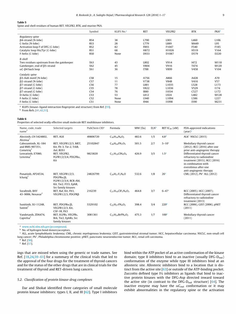
R. Roskoski Jr., A. Sadeghi-Nejad / Pharmacological Research 128 (2018) 1–17 9
Table 5Spine and shell residues of human RET, VEGFR2, BTK, and murine PKA.
Symbol KLIFS No.a RET VEGFR2 BTK PKAb
Regulatory spine�4-strand (N-lobe) RS4 38 L790 L901 L460 L106C-helix (N-lobe) RS3 28 L779 L889 M449 L95Activation loop F of DFG (C-lobe) RS2 82 F893 F1047 F540 F185Catalytic loop His/Tyr (C-lobe) RS1 68 H872 H1026 H519 Y164F-helix (C-lobe) RS0 None D933 D1087 D579 D220
R-shellTwo residues upstream from the gatekeeper Sh3 43 L802 V914 I472 M118Gatekeeper, end of �5-strand Sh2 45 V804 V916 T474 M120�C-�4 back loop Sh1 36 I788 V899 V458 V104
Catalytic spine�3-AxK motif (N-lobe) CS8 15 A756 A866 A428 A70�2-strand (N-lobe) CS7 11 V738 V848 V416 V57�7-strand (C-lobe) CS6 77 L881 L1035 L528 L173�7-strand (C-lobe) CS5 78 V822 L1036 V529 I174�7-strand (C-lobe) CS4 76 I880 I1034 C527 L172D-helix (C-lobe) CS3 53 L812 L924 L482 M128F-helix (C-lobe) CS2 None L940 L1094 L586 L227F-helix (C-lobe) CS1 None I944 I1098 I590 M231
a KLIFS (kinase–ligand interaction fingerprint and structure) from Ref. [55].b From Refs. [41,42,51].
Table 6Properties of selected orally effective small molecule RET multikinase inhibitors.
Name, code, tradename®
Selected targets PubChem CIDa Formula MW (Da) D/Ab RET IC50 (nM) FDA-approved indications(year)c
Alectinib, CH 5424802,Alcensa®
RET, ALK 49806720 C30H34N4O2 482.6 1/5 4.8d ALK+ NSCLC (2015)
Cabozantinib, XL-184and BMS-907351,Cometriq®
RET, VEGFR1/2/3, MET,Kit, Flt-3, Tie-2, TrkB,Axl, ROS1
25102847 C28H24FN3O5 501.5 2/7 5–10e Medullary thyroid cancer(2012); RCC (2016) after oneprior anti-angiogenic therapy
Lenvatinib, E7080,Lenvima®
RET, VEGFR2,FGFR1/2/3/4, PDGFR�,Kit
9823820 C21H19ClN4O4 426.9 3/5 1.5e Differentiated thyroid cancerrefractory to radioiodinetreatment (2015); RCC (2016)in combination witheverolimus after oneanti-angiogenic therapy
Ponatinib, AP24534,Iclusig®
RET, VEGFR1/2/3,PDGFR�/�,FGFR1/2/3/4, BCR-Abl,Kit, Tie2, Fl33, EphR,Src family kinases
24826799 C29H27F3N6O 532.6 1/8 26e CML (2012), Ph+ ALL (2012)
Sorafenib, BAY43–9006, Nexavar®
RET, Raf, Kit, Flt3,VEGFR1/2/3, PDGFR�
216239 C21H16ClF3N4O3 464.8 3/7 6–47e RCC (2005); HCC (2007);Differentiated thyroid cancerrefractory to radioiodinetreatment (2013)
Sunitinib, SU-11248,Sutent®
RET, PDGFR�/�,VEGFR1/2/3, Kit,CSF-1R, Flt3
5329102 C22H27FN4O2 398.4 3/4 220e RCC (2006), GIST (2006), pNET(2011)
Vandetanib, ZD6474,Caprelsa®
RET, EGFRs, VEGFRs,Brk, Tie2, EphRs, Srcfamily kinases
3081361 C22H24BrFN4O2 475.3 1/7 100e Medullary thyroid cancer(2011)
a www.ncbi.nlm.nih.gov/pccompound.b No. of hydrogen bond donors/acceptors.
, gastl rine tu
iRtat
5
p
c ALL, acute lymphoblastic leukemia; CML, chronic myelogenous leukemia; GISTung cancer; Ph+, Philadelphia chromosome positive; pNET, pancreatic neuroendoc
d Ref. [56].e Ref. [57].
ngs that are missed when using the generic or trade names. Seeef. [18,24,59–61] for a summary of the clinical trials that led tohe approval of the four drugs for the treatment of thyroid cancersnd for the status of the other drugs that are in clinical trials for thereatment of thyroid and RET-driven lung cancers.
.2. Classification of protein kinase-drug complexes
Dar and Shokat identified three categories of small moleculerotein kinase inhibitors: types I, II, and III [62]. Type I inhibitors
rointestinal stromal tumor; HCC, hepatocellular carcinoma; NSCLC, non-small cellmor; RCC, renal cell carcinoma.
bind within the ATP pocket of an active conformation of the kinasedomain; type II inhibitors bind to an inactive (usually DFG-Dout)conformation of the enzyme while type III inhibitors bind at anallosteric site. Allosteric inhibitors bind to a location that is dis-tinct from the active site [63] or outside of the ATP-binding pocket.Zuccotto defined type I½ inhibitors as ligands that bind to inac-
tive protein kinases with the DFG-Asp directed inward towardthe active site (in contrast to the DFG-Dout structure) [64]. Theinactive enzyme may have the �Cout conformation or it mayexhibit abnormalities in the regulatory spine or the activation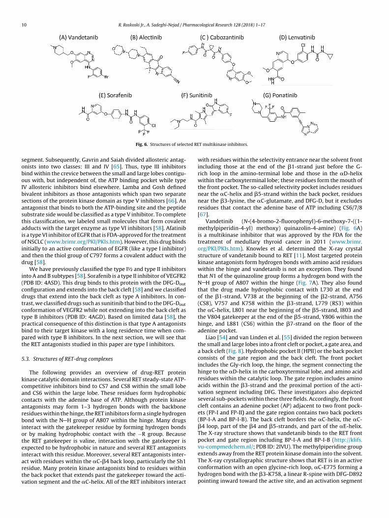
10 R. Roskoski Jr., A. Sadeghi-Nejad / Pharmacological Research 128 (2018) 1–17
ted RE
soboIbsastaioiad
i(cdtctpbpt
5
kcacarbioteiartv
Fig. 6. Structures of selec
egment. Subsequently, Gavrin and Saiah divided allosteric antag-nists into two classes: III and IV [65]. Thus, type III inhibitorsind within the crevice between the small and large lobes contigu-us with, but independent of, the ATP binding pocket while type
V allosteric inhibitors bind elsewhere. Lamba and Gosh definedivalent inhibitors as those antagonists which span two separateections of the protein kinase domain as type V inhibitors [66]. Anntagonist that binds to both the ATP-binding site and the peptideubstrate side would be classified as a type V inhibitor. To completehis classification, we labeled small molecules that form covalentdducts with the target enzyme as type VI inhibitors [58]. Afatinibs a type VI inhibitor of EGFR that is FDA-approved for the treatmentf NSCLC (www.brimr.org/PKI/PKIs.htm). However, this drug binds
nitially to an active conformation of EGFR (like a type I inhibitor)nd then the thiol group of C797 forms a covalent adduct with therug [58].
We have previously classified the type I½ and type II inhibitorsnto A and B subtypes [58]. Sorafenib is a type II inhibitor of VEGFR2PDB ID: 4ASD). This drug binds to this protein with the DFG-Dout
onfiguration and extends into the back cleft [58] and we classifiedrugs that extend into the back cleft as type A inhibitors. In con-rast, we classified drugs such as sunitinib that bind to the DFG-Dout
onformation of VEGFR2 while not extending into the back cleft asype B inhibitors (PDB ID: 4AGD). Based on limited data [58], theractical consequence of this distinction is that type A antagonistsind to their target kinase with a long residence time when com-ared with type B inhibitors. In the next section, we will see thathe RET antagonists studied in this paper are type I inhibitors.
.3. Structures of RET-drug complexes
The following provides an overview of drug-RET proteininase catalytic domain interactions. Several RET steady-state ATP-ompetitive inhibitors bind to CS7 and CS8 within the small lobend CS6 within the large lobe. These residues form hydrophobicontacts with the adenine base of ATP. Although protein kinasentagonists may form 1–3 hydrogen bonds with the backboneesidues within the hinge, the RET inhibitors form a single hydrogenond with the N H group of A807 within the hinge. Many drugs
nteract with the gatekeeper residue by forming hydrogen bondsr by making hydrophobic contact with the −R group. Becausehe RET gatekeeper is valine, interaction with the gatekeeper isxpected to be hydrophobic in nature and several RET antagonists
nteract with this residue. Moreover, several RET antagonists inter-ct with residues within the �C-�4 back loop, particularly the Sh1esidue. Many protein kinase antagonists bind to residues withinhe back pocket that extends past the gatekeeper toward the acti-ation segment and the �C-helix. All of the RET inhibitors interactT multikinase inhibitors.
with residues within the selectivity entrance near the solvent frontincluding those at the end of the �1-strand just before the G-rich loop in the amino-terminal lobe and those in the �D-helixwithin the carboxyterminal lobe; these residues form the mouth ofthe front pocket. The so-called selectivity pocket includes residuesnear the �C-helix and �5-strand within the back pocket, residuesnear the �3-lysine, the �C-glutamate, and DFG-D, but it excludesresidues that contact the adenine base of ATP including CS6/7/8[67].
Vandetinib (N-(4-bromo-2-fluorophenyl)-6-methoxy-7-((1-methylpiperidin-4-yl) methoxy) quinazolin-4-amine) (Fig. 6A)is a multikinase inhibitor that was approved by the FDA for thetreatment of medullary thyroid cancer in 2011 (www.brimr.org/PKI/PKIs.htm). Knowles et al. determined the X-ray crystalstructure of vandetanib bound to RET [11]. Most targeted proteinkinase antagonists form hydrogen bonds with amino acid residueswithin the hinge and vandetanib is not an exception. They foundthat N1 of the quinazoline group forms a hydrogen bond with theN H group of A807 within the hinge (Fig. 7A). They also foundthat the drug made hydrophobic contact with L730 at the endof the �1-strand, V738 at the beginning of the �2-strand, A756(CS8), V757 and K758 within the �3-strand, L779 (RS3) withinthe �C-helix, L801 near the beginning of the �5-strand, I803 andthe V804 gatekeeper at the end of the �5-strand, Y806 within thehinge, and L881 (CS6) within the �7-strand on the floor of theadenine pocket.
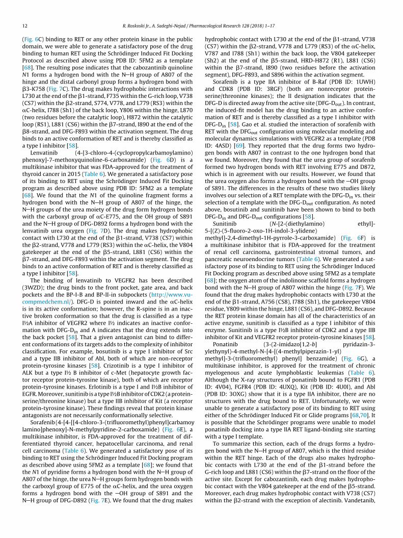
Liao [54] and van Linden et al. [55] divided the region betweenthe small and large lobes into a front cleft or pocket, a gate area, anda back cleft (Fig. 8). Hydrophobic pocket II (HPII) or the back pocketconsists of the gate region and the back cleft. The front pocketincludes the Gly-rich loop, the hinge, the segment connecting thehinge to the �D-helix in the carboxyterminal lobe, and amino acidresidues within the catalytic loop. The gate region includes aminoacids within the �3-strand and the proximal portion of the acti-vation segment including DFG. These investigators also depictedseveral sub-pockets within these three fields. Accordingly, the frontcleft contains an adenine pocket (AP) adjacent to two front pock-ets (FP-I and FP-II) and the gate region contains two back pockets(BP-I-A and BP-I-B). The back cleft borders the �C-helix, the �C-�4 loop, part of the �4 and �5-strands, and part of the �E-helix.The X-ray structure shows that vandetanib binds to the RET frontpocket and gate region including BP-I-A and BP-I-B (http://klifs.vu-compmedchem.nl/; PDB ID: 2IVU). The methylpiperidine group
extends away from the RET protein kinase domain into the solvent.The X-ray crystallographic structure shows that RET is in an activeconformation with an open glycine-rich loop, �C-E775 forming ahydrogen bond with the �3-K758, a linear R-spine with DFG-D892pointing inward toward the active site, and an activation segment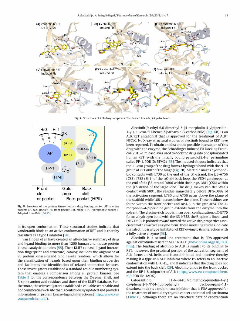
R. Roskoski Jr., A. Sadeghi-Nejad / Pharmacological Research 128 (2018) 1–17 11
Fig. 7. Structures of RET-drug complexes.
FpA
ivc
akt8taTtTRtnic
Cabozantinib (1-N-[4-(6,7-dimethoxyquinolin-4-yl)
ig. 8. Structure of the protein kinase domain drug binding pocket. AP, adenineocket; BP, back pocket; FP, front pocket; Hn, hinge; HP, Hydrophobic pocket II.dapted from Refs. [54,55].
n its open conformation. These structural studies indicate thatandetanib binds to an active conformation of RET and is therebylassified as a type I inhibitor [58].
van Linden et al. have created an all-inclusive summary of drugnd ligand binding to more than 1200 human and mouse proteininase catalytic domains [55]. Their KLIFS (kinase–ligand interac-ion fingerprint and structure) catalog includes the alignment of5 protein kinase-ligand binding-site residues, which allows forhe classification of ligands based upon their binding propertiesnd facilitates the identification of related interaction features.hese investigators established a standard residue numbering sys-em that enables a comparison among all protein kinases. Seeable 5 for the correspondence between the C-spine, Shell, and-spine amino acid residues and those of the KLIFS database. Fur-
hermore, these investigators established a valuable searchable andoncommercial web site that is continuously updated and providesnformation on protein kinase-ligand interactions (http://www.vu-ompmedchem.nl/).
The dashed lines depict polar bonds.
Alectinib (9-ethyl-6,6-dimethyl-8-(4-morpholin-4-ylpiperidin-1-yl)-11-oxo-5H-benzo[b]carbazole-3-carbohitrile) (Fig. 6B) is anALK/RET antagonist that is approved for the treatment of ALK+
NSCLC. No X-ray structural studies of alectinib bound to RET havebeen reported. To obtain an idea on the possible interaction of thisdrug with the enzyme, the Schrödinger Induced Fit Docking Proto-col (2016-1 release) was used to dock the drug into phosphorylatedhuman RET (with the initially bound pyrazolo[3,4-d] pyrimidinecalled PP-1, PDB ID: 5FM2) [68]. The induced-fit pose indicates thatthe 11-oxo group of the drug forms a hydrogen bond with the N Hgroup of RET A807 of the hinge (Fig. 7B). Alectinib makes hydropho-bic contacts with L730 at the end of the �1-strand, the �3-A756(CS8), I788 (Sh1) of the �C-�4 back loop, the V804 gatekeeper atthe end of the �5-strand, Y806 within the hinge, L881 (CS6) withinthe �7-strand of the large lobe. The drug makes van der Waalscontact with S891, the residue immediately before DFG-D892 ofthe activation segment. L730 and A756 occur above the plane ofthe scaffold while L881 occurs below the plane. These residues arefound within the front pocket and BP-I-B in the gate area. The 4-morpholin-4-piperidine group extends from the enzyme into thesolvent. The glycine-rich loop is in an open configuration, �C-E775forms a hydrogen bond with the �3-K758, the R-spine is linear, andDFG-D892 is pointed inward toward the active site, properties asso-ciated with an active enzyme form. These modeling studies indicatethat alectinib is a type I inhibitor of RET owing to its interaction witha fully active enzyme [58].
Alectinib is a second-line treatment that is FDA-approvedagainst crizotinib-resistant ALK+ NSCLC (www.brimr.org/PKI/PKIs.htm). The binding of alectinib to ALK is similar to its binding toRET; however, the proximal portion of the activation segment ofALK forms an AL-helix and is autoinhibited and inactive therebymaking it a type I½B ALK inhibitor where I½ refers to an inactiveconformation with DFG-Din and B indicates that the drug does notextend into the back cleft [29]. Alectinib binds to the front pocketand the BP-I-B subpocket of ALK (http://www.vu-compmedchem.nl/; PDB ID: 3AOX).
oxyphenyl]-1-N’-(4-fluorophenyl) cyclopropane-1,1-dicarboxamide) is a multikinase inhibitor that is FDA approved forthe treatment of medullary thyroid cancer and renal cell carcinoma(Table 6). Although there are no structural data of cabozantinib
1 arma
(dbP[Nh�L(�(l�ba
pmtop[hNwalctg�ba
(pcitImtecapAtpEspa
lmfcbatAtfN
2 R. Roskoski Jr., A. Sadeghi-Nejad / Ph
Fig. 6C) binding to RET or any other protein kinase in the publicomain, we were able to generate a satisfactory pose of the druginding to human RET using the Schrödinger Induced Fit Dockingrotocol as described above using PDB ID: 5FM2 as a template68]. The resulting pose indicates that the cabozantinib quinoline1 forms a hydrogen bond with the N H group of A807 of theinge and the distal carbonyl group forms a hydrogen bond with3-K758 (Fig. 7C). The drug makes hydrophobic interactions with730 at the end of the �1-strand, F735 within the G-rich loop, V738CS7) within the �2-strand, S774, V778, and L779 (RS3) within theC-helix, I788 (Sh1) of the back loop, Y806 within the hinge, L870
two residues before the catalytic loop), H872 within the catalyticoop (RS1), L881 (CS6) within the �7-strand, I890 at the end of the8-strand, and DFG-F893 within the activation segment. The druginds to an active conformation of RET and is thereby classified as
type I inhibitor [58].Lenvatinib (4-[3-chloro-4-(cyclopropylcarbamoylamino)
henoxy]-7-methoxyquinoline-6-carboxamide) (Fig. 6D) is aultikinase inhibitor that was FDA-approved for the treatment of
hyroid cancer in 2015 (Table 6). We generated a satisfactory posef its binding to RET using the Schrödinger Induced Fit Dockingrogram as described above using PDB ID: 5FM2 as a template68]. We found that the N1 of the quinoline fragment forms aydrogen bond with the N H group of A807 of the hinge, the
H groups of the urea moiety of the drug form hydrogen bondsith the carboxyl group of �C-E775, and the OH group of S891
nd the N H group of DFG-D892 forms a hydrogen bond with theenvatinib urea oxygen (Fig. 7D). The drug makes hydrophobicontact with L730 at the end of the �1-strand, V738 (CS7) withinhe �2-strand, V778 and L779 (RS3) within the �C-helix, the V804atekeeper at the end of the �5-strand, L881 (CS6) within the7-strand, and DFG-F893 within the activation segment. The druginds to an active conformation of RET and is thereby classified as
type I inhibitor [58].The binding of lenvatinib to VEGFR2 has been described
3WZD); the drug binds to the front pocket, gate area, and backockets and the BP-I-B and BP-II-in subpockets (http://www.vu-ompmedchem.nl/). DFG-D is pointed inward and the �C-helixs in its active conformation; however, the R-spine is in an inac-ive broken conformation so that the drug is classified as a type½A inhibitor of VEGFR2 where I½ indicates an inactive confor-
ation with DFG-Din and A indicates that the drug extends intohe back pocket [58]. That a given antagonist can bind to differ-nt conformations of its targets adds to the complexity of inhibitorlassification. For example, bosutinib is a type I inhibitor of Srcnd a type IIB inhibitor of Abl, both of which are non-receptorrotein-tyrosine kinases [58]. Crizotinib is a type I inhibitor ofLK but a type I½ B inhibitor of c-Met (hepatocyte growth fac-
or receptor protein-tyrosine kinase), both of which are receptorrotein-tyrosine kinases. Erlotinib is a type I and I½B inhibitor ofGFR. Moreover, sunitinib is a type I½B inhibitor of CDK2 (a protein-erine/threonine kinase) but a type IIB inhibitor of Kit (a receptorrotein-tyrosine kinase). These findings reveal that protein kinasentagonists are not necessarily conformationally selective.
Sorafenib (4-[4-[[4-chloro-3-(trifluoromethyl)phenyl]carbamoyamino]phenoxy]-N-methylpyridine-2-carboxamide) (Fig. 6E), a
ultikinase inhibitor, is FDA-approved for the treatment of dif-erentiated thyroid cancer, hepatocellular carcinoma, and renalell carcinoma (Table 6). We generated a satisfactory pose of itsinding to RET using the Schrödinger Induced Fit Docking programs described above using 5FM2 as a template [68]; we found that
he N1 of pyridine forms a hydrogen bond with the N H group of807 of the hinge, the urea N H groups form hydrogen bonds withhe carboxyl group of E775 of the �C-helix, and the urea oxygenorms a hydrogen bond with the OH group of S891 and the
H group of DFG-D892 (Fig. 7E). We found that the drug makes
cological Research 128 (2018) 1–17
hydrophobic contact with L730 at the end of the �1-strand, V738(CS7) within the �2-strand, V778 and L779 (RS3) of the �C-helix,V787 and I788 (Sh1) within the back loop, the V804 gatekeeper(Sh2) at the end of the �5-strand, HRD-H872 (R1), L881 (CS6)within the �7-strand, I890 (two residues before the activationsegment), DFG-F893, and S896 within the activation segment.
Sorafenib is a type IIA inhibitor of B-Raf (PDB ID: 1UWH)and CDK8 (PDB ID: 3RGF) (both are nonreceptor protein-serine/threonine kinases); the II designation indicates that theDFG-D is directed away from the active site (DFG-Dout). In contrast,the induced-fit model has the drug binding to an active confor-mation of RET and is thereby classified as a type I inhibitor withDFG-Din [58]. Gao et al. studied the interaction of sorafenib withRET with the DFGout configuration using molecular modeling andmolecular dynamics simulations with VEGFR2 as a template (PDBID: 4ASD) [69]. They reported that the drug forms two hydro-gen bonds with A807 in contrast to the one hydrogen bond thatwe found. Moreover, they found that the urea group of sorafenibformed two hydrogen bonds with RET involving E775 and D872,which is in agreement with our results. However, we found thatthe urea oxygen also forms a hydrogen bond with the OH groupof S891. The differences in the results of these two studies likelyinvolves our selection of a RET template with the DFG-Din vs. theirselection of a template with the DFG-Dout configuration. As notedabove, bosutinib and sunitinib have been shown to bind to bothDFG-Din and DFG-Dout configurations [58].
Sunitinib (N-[2-(diethylamino) ethyl]-5-[(Z)-(5-fluoro-2-oxo-1H-indol-3-ylidene)methyl]-2,4-dimethyl-1H-pyrrole-3-carboxamide) (Fig. 6F) isa multikinase inhibitor that is FDA-approved for the treatmentof renal cell carcinoma, gastrointestinal stromal tumors, andpancreatic neuroendocrine tumors (Table 6). We generated a sat-isfactory pose of its binding to RET using the Schrödinger InducedFit Docking program as described above using 5FM2 as a template[68]; the oxygen atom of the indolinone scaffold forms a hydrogenbond with the N H group of A807 within the hinge (Fig. 7F). Wefound that the drug makes hydrophobic contacts with L730 at theend of the �1-strand, A756 (CS8), I788 (Sh1), the gatekeeper V804residue, Y809 within the hinge, L881 (CS6), and DFG-D892. Becausethe RET protein kinase domain has all of the characteristics of anactive enzyme, sunitinib is classified as a type I inhibitor of thisenzyme. Sunitinib is a type I½B inhibitor of CDK2 and a type IIBinhibitor of Kit and VEGFR2 receptor protein-tyrosine kinases [58].
Ponatinib (3-(2-imidazo[1,2-b] pyridazin-3-ylethynyl)-4-methyl-N-[4-[(4-methylpiperazin-1-yl)methyl]-3-(trifluoromethyl) phenyl] benzamide) (Fig. 6G), amultikinase inhibitor, is approved for the treatment of chronicmyelogenous and acute lymphoblastic leukemias (Table 6).Although the X-ray structures of ponatinib bound to FGFR1 (PDBID: 4V04), FGFR4 (PDB ID: 4UXQ), Kit (PDB ID: 4U0I), and Abl(PDB ID: 3OXG) show that it is a type IIA inhibitor, there are nostructures with the drug bound to RET. Unfortunately, we wereunable to generate a satisfactory pose of its binding to RET usingeither of the Schrödinger Induced Fit or Glide programs [68,70]. Itis possible that the Schrödinger programs were unable to modelponatinib docking into a type IIA RET ligand-binding site startingwith a type I template.
To summarize this section, each of the drugs forms a hydro-gen bond with the N H group of A807, which is the third residuewithin the RET hinge. Each of the drugs also makes hydropho-bic contacts with L730 at the end of the �1-strand before the
G-rich loop and L881 (CS6) within the �7-strand on the floor of theactive site. Except for cabozantinib, each drug makes hydropho-bic contact with the V804 gatekeeper at the end of the �5-strand.Moreover, each drug makes hydrophobic contact with V738 (CS7)within the �2-strand with the exception of alectinib. Vandetanib,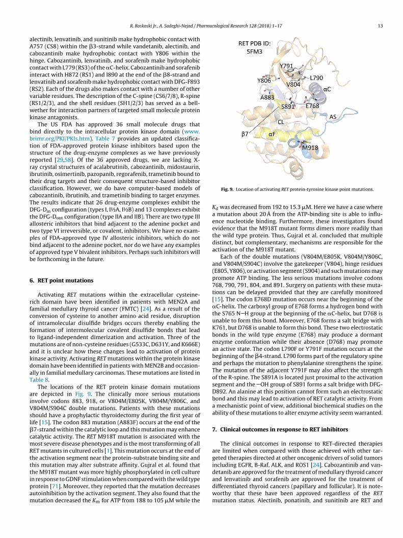
armacological Research 128 (2018) 1–17 13
aAchcil(v(wk
bbtsrritccTDtatpbob
6
rfcoftmakdaT
aiVsl�cmRtttipam
R. Roskoski Jr., A. Sadeghi-Nejad / Ph
lectinib, lenvatinib, and sunitinib make hydrophobic contact with757 (CS8) within the �3-strand while vandetanib, alectinib, andabozantinib make hydrophobic contact with Y806 within theinge. Cabozantinib, lenvatinib, and sorafenib make hydrophobicontact with L779 (RS3) of the �C-helix. Cabozantinib and sorafenibnteract with H872 (RS1) and I890 at the end of the �8-strand andenvatinib and sorafenib make hydrophobic contact with DFG-F893RS2). Each of the drugs also makes contact with a number of otherariable residues. The description of the C-spine (CS6/7/8), R-spineRS1/2/3), and the shell residues (SH1/2/3) has served as a bell-ether for interaction partners of targeted small molecule protein
inase antagonists.The US FDA has approved 36 small molecule drugs that
ind directly to the intracellular protein kinase domain (www.rimr.org/PKI/PKIs.htm). Table 7 provides an updated classifica-ion of FDA-approved protein kinase inhibitors based upon thetructure of the drug-enzyme complexes as we have previouslyeported [29,58]. Of the 36 approved drugs, we are lacking X-ay crystal structures of acalabrutinib, cabozantinib, midostaurin,brutinib, osimertinib, pazopanib, regorafenib, trametinib bound toheir drug targets and their consequent structure-based inhibitorlassification. However, we do have computer-based models ofabozantinib, ibrutinib, and trametinib binding to target enzymes.he results indicate that 26 drug-enzyme complexes exhibit theFG-Din configuration (types I, I½A, I½B) and 13 complexes exhibit
he DFG-Dout configuration (type IIA and IIB). There are two type IIIllosteric inhibitors that bind adjacent to the adenine pocket andwo type VI irreversible, or covalent, inhibitors. We have no exam-les of FDA-approved type IV allosteric inhibitors, which do notind adjacent to the adenine pocket, nor do we have any examplesf approved type V bivalent inhibitors. Perhaps such inhibitors wille forthcoming in the future.
. RET point mutations
Activating RET mutations within the extracellular cysteine-ich domain have been identified in patients with MEN2A andamilial medullary thyroid cancer (FMTC) [24]. As a result of theonversion of cysteine to another amino acid residue, disruptionf intramolecular disulfide bridges occurs thereby enabling theormation of intermolecular covalent disulfide bonds that leado ligand-independent dimerization and activation. Three of the
utations are of non-cysteine residues (G533C, D631Y, and K666E)nd it is unclear how these changes lead to activation of proteininase activity. Activating RET mutations within the protein kinaseomain have been identified in patients with MEN2B and occasion-lly in familial medullary carcinomas. These mutations are listed inable 8.
The locations of the RET protein kinase domain mutationsre depicted in Fig. 9. The clinically more serious mutationsnvolve codons 883, 918, or V804M/E805K, V804M/Y806C, and804M/S904C double mutations. Patients with these mutationshould have a prophylactic thyroidectomy during the first year ofife [15]. The codon 883 mutation (A883F) occurs at the end of the7-strand within the catalytic loop and this mutation may enhanceatalytic activity. The RET M918T mutation is associated with theost severe disease phenotypes and is the most transforming of all
ET mutants in cultured cells [1]. This mutation occurs at the end ofhe activation segment near the protein-substrate binding site andhis mutation may alter substrate affinity. Gujral et al. found that
he M918T mutant was more highly phosphorylated in cell culturen response to GDNF stimulation when compared with the wild typerotein [71]. Moreover, they reported that the mutation decreasesutoinhibition by the activation segment. They also found that theutation decreased the Km for ATP from 188 to 105 �M while theFig. 9. Location of activating RET protein-tyrosine kinase point mutations.
Kd was decreased from 192 to 15.3 �M. Here we have a case wherea mutation about 20 Å from the ATP-binding site is able to influ-ence nucleotide binding. Furthermore, these investigators foundevidence that the M918T mutant forms dimers more readily thanthe wild type protein. Thus, Gujral et al. concluded that multipledistinct, but complementary, mechanisms are responsible for theactivation of the M918T mutant.
Each of the double mutations (V804M/E805K, V804M/Y806C,and V804M/S904C) involve the gatekeeper (V804), hinge residues(E805, Y806), or activation segment (S904) and such mutations maypromote ATP binding. The less serious mutations involve codons768, 790, 791, 804, and 891. Surgery on patients with these muta-tions can be delayed provided that they are carefully monitored[15]. The codon E768D mutation occurs near the beginning of the�C-helix. The carboxyl group of E768 forms a hydrogen bond withthe S765 N H group at the beginning of the �C-helix, but D768 isunable to form this bond. Moreover, E768 forms a salt bridge withK761, but D768 is unable to form this bond. These two electrostaticbonds in the wild type enzyme (E768) may produce a dormantenzyme conformation while their absence (D768) may promotean active state. The codon L790F or Y791F mutation occurs at thebeginning of the �4-strand. L790 forms part of the regulatory spineand perhaps the mutation to phenylalanine strengthens the spine.The mutation of the adjacent Y791F may also affect the strengthof the R-spine. The S891A is located just proximal to the activationsegment and the OH group of S891 forms a salt bridge with DFG-D892. An alanine at this position cannot form such an electrostaticbond and this may lead to activation of RET catalytic activity. Froma mechanistic point of view, additional biochemical studies on theability of these mutations to alter enzyme activity seem warranted.
7. Clinical outcomes in response to RET inhibitors
The clinical outcomes in response to RET-directed therapiesare limited when compared with those achieved with other tar-geted therapies directed at other oncogenic drivers of solid tumorsincluding EGFR, B-Raf, ALK, and ROS1 [24]. Cabozantinib and van-
detanib are approved for the treatment of medullary thyroid cancerand lenvatinib and sorafenib are approved for the treatment ofdifferentiated thyroid cancers (papillary and follicular). It is note-worthy that these have been approved regardless of the RETmutation status. Alectinib, ponatinib, and sunitinib are RET and
14 R. Roskoski Jr., A. Sadeghi-Nejad / Pharmacological Research 128 (2018) 1–17
Table 7Drug-protein kinase interactions.
Drug-enzymea PDB ID DFG-D ASb �C R-Spine GKc Inhibitor class Pockets and sub-pockets occupiedd
Alectinib-RET Model in open in active V I F, BP-I-Be
Bosutinib-Src 4MXO in open in active T I F, G, BP-I-A/BCabozantinib-RET Model in open in active V I F, G, B, BP-I-B, BP-II-in, BP-II-Be
Dasatinib-Abl 2GQG in open in active T I F, FP-IErlotinib-EGFR 1M17 in open in active T I F, G, BP-I-A/BGefitinib-EGFR 2ITY in open in active T I F, G, BP-I-A/BLenvatinib-RET Model in open in active V I F, G, B, BP-I-B, BP-II-ine
Palbociclib-CDK6 2EUF in open in active F I FRuxolitinib-Srcf 4U5J in open in active T I Fe
Sorafenib-RET Model in open in active V I F, G, B, BP-I-B, BP-II-ine
Sunitinib-RET Model in open in active V I F, BP-I-Be
Tofacitinib-JAK3 3LXK in open in active M I F, FP-I/IITofacitinib-JAK1 3EYG in open in active M I F, FP-I/IIVandetanib-RET 2IVU in open in active V I F, G, BP-I-A/B
Dabrafenib-B-Raf 5CSW in closed out RS3→ T I ½A F, G, B, FP-II, BP-I-A, BP-I-B, BP-II-in, BP-II-A-inDasatinib-Lyng 2ZVA in open in RS2/3/4 up T I ½A F, G, B, BP-I-A/BLapatinib-EGFR 1XKK in closed out RS2/3→ T I ½A F, G, B, BP-I-A/B, II in, IIA inLenvatinib-VEGFR 3WZD in ? in RS3/4 up V I ½A F, G, B, IB, II inVemurafenib-B-Raf 3OG7 in ? out RS3→ T I ½A F, G, B
Abemaciclib-CDK6 5L2S in ? out RS3→ F I ½B F, FP-IIAlectinib-ALK 3AOX in closed in active L I ½B F, FP-ICeritinib-ALK 4MKC in closed in active L I ½B F, FP-ICrizotinib-ALK 2XP2 in closed in active L I ½B F, FP-ICrizotinib-Met 2WGJ in closed out ←−RS4 L I ½B F, FP-IErlotinib-EGFR 4HJO in closed out RS2/3→ T I ½B F, G, BP-I-A/BSunitinib- CDK2 3TI1 in closed out RS3→ F I ½B F
Axitinib-VEGFR 4AG8 out closed in ←−RS2/4 V IIA F, G, B, BP-I-B, II outImatinib-Ablg 1IEP out closed in ←−RS2 T IIA F, G, B, BP-I-A/B, II out, IVImatinib-Kit 1T46 out closed in ←−RS2 T IIA F, G, B, BP-I-A/B, II out, IVNilotinib-Abl 3CS9 out closed in ←−RS2 T IIA F, G, B, BP-I-A/B, II out, III, VPonatinib-Ablg 3OXZ out closed in ←−RS2 T IIA F, G, B, BP-I-A/B, II out, III, IVPonatinib-Kit 4U0I out closed in ←−RS2 T IIA F, G, B, BP-I-A/B, II out, III, IVPonatinib-B-Raf 1UWH out ? in ←−RS2/4 T IIA F, G, B, BP-I-B, II out, IIISorafenib-CDK8 3RGF out ? in ←−RS2/4 F IIA F, G, B, BP-I-B, II out, IIISorafenib-VEGFR 4ASD out closed in ←−RS2 V IIA F, G, B, BP-I-B, II-out, III
Bosutinib-Abl 3UE4 out open in ←−RS2 T IIB F, G, BP-I-A/BNintedanib-VEGFR2 3C7Q out closed in ←−RS2 V IIB F, G, BP-I-BSunitinib-Kit 3G0E out closed in ←−RS2 T IIB FSunitinib-VEGFR 4AGD out closed in ←−RS2 V IIB F, BP-I-B
Cobimetanib-MEK1 4AN2 in closed out RS3→ M III F,G, B, BP-II-inTrametinib-MEK1 Modelh in closed out RS3→ M III F,G, B, BP-II-ine
Afatinib-EGFR 4G5J in open in active T VI F, G, BP-I-A/BIbrutinib-BTK Modeli in closed in inactive T VI F, G, B, BP-I-A/Be
a All human proteins unless otherwise noted.b Activation segment.c Gatekeeper residue.d F, front cleft; G, gate area; B, back cleft; from http://klifs.vu-compmedchem.nl/unless otherwise noted.e Inferred.f Chicken enzyme.g Mouse enzyme.h Ref. [47].i Ref. [44].
Table 8RET point mutationsa.
Mutations in the extracellular cysteine-rich segment Protein kinase domain mutations
Mutation Mutation Location
Exon 8 G533C Exon 13 E768D Fourth residue of the �C-helixExon 10 C609F/G/R/S/Y Exon 13 L790F (RS4) Beginning of the �4-strandExon 10 C611F/G/S/Y/W Exon 13 Y791F Second residue of the �4-strandExon 10 C618F/R/S Exon 14 V804M/L (Sh2) Gatekeeper residueExon 10 C620F/R/S Exon 14 Y806C Hinge residueExon 11 C630R/Y Exon 15 A883F Last residue of the �7-strandExon 11 D631Y Exon 15 S891A Between �8-strand and activation segmentExon 11 C634/F/G/R/W/Y Exon 16 M918T End of the activation segmentExon 11 K666E
a Data from Ref. [24].

armac
mt
mVcaoadniBet
ttuDTas
tyctasfw
pompmmmMaRa
cpAaratoi
8
cegktig
R. Roskoski Jr., A. Sadeghi-Nejad / Ph
ultikinase inhibitors that have been approved for other indica-ions (www.brimr.org/PKI/PKIs.htm).
The leading hypothesis to explain the modest response of theseultikinase inhibitors is related to the concomitant inhibition of
EGFR2, which limits the permissible dosage [24]. For example, atlinically achievable concentrations, cabozantinib and vandetanibre more effective inhibitors of VEGFR2 than of RET. In the casef cabozantinib, its IC50 value for VEGFR2 is 0.035 nM vs. 5.2 nMgainst RET, which is nearly 150 times as large [72]. None of therugs listed in Table 6 were designed primarily as RET antago-ists. Development of antagonists that target RET specifically may
mprove the effectiveness in the treatment of RET-driven cancers.LU-667 and LOXO-292, two RET specific antagonists, are in thearly developmental phase and have entered clinical trials for bothhyroid and lung cancers (www.clinicaltrials.gov) [24].
Li et al. reported that RXDX-105 (CEP-32496), which was ini-ially developed as a B-Raf inhibitor, exhibits an 800-fold selectivityoward RET vs. VEGFR2 (0.33 vs. 258 nM IC50 values) [73]. Basedpon modeling studies, they reported that RXDX-105 binds to theFG-Dout conformation and is classified as a type II RET inhibitor.his drug is in clinical trials in patients with advanced lung cancernd other solid tumors (www.clinicaltrials.gov). See Ref. [60] for aummary of the development of other RET inhibitors.
The estimated incidence of new cases associated with RET muta-ions or RET-fusion proteins in the United States is about 10,000 perear (Table 3). This is about the same as number of new cases ofhronic myelogenous leukemia for which imatinib and second andhird generation of BCR-Abl non-receptor protein-tyrosine kinasentagonists have proven clinically efficacious and commerciallyuccessful [33]. Accordingly, such findings suggest that the searchor specific antagonists targeting RET-driven neoplasms is worth-
hile.In addition to the inhibition of wild type RET, studies have been
erformed on the ability of antagonists to inhibit RET mutants thatccur in sporadic and familial medullary thyroid carcinomas andultiple endocrine neoplasia type 2 syndromes [24]. For exam-
le, alectinib is a good inhibitor of the RET M918T, V804L, or Y791utants, ponatinib is effective against the RET V804M gatekeeperutant, and sunitinib is effective against the V804L gatekeeperutant. Moreover, cabozantinib is an effective inhibitor of the RET918T mutant, vandetanib is a good inhibitor of the RET C634W
nd M918T mutants while sorafenib is a potent antagonist of theET C634R and M918T mutants. In contrast, lenvatinib is ineffectivegainst the more common RET mutants.
As noted by Winer and colleagues “Biologically, the cancerell is notoriously wily; each time we throw an obstacle in itsath, it finds an alternate route that must then be blocked” [74].cquired resistance to RET antagonists occurs, but little is knownbout the resistance mechanisms. To date, no mutations have beeneported in tumor samples from patients who have developedcquired resistance to these multikinase inhibitors [24]. The activa-ion of bypass pathways is a likely mechanism for the developmentf some cases of acquired resistance following RET multikinase
nhibitor treatment [75].
. Epilogue
Manning et al. reported that the human protein kinase lineageonsists of 518 members that include 478 typical and 40 atypicalnzymes [5]. Based upon the identity of the phosphorylated OH
roup, these enzymes are classified as protein-serine/threonineinases (385 members), protein-tyrosine kinases (90), and protein-yrosine kinase like enzymes (43). The protein-tyrosine kinasesnclude both non-receptor (32) and receptor (58) proteins. A smallroup of enzymes such as MEK1/2, which catalyze the phosphory-ological Research 128 (2018) 1–17 15
lation of both threonine and tyrosine residues within the activationsegment of target proteins, are classified as dual specificity kinases.Although RET is only distantly related to MEK1/2, Plaza-Menachoreported that RET is a dual specificity kinase with the ability to cat-alyze the phosphorylation of both tyrosyl and seryl residues [9].Furthermore, Plaza-Menacho described an active structure of RETwith an open glycine-rich loop and an inactive structure of RETwith a closed glycine-rich loop that is unable to bind ATP [7]. Thisfinding adds another parameter in addition to the conformation ofthe �C-helix, the activation segment, and the regulatory spine indetermining the differences between active and dormant proteinkinases. RET is also an unusual receptor protein-tyrosine kinaseowing to the requirement of GDNF family receptor-� co-receptorsnecessary for ligand-mediated activation.
Dysregulation of protein kinases plays a fundamental role inthe pathogenesis of human illness, including autoimmune, can-cer, inflammatory, and nervous system disorders. Accordingly, thisfamily of enzymes has become one of the most important drug tar-gets over the past two decades [59,76]. There are currently morethan 250 protein kinase antagonists in various stages of clinicaldevelopment worldwide. The 36 FDA-approved drugs target about20 different protein kinases within the family with 518 members(www.brimr.org/PKI/PKIs.htm). Most of these approved drugs areused for the treatment of neoplastic disorders. Owing to their role inmany aspects of cellular signaling, a much wider range of diseasesshould be amenable to treatments with protein kinase antagonists[77]. Accordingly, we can expect future advances in clinical effec-tiveness and the subsequent approval of additional drugs targeting(i) more protein kinases and (ii) other illnesses such as hypertensionand Parkinson disease [76].
Conflict of interest
The authors are unaware of any affiliations, memberships, orfinancial holdings that might be perceived as affecting the objec-tivity of this review.
Acknowledgments
The colored figures in this paper were checked to ensure thattheir perception was accurately conveyed to colorblind readers[78]. The authors thank Laura M. Roskoski for providing edito-rial and bibliographic assistance. We also thank Josie Rudnicki andJasper Martinsek help in preparing the figures and Pasha Brezinaand W.S. Sheppard for their help in structural analyses.
References
[1] L.M. Mulligan, RET revisited: expanding the oncogenic portfolio, Nat. Rev.Cancer 14 (2014) 173–186.
[2] M. Takahashi, J. Ritz, G.M. Cooper, Activation of a novel human transforminggene, ret, by DNA rearrangement, Cell 42 (1985) 581–588.
[3] Y. Ishizaka, F. Itoh, T. Tahira, I. Ikeda, T. Sugimura, J. Tucker, et al., Human retproto-oncogene mapped to chromosome 10q11.2, Oncogene 4 (1989)1519–1521.
[4] P. Durbee, C.V. Marcos-Gutierrez, C. Kilkenny, M. Grigoriou, K. Wartiowaara,P. Suvanto, et al., GDNF signalling through the Ret receptor tyrosine kinase,Nature 381 (1996) 789–793.
[5] G. Manning, D.B. Whyte, R. Martinez, T. Hunter, S. Sudarsanam, The proteinkinase complement of the human genome, Science 298 (2002) 1912–1934.
[6] M.A. Lemmon, J. Schlessinger, Cell signaling by receptor tyrosine kinases, Cell141 (2010) 1117–1134.
[7] I. Plaza-Menacho, K. Barnouin, K. Goodman, R.J. Martínez-Torres, A. Borg, J.
Murray-Rust, et al., Oncogenic RET kinase domain mutations perturb theautophosphorylation trajectory by enhancing substrate presentation in trans,Mol. Cell 53 (2014) 738–751.[8] I. Plaza-Menacho, L. Mologni, N.Q. McDonald, Mechanisms of RET signaling incancer: current and future implications for targeted therapy, Cell. Signal. 26(2014) 1743–1752.

1 arma
[
[
[
[
[
[
[
[
[
[
[
[
[
[
[
[
[
[
[
[
[
[
[
[
[
[
[
[
[
[
[
[
[
[
[
[
[
[
[
[
[
[
[
[
[
[
[
[
[
[
[
[
[
[
[
[
[
[
[
[
6 R. Roskoski Jr., A. Sadeghi-Nejad / Ph
[9] I. Plaza-Menacho, K. Barnouin, R. Barry, A. Borg, M. Orme, R. Chauhan, et al.,RET functions as a dual-specificity kinase that requires allosteric inputs fromjuxtamembrane elements, Cell Rep. 17 (2016) 3319–3332.
10] X. Liu, Q.C. Vega, R.A. Decker, A. Pandey, C.A. Worby, J.E. Dixon, Oncogenic RETreceptors display different autophosphorylation sites and substrate bindingspecificities, J. Biol. Chem. 271 (1996) 5309–5312.
11] P.P. Knowles, J. Murray-Rust, S. Kjaer, R.P. Scott, S. Hanrahan, M. Santoro,et al., Structure and chemical inhibition of the RET tyrosine kinase domain, J.Biol. Chem. 281 (2006) 33577–33587.
12] Y. Kawamoto, K. Takeda, Y. Okuno, Y. Yamakawa, Y. Ito, R. Taguchi, et al.,Identification of RET autophosphorylation sites by mass spectrometry, J. Biol.Chem. 279 (2004) 14213–14224.
13] J. Anders, S. Kjar, C.F. Ibánez, Molecular modeling of the extracellular domainof the RET receptor tyrosine kinase reveals multiple cadherin-like domainsand a calcium-binding site, J. Biol. Chem. 276 (2001) 35808–35817.
14] R.L. Siegel, K.D. Miller, A. Jemal, Cancer Statistics, 2017, CA Cancer J. Clin. 67(2017) 7–30.
15] L. Kwatampora, S.P. Weitzman, M.A. Habra, N.L. Busaidy, Endocrinemalignancies, in: H.M. Kantarjian, R.A. Wolff (Eds.), The MD Anderson Manualof Medical Oncology, third ed., McGraw-Hill Education, New York, 2016, pp.903–932.
16] J.A. Fagin, S.A. Wells Jr., Biologic and clinical perspectives on thyroid cancer, N.Engl. J. Med. 375 (2016) 1054–1067.
17] J.F. Gainor, A.T. Shaw, Novel targets in non-small cell lung cancer: ROS1 andRET fusions, Oncologist 18 (2013) 865–875.
18] A.T. Shaw, P.P. Hsu, M.M. Awad, J.A. Engelman, Tyrosine kinase generearrangements in epithelial malignancies, Nat. Rev. Cancer 13 (2013)772–787.
19] G.W. Krampitz, J.A. Norton, RET gene mutations (genotype and phenotype) ofmultiple endocrine neoplasia type 2 and familial medullary thyroidcarcinoma, Cancer 120 (2014) 1920–1931.
20] C. Romei, R. Ciampi, R. Elisei, A comprehensive overview of the role of the RETproto-oncogene in thyroid carcinoma, Nat. Rev. Endocrinol. 12 (2016)192–202.
21] H. Donis-Keller, S. Dou, D. Chi, K.M. Carlson, K. Toshima, T.C. Lairmore, et al.,Mutations in the RET proto-oncogene are associated with MEN 2A and FMTC,Hum. Mol. Genet. 2 (1993) 851–856.
22] R.M. Hofstra, R.M. Landsvater, I. Ceccherini, R.P. Stulp, T. Stelwagen, Y. Luo,et al., A mutation in the RET proto-oncogene associated with multipleendocrine neoplasia type 2B and sporadic medullary thyroid carcinoma,Nature 367 (1994) 375–376.
23] L.M. Mulligan, J.B. Kwok, C.S. Healey, M.J. Elsdon, C. Eng, E. Gardner, et al.,Germ-line mutations of the RET proto-oncogene in multiple endocrineneoplasia type 2A, Nature 363 (1993) 458–460.
24] A. Drilon, Z.I. Hu, G.G.Y. Lai, D.S.W. Tan, Targeting RET-driven cancers: lessonsfrom evolving preclinical and clinical landscapes, Nat. Rev. Clin. Oncol. (2017),http://dx.doi.org/10.1038/nrclinonc.2017.188 (Epub ahead of print).
25] M. Grieco, M. Santoro, M.T. Berlingieri, R.M. Melillo, R. Donghi, I. Bongarzone,et al., PTC is a novel rearranged form of the ret proto-oncogene and isfrequently detected in vivo in human thyroid papillary carcinomas, Cell 60(1990) 557–563.
26] T. Kohno, H. Ichikawa, Y. Totoki, K. Yasuda, M. Hiramoto, T. Nammo, et al.,KIF5B-RET fusions in lung adenocarcinoma, Nat. Med. 18 (2012) 375–377.
27] D. Lipson, M. Capelletti, R. Yelensky, G. Otto, A. Parker, M. Jarosz, et al.,Identification of new ALK and RET gene fusions from colorectal and lungcancer biopsies, Nat. Med. 18 (2012) 382–384.
28] A.M. Schram, M.T. Chang, P. Jonsson, A. Drilon, Fusions in solid tumours:diagnostic strategies, targeted therapy, and acquired resistance, Nat. Rev. Clin.Oncol. 14 (2017) 735–748.
29] R. Roskoski Jr., Anaplastic lymphoma kinase (ALK) in the treatment ofALK-driven lung cancers, Pharmacol. Res. 117 (2017) 343–356.
30] S.K. Hanks, T. Hunter, Protein kinases 6: The eukaryotic protein kinasesuperfamily: kinase (catalytic) domain structure and classification, FASEB J. 9(1995) 576–596.
31] D.R. Knighton, J.H. Zheng, L.F. Ten Eyck, V.A. Ashford, N.H. Xuong, S.S. Taylor,et al., Crystal structure of the catalytic subunit of cyclic adenosinemonophosphate-dependent protein kinase, Science 253 (1991) 407–414.
32] D.R. Knighton, J.H. Zheng, L.F. Ten Eyck, N.H. Xuong, S.S. Taylor, J.M. Sowadski,Structure of a peptide inhibitor bound to the catalytic subunit of cyclicadenosine monophosphate-dependent protein kinase, Science 253 (1991)414–420.
33] R. Roskoski Jr., A historical overview of protein kinases and their targetedsmall molecule inhibitors, Pharmacol. Res. 100 (2015) 1–23.
34] D. Bajusz, G.G. Ferenczy, G.M. Keser, Structure-based virtual screeningapproaches in kinase-directed drug discovery, Curr. Top. Med. Chem. 17(2017) 2235–2259.
35] R. Roskoski Jr., Src protein-tyrosine kinase structure, mechanism, and smallmolecule inhibitors, Pharmacol. Res. 94 (2015) 9–25.
36] S.S. Taylor, M.M. Keshwani, J.M. Steichen, A.P. Kornev, Evolution of theeukaryotic protein kinases as dynamic molecular switches, Philos. Trans. R
Soc. Lond. B Biol. Sci. 367 (2012) 2517–2528.37] B. Nolen, S. Taylor, G. Ghosh, Regulation of protein kinases; controlling activitythrough activation segment conformation, Mol. Cell 15 (2004) 661–675.
38] R. Roskoski Jr., ErbB/HER protein-tyrosine kinases: structures and smallmolecule inhibitors, Pharmacol. Res. 79 (2014) 34–74.
[
[
cological Research 128 (2018) 1–17
39] R. Roskoski Jr., MEK1/2 dual-specificity protein kinases: structure andregulation, Biochem. Biophys. Res. Commun. 417 (2012) 5–10.
40] J. Zhou, J.A. Adams, Participation of ADP dissociation in the rate-determiningstep in cAMP-dependent protein kinase, Biochemistry 36 (1997)15733–15738.
41] A.P. Kornev, N.M. Haste, S.S. Taylor, L.F. Ten Eyck, Surface comparison of activeand inactive protein kinases identifies a conserved activation mechanism,Proc. Natl. Acad. Sci. U. S. A. 103 (2006) 17783–17788.
42] A.P. Kornev, S.S. Taylor, L.F. Ten Eyck, A helix scaffold for the assembly ofactive protein kinases, Proc. Natl. Acad. Sci. U. S. A. 105 (2008) 14377–14382.
43] R. Roskoski Jr., Anaplastic lymphoma kinase (ALK): structure, oncogenicactivation, and pharmacological inhibition, Pharmacol. Res. 69 (2013) 69–94.
44] R. Roskoski Jr., Ibrutinib inhibition of Bruton protein-tyrosine kinase (BTK) inthe treatment of B cell neoplasms, Pharmacol. Res. 113 (2016) 395–408.
45] R. Roskoski Jr., Cyclin-dependent protein kinase inhibitors includingpalbociclib as anticancer drugs, Pharmacol. Res. 111 (2016) 784–803.
46] R. Roskoski Jr., ERK1/2 MAP kinases: structure, function, and regulation,Pharmacol. Res. 66 (2012) 105–143.
47] R. Roskoski Jr., Allosteric MEK1/2 inhibitors including cobimetanib andtrametinib in the treatment of cutaneous melanomas, Pharmacol. Res. 117(2017) 20–31.
48] R. Roskoski Jr., Janus kinase (JAK) inhibitors in the treatment of inflammatoryand neoplastic diseases, Pharmacol. Res. 111 (2016) 784–803.
49] R. Roskoski Jr., ROS1 protein-tyrosine kinase inhibitors in the treatment ofROS1 fusion protein-driven non-small cell lung cancers, Pharmacol. Res. 121(2017) 202–212.
50] R. Roskoski Jr., Vascular endothelial growth factor (VEGF) and VEGF receptorinhibitors in the treatment of renal cell carcinomas, Pharmacol. Res. 120(2017) 116–132.
51] H.S. Meharena, P. Chang, M.M. Keshwani, K. Oruganty, A.K. Nene, N. Kannan,et al., Deciphering the structural basis of eukaryotic protein kinase regulation,PLoS Biol. 11 (2013) e1001690.
52] K. Shah, Y. Liu, C. Deirmengian, K.M. Shokat, Engineering unnatural nucleotidespecificity for Rous sarcoma virus tyrosine kinase to uniquely label its directsubstrates, Proc. Natl. Acad. Sci. U. S. A. 94 (1997) 3565–3570.
53] Y. Liu, K. Shah, F. Yang, L. Witucki, K.M. Shokat, A molecular gate whichcontrols unnatural ATP analogue recognition by the tyrosine kinase v-Src,Bioorg. Med. Chem. 6 (1998) 1219–1226.
54] J.J. Liao, Molecular recognition of protein kinase binding pockets for design ofpotent and selective kinase inhibitors, J. Med. Chem. 50 (2007) 409–424.
55] O.P. van Linden, A.J. Kooistra, R. Leurs, I.J. de Esch, C. de Graaf, KLIFS: aknowledge-based structural database to navigate kinase-ligand interactionspace, J. Med. Chem. 57 (2014) 249–277.
56] T. Kodama, T. Tsukaguchi, Y. Satoh, M. Yoshida, Y. Watanabe, O. Kondoh, et al.,Alectinib shows potent antitumor activity against RET-rearranged non-smallcell lung cancer, Mol. Cancer Ther. 13 (2014) 2910–2918.
57] M. Song, Progress in discovery of KIF5B-RET kinase inhibitors for thetreatment of non-small-cell lung cancer, J. Med. Chem. 58 (2015) 3672–3681.
58] R. Roskoski Jr., Classification of small molecule protein kinase inhibitors basedupon the structures of their drug-enzyme complexes, Pharmacol. Res. 103(2016) 26–48.
59] P.M. Fischer, Approved and experimental small-molecule oncology kinaseinhibitor drugs: a mid-2016 overview, Med. Res. Rev. 37 (2017) 314–367.
60] L. Mologni, C. Gambacorti-Passerini, P. Goekjian, L. Scapozza, RET kinaseinhibitors: a review of recent patents (2012–2015), Expert Opin. Ther. Pat 27(2017) 91–99.
61] J. Krajewska, T. Gawlik, B. Jarzab, Advances in small molecule therapy fortreating metastatic thyroid cancer, Expert Opin. Pharmacother. 18 (2017)1049–1060.
62] A.C. Dar, K.M. Shokat, The evolution of protein kinase inhibitors fromantagonists to agonists of cellular signaling, Annu. Rev. Biochem 80 (2011)769–795.
63] J. Monod, J.P. Changeux, F. Jacob, Allosteric proteins and cellular controlsystems, J. Mol. Biol. 6 (1963) 306–329.
64] F. Zuccotto, E. Ardini, E. Casale, M. Angiolini, Through the gatekeeper door:exploiting the active kinase conformation, J. Med. Chem. 53 (2010)2691–2694.
65] L.K. Gavrin, E. Saiah, Approaches to discover non-ATP site inhibitors, Med.Chem. Commun. 4 (2013) 41.
66] V. Lamba, I. Ghosh, New directions in targeting protein kinases: focusing upontrue allosteric and bivalent inhibitors, Curr. Pharm. Des. 18 (2012) 2936–2945.
67] Z. Zhao, L. Xie, L. Xie, P.E. Bourne, Delineation of polypharmacology across thehuman structural kinome using a functional site interaction fingerprintapproach, J. Med. Chem. 59 (2016) 4326–4341.
68] W. Sherman, T. Day, M.P. Jacobson, R.A. Friesner, R. Farid, Novel procedure formodeling ligand/receptor induced fit effects, J. Med. Chem. 49 (2006)534–553.
69] C. Gao, M. Grøtli, L.A. Eriksson, Characterization of interactions andpharmacophore development for DFG-out inhibitors to RET tyrosine kinase, J.Mol. Model. 21 (2015) 167.
70] R.A. Friesner, J.L. Banks, R.B. Murphy, T.A. Halgren, J.J. Klicic, D.T. Mainz, et al.,Glide: a new approach for rapid, accurate docking and scoring: 1. Method andassessment of docking accuracy, J. Med. Chem. 47 (2004) 1739–1749.
71] T.S. Gujral, V.K. Singh, Z. Jia, L.M. Mulligan, Molecular mechanisms of RETreceptor-mediated oncogenesis in multiple endocrine neoplasia 2B, CancerRes. 66 (2006) 10741–10749.

armac
[
[
[
[
[Chem. Biol. 8 (2013) 96–104.
[77] D.H. Drewry, C.I. Wells, D.M. Andrews, R. Angell, H. Al-Ali, A.D. Axtman, et al.,
R. Roskoski Jr., A. Sadeghi-Nejad / Ph
72] F. Bentzien, M. Zuzow, N. Heald, A. Gibson, Y. Shi, L. Goon, et al., In vitro andin vivo activity of cabozantinib (XL184), an inhibitor of RET, MET, and VEGFR2,in a model of medullary thyroid cancer, Thyroid 23 (2013) 1569–1577.
73] G.G. Li, R. Somwar, J. Joseph, R.S. Smith, T. Hayashi, L. Martin, et al., Antitumoractivity of RXDX-105 in multiple cancer types with RET rearrangements or
mutations, Clin. Cancer Res. 23 (2017) 2981–2990.74] E. Winer, J. Gralow, L. Diller, B. Karlan, P. Loehrer, L. Pierce, et al., Clinicalcancer advances 2008: major research advances in cancer treatment,prevention, and screening–a report from the American Society of ClinicalOncology. J Clin Oncol 27:812-26. Erratum in, J. Clin. Oncol. 2009 (27) (2009)3070–3071.
[
ological Research 128 (2018) 1–17 17
75] M.J. Niederst, J.A. Engelman, Bypass mechanisms of resistance to receptortyrosine kinase inhibition in lung cancer, Sci. Signal. 6 (294) (2013) re6.
76] P. Cohen, D.R. Alessi, Kinase drug discovery −what’s next in the field? ACS
Progress towards a public chemogenomic set for protein kinases and a call forcontributions, PLoS One 12 (8) (2017) e0181585.
78] R. Roskoski Jr., Guidelines for preparing color figures for everyone includingthe colorblind, Pharmacol. Res. 119 (2017) 240–241.