role of the immunohistochemical marker (ki67) in diagnosis
TRANSCRIPT

136 IMJM Volume 18 No.3, December 2019
ORIGINAL ARTICLE
Role of the Immunohistochemical Marker (Ki67) in
Diagnosis and Classification of Hydatidiform Mole Hasan Abu Deka FFa , Abd Ali Al Saeng ZHb, Khalid Almukhtar Zc
aDepartment of pathology, Collage of Medicine, Kerbela University,Iraq bDepartment of Pathology, Medical city,Iraq cDepartment of Pathology, Collage of Medicine, Baghdad University, Iraq
ABSTRACT
Introduction: Since the hallmark of gestational trophoblastic disease is trophoblastic proliferation, Ki67
is regarded as the best marker in studying hydatidiform mole.This study was conducted to evaluate the role
of this proliferative marker in distinguishing among hydropic abortion, partial and complete hydatidiform
mole. Materials and methods: This is a cross sectional study involving the application of Ki67 on a total
of 90 histological samples of curetting materials from molar (partial and complete mole) and non molar
hydropic abortion belong to Iraqi females, so three study groups were created. Immunohistochemical
expression in villous cytotrophoblasts, syncytiotrophoblasts and stromal cells were recorded separately by
three independent observers and the results were correlated statically. Results: The mean number of
stained nuclei of villous cytotrophoblasts and stromal cells was the highest in complete mole and the lowest
in non molar hydropic abortion. There is a significant statistical relationship regarding Ki67 labeling index in
villous cytotrophoblasts between partial moles and hydropic abortion, complete mole and partial moles,
hydropic abortion and complete mole. Regarding Ki67 labelling index in villous stromal cells, a significant
statistical relationship achieved when the correlation done between partial mole and hydropic abortions,
hydropic abortion and complete mole, while a non significant statistical relationship was achieved if the
correlation done between partial and complete mole. All villous syncytiotrophoblasts showed negative
results. Conclusion: Ki-67 labeling index in villous cytotrophblastic cells are useful in separating between
partial moles and hydropic abortion, partial mole and complete mole, hydropic abortion and complete
mole. While Ki-67 labeling index in villous stromal cells is only useful in separating between partial moles
and hydropic abortion, hydropic abortion and complete mole.
KEYWORDS: Gestational trophoblastic disease, partial mole, complete mole, hydropic abortion
Corresponding author:
Dr. Farah Falah Hassan Abu Deka
Department of Pathology,
College of Medicine, Kerbela University,
Al Sarafia Street, Bagdad Iraq.
Tel No: +07706084653
Email: [email protected]
INTRODUCTION
Gestational trophoblastic diseases (GTD) are groups
of disorders of fertilization that include complete
and partial molar pregnancies, invasive mole,
choriocarcinoma and the very rare placental site
trophoblastic tumor (PSTT).24 Hydatidiform mole is
regarded as the most common type of GTD.1 There
are two subtypes of hydatidiform mole, partial and
complete mole, the distinction between them relies
mainly on both genetic and histological criteria. 9,22
At gynecological department trophoblastic diseases
often diagnosed when the serum human chorionic
gonadotrophin levels plateau or rise in patients
being observed after the an initial suspicion of
hydatidiform mole.16 Most hydatidiform mole are
sporadic however a familial syndrome of recurrent
hydatidiform mole has been recorded.10 Genetically
a complete mole is diploid without maternal
contribution, whereas a partial mole is triploid with
a maternal chromosome complement.15 It is very
important to distinguish complete hydatidiform mole
from partial hydatidiform moles, and non-molar
hydropic abortion, because the risk of persistent
trophoblastic disease is very high in complete
one.4,14 The histomorphologic separation between
hydropic abortion, partial and complete mole
sometime very difficult 12 and the fact that
abortion due to chromosomal abnormalities

IMJM Volume 18 No.3, December 2019
137
sometimes produce a histological feature
indistinguishable from partial mole made the
bmatter worse.7,18 Partial mole is differentiated
histologically from complete mole by the presence
of two types of chorionic villi (enlarged, hydropic,
and normal sized villi with sclerotic stroma); mild
focal trophoblastic hyperplasia; villus scalloping
often containing trophoblastic inclusions; and
presence of fetal red blood cells in the stromal
villous vasculature.7 On the other hand complete
mole characterize by diffuse villous enlargement
with marked hydropic changes, cistern formation
and marked trophoblastic hyperplasia usually in
a circumferential pattern with a remarkable
cytological atypia.22 Hydropic abortion is regarded
as a benign degenerative changes characterized by
swelling of villi associated with minimal to absent
cistern formation, neither abnormal trophoblastic
proliferation nor atypia is present.24
DNA polymorphisms and polymerase chain reaction
are probably the best accepted method of genetic
analysis of gestational trophoblastic diseases,
but it is time-consuming, expensive, requires
skilled personnel and special facilities, it required
both maternal and paternal blood samples in
addition to molar tissue.14 One of the advantages
immunohistochemistry is the ability to apply them
retrospectively to sections of routinely formalin-
fixed and paraffin-embedded tissue that
preclude the need for expensive or sophisticated
equipments.18 Recent studies revealed that
expression of the paternally imprinted gene (p57) is
of particular help in differentiation between
partial and complete mole, however it doesn’t
differentiate between partial mole and hydropic
abortion and the positively stained villous
intermediate trophoblasts sometimes account for
false interpretation.11 Histologically confirmed
cases ofhydatidiform mole should be included in
the formal serum human chorionic gonadotropin
follow-up and management of GTD should be
established.19,21 Ki-67 gene encodes a nuclear
protein having 2 isoforms, Ki-67 immunoreactivity
presents in all cell cycle phases except in
the quiescent G0 phase thus it reflects
cellular proliferation,8 Over expression is
frequently seen in various malignancy and
associated with worse overall survival in
bladder, brain, breast, kidney, lung, ovary,
prostate, and thyroid cancer 21, Furthermore Ki-67
reactivity is established in addition to other
parameters in the World Health Organization’s
recommended grading system for neuroendocrine
tumors.13 Because Ki-67 has been established as a
valuable reflection of the tissue proliferative
compartment and thus could be of value in studying
the biologic behavior of the gestational
trophoblastic disease.17 Its expression in gestational
trophoblastic diseases became an interested topic
for many gynecological and pathological researchers
worldwide with a variable recorded results. This
study was conducted to evaluate the role of this
proliferative marker in distinguishing among Iraqi
females with hydropic abortion, partial and
complete mole and to compare with the results of
previous similar worldwide studies.
MATERIALS AND METHODS
Materials selection
In this cross sectional study, a total of 90 blocks of
formalin-fixed, paraffin-embedded tissue for cases
diagnosed as molar pregnancy and hydropic abortion
were retrieved from the patient archive in the
histopathology department of teaching laboratories
in the medical city complex at Bagdad (in the
period from January 2016 to February 2017). The
only clinical information available for each case was
a female,s age. From each block two histological
sections were made, one stained with routine
hematoxyline and eosine for morphological
evaluation and the other for immunohistochemical
staining with Ki67 antibody using
slandered immunohistochemical protocols. After
morphological reevaluation (thorough searching at
different magnifications) by three pathologists who
had no idea about the previous diagnosis using the
published criteria for diagnosis and classifications of
hydatidiform mole, 30 cases were agreed to be
partial mole and 30 complete mole and 30 hydropic
abortion, so three study groups were made.
Immunohistochemistry staining
Immunohistochemical staining was performed by the
application of a commercially available Ki67
immunohistochemical kit (Clone MIB-1, mouse
monoclonal antibody, dilution 1:75-1:50, Dako) with
envision detection system. A section from tonsil
regarded as a positive control, negative control
slides were prepared by omitting the primary
antibody. In order to perform immunohistochemical

138 IMJM Volume 18 No.3, December 2019
staining on each block, 5 micrometer sections were
made and applied the section on a positive charge
dewaxing paraffin-embedded sections, after
antigen retrieval was performed the tissue section
was covered with peroxide blocking solution for 5
minutes followed by incubation of Primary antibody
for 30 minutes, then the tissue section was covered
with PolyExcel poly HRP and incubate for 30
minutes at room temperature, working solution and
incubate for 5 minutes at room temperature, and
lastly Hematoxyline stain was applied.
Histopathological observation
All immunohistochemically stained sections were
evaluated independently by the same three
pathologists under a light microscope using
a microscope (Leica DM500), only sections
that contain more than ten chorionic villi
were evaluated. Immunohistochemical expression
analyses for villous cytotrophoblasts,
syncytiotrophoblasts and stromal cells were made
separately in the most representative area for Ki-67
(the area with the most concentrated
immunohistochemically labeled nuclei of
trophoblastic cells in medium sized villi), and the
results described as (number of positive nuclei per
total number of nuclei counting 100 cells of each
population).8 Only nuclear staining plus mitotic
figures should be included in the Ki67 scoring, the
median time required for nuclei counting was 4
minutes.
STATISTICAL ANALYSIS
One way ANOVA with post hoc Tukey’s test used to
compare mean values of nuclei stained with Ki67 of
different study groups. Results were expressed as
mean ± standard deviation. The differences were
considered statistically significant when p value
<0.05. All statistical tests were done by using IBM
SPSS v. 24 software.
Photographs taken in this study by the camera
(Leica Icc 50E).
This study was approved by the ethical committee
of the national center for educational laboratories
and done in accordance with it is institutional
policy in which patient consent was taken at the
beginning of consultation to teaching laboratories
for any future retrospective studies.
RESULTS
The study enrolled 90 cases of hydatidiform moles,
30 were the partial mole and 30 were the complete
mole and 30 were hydropic abortion (figure 1, 2 and
3), so three study groups were created, age range of
cases diagnosed as hydatidiform mole was from 15
years to 48 years with a mean age 27.13 ± 1.28
years.
Figure 1: Hematoxyline and Eosin stained tissue section
showing the histological features of hydropic abortion
(100x)
Figure 2: Hematoxyline and Eosin stained tissue section showing the histological features of partial mole (100x)
The application of Immunohistochemical staining
with Ki67 antibody (Figure 4, 5 and 6) revealed
varying degrees of positivity in both villous
cytotrophoblasts and stromal cells, minimum,
maximum and the mean numbers of stained nuclei
for each study group were shown in Table I and II.
None of syncytiotrophoblasts reacted with this
antigen. Strong immunoreactivity in the nuclei of
decidual cells, regarded as a positive internal
control.
IMJM Volume 18 No.3, December 2019
139
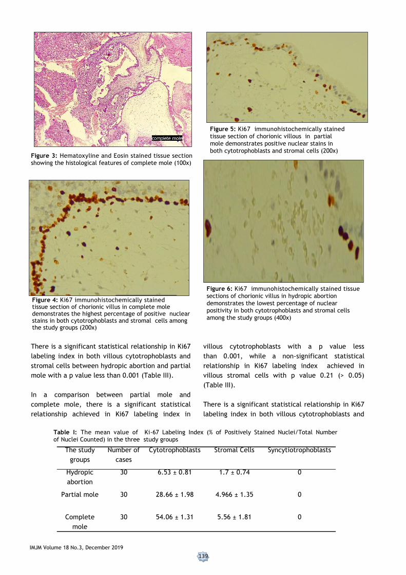
Figure 3: Hematoxyline and Eosin stained tissue section showing the histological features of complete mole (100x)
Figure 6: Ki67 immunohistochemically stained tissue sections of chorionic villus in hydropic abortion
demonstrates the lowest percentage of nuclear positivity in both cytotrophoblasts and stromal cells among the study groups (400x)
Figure 5: Ki67 immunohistochemically stained tissue section of chorionic villous in partial
mole demonstrates positive nuclear stains in both cytotrophoblasts and stromal cells (200x)
Figure 4: Ki67 immunohistochemically stained tissue section of chorionic villus in complete mole demonstrates the highest percentage of positive nuclear stains in both cytotrophoblasts and stromal cells among the study groups (200x)
There is a significant statistical relationship in Ki67
labeling index in both villous cytotrophoblasts and
stromal cells between hydropic abortion and partial
mole with a p value less than 0.001 (Table III).
In a comparison between partial mole and
complete mole, there is a significant statistical
relationship achieved in Ki67 labeling index in
The study
groups
Number of
cases
Cytotrophoblasts Stromal Cells Syncytiotrophoblasts
Hydropic
abortion
30 6.53 ± 0.81 1.7 ± 0.74 0
Partial mole 30 28.66 ± 1.98 4.966 ± 1.35 0
Complete
mole
30 54.06 ± 1.31 5.56 ± 1.81 0
villous cytotrophoblasts with a p value less
than 0.001, while a non-significant statistical
relationship in Ki67 labeling index achieved in
villous stromal cells with p value 0.21 (> 0.05)
(Table III).
There is a significant statistical relationship in Ki67
labeling index in both villous cytotrophoblasts and
Table I: The mean value of Ki-67 Labeling Index (% of Positively Stained Nuclei/Total Number of Nuclei Counted) in the three study groups

140 IMJM Volume 18 No.3, December 2019
Table II: Minimal and maximal numbers of positively stained cytotrophoblastic and stromal cells nuclei among the study groups.
stromal cells between hydropic abortion and
complete mole with a p value less than 0.001
(Table III).
The distribution of Ki-67 labeling index of villous
cytotrophoblasts among the three study groups
showed that all cases of hydropic abortions
displayed less than 20% index, all cases of partial
mole displayed between 21 to 40% index and all
cases of complete mole displayed more than 40%
index. While in regard to villous stromal cells all
the study groups displayed Ki67 index below 20%
(Table IV).
Non molar and molar villi Ki67 staining in cytotrophoblastic nuclei
Ki67 staining in stromal cell nuclei
The study groups Total number of cases
Minimal number of positive cells
Maximum number of positive cells
Minimal number of
positive cells
Maximum number of
positive cells
Hydrobic abortion 30 5 8 1 3
Partial mole 30 25 31 3 8
Complete mole 30 50 55 3 9
DISCUSSION
The strong recommendations for the use of
immunohistochemistry in differentiation between
hydropic abortion and partial mole or between types
of hydatidiform mole, especially in early pregnancy
are the aim of this study. Among the
immunohistochemical antibodies, (Ki-67) regarded
as a valuable marker for detecting tissue
proliferation and it is widely available than p57 in
most immunohistochemical laboratories. This study
showed that the mean age of women with molar
pregnancy was in the third decade of life which
Table III: Results of Statistical Analysis to Compare the mean value Ki-67 Expression between the three study
Groups
Comparison between
each two study groups
Results of statistical analysis
Cytotrophoblasts (p value)
Stromal
cells (p value)
Hydropic abortion
and partial mole
<0.001* <0.001*
Partial mole and com-
plete mole
<0.001* 0.21**
Hydropic abortion and
complete mole
<0.001* <0.001*
agreed with two Iraqi studies done by Al-Baldawi2
and Taboo23 and a worldwide study done by Thama
et al.3 So all agreed that gestational trophoblastic
disease is a disease of young female.
The application of Immunohistochemical staining
with Ki67 antibody revealed varying degrees of
positivity in both villous cytotrophoblasts and
stromal cells, while none of syncytiotrophoblasts
reacted with this antigen, this results agreed with
three studies worldwide.13,20,5 Because in contrast to
cytotrophoblasts which are stem cells,
syncytiotrophoblasts are terminally differentiated
cells that secrete most of placental hormones. A
study done by Erfanian et al.8 surprisingly showed the
positivity also present in syncytiotrophoblasts.
Comparison of Ki67 labeling index in villous
cytotrophoblasts between hydropic abortion and
partial mole and between partial and complete
mole revealed a significant statistical relationship
with p value (< 0.05). This result agreed with
studies done by Erfanian et al., Khooei et al and
Chen et al. 8,13,5, but disagrees with Cheville et al.6
which stated that growth fraction (number of
positive cells/total number of cells) of Ki67 in
cytotrophoblastic cells was useful in separating
complete mole from partial mole, but not partial
moles from hydropic abortion.
Erfanian et al. and Khooei et al.8,13 found a
nonsignificant statistical relationship by correlation
of Ki67 labeling index in villous stromal cells
between hydropic abortion and partial mole and
between partial and complete mole.

IMJM Volume 18 No.3, December 2019
141
Table IV: Distribution of the numbers of positive nuclear staining with Ki67 between the three study groups (H:hydropic abortion; P:partialmole; C:complete mole)
Ki-67 Labeling Index
Hydrobic abortion Partial mole Complete mole
Cytotrophoblasts Stromal cells
Cytotrophoblasts Stromal cells
Cytotrophoblasts Stromal cells
0-20 30 30 0 30 0 30
21-40 0 0 30 0 0 0
More than 40 0 0 0 0 30 0
Total 30 30 30 30 30 30
However this study showed that there is a
significant statistical relationship regarding Ki67
labeling index in villous stromal cells between
hydropic abortion and partial mole while a non
significant relationship achieved if the comparison
done between partial and complete mole, there is
no such previous similar Iraqi study on Ki67
expression in gestational trophoblastic disease .The
variations in the reported results between studies
may be related to many factors such as technical
factors like the time of fixation (because the tissues
used in these studies were archival retrieved with
no standardized fixation time), variations in
detection methods and sample size. Furthermore,
the difference in gestational age between samples
may affect the results of immunohistochemistry.
There is a growing interest to apply a special Ki67
scoring system for each type of malignancy as a
powerful prognostic parameter, this matter became
routinely reported, particularly in breast, brain and
neuroendocrine tumors.13 Our results can be
regarded as an early attempt to create a
comprehensive index in the form of a three tied
score advised to be used during histomorphological
evaluation of all curetting materials
morphologically suspected to be of molar type
products of conception for both diagnostic and
prognostic purposes as below:
Score 1(+) less than 20%, representing cases of
hydropic abortion.
Score 2(++) 21-40%, representing cases of partial
mole.
Score 3(+++) above 40%, representing cases of
complete mole.
This applications if became standardized will
reduce the interobserver variations sometime
associated with diagnosis and classification of
hydatidiform mole and increase diagnostic accuracy
of the complete mole because the higher association
with persistent trophoblastic disease and
choriocarcinoma which necessitates a thorough
follow up.
CONCLUSION
Ki-67 labeling index in villous cytotrophblastic cells
is useful in separating between hydropic abortion
and partial mole, partial and complete mole,
hydropic abortion and complete mole. While Ki-67
labeling index in villous stromal cells is useful in
separating between hydropic abortion and partial
mole, hydropic abortion and complete mole. The
results of this study will help to raise awareness
among Iraqi pathologists to apply (Ki67) in all
curetting materials of molar pregnancies, especially
if the morphological features of distinction were not
clear cut.
CONFLICT OF INTEREST
None
REFERENCES
1. A Sita-Lumsden, D Short , I Lindsay , N J
Sebire , D Adjogatse , M J Seckl & P M
Savage .Treatment outcomes for 618 women
with gestational trophoblastic tumors
following a molar pregnancy at the Charing
Cross Hospital, 2000–2009. British Journal of
Cancer 2012, 107:1810–1814/
2. Al-Baldawi R. Retrospective Study On
Management Of Gestational Trophoblastic
Disease In Baghdad Teaching Hospital J Fac
Med Baghdad Vol ,2013. 48 (3) : 262-66.
3. B.W.L. Thama, J.E. Everardb, J.A. Tidyc, D.
Drewd, B.W. Hancock Gestational
trophoblastic disease in the Asian population
142 IMJM Volume 18 No.3, December 2019
of Northern England and North Wales BJOG:
an International Journal of Obstetrics and
Gynaecology, 2003;110 : 555–559.
4. Carey L, Nash BM, Wright DC. Molecular
genetic studies of complete hydatidiform
moles. Transl Pediatr. 2015; 4(2):181-8.
5. Chen Y , Shen D , Gu Y , Zhong P, Xie J , So
Q .The diagnostic value of Ki-67, P53 and P63
in distinguishing partial Hydatidiform mole
from hydropic abortion Wiener klinische
Wochenschrift, ,2012 ;124(5)184–7.
6. Cheville JC, Robinson R, Benda JA. Evaluation
of Ki-67 (MIB-1) in placentas with hydropic
change and partial and complete
hydatidiform mole. Pediatr Pathol Lab Med.
1966;16(1):41–50.
7. Clement PB, Young RH. Atlas of Gynecologic
surgical pathology. Clement PB, Young RH.
(eds). 3rd ed. Saunders Elsevier, London
2014;272-86.
8. Erfanian M, Sharifi N, and Ali A. P63 and
Ki-67 expression in trophoblastic disease and
spontaneous abortion J Res Med Sci.2009; 14
(6): 375–84.
9. Fowler DJ, Lindsay I, Seckl MJ and Sebire NJ,.
Routine pre-evacuation ultrasound diagnosis
of hydatidiform mole: experience of more
than 1000 cases from a regional referral
center. Ultrasound Obstet Gynecol
2006 ;27:56–60.
10. Garner EI, Goldstein DP, Feltmate CM and
Berkowitz RS. Gestational trophoblastic
disease.Clin Obstet Gynecol 2007; 50(1):112-
22.
11. Ghanshyam Biyani and Pradeep Bhatia.
Mortality in hydatidiform mole: Should we
blame thyroid?. Indian J Anaesth 2011;55(6):
628–29.
12. Jessica Fernández , Rafael Cortés, Aleydah
Salazar, Aníbal Pulido, Pablo Dabed and
Victoria García. p57 kip2
immunohistochemistry: ancillary technique in
hydatidiform moles diagnosis. BMC Proc
2013 ,7: 33.
13. Khooei A , Pasdar FA , Fazel A , Mahmoud
iM , Nikravesh MR , Delui MK and Pourheydar
B . Ki-67 Expression in Hydatidiform Moles
and Hydropic Abortions Iran Red Crescent
Med J ,2013;15(7): 590–594.
14. Kubelka-Sabit K, Jasar D, Filipovski V,
Bozinovski G, Plaseska-Karanfilska D.
Molecular and histological characteristics of
early triploid and partial molar pregnancies.
Pol J Pathol 2017; 68(2):138-143.
15. Lai CY, Chan KY, Khoo US, Ngan HY, Xue WC,
Chiu PM et tal. . Analysis of gestational
trophoblastic disease by genotyping and
chromosome in situ hybridization. Mod Pathol
2004;17(1):40-8.
16. Lurain JR. Advances in management of high-
risk gestational trophoblastic tumors. J Reprod
Med. 2002;47(6):451-9.
17. Mahzouni P, Mokhtari M, Amirmansour B..
Diffrentiation between reactive gliosis and
astrocytomas by MIB-1/Ki67 immunostaining. J
Res Med Sci.2007;12(5):241–5.
18. McConnell TG , Murphy KM, Hafez M, Vang R,
Ronnett BM. Diagnosis and subclassification of
hydatidiform moles using p57
immunohistochemistry and molecular
genotyping: validation and prospective
analysis in routine and consultation practice
settings with development of an algorithmic
approach.Am J Surg Pathol 2009 ;33(6):805-17.
19. Neil J Sebire, Philippa C May, Baljeet Kaur,
Michael J Seckl, and Rosemary A
Fisher.Abnormal villous morphology mimicking
a hydatidiform mole associated with paternal
trisomy of chromosomes 3,7,8 and unipaternal
disomy of chromosome 11 Diagn
Pathol,2016;11: 20.
20. Olvera M, Harris S, Amezcua CA, McCourty A,
Rezk S, Koo C et al.. Immunohistochemical
expression of cell cycle proteins E2F-1, Cdk-2,
Cyclin E, p27 (kip1), and Ki-67 in normal
placenta and gestational trophoblastic
disease. Mod Pathol ,2001;14(10):1036–42.
21. Rosai J. Ackerman's surgical pathology. 10 ed.
Female reproductive system St Louis:
Mosby,2011; pp. 1639–45.
22. Smith HO, Kohorn E, Cole LA. Choriocarcinoma
and gestational trophoblastic disease. Obstet
Gynecol Clin North Am 2005 ;32(4):661-84.
23. Taboo ZA . A Prospective Study of Gestational
Trophoblastic Disease in Al-Mosul City.The
Iraqi post graduate medical journa. 2013;12
(2):268-76.
24. Tham BWL, Everard JE, Tidy JA, Drew D,
Hancock BW. Gestational trophoblastic disease
in the Asian population of Northern England
and North Wales. BJOG 2003 ;110:555–9.