rom j morphol embryol r j m e case report romanian … · fronto-parietal lobes, accompanied by...
TRANSCRIPT
Rom J Morphol Embryol 2017, 58(4):1555–1559
ISSN (print) 1220–0522 ISSN (online) 2066–8279
CCAASSEE RREEPPOORRTT
Cyclopia and proboscis – the extreme end of holoprosencephaly
ANDREI MIHAI MĂLUŢAN1), MARINA DUDEA1), RĂZVAN CIORTEA1), MIHAELA MUREŞAN2), CARMEN ELENA BUCURI1), CARINA MIHU1), DAN MIHU1)
1)2nd Obstetrics and Gynecology Department, “Iuliu Haţieganu” University of Medicine and Pharmacy, Cluj-Napoca, Romania
2)Department of Pathology, Emergency County Hospital, Cluj-Napoca, Romania
Abstract Holoprosencephaly (HPE), a major congenital abnormality in brain development is characterized by the absence or incomplete cleavage of prosencephalon into separate hemispheres, with cyclopia as the extreme manifestation of HPE, presenting as a failure of embryonic prosencephalon to properly divide the orbits of the eye in two cavities. We report the case of a 15-year-old pregnant patient, who delivered a 34-week living fetus with alobar HPE, cyclopia and proboscis. The patient did not have any routine scans during pregnancy; her first obstetrical exam was performed at 29 weeks of gestation (WG), when a prenatal ultrasound found a fetus with alobar HPE, cyclopia, proboscis, polydactyly and single umbilical artery. Despite adequate medical and genetic counseling, the patient and her legal representative refused further investigations – magnetic resonance imaging and genetic testing. She was admitted to the hospital at 34 WG for premature rupture of membranes, with clear amniotic fluid. Twenty-four hours later, she delivered vaginally a living male fetus, weighing 1995 g. Macroscopic examination revealed umbilical cord with two vessels, fetal proboscis, cyclopia, low implanted ears, bilateral polydactyly of the upper limbs, spina bifida occulta in the sacral region. The newborn lived for 40 minutes. Microscopy of the eyeball revealed choroid, ciliary body and conjunctiva structures, with no identification of the retina, and no evidence of the optic nerve in the fragments obtained from the optic chiasm region. This case underlines the importance of early obstetrical examinations during pregnancy and raises concerns about the ethics of allowing therapeutic termination of pregnancy after 24 WG in selected cases.
Keywords: alobar holoprosencephaly, cyclopia, proboscis, live newborn, polydactyly, spina bifida occulta.
Introduction
Holoprosencephaly (HPE) is a major congenital malformation, characterized by the absence or incomplete cleavage of the prosencephalon into separate hemispheres [1]. It occurs between the 18th and 28th day of gestation [2]. Depending on the severity of the condition, HPE is subdivided in the lobar, semilobar and alobar forms.
Cyclopia is an extreme manifestation of HPE, defined as the failure of the embryonic prosencephalon to properly divide the orbits of the eye in two cavities [3, 4]. While HPE affects one in 16 000 live newborns, cyclopia is seen as rarely as one in 100 000 newborns, including stillbirth [5, 6]. Cyclopia, usually incompatible with postnatal life, is most frequently diagnosed during the first trimester ultrasound scan, allowing early termination of pregnancy and thus avoiding maternal psychological trauma of giving birth to a deformed fetus [7].
This case report aims to present a rare case of a fetus with alobar HPE, showing typical facial manifestations, such as cyclopia and proboscis, and associating extra-cranial malformations.
Case presentation
We report the case of a 15-year-old pregnant woman, who delivered a 34-week living fetus with alobar HPE, cyclopia and proboscis.
This case report was approved by the Local Ethics
Committee of “Iuliu Haţieganu” University of Medicine and Pharmacy, Cluj-Napoca, Romania, and signed informed consent was obtained from the patient for publication of this case report and accompanying images.
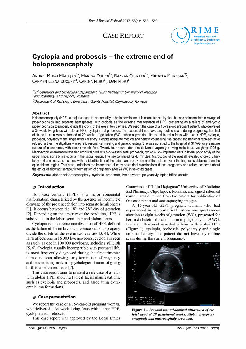
A 15-year-old G2P1 pregnant woman, who had experienced in her obstetrical history one spontaneous abortion at eight weeks of gestation (WG), presented for her first obstetrical examination in pregnancy at 29 WG. Prenatal ultrasound revealed a fetus with alobar HPE (Figure 1), cyclopia, proboscis, polydactyly and single umbilical artery. The patient did not have any routine scans during the current pregnancy.
Figure 1 – Prenatal transabdominal ultrasound of the fetal head at 29 gestational weeks. Alobar holopros-encephaly and macrocephaly are noted.
R J M ERomanian Journal of
Morphology & Embryologyhttp://www.rjme.ro/

Andrei Mihai Măluţan et al.
1556
Despite adequate medical and genetic counseling, the patient and her legal representative (mother) refused further investigations – magnetic resonance imaging (MRI) examination and genetic testing. There was no history of consanguinity between her and her partner, no family history of structural abnormalities and no exposure to known teratogens during pregnancy.
She was admitted to the hospital at 34 WG, for premature rupture of membranes, with clear amniotic fluid. Maternal blood tests revealed laboratory results within normal range (hemoglobin, red blood cells, leukocytes, platelets, hepatic and renal function, C-reactive protein). Microbiological examination of the cervical secretion was performed and no pathological agents were iden-tified. Ampicillin therapy (intravenous administration, 4 g/24 hours) was initiated.
Twenty-four hours later, she delivered vaginally, a living male fetus, weighing 1995 g. The clinical examination
of the newborn and umbilical cord revealed: cyclopia, proboscis, low implanted ears (Figure 2), polydactyly of the upper limbs (Figure 3), umbilical cord presenting one vein and one artery (Figure 4). The newborn lived for 40 minutes.
Pathological examination of the newborn and fetal annexes was performed. We used Hematoxylin and Eosin (HE) staining and Leica ICC50 HD microscope for examination.
Macroscopic examination revealed: umbilical cord with two vessels; fetal proboscis, measuring 4.2 cm; cyclopia; low implanted ears; bilateral polydactyly of the upper limbs with six fingers; spina bifida occulta in the sacral region (Figure 5). The fetal skull presented no anterior fontanelle, the bones being stacked at that level. The opening of the skull externalized 250 mL of serous liquid and alobar HPE was noted.
Figure 2 – Facial photograph of the newborn with alobar holoprosencephaly shows cyclopia, proboscis and
low implanted ears.
Figure 3 – Photograph of the newborn’s hand shows polydactyly with
six fingers.
Figure 4 – Photograph showing umbilical cord
with one artery and one vein.
Figure 5 – Macroscopic pathology image demonstrating the presence
of spina bifida occulta in the sacral region.
At microscopy, the eyeball examination revealed choroid, ciliary body and conjunctiva structures, with no identification of retinal tissue (Figure 6). There was no evidence of the optic nerve in the fragments obtained from the optic chiasm region. The cerebral parenchyma presented subarachnoid and periventricular areas of hemorrhage, thrombosis and necrosis (Figure 7), along with abnormal stratification areas, immature neural tube structures and areas of high neuronal density (Figure 8). There was a normally developed cerebellum.
Te proboscis microscopic examination, noted respi-ratory epithelium, along with mucous glands in the sub-mucosa, cartilage fragments and compact bone lamellae. There was meconium-stained amniotic fluid in the pulmonary parenchyma (Figure 9).
The fetal annexes’ examination has revealed umbilical cord with two vascular structures, one artery and one vein (Figure 10), placenta with medium and large size chorionic villi, corresponding to the 3rd trimester of pregnancy, presenting areas of necrosis and hemorrhage, diffuse neutrophilic inflammatory infiltrate and focal calcifi-cations (Figure 11), amniotic membranes with hematic infiltrates, diffuse mixed inflammatory infiltrate and areas of necrosis and abscess, thus pointing to a stage 3 chorioamnionitis (Figure 12).
Figure 6 – Cross-section showing eyeball with internal surface covered by the pigment epithelium layer, other retinal layers being desquamated (HE staining, ×40).
Discussion
HPE is a rare fetal developmental disorder in which, depending on the prosencephalon division, the most severe form is alobar HPE, characterized by an absence in the lateral ventricles division, which appear as a single ventricle [8], fusion of the thalami, absence of the

Cyclopia and proboscis – the extreme end of holoprosencephaly
1557
corpus callosum, a small brain with abnormal gyral pattern and absence of olfactory nerves and optic tracts. Midline facial anomalies are frequent and severe, such as cyclopia and proboscis (an anterior appendage-like structure in the form of a non-functioning nose), median cleft lip and palate. Cyclopia itself is always an external manifestation of deep brain malformation [2, 9]. The semilobar HPE is
characterized by a partial separation of the hemispheres; most commonly, the fusion involves the thalami and the fronto-parietal lobes, accompanied by corpus callosum agenesis or hypoplasia. Lobar HPE is the least affected subtype, with fusion of the ventral frontal lobes but well developed lobes and almost complete separation of the hemispheres [2].
Figure 7 – Microscopic brain images showing (a) areas with cerebral hemorrhage, necrosis and edema, and (b) a longitudinal section of a cerebral blood vessel, showing early thrombosis. HE staining: (a) ×100; (b) ×40.
Figure 8 – Microscopic brain image showing an area with immature nervous tissue with neural tube structures and gliosis (HE staining, ×100).
Figure 9 – Microscopic section of the fetal lung in saccular phase, with air spaces containing meconium and keratin fragments (HE staining, ×400).
Figure 10 – Umbilical cord section showing umbilical vein (partially) and a single umbilical artery (HE staining, ×40).
Figure 11 – Detail from a placental section, showing areas with inflammatory infiltrate, hemorrhage and necrosis (HE staining, ×200).

Andrei Mihai Măluţan et al.
1558
Figure 12 – Detail from an amniotic membrane section showing severe chorioamnionitis with abscess formation (HE staining, ×200).
The prognosis of HPE depends on the type and accompanying malformations. Almost all alobar and semilobar forms are incompatible with extrauterine life. Patients affected by milder forms tend to survive beyond infancy, but they often present complications that require neurosurgical intervention and medical management for other associated systemic anomalies [10]. In our case, the fetus, presenting the alobar form of HPE, was born at 34 WG and lived for 40 minutes. Extracranial malfor-mations were noted, including cyclopia and proboscis, low implanted ears, bilateral polydactyly of the upper limbs and spina bifida occulta in the sacral region.
Early diagnosis is extremely important, allowing early termination of pregnancy and thus avoiding maternal psychological trauma of giving birth to a deformed fetus [7].
The main screening diagnostic method is prenatal ultrasound (US), usually revealing characteristic images in the first trimester. When HPE is suspected by US, careful intrauterine scanning of the face will allow a more accurate diagnosis. One has to remember the well-known phrase, “the face predicts the brain” [11]. More information of the development of the brain structures can be obtained by in utero MRI, which can be routinely performed in suspected cases of HPE [10]. In our case, the patient did not have the routine scans during pregnancy, and her first obstetrical examination was performed at 29 WG, when ultrasound revealed multiple images suggestive for the diagnosis of fetal alobar HPE. A single spherical forebrain structure with a single ventricle, cyclopia and proboscis were noted. The patient and her legal representative (mother) refused to undergo the MRI examination.
At this gestational age (29 WG), according to the legislation in most countries and in ours, the therapeutic termination of the pregnancy could not be performed and the patient had to continue the pregnancy, delivering vaginally at 34 WG a male fetus, that lived for 40 minutes.
On the other side, the etiology of HPE remains unclear. Maternal diabetes mellitus is a known risk factor [12]. Other environmental factors, such as maternal alcohol use, infections during pregnancy [TORCHs – Toxoplasmosis, Other (syphilis, varicella-zoster, parvovirus B19), Rubella,
Cytomegalovirus (CMV), and Herpes infections], drugs during pregnancy, have been implicated, but not proven to be causative.
Many single-gene disorders (18–25%) can result in syndromes with a variable incidence of HPE, such as Pallister–Hall, Rubinstein–Taybi, Kallmann, Smith–Lemli–Opitz [13, 14].
However, the most frequent findings are chromosomal abnormalities, present in up to 41% of the cases [15]; of these, trisomy 13 is encountered in up to 75%, triploidy in 20%, and trisomy 18 in 12% of the HPE cases with aneuploidy [16–18]. In our case, regarding the clinical and pathological findings in the fetus, trisomy 13 could be suspected, but despite adequate medical and genetic counseling, the patient and her legal representative (mother) refused genetic testing.
The originality of our case stands in the fact, that it reports an advanced pregnancy with a fetus presenting alobar HPE, with extreme facial manifestations such as cyclopia and proboscis, associated with multiple extra-cranial anomalies, comprising low implanted ears, bilateral polydactyly of the upper limbs and spina bifida occulta in the sacral region, finalized at 34 WG with a spontaneous delivery, of a fetus that lived for 40 minutes. Even though we cannot presume that this is the first such case reported, it is still a rare report due to the multiple extra-cranial malformations associated, and to the fact that the fetus was born alive.
Conclusions
We report a rare case of alobar HPE, presenting cyclopia and proboscis, associating extracranial malformations like low implanted ears, bilateral polydactyly of the upper limbs and spina bifida occulta in the sacral region, in which the patient had to continue the pregnancy to a spontaneous delivery of a live fetus due to a delayed diagnosis. This case underlines the importance of early obstetrical examinations during pregnancy and raises concerns about the ethics of allowing therapeutic termi-nation of pregnancy after 24 WG in selected cases.
Conflict of interests The authors declare that there is no conflict of interests
regarding the publication of this paper.
References [1] Nyberg DA, Mack LA, Bronstein A, Hirsch J, Pagon RA.
Holoprosencephaly: prenatal sonographic diagnosis. AJR Am J Roentgerol, 1987, 149(5):1051–1058.
[2] Swatek J, Szumiło J, Burdan F. Alobar holoprosencephaly with cyclopia – autopsy-based observations from one medical center. Reprod Toxicol, 2013, 41:80–85.
[3] Golovataya EI, Pribushenya OV, Trebka EG, Novikova IV, Lurie IW. Cyclopia and other defects in a fetus with unique chromosomal rearrangement. Genet Couns, 2015, 26(3): 359–364.
[4] Sharma D, Yadav J, Garg E. Cyclopia syndrome. BMJ Case Rep, 2014, 2014:bcr2014203535.
[5] Källén B, Castilla EE, Lancaster PA, Mutchinick O, Knudsen LB, Martínez-Frías ML, Mastroiacovo P, Robert E. The cyclops and the mermaid: an epidemiological study of two types of rare malformation. J Med Genet, 1992, 29(1):30–35.
[6] Roach E, DeMyer W, Conneally PM, Palmer C, Merritt AD. Holoprosencephaly: birth data, genetic and demographic analysis of 30 families. Birth Defects Orig Artic Ser, 1975, 11(2):294–313.
Cyclopia and proboscis – the extreme end of holoprosencephaly
1559
[7] Raman R, Mukunda Jagadesh G. Antenatal diagnosis of alobar holoprosencephaly. Case Rep Radiol, 2014, 2014: 724671.
[8] Genç M, Genç B, Solak A, Alkiliç L, Uyar M. Alobar holo-prosencephaly, proboscis and cyclopia in a chromosomally normal fetus: prenatal diagnosis and fetal outcome. Ital J Anat Embryol, 2015, 120(2):83–88.
[9] Nazer Herrera J, Cifuentes Ovalle L, Cortez López A. [Frequency of holoprosencephaly in Chile]. Rev Med Chil, 2015, 143(7):874–879.
[10] Kaliaperumal C, Ndoro S, Mandiwanza T, Reidy F, McAuliffe F, Caird J, Crimmins D. Holoprosencephaly: antenatal and postnatal diagnosis and outcome. Childs Nerv Syst, 2016, 32(5):801–809.
[11] Salama GSA, Kaabneh MAF, Al-Raqad MK, Al-Abdallah IMH, Shakkoury AGA, Halaseh RAA. Cyclopia: a rare condition with unusual presentation – a case report. Clin Med Insights Pediatr, 2015, 9:19–23.
[12] Barr M Jr, Hanson JW, Currey K, Sharp S, Toriello H, Schmickel RD, Wilson GN. Holoprosencephaly in infants of diabetic mothers. J Pediatr, 1983, 102(4):565–568.
[13] Chandra SR, Daryappa MM, Mukheem Mudabbir MA, Pooja M, Arivazhagan A. Pallister–Hall syndrome. J Pediatr Neurosci, 2017, 12(3):276–279.
[14] Coletă E, Siminel M, Gheonea M. Holoprosencephaly sequence. Rom J Morphol Embryol, 2011, 52(2):725–728.
[15] Solomon BD, Rosenbaum KN, Meck JM, Muenke M. Holo-prosencephaly due to numeric chromosome abnormalities. Am J Med Genet C Semin Med Genet, 2010, 154C(1):146–148.
[16] Chen CP, Huang MC, Chern SR, Kuo YL, Chen YN, Wu PS, Chen LF, Pan CW, Wang W. Distal 3p duplication and terminal 7q deletion associated with nuchal edema and cyclopia in a fetus and a review of the literature. Taiwan J Obstet Gynecol, 2015, 54(3):297–302.
[17] Gondré-Lewis MC, Gboluaje T, Reid SN, Lin S, Wang P, Green W, Diogo R, Fidélia-Lambert MN, Herman MM. The human brain and face: mechanisms of cranial, neurological and facial development revealed through malformations of holoprosencephaly, cyclopia and aberrations in chromosome 18. J Anat, 2015, 227(3):255–267.
[18] Bozkurt O, Kanmaz HG, Sahin S, Canpolat FE, Uras N, Oguz SS, Dilmen U. Cyclopia, proboscis and alobar holoprosencephaly representative for trisomy 13. Genet Couns, 2014, 25(3): 349–351.
Corresponding author Marina Dudea, MD, PhD Student, 2nd Obstetrics and Gynecology Department, “Iuliu Haţieganu” University of Medicine and Pharmacy, 55 21 Decembrie 1989 Avenue, 400610 Cluj-Napoca, Romania; Phone +40745–779 832, Fax +40264–596 893, e-mail: [email protected] Received: May 31, 2017
Accepted: January 22, 2018