roots international magazine of endodontology vol 9 issue 4 2013
TRANSCRIPT

rootsinternational magazine ofendodontology42013
i s sn 2193-4673 Vol. 9 • Issue 4/2013
| CE articleDiagnosis 2013: The things you need to know for successful endodontic treatment
| techniqueBioactive endodontic obturation: Combining the new with the tried and true
| specialLaser versus conventional therapies
RO0413_01_Titel 28.11.13 13:11 Seite 1


I 03
editorial _ roots I
roots4_2013
Dr George Freedman
_One of the most innovative technologies widely used in medicine, kind to tissue and excellent forhealing, has only recently begun to make a significant dental impact. Dental lasers have been commer-cially available for several decades, but the profession has been slow to incorporate this technology intothe practice. Lasers, extensively documented in the academic and clinical dental literature, have long beenperceived by practitioners as too limited in intra-oral applications, too complicated and too expensive.In recent years, ease of use, scientific research and documentation, and greater affordability have converged to make lasers essential for every dental practice.
Lasers were first indicated for soft-tissue treatment and management. Diode technology has reducedthe initial financial investment and made lasers largely affordable for most practices. More recently, lasertechnologies have been successfully incorporated into endodontic procedures.
The success of intra-radicular endodontic treatment is dependent upon the cleaning and shaping ofthe root-canal space, disinfection of the root-canal space and 3-D obturation of the root-canal system.Many technologies have been utilised to accomplish these tasks: instrumentation systems, irrigants, intra-canal medications, and a host of obturation materials. Unfortunately, conventional endodontictherapy is still observed to fail on occasion owing to incomplete disinfection and subsequent reinfection.Bacteria may also be found outside the tooth’s root-canal system at the apex and elsewhere on the rootsurface. These extra-radicular bacteria cannot be eliminated with conventional therapies, and the resid-ual contamination maintains the active infectious process.
Laser-assisted endodontic therapy, undertaken after access and mechanical preparation, overcomesthe inherent difficulties of existing treatment. Lasers must be considered additions to the existing endodontic armamentarium rather than as stand-alone instruments. The benefits of the variously doc-umented endodontic laser therapies include patient comfort, effective debridement, and penetratingdisinfection. Laser therapy avoids vibration, facilitating anaesthesia and eliminating microfractures. The energy of the laser and its associated hydro-photonic activity efficiently remove pulpal tissue, thesmear layer and bacteria from the canal walls three-dimensionally, typically without physical contactand without the risk of over-instrumentation beyond the apex.
While the future mainstream laser tools and techniques are still in the process of development anddefinition, the mounting scientific and clinical evidence indicates that photoactivated debridement anddisinfection instruments cannot be dismissed. Dentists who perform endodontic therapy must considerintegrating endodontic lasers into their practices. Lasers have arrived in endodontics!
Dr George Freedman (DDS, BSc, Fellow of the American Academy of Cosmetic Dentistry, American College of Dentists, and International Academy for Dental-Facial Esthetics)
Lasers are mainstreamin endodontics
RO0413_03_Editorial 28.11.13 13:12 Seite 1

I editorial
03 Lasers are mainstream in endodontics| Dr George Freedman
I CE article
06 Diagnosis 2013: The things you need to know for successful endodontic treatment| Dr Thomas Jovicich
I case report
12 CBCT in endodontic treatment of fused second and thirdmandibular molars| Dr Andreas Krokidis & Dr Riccardo Tonini
I technique
16 Bioactive endodontic obturation: Combining the newwith the tried and true| Dr Gary Glassman
I special
26 SEM analysis of the laser activation of final irrigants forsmear layer removal| Dr Vivek Hegde, Dr Naresh Thukral, Dr Sucheta Sathe,
Dr Shachi Goenka & Dr Paresh Jain
30 Laser versus conventional therapies| Cristiane Meira Assunção, Joanna Tatith Pereira,
Renata Schlesner Oliveira & Dr Jonas de Almeida Rodrigues
34 Treatment of aphthous stomatitis using low-level lasertherapy| Pedro J. Muńoz Sánchez, Cuba, José Luis Capote Femenias
& Jan Tunér
I meetings
38 Navigating canal system – The 16th ESE biennial congress
40 International Events
I about the publisher
41 | submission guidelines42 | imprint
I content _ roots
page 26 page 30 page 38
page 6 page 12 page 16
Frontal and lateral views of a 3-D reconstruction of a maxillary first premolar showing a three-rooted canal system.
This micro-CT image was developed as part of the Root CanalAnatomy Project http://rootcanalanatomy.blogspot.com in the
Laboratory of Endodontics of the University of Sao Paulo inRibeirao Preto, Brazil by Prof. Marco Versiani, Prof. Jesus Pécora
& Prof. Manoel Sousa-Neto.
04 I roots4_2013
RO0413_04_Content 28.11.13 13:12 Seite 1

1 Year Clinical Masters Programin Aesthetic and Restorative Dentistry13 days of intensive live training with the Mastersin Santorini (GR), Geneva (CH), Pesaro (IT)
Three on location sessions with live patient treatment, hands on practice plus online learning and online mentoring under the Masters’ supervision.
Tribune America LLC is the ADA CERP provider. ADA CERP is a service of the American Dental Association to assist dental professionals in identifying quality providers of continuing dental education. ADA CERP does not approve or endorse individual courses or instructors, nor does it imply acceptance of credit hours by boards of dentistry.
Learn from the Masters of Aesthetic and Restorative Dentistry:
Collaborate on your casesand access hours of premium video training and live webinars
University of the Pacificyou will receive a certificate from the University of the Pacific
Latest iPad with coursesall early birds receive an iPad preloadedwith premium dental courses
ADA CERPC.E. CREDITS150
Registration information:
13 days of live training with the Mastersin Santorini, Geneva, Pesaro + self study
Curriculum fee: € 9,900 contact us at tel.: +49-341-48474-302 / email: request@tribunecme(€ 900 when registering, € 3,000 prior to each session)

06 I
I CE article _ retreatment
_The goal of endodontic treatment is for the cli-nician to achieve an effective cleaning and debride-ment of the root canal system, including the smearlayer and all of its mechanical and bacterial byprod-ucts. Traditionally this is accomplished via mechani-cal instrumentation in conjunction with chemical irrigants together and actively engaged to completelydebride and sterilize the root canal system.
The root canal system is a vast and complex three-dimensional structure comprising deltas and lateralcanals, along with multiple branches off of the mainroot canal system (Figs. 1, 2, 9).
Before the clinician can begin to treat a patient in need of endodontic treatment, he or she first mustcome up with the proper diagnosis. Once the diagno-sis has been made, it then must be integrated with thetreatment plan. Taking that treatment plan and pre-senting it to the patient creates the next challenge:creating value for the patient. One of my most diffi-cult challenges as a working endodontist is creating
value for the patient in my chair who has no pain andis here because his or her dentist “saw something” onthe radiograph. Pain is the greatest patient motivatorwe have in dentistry today.
The focus of this article is on diagnosis, and it is my goal to provide the reader with a good grasp of diagnosis as it relates to endodontic treatment.
Endodontics is all about vision. You have it. I haveit. The dentist down the street has it. Doing root canalstoday is all about having the confidence to make theproper diagnosis. This is achieved through repetition.The more you do it, the easier it becomes. In addition,you need consistency that is achieved through posi-tive reinforcement. Once you believe you can do it and the results support that, you then develop com-petence. This allows you to retain the skills you haveworked hard to hone. The most important trait to utilize in clinical practice today is common sense. This is what separates the true artisans from toothmechanics.
The key component to endodontic treatment is diagnosis. It is based upon using a multifocal approachthat involves:_patient report,_medical and dental history,_clinical signs and symptoms,_diagnostic testing,_radiographic findings,_restorability.
Fig. 1_Maxillary molar. Note the
complex anatomy and multiple
portals of exit. (Photos/Provided by
Thomas Jovicich, MS, DMD)
Fig. 2_Mandibular molar.
Note the curvature along with
the multiple portals of exit.
Fig. 3a_Maxillary central incisor
with a periapical lesion. This is
a markedly calcified canal.
Fig. 3b_Maxillary central incisor
with completed root canal using
Sybron TFA rotary nickel titanium
instruments, Sealapex sealer.
Note the multiple portals of exit
in the apical region.
roots4_2013
Diagnosis 2013: The things you need to know for successfulendodontic treatmentAuthor_ Dr Thomas Jovicich, USA
This article qualifies for CE credit. To take the CE quiz, log on towww.dtstudyclub.com. Click on ‘CE articles’ and search for thisedition of the magazine. If you are not registered with the site,you will be asked to do so before taking the quiz. You may alsoaccess the quiz by using the QR code below.
_ce credit roots
Fig. 1 Fig. 2 Fig. 3a Fig. 3b
RO0413_06-10_Jovicich 28.11.13 13:13 Seite 1

I 07
CE article _ retreatment I
roots4_2013
Taking and collating all of this information will allow the clinician to arrive at a proper and thoroughdiagnosis. Let’s break these down and delve into whatneeds to be done.
_Patient report
This is the first opportunity to create a road map to a diagnosis. The goal is to ascertain the nature ofthe problem. Step one: Ask the patient the where the pain is located. Once you’ve localized the area, it’simperative to ask a few more questions. The nextquestion should involve determining pulpal vitalitythrough the use of an ice pencil.
Other times the patient will volunteer this infor-mation with a statement like: “The minute I put any-thing cold on this tooth, the pain is present and quiteintense.” This information suggests that the pain may be pulpal in origin. Because the trigeminal nerveis involved in endodontics, it is important to deter-mine any type of radiating pain. It is not uncommonfor maxillary pain to radiate from the mandibular areaand vice versa. A final area of feedback I want from patients relates to biting and chewing.
The patient’s report is the foundation upon whichwe begin the diagnostic procedure. Asking probingand leading questions in “plain English” will allow thepatient to give you critical diagnostic information.
_Medical and dental history
Once you have the patient’s report, probing his orher medical and dental history gives clarity to thebackground. What are the patient’s medical allergies?What recent dental treatment has the patient had?Was there any mention of restorations placed thatwere near or at the pulp?
Many times a patient will mention having heardthe dentist tell his assistant that they were close to the pulp during the excavation of decay. Asking de-tailed questions enables you to enrich the diagnosticcanvas as to why the patient is sitting in your chair.
_Clinical signs and symptoms
By this point, you have listened to the patient’schief complaint and you have taken radiographs ordigital images. It’s time to “test” the patient. The “bitetest” involves having the patient attempt to repro-duce the pain through biting on an orangewood stickor a cotton swab or a wet cotton roll. If there is pain to bite, you are dealing with some degree of pulpal inflammation with secondary involvement of the periodontal ligament. Once you have this informa-tion, the next step is to look at your digital imaging
and analyze the relationship of the periodontal liga-ment (pdl) to the root. Is there a thickening? Is there a widening?
If the patient reports pain to bite upon release, thisinfers that there may be some structural root damage(Figs. 5a & b). At that point is it essential to look at theocclusal surface of the tooth, account for the type and age of any restoration and inquire if any recentdentistry has been done. In addition, it is imperativeto probe the suspected tooth.
Probing from buccal to lingual with at least fourmeasurements per side is the best barometer to assessperiodontal health. If you find an isolated defect inany single probing, you are most likely dealing with afracture of the root. Endodontic treatment to confirmor rule out a fracture is indicated in these clinical situations.
_Diagnostic testing
The percussion test involves using the blunt end of a mouth mirror or periodontal probe to assess forperiodontal inflammation. It is imperative that the cli-nician gets a frame of reference. This is accomplishedby testing the same tooth on the opposite side of thearch. In addition, it is prudent to test the suspectedtooth as well as the teeth on either side. Testing shouldinvolve both the occlusal and facial surfaces.
Thermal tests utilizing hot or cold are the defini-tive modality to assess pulpal vitality. There are a myr-iad of ways to test with cold, including CO2 systems,refrigerant sprays and ice cubes (pellets). I believe icepellets are the best way to test for cold symptoms. Inour practice, we use anesthetic carpules that are filledup with water and frozen.
This method is cheap, efficient and plentiful. Thegoal is to reproduce the patient’s symptoms. Manypatients who report pulpal hyperemia have managedthis symptom by utilizing the opposite side of theirmouth. Temperature symptoms are a major motivatorfor patients to seek dental care.
Fig. 4a_The presence of caries
under the margin of a restoration.
The caries extend to the pulp and
will need endodontic treatment.
Fig. 4b_The endodontic treatment is
completed. In this case, the patient
was lost to the practice for three
years and came back when his face
was swollen because of incomplete
treatment.
Fig. 4a Fig. 4b
RO0413_06-10_Jovicich 28.11.13 13:13 Seite 2

08 I
I CE article _ retreatment
Testing with ice involves establishing a baseline to cold. Typically, I chose to test the same tooth on the opposite side or the maxillary central incisor. I askpatients to tell me when they feel an “electrical shockor jolt” to the tooth. As soon as they do that, I removethe ice from the tooth. This is easily accomplished onthe buccal surface of the tooth at the margin of thegingiva. When porcelain restorations are present, Istrive to put the ice right at the margin on or aboveany metal margins.
Sometimes it is necessary to apply the ice on thelingual aspect of the tooth. As unresponsive as porce-lain restorations can be, the clinician needs to beaware that pulp testing gold restorations can have theopposite effect. This is because of the metallurgicalproperties of gold. It is an amazing conductor of tem-perature. Always forewarn the patient when testinggold-restored teeth.
Ask the patient if the cold on the tooth reproducedhis or her pain. Also, ask if the pain lingered after youremoved the ice from the test site. If the pain it is lingering, it is a sign of irreversible pulpitis.
In some cases the pain can and does radiate alongthe pathway of the trigeminal nerve. Sometimes, especially in the maxilla, referred pain can be relatedto sinus issues, such as sinusitis, allergic rhinitis andrhinovirus.
If the patient does not respond to any thermaltests, both hot and cold, it is a sign that the pulp isnecrotic, dying or infected. In this instance, studying
the digital imaging may aid the diagnosis. One caveat:It is possible to have a necrotic pulp without beingable to quantify it via digital images In many incipientpathology issues, it takes approximately 90 to 120days for breakdown to manifest itself on imaging. Today’s cone-beam imaging technology can shortenthat process to 30 days. It is not uncommon to have apatient in the chair with symptoms that you cannotquantify radiographically.
_Radiographic findings
Radiographic findings (Figs. 8a & b) are the roadmap for endodontics. Thorough study and evaluationof imaging allows the clinician to determine a multi-tude of facts about the tooth in question. What doesthe image reveal? Can you see if there is a widening ofthe pdl? If there is a widening of the pdl, it is essentialto have the patient bite down on a bite stick.
Once he or she does that, you must ask if the pain,if present, is worse upon bite or upon release of bite.The latter is highly correlated with root fracture. Oncethat is confirmed, the next step is to prepare the patient for a root canal.
The dentist must convincingly explain the proce-dure’s value as well as caution the patient about the possibility of losing the tooth due to the fractureextending apical from the cementoenamel junction(CEJ). Is there a lesion (Figs. 3a & b) present? This information allows me to frame my diagnostic ques-tions to the patient. These include: Is the tooth sensi-tive to cold? I know from the lesion that the answer to that should be no. If, however, the answer is yes, itautomatically triggers my mind to look for anothertooth.
Generally, speaking teeth with lesions of endo -dontic origin (LEOs) test non-vital to thermal or elec-tric pulp testing. In sequencing, I first ask for the patient’s report, followed by radiographic findings,which I then augment with clinical testing to tie it alltogether and arrive at a diagnosis. Lastly, are cariespresent? The location of caries is a determining factoras to whether a root canal is needed (Figs. 4a & b).
Fig. 5a_Cracked tooth syndrome.
Pre-treatment radiograph.
Fig. 5b_What can happen in a
cracked tooth when you obturate
with warm, vertical condensation of
gutta-percha.
Fig. 6_Well-done endodontic
treatment of tooth #6. Notice the
multiple portals of exit as they relate
to the presence of lesions.
Fig. 7_Know when to say when.
This dentist attempted to do an
endodontic procedure that should
not have been done.
roots4_2013
Fig. 5a Fig. 5b
Fig. 6 Fig. 7
RO0413_06-10_Jovicich 29.11.13 14:33 Seite 3

I 09
CE article _ retreatment I
roots4_2013
_Restorability
Restorability is an issue that has been a hot topicin dentistry for years. Its meaning has evolved as technology has become the backbone of modern den-tistry. Prior to the incorporation of implant dentistry,restorability had a very different meaning. Dentistswere much more motivated to save teeth. Options andcreativity were necessary for clinical success, both inendodontics as well as in restorative dentistry.
Technology has taken away one form of resource-fulness and replaced it with the promise of a panacea.It has become far too easy for general dentists to recommend removal of a tooth to a patient with thepromise that an implant will save the day.
Historically speaking, the diagnosis of a tooth being non-restorable came after a myriad of attemptsto save the tooth. Every aspect of dentistry came intoplay. Periodontists did osseous surgery and root amputations. Endodontists performed conventionalendodontics and, if necessary, surgical interventionto do everything possible to save the tooth. Decisionsinvolving the long-term prognosis of the tooth wererelevant. Decisions about the type of restoration werediscussed. Decisions about the osseous health of theroots and surrounding bone structures were relevant.
The goal of every specialist is to be an extension of the general dentist’s practice. To that end, decidingwhether a tooth was restorable or not was, at a mini-mum, a conversation to be had between the special-ist and the general dentist.
Leap forward to the new millennium, and dentistsno longer fight to save teeth. Dentists realize the financial windfall that implants offer their practices.Dentists can attend a myriad of continuing education
courses over a weekend and on Monday become nascent implantologists. This fact makes diagnosisand saving a tooth the most important facet ofrestorative dentistry moving forward.
Treatment planning and restorability are integral tosuccess both for the patient and the dentist. A patientin pain presents a unique opportunity for the dentist.Many questions need to be asked and answered.Among them: What can the dentist do to manage thepain? What is the cause of the pain? How long has thepatient been in pain? Once the initial triage phase iscomplete, other factors must be addressed. These include: Is the tooth restorable? If endodontic treat-ment is indicated, what further treatment will beneeded? Is there a need for periodontal intervention?If so, what type of treatment is it? Osseous surgery?Does the tooth need crownlengthening surgery? Howwill these procedures affect the adjacent teeth?
The above paragraph speaks volumes as to thecomplexities of treatment planning in dentistry today.Every day in offices around the world, a patient visitshis or her dentist in pain. How the dentist responds tothis will go a long way in determining the patient’sdental well-being. A well rounded practice with highmoral fiber will enable the dentist and patient to worksynergistically to develop a realistic treatment plan.
The last essential ingredient to success is that thedentist knows “when to say when” (Fig. 7). As a spe-cialist and lecturer, I believe that if a general dentistdoes roughly 80 per cent of the endodontic cases thatwalk in the door of his practice and refers out the remaining 20 per cent, he or she will have a very busyendodontic practice. In the past five years, especiallysince the decline in the economy and busyness ofpractices, more than 50 per cent of my practice consists of retreatment. The general dentist shouldhave never attempted more than half of those cases.I can only speculate how much more there would beif dentists didn’t have implants to fall back upon.
_Implants vs. endodontic treatment
The next aspect of the diagnostic conundrum is theincreasing role implants play in treatment planning.When I first began practicing endodontics in 1988,implants were in their nascent stages. If a patient had a root canal and continued to experience pain ordiscomfort, both the dentist and the endodontist had a myriad of choices, from retreatment to surgicalcorrection. In 2013, the knee-jerk reaction to placingimplants has never been greater. More and more gen-eral dentists go to weekend “seminars/courses,” andon Monday morning they are placing implants. Muchof this is based on the financially lucrative aspect ofimplant dentistry.
‘In modern endodontics, as technology advances andwe bring on file systems thatshape more efficiently andsafely—and we develop a
greater understanding of therole of irrigation in endo -
dontics—we can offerhigher success rates than at
any time in history.’
RO0413_06-10_Jovicich 28.11.13 13:13 Seite 4

10 I
I CE article _ retreatment
This has created polarizing arguments: save thetooth via endodontic treatment, or extract the toothand place an implant. Too soon today, dentists will optto extract a tooth that has a questionable prognosisin favour of placing an implant. It is my opinion thatdentists should exhaust all possible options beforeopting to place an implant. Recently, I treated two ofmy colleagues with cracked teeth who wanted to exhaust every option (both were treated surgically).Ironically, they are two dentists who are heavy intoimplant dentistry. There has never been a better timeto employ the “Golden Rule” for treatment planning.
What are the factors involved in the decision? Isthere enough bone to support an implant? Will youhave to augment or condition the site? If you elect todo endodontic treatment and it fails, are you willingto surgically try to save the tooth? If so, and it still fails because of a fracture, by doing surgery have youdestroyed the bone? Can the patient afford to placean implant? And are they prepared for the amount oftime they may be edentulous in that spot? All of thesesituations merit a thorough and honest discussionwith the patient. In addition, the dentist needs to take into consideration the patient’s motivation to go through these procedures. Many times I speak to patients about implants, and they are surprised by thecost and shocked by the time it will take before theyhave an implant crown functioning in their mouths.
In modern endodontics, as technology advancesand we bring on file systems that shape more effi-ciently and safely—and we develop a greater under-standing of the role of irrigation in endodontics — wecan offer higher success rates than at any time in history. This paradigm starts with understanding thepatient’s symptoms and medical contraindications,correlating them with the proper diagnosis and thenhaving the ability to honestly look in the mirror and de-cide that you can perform this treatment successfully.
These are the core decisions that need to occur onevery level of dentistry. Successful implementation ofthese values and diagnostic procedures will lead to aprofitable and stress-free practice.
_Summary
Does the dentist have all of the salient dental facts?By asking for the patient’s symptoms, you begin thediagnostic process. From there the journey begins.Next, does the dentist understand the patient’s chiefcomplaint and symptoms? Once I understand whatthe patient is in my chair for, I calculate a path that willget me the most diagnostic information. I will need touse imaging, thermal sensitivity tests and bite tests.Imaging gives me the direction. Once I determine the vitality and take the periodontal health into consideration, it’s time to discuss the diagnosis andtreatment options with the patient.
I always present treatment in sequences. The firstoption for the patient would be to take my findings“under advisement.” Those are patients who typicallydo not present with pain and at that moment in timedo not appreciate the need for a root canal. I neverworry about those people, because nine times out of10 they will be back in my chair sooner rather thanlater. The second choice revolves around the need forendodontic treatment.
With this option, I create value for the need fortreatment. Couple that with the patient being in painand wanting relief, and the decision and diagnosis iseasy for this patient type. The third option I give eachand every patient involves letting him or her knowthat extraction is a viable option for his or her tooth.With that, I explain if the site is a good candidate toreceive an implant and give him or her informationon the time, cost and procedure involved in placingan implant. It is legally very important that your consultation and diagnosis involve every possibleoption.
In sum, the goal of diagnosis is to be able to collatethe patient’s chief complaint with his or her clinicalsymptoms. Once that is done, the dentist movesthrough a logical progression of treatment options,with the goal of providing excellence (Fig. 6). In thisparadigm, both the patient and the dentist benefitfrom superior service and treatment._
Fig. 8a_Initial digital image with a
patient whose chief complaint was
mild pain to bite and chew.
Fig. 8b_Digital photo of the tooth
after I extracted it, showing a gross
negligence. The tooth was perforated
through the furcation, and gutta -
percha was placed in what the dentist
thought was the root canal system.
Fig. 9_The complexities of maxillary
molar endodontics and multiple
portals of exit. Of note, I was never
able to shape the MB2 canal.
roots4_2013
Dr Thomas Jovicich, MS,DMD, is director of theWest Valley EndodonticGroup, located in the SanFernando Valley of California.In addition to working in hisprivate practice, Jovicichhas been a key opinionleader for Sybron DentalSpecialties since 2000. He lectures around theworld on current conceptsand theories in endodon-tics. Jovicich also hosts alearning lab in his office fordentists, teaching them endodontics on their patientsutilizing the latest state-of-the-art technology andmaterials through the surgical microscope. He may be contacted [email protected]
_author roots
Fig. 8a Fig. 8b Fig. 9
RO0413_06-10_Jovicich 28.11.13 13:13 Seite 5

October 9-14, 2014 | San Antonio, Texas, USA
Education: October 9-12 | Exhibition: October 9-11
To learn more, visit ADA.org/meeting.
ExhibitionResearch and purchase dental products and services at a discount
ConnectionsMingle with colleagues from across the world
EducationParticipate in challenging CE courses that fit into your schedule and budget

12 I
I case report _ CBCT diagnostics
_Abstract
The aim of this article is to report a rare anatomiccase and the contribution of new technologies in best resolving it. Fusion is defined as the union of twoseparate tooth germs at any stage of tooth develop-ment. Planning treatment for this condition can bedifficult and requires all diagnostic means available.A 45-year-old female patient presenting with a fusedsecond and third molar underwent endodontic treat-ment and direct restoration after CBCT imaging re-vealed a direct relationship between the two germs.The treatment was successful once the correct diag-nosis had been made.
_Introduction
Fusion is defined as the union of two separatetooth germs at any stage of tooth development.Fused elements may be attached at the dentine orenamel. This process involves the epithelial and mes-enchymal germ layers, and results in irregular toothmorphology.1 Depending on the stage of develop-ment in which the fusion occurs, pulp chambers andcanals may be linked or separated.
The reason for this phenomenon is unknown, butgenetic factors, physical forces, pressure, and traumamay be influencing factors.2 The prevalence of dentalfusion is higher in primary dentition (0.5–2.5%) thanin permanent dentition (0.1%); in both cases, the anterior region has the highest prevalence.3 The inci-dence is the same between males and females.
Cases of affected posterior teeth are rare in the literature. Most posterior teeth are fused with fourthmolars (supernumerary). Fusion between premolarsand molars or second and third molars has also been reported, but is less common. In some reported cases, teeth are bilaterally fused with supernumerarymolars.4–9 In these cases, the number of teeth in thedental arch is also normal and differentiation fromgemination is clinically difficult or impossible. A di-
Fig. 1_Initial clinical situation.
Observe the plaque in the lingual side
in the fusion area and discoloration
due to caries.
Fig. 2_Initial X-ray situation.
roots4_2013
CBCT in endodontic treatment of fused secondand third mandibular molarsAuthors_ Dr Andreas Krokidis, Greece, & Dr Riccardo Tonini, Italy
Fig. 3_Reconstruction.
Fig. 1 Fig. 2
Fig. 3
RO0413_12-15_Krokidis 28.11.13 13:14 Seite 1
I 13
case report _ CBCT diagnostics I
roots4_2013
agnostic consideration, but not a set rule, is that supernumerary teeth are often slightly aberrant andhave a cone-shaped clinical appearance. Thus, fusionbetween a supernumerary and a normal tooth willgenerally involve differences in the two halves of the joined crown. However, in gemination cases, thetwo halves of the joined crown are commonly mirrorimages.9
Periodontic problems occur as a part of thepathology in these cases.5–8 A high prevalence ofcaries also occurs due to anatomically abnormalplaque retention. In the anterior region, an anti-aes-thetic effect occurs owing to the abnormal anatomy.In contrast, crowding and occlusal dysfunction mayoccur in the posterior region, especially in cases withsupernumerary teeth, which often leads to tooth extraction.5,10,11
Fused teeth are usually asymptomatic. The collab-oration of practitioners with expertise in multiple areas of dentistry is important to create or achievefunctional and aesthetic success in these cases. Sev-eral treatment methods have been described in theliterature with respect to the different types and morphological variations of fused teeth, includingendodontic, restorative, surgical, periodontal, and orthodontic treatment.3–6,10–12
In cases in which endodontic therapy is indicated,clinicians must be very careful during access becauseanatomy is not predetermined and canals may be displaced from their normal position, depending onthe position of the two germs and whether the teethinvolved are part of the normal dentition or supernu-merary. For this reason, clinicians should examine theelement meticulously, both clinically and radiograph-ically. This case report demonstrates the usefulness ofa CBCT scan in addition to conventional intra-oral X-rays from different projections in diagnosing anddesigning appropriate treatment for this rare case.13,14
_Case presentation
A 45-year-old woman was referred by an oral sur-geon who had proposed an extraction of the lastmandibular molar because of pain and abnormalanatomy. The patient complained of pulsing pain inthe right side of the oral cavity, which extended to theear region and worsened at night.
After a comprehensive extra-oral and intra-oralexamination, the pain was found to be localised to theregion of teeth 47 and 48 (Fig. 1). Both cold and hotstimuli consistently caused pain in those teeth. An ob-vious anatomic abnormality noted during the clinicalexamination was confirmed with intra-oral X-rays
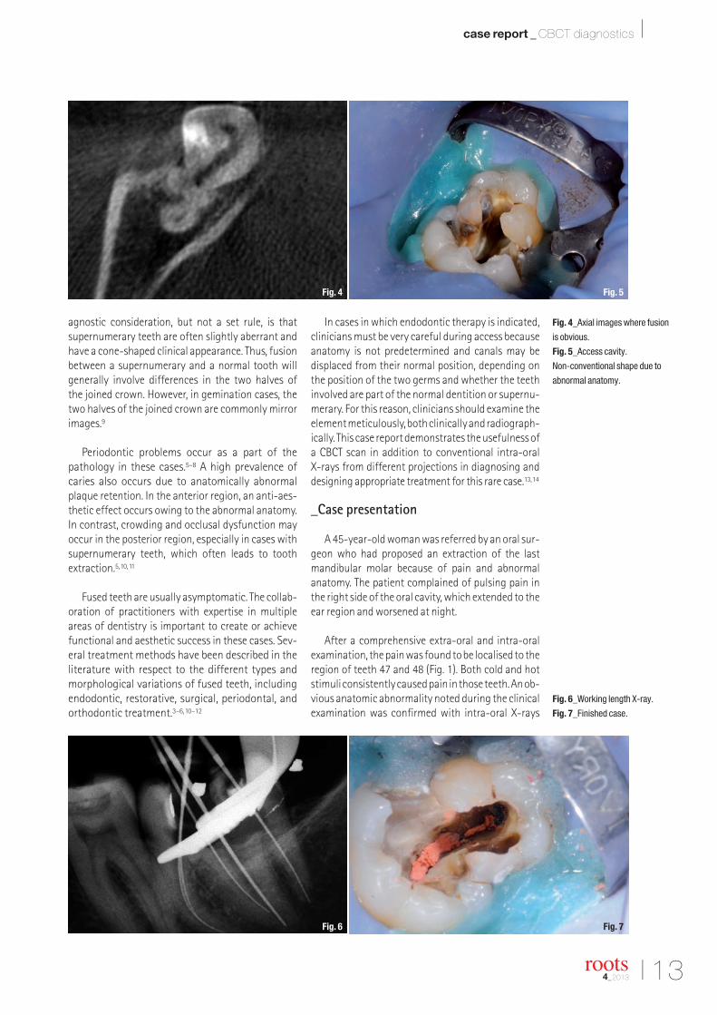
Fig. 4_Axial images where fusion
is obvious.
Fig. 5_Access cavity.
Non-conventional shape due to
abnormal anatomy.
Fig. 6_Working length X-ray.
Fig. 7_Finished case.
Fig. 4 Fig. 5
Fig. 6 Fig. 7
RO0413_12-15_Krokidis 28.11.13 13:14 Seite 2

14 I
I case report _ CBCT diagnostics
using a parallel-cone technique and various projec-tions. The X-ray (Fig. 2) also revealed a deep amal-gam restoration extending into the pulp chamber,which had been infiltrated, and distal caries in thefused tooth. A deep carious lesion was also observedon tooth 46, but a simple filling was scheduled be-cause the tooth responded normally to cold and hotstimuli.
In this case, the treatment plan was determined to be root-canal therapy for the pulpitis in the fusedtooth and a direct restoration for the same tooth. Inaddition, dental hygiene sessions were scheduled forthe patient because of generalised plaque and toavoid worsening of periodontal conditions in the area of the fused tooth. Direct restorations were alsoarranged with the general practitioner to avoid anyother pulp implications in other teeth with marked infiltrated restorations.
Initially, the treatment plan was targeted at theroot-canal therapy of the fused tooth, which was urgent. In order to clarify the anatomy of this element,a CBCT examination was also performed; it revealedtwo independent mesial roots (lingual and buccal)and a single distal root. The fused root in the middleinvolved two independent canals ending in the samearea (Figs. 3 & 4).
After anaesthetic with 1:100,000 lidocaine hadbeen administered, the tooth was isolated with a rubber dam (KKD, Sympatic Dam). Because of the ab-normal anatomy, the use of a liquid photopolymeris-ing dam (DAM COOL, Danville Materials) was neces-sary to seal gaps completely and to avoid leakage ofsaliva into the treated tooth and sodium hypochloriteinto the patient’s mouth. An extended access cavityusing a 1.2mm cylindrical bur and a #2 Start-X ultra-sonic tip (DENTSPLY Maillefer) was created to visu-alise all five orifices (Fig. 5).
Once the surface was clean and canals were visible, negotiation with hand files (K-files) and Path-Files (DENTSPLY Maillefer) was performed to ensurepatency of the canals. First #10 and #08 K-files (ifneeded) were alternated along the canals with copi-ous irrigation with sodium hypochlorite and using17% EDTA gel (B&L Biotech) until the #10 file was atthe apex. Working length was measured with an apexlocator (Root ZX, Morita). Afterwards #1–3 PathFileswere used until the #3 file reached working length inall five canals. Once patency had been confirmed,working length was also confirmed radiographically(Fig. 6).
The next step was to shape the canals using reciprocating files (WaveOne, DENTSPLY Tulsa Dental
Fig. 8_X-rays of the finished case.
Fig. 9_After restoration.
Fig. 10_After restoration.
Fig. 11_One-year recall X-ray.
roots4_2013
Fig. 8 Fig. 9
Fig. 10 Fig. 11
RO0413_12-15_Krokidis 28.11.13 13:14 Seite 3

I 15
case report _ CBCT diagnostics I
roots4_2013
Specialties) with a single-file reciprocating tech-nique. Since the anatomy was slightly different, theshaping technique was changed. After the primaryfile (25.08, red code), apical gauging was performedwith manual NiTi K-files (ISO) to measure the apicalrestriction diameter. For the distal canal, the large filewas also needed. Throughout the procedure, irriga-tion with preheated 5.25% sodium hypochlorite was performed with 30g irrigating needles (NaviTip,Ultradent) and the irrigant was activated with IrriSafefiles (ACTEON).15–17 Once the shaping had been com-pleted, apical diameter was confirmed through apicalgauging, and cones were fitted. Irrigation with pre-heated and activated 17% EDTA solution (Vista Den-tal Products) was used to remove inorganic debrisfrom the canals. Canals were then dried with papercones and the roots were sealed with vertical con-densation of hot gutta-percha (Endo-�2 B&L Biotech)with standardised gutta-percha cones and Pulp CanalSealer. Back-filling was performed with warm liquidgutta-percha (SuperEndo-�B&L Biotech; Figs. 7 & 8).The treatment was completed with a direct compos-ite restoration (Figs. 9 & 10). All treatment was per-formed under clinical microscope (OMNI pico, Zeiss).
The patient kept to her treatment plan and at-tended several recall appointments after the root-canal therapy. She also attended six-monthly oral hygiene appointments with the dental hygienist (Figs. 11–13).
_Discussion
Treatment planning for rare conditions such asfused teeth is fundamental to the success of eachcase. For this reason, clinicians must consider everyparameter before starting treatment. In this case, atooth extraction would have been the likely outcomewithout a CBCT examination. Because the fused teethcomplex did not involve any occlusal or periodontalproblems, the extraction would have caused signifi-cant biological damage and held significant financialimplications.
Once a treatment plan was in place, a CBCT scanwas very helpful in determining the exact position ofthe canals and in designing the access cavity accord-ing to the exact anatomy, which was different fromthat of a normal single tooth. The single-file recipro-cating technique chosen for this case was adapted to the need of the tooth. Since the anatomy was complex, the direct use of a large file in the distal root might have failed. Had different diameters beenestablished during apical gauging, the shaping tech-nique would have been changed and more fileswould have been introduced. For this reason theshaping technique was modified using more files forthis particular root.
_Conclusion
In conclusion, this case demonstrates the impor-tance of treatment planning. In designing a treatmentplan, all diagnostic methods should be considered. Inthis case, a CBCT examination resulted in a successfuland predictable treatment._
Editorial note: A complete list of references is available from the publisher.
Fig. 12_One-year recall.
Fig. 13_Four-year recall.
Andreas Krokidis, DDS, MSc, is a research associate at the National and Kapodistrian Univer-sity of Athens in Greece. He can be contacted at [email protected]
Riccardo Tonini, DDS, MSc, is in private practicein Brescia in Italy.
_contact roots
Fig. 12 Fig. 13
RO0413_12-15_Krokidis 28.11.13 13:14 Seite 4
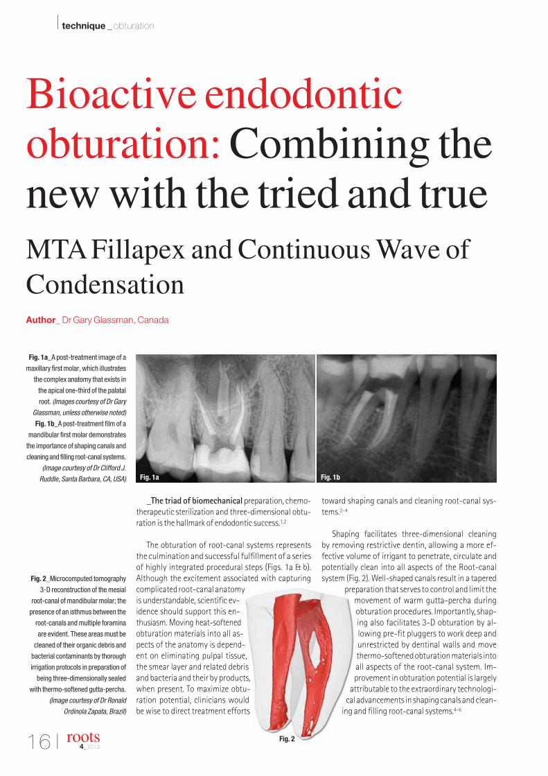
Fig. 2_Microcomputed tomography
3-D reconstruction of the mesial
root-canal of mandibular molar; the
presence of an isthmus between the
root-canals and multiple foramina
are evident. These areas must be
cleaned of their organic debris and
bacterial contaminants by thorough
irrigation protocols in preparation of
being three-dimensionally sealed
with thermo-softened gutta-percha.
(Image courtesy of Dr Ronald
Ordinola Zapata, Brazil)
16 I
I technique _ obturation
_The triad of biomechanicalpreparation, chemo -therapeutic sterilization and three-dimensional obtu-ration is the hallmark of endodontic success.1,2
The obturation of root-canal systems representsthe culmination and successful fulfillment of a seriesof highly integrated procedural steps (Figs. 1a & b). Although the excitement associated with capturingcomplicated root-canal anatomyis understandable, scientific ev-idence should support this en-thusiasm. Moving heat-softenedobturation materials into all as-pects of the anatomy is depend-ent on eliminating pulpal tissue,the smear layer and related debrisand bacteria and their by products,when present. To maximize obtu-ration potential, clinicians wouldbe wise to direct treatment efforts
toward shaping canals and cleaning root-canal sys-tems.2–4
Shaping facilitates three-dimensional cleaning by removing restrictive dentin, allowing a more ef-fective volume of irrigant to penetrate, circulate andpotentially clean into all aspects of the Root-canalsystem (Fig. 2). Well-shaped canals result in a tapered
preparation that serves to control and limit themovement of warm gutta-percha duringobturation procedures. Importantly, shap-ing also facilitates 3-D obturation by al-lowing pre-fit pluggers to work deep andunrestricted by dentinal walls and movethermo-softened obturation materials intoall aspects of the root-canal system. Im-provement in obturation potential is largely
attributable to the extraordinary technologi-cal advancements in shaping canals and clean-
ing and filling root-canal systems.4–6
roots4_2013
Bioactive endodontic obturation: Combining thenew with the tried and trueMTA Fillapex and Continuous Wave ofCondensationAuthor_ Dr Gary Glassman, Canada
Fig. 1a_A post-treatment image of a
maxillary first molar, which illustrates
the complex anatomy that exists in
the apical one-third of the palatal
root. (Images courtesy of Dr Gary
Glassman, unless otherwise noted)
Fig. 1b_A post-treatment film of a
mandibular first molar demonstrates
the importance of shaping canals and
cleaning and filling root-canal systems.
(Image courtesy of Dr Clifford J.
Ruddle, Santa Barbara, CA, USA) Fig. 1a Fig. 1b
Fig. 2
RO0413_16-24_Glassman 28.11.13 13:15 Seite 1
In the article “Filling Root-canals in Three Dimen-sions,”7 Dr Herb Schilder stated that while there wasmerit in all obturation techniques available at thattime, “when used well … vertical condensation of warmgutta-percha produces consistently dense, dimen-sionally stable, three-dimensional root-canal fillings.”This landmark article gave birth to a paradigm shift innot only a variety of warm gutta-percha techniques,but in a new approach to cleaning and shaping canals,as well as irrigation protocols.8
In addition to the classic “Schilder technique” ofobturation, there is Steve Buchanan’s “ContinuousWave of Condensation” technique9 and variationsthereof. Vertical condensation of gutta-percha is nowone of the most-trusted obturation methods of ourtime. It is taught in most of the graduate endodonticprograms in North America and in a growing numberof undergrad programs as well. Its success rate is welldocumented.8,10
This article will feature the Elements ObturationUnit (Axis|Sybron Endo, USA) that may be used to fillroot-canals systems (Fig. 3a) using the ContinuousWave of Condensation technique and a new mineraltrioxide aggregate-based endodontic sealer that isbiocompatible and bioactive, called MTA Fillapex(MTA-F; Angelus, Londrina, Brazil) (Fig. 3b). Mineraltrioxide aggregate was developed at Loma Linda uni-versity and in 1998 received approval from the FDA for human use.11,12
Since then, MTA has shown excellent biologicalproperties in several in vivo and in vitro studies.13–18
In cell culture systems, for example, MTA has been shown to enhance proliferation of periodontalligament fibroblasts,15 to induce differentiation of osteoblasts16,17 and to stimulate mineralization ofdental pulp.
In an effort to expand its applicability in endo -dontics, MTA-based root-canal sealershave been proposed, such as MTA Fillapex.19–22
MTA Fillapex is an endodonticsealer that combines the proven ad-vantage of MTA with a superior canalobturation product. Its formulationin the paste/paste system allows acomplete filling of the entire root-canal, including accessory and lateralcanals. MTA, present in the compo-sition of MTA Fillapex, is more sta-ble than calcium hydroxide, pro-viding constant release of calciumions for the tissues and maintaining a pH
that elicits antibacterial effects. The tissue recoveryand the lack of inflammatory response are optimizedby the use of MTA and disalicylate resin. The productis eugenol-free and will not interfere with adhesiveprocedures inside the root-canal.
The two-paste system contains tricalcium silicate,dicalcium silicate, calcium oxide and tricalcium alu-minate, a salicylate resin, a natural resin and bismuthoxide as a radiopacifing agent. The combination ofthese components has been shown to have bioactivepotential in its ability to stimulate nucleation sites for the formation of apatite crystals in human os-teoblast-like cell culture.22
The two pastes of MTA Fillapex are mixed in equalvolumes and dispensed on a glass slab. Its averageworking time is 35 minutes, with an average settingtime of 130 minutes.
The chemical reaction that promotessetting in MTA Fillapex is nota polymerization reactionbetween pastes but a com-
I 17roots4_2013
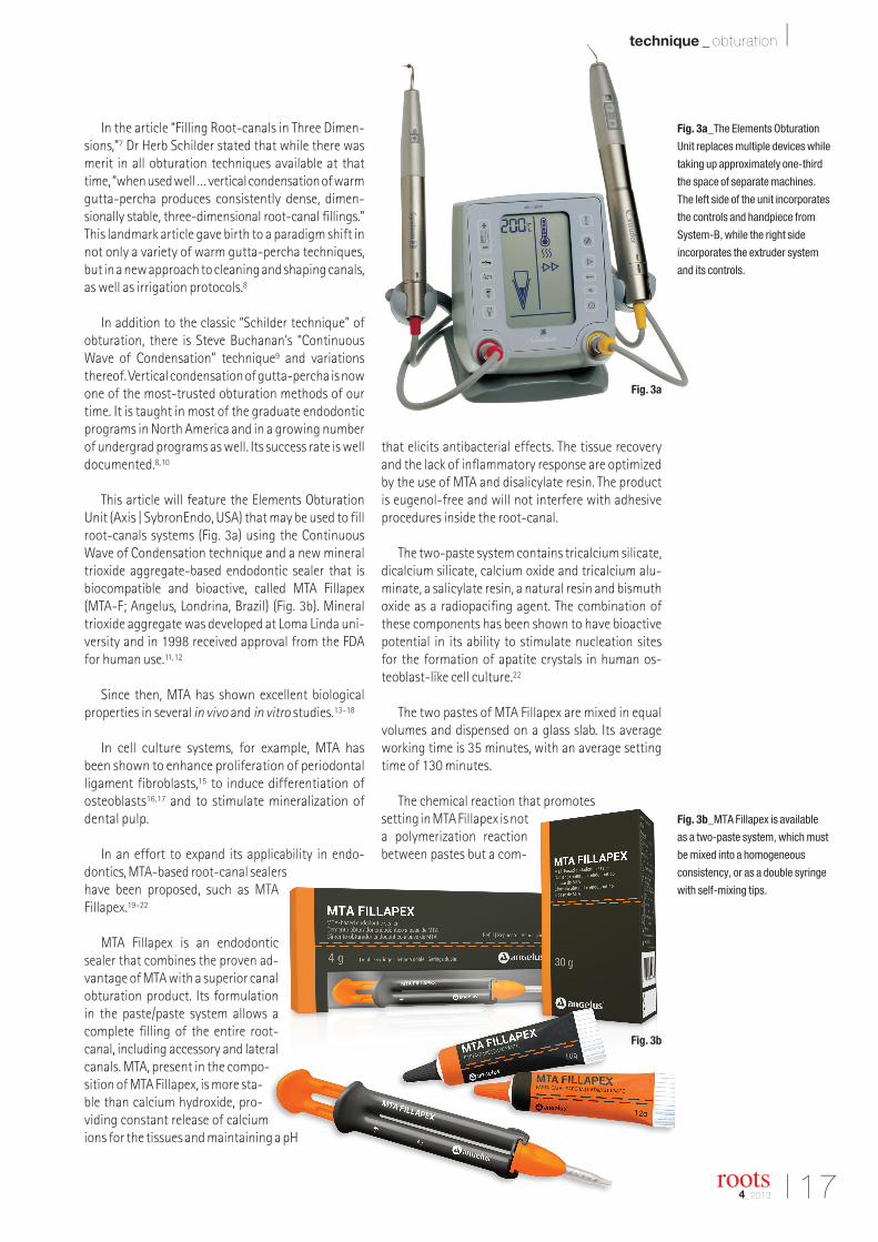
Fig. 3b_MTA Fillapex is available
as a two-paste system, which must
be mixed into a homogeneous
consistency, or as a double syringe
with self-mixing tips.
technique _ obturation I
Fig. 3a_The Elements Obturation
Unit replaces multiple devices while
taking up approximately one-third
the space of separate machines.
The left side of the unit incorporates
the controls and handpiece from
System-B, while the right side
incorporates the extruder system
and its controls.
Fig. 3a
Fig. 3b
RO0413_16-24_Glassman 29.11.13 11:32 Seite 2

18 I
I technique _ obturation
plexation reaction. The complexation reaction is anautocatalytic process. A chain reaction is initiated by water molecules in the external medium that hasan intrinsic process of self-acceleration. The com-plexation reaction is also a chelation reaction whereCa(OH)2 contacts thedisalicylate resin, resulting in the entrapment of calcium ions in the compound. Inaddition to salicylate, Ca(OH)2 is fundamental. Themajor source of Ca(OH)2 responsible for the MTA Fillapex reaction is from the hydration of free CaO,which is in high concentration in the formula. It istherefore concluded that the moisture present in thedentin tubules hydrates free CaO, forming Ca(OH)2,which will react with the salicylate and promote thesetting.23
_The Continuous Wave of Condensationtechnique
This technique allows a single-taperedelectric heat plugger to capture awave of condensation at the orificeof a canal and ride it, without re-lease, to the apical extent of downpacking in a single, continuous move-ment. Because the tip moves througha viscosity-controlled material intoa tapered-like canal form, thevelocity of the thermo-soft-ened gutta-percha and sealermoving into the root-canal sys-tem actually accelerates as thedownpacking progresses, movingsoftened gutta-percha into extremelysmall ramifications (Figs. 4a, b).
The continuously tapered root-canal preparationfacilitates the fit of a suitably sized gutta-perchacone, preferably fine-medium or medium. A clevertool to assist with the cone fit, especially if you choosenot to use pre-sized cones or prefer nonstandardized
cones, is a gutta-percha gauge such as the Tip Snip(Axis|Sybron Endo, USA) (Fig. 5). This allows you tocustomize a non-standardized or tapered cone to aprecise apical diameter. The master cone is fit in afluid-filled canal to more closely simulate the lubri-cation effect that sealer will provide when sliding thebuttered master cone into the prepared canal.
Further, the master cone should be able to be insertedto the full working length and exhibit apical tugbackupon removal. It is simple to fit a master cone into apatent, smoothly tapered and well-prepared canal.4
The intimacy of diametrical fit between the coneand the canal space is confirmed radiographically(Fig. 6). The cone is then trimmed about 0.5 to 1mmfrom radiographic terminus, so that its most apicalend is just short of the working length to accom-modate vertical movement of the vertically con-
densed gutta-percha cone.
The System-B 0.06 or 0.08 taper,0.5mm plugger should fit to within 4 to6mm from most canal termini and is
pre-fit to its binding point in the canal,and the rubber stop is adjusted adjacent to
a reference point (Fig. 7).
Difficulties in achieving adequateplugger depth are because of defi-cient deep shape in the canal prepa-
ration (inadequate enlargement 3 to4mm shy of the terminus).
Stainless-steel Buchanan pluggers (Axis|Sybron -Endo, USA) are pre-fit into the canals to their bindingpoint. Rubber stoppers are adjusted on these pluggersto the occlusal reference point, corresponding to2mm short of the apical binding point. These pluggersare placed aside to be used later in the backfill phaseof canal obturation (Fig. 8).
Figs. 4a & b_Gutta-percha
and sealer can move into extremely
small canal ramifications by virtue
of the vertical and lateral forces
created during the simultaneous
warming and condensation
of the gutta-percha.
Fig. 5_The Tip Snip can be used
to customize the apical size of the
master gutta-percha cone.
roots4_2013
Fig. 4a Fig. 4b
Fig. 5
RO0413_16-24_Glassman 28.11.13 13:15 Seite 3

www.idem-singapore.com
THE BUSINESS OF DENTISTRY
To Exhibit
Koelnmesse Pte Ltd
Stephanie Sim
Tel: +65 6500 6723
To Visit
Koelnmesse Pte Ltd
Andrea Berghoff
Tel: +65 6500 6706
IDEM Singapore offers an unrivalled opportunity to reach out to the dental fraternity in the Asia-Pacific region. With a powerful combination of an extensive international trade exhibition and a world-class scientific conference, IDEM Singapore has been a cornerstone event in the dental community calendar since 2000. It is a “must-attend” for dental practitioners and professionals in the Asia-Pacific looking for the latest cutting edge technology and innovations in dental solutions and services.
YOUR GATEWAY TO THE ASIA PACIFIC’S DENTAL MARKETSIDEM Singapore is a highly targeted trade exhibition and conference that offers exhibitors unrivalled prospects to meet and do business with the dental fraternity in the Asia-Pacific region. Capitalize on this unique opportunity to showcase your products and solutions to the dental community in Asia-Pacific.
ONE-STOP SHOPPING AND BUSINESS NETWORKINGWith more than 450 exhibitors from over 36 countries in one location - See, learn and shop for the latest and best deals in dental technology at IDEM Singapore 2014. For the traders and distribution houses, IDEM Singapore 2014 will also feature many new exhibitors globally, using this exhibition as a platform to seek distributors in Asia. Meet dental professionals from all over the Asia-Pacific region. Establish contacts, exchange ideas and socialise with colleagues both familiar and new from the regional dental fraternity. For a full list of exhibitors, please visit our website. Register online to visit the trade exhibition for free.
A CONTINUAL EDUCATION PROGRAM THAT IS TAILORED TO YOUR NEEDSIn four power-packed days of lectures and workshops, IDEM Singapore 2014 caters to Dentists and the rest of the dental team, including Dental Technicians, Dental Hygienists and Dental Therapists. A diverse range of topics and educational sessions will be presented, so you can tailor a valuable program that is relevant to your needs.
APRIL 4 - 6, 2014Pre-Congress Day: April 3, 2014
Singapore Dental Association
Co-organizerHeld InEndorsed By In Co-operation WithSupported By
REGISTER ONLINE NOW!Enjoy free entry to the Trade Fair & Conference Early Bird rates
More than 80% of the 16,000 sqm of exhibiting space has been booked - secure your booth space now!

_Sealer and master cone placement
MTA Fillapex can be used for the warm gutta-per-cha with vertical condensation technique and affordsseveral advantages.23
The presence of MTA in the formula along with itscalcium ion release allows the formation of new tis-sue, including root cementum without causing an inflammatory reaction. Perfect radiographic visuali-zation is possible because of its high radiopacity, andits excellent flow properties make MTA Fillapex suit-able to penetrate and fill lateral and accessory canals.Upon setting, MTA Fillapex expands, thereby provid-ing an excellent seal of the root-canal, avoiding thepenetration of tissue fluids and/or bacterial reconta-mination. It is available in a two-paste system, which
allows easy handling, insertion and adequate workingtime to be used by both specialists and/or generalpractitioners. If retreatment is necessary it is easily removed particularly when used with GP points.
The amount of sealer used in this obturation tech-nique should be minimal.
The radicular portion of the master cone is lightlybuttered with sealer and gently swirled as it is slowlyslid to length. Placing the master cone in this mannerwill serve to more evenly distribute sealer along thewalls of the preparation and, importantly, allow surplus sealer to harmlessly vent coronally. To be con-fident that there is sufficient sealer, the master coneis removed and its radicular surfaces inspected to ensure it is evenly coated with sealer. If the mastercone is devoid of sealer, then simply re-butter and re-insert this cone to ensure there is sufficient sealerpresent. When the master cone is evenly coated withsealer and fully seated, obturation can commence.4
The canal is dried and the master cone is cemented inthe canal with sealer (Fig. 9).
The System-B handpiece is activated by depress-ing the button with a gloved finger. The tip will heatinstantly, and the LED indicator on the handpiece willilluminate. The tip will remain heated only as long asthe button is depressed. A “time-out” feature assiststhe clinician by shutting off the energy to the tip afterfour seconds. This will aid in avoiding overheating ofthe tooth and/or tissue. The handpiece will need to be reactivated to resume heating beyond the presetduration.
Fig. 6_A non-standardized
(finemedium or medium) gutta-
percha cone is fit into the tapered
root-canal preparation, making sure
that “apical tugback” has been
achieved 0.5 to 1mm short of the
working length (distance from apical
reference point will vary with canal
curvature and size).
Fig. 7_It is essential that appropriate
System-B plugger is pre-fit into each
canal to its binding point. A rubber
stop must be placed and adjusted to
the appropriate coronal reference
point for each canal.
Figs. 8a–c_Buchanan pluggers
may be pre-fit into the canals to
their binding point. Rubber stoppers
are adjusted on these pluggers to the
occlusal reference point correspon-
ding to 2 mm short of the apical
binding point.
Fig. 6 Fig. 7
Fig. 8a Fig. 8b Fig. 8c
20 I
I technique _ obturation
roots4_2013
RO0413_16-24_Glassman 28.11.13 13:15 Seite 4

The 10th World Endodontic Congress IFEA
Endodontic Excellence at the Apex of Africa
2016Cape Town, South Africa
International Federation of Endodontic Associations
IFEA_JanFeb 2013_Layout 1 2013/02/04 3:12 PM Page 1
22 I
I technique _ obturation
The master cone is seared at the orifice of thecanals with the activated System-B plugger and thengently “seated” with a larger stainless-steel Buchananplugger. The plugger is driven through the center ofthe gutta-percha in a single motion (about one to twoseconds), to a point about 3 to 4mm shy of its apicalbinding point (Figs. 10 & 11).
While maintaining pressure on the plugger, theactivation button on the System-B is released and the plugger slows its apical movement as the pluggertip cools (about one second) to within 2mm from itsapical binding point. After the plugger stops short ofits binding point, apical pressure on the plugger issustained until the apical mass of gutta-percha hasset (5 to 10 seconds), to prevent any shrinkage thatoccurs upon cooling (Fig. 12).
_Separation burst
After the apical mass has set, the activation buttonon the System-B is depressed again, for a one-secondsurge of heat. Pause for one second after this separa-tion burst, and then remove the heated plugger and themiddle and coronal gutta-percha, leaving behind the 4 to 6mm apical plug of gutta-percha (Figs. 13 & 14).Because these pluggers heat from their tips, this sep-aration burst of heat allows for quick, sure severanceof the plugger from the already condensed and set api-cal mass of gutta-percha, minimizing the possibilityof pulling the master cone out. Be certain to limit thelength of this heat burst, as the goal is separation fromthe apical mass of gutta-percha without reheating.
Clinicians must be very alert during the first sec-ond of the downpack that the binding point is notreached before completion of the downpack. If heat isheld for too long, the plugger drops to its bindingpoint in the canal and then cannot maintain conden-sation pressure on the apical mass of gutta-perchaduring cooling, possibly allowing it to pull away fromthe canal walls. If binding length is reached by mistake,the heat plugger should be removed immediately, andthe small end of the nickel-titanium end of a Buchananhand plugger (Sybron Endo, USA) should be used tocondense the apical mass of gutta-percha until set.
_Backfilling
The Elements Obturation Unit (Fig. 3a) has an ex-truder handpiece that accommodates disposable pre-loaded cartridges of gutta-percha of varying densi-ties and is use to back fill the root-canal space. Theyare available in easy-flow, normal-flow and heavy-body-flow viscosities. The applicator tips are availablein 20-, 23- and 25-gauge diameters. There is enoughgutta-percha in the disposable cartridges to fill an average four-canal molar. The author prefers to use
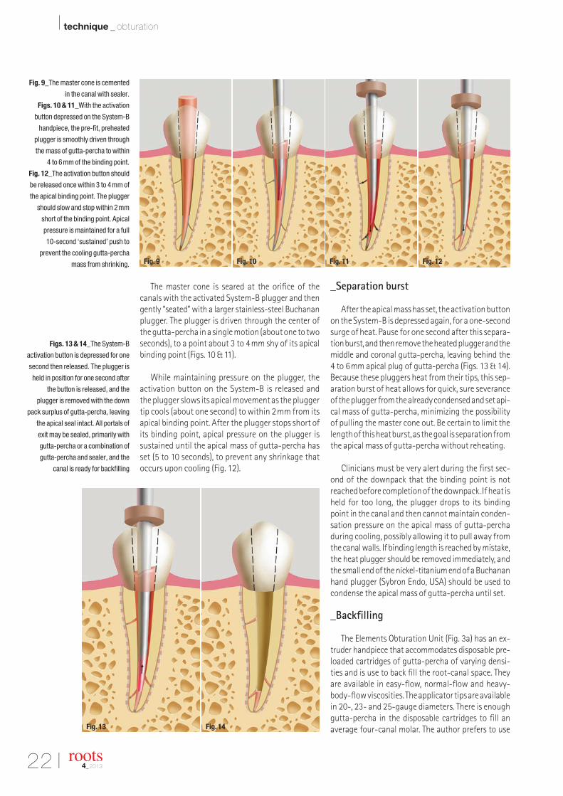
Fig. 9_The master cone is cemented
in the canal with sealer.
Figs. 10 & 11_With the activation
button depressed on the System-B
handpiece, the pre-fit, preheated
plugger is smoothly driven through
the mass of gutta-percha to within
4 to 6mm of the binding point.
Fig. 12_The activation button should
be released once within 3 to 4mm of
the apical binding point. The plugger
should slow and stop within 2mm
short of the binding point. Apical
pressure is maintained for a full
10-second ‘sustained’ push to
prevent the cooling gutta-percha
mass from shrinking.
Figs. 13 & 14_The System-B
activation button is depressed for one
second then released. The plugger is
held in position for one second after
the button is released, and the
plugger is removed with the down
pack surplus of gutta-percha, leaving
the apical seal intact. All portals of
exit may be sealed, primarily with
gutta-percha or a combination of
gutta-percha and sealer, and the
canal is ready for backfilling
roots4_2013
Fig. 9 Fig. 10 Fig. 11 Fig. 12
Fig. 13 Fig. 14
RO0413_16-24_Glassman 28.11.13 13:15 Seite 5
I 23
technique _ obturation I
roots4_2013
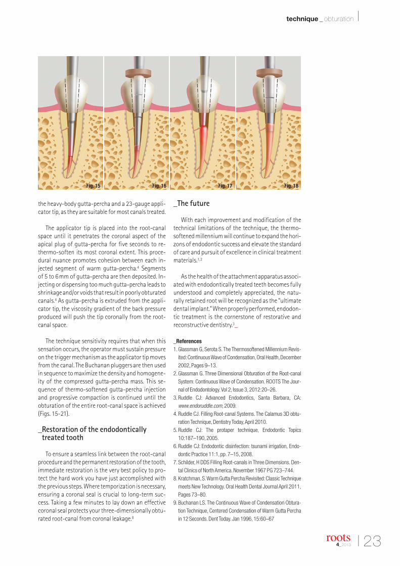
the heavy-body gutta-percha and a 23-gauge appli-cator tip, as they are suitable for most canals treated.
The applicator tip is placed into the root-canalspace until it penetrates the coronal aspect of the apical plug of gutta-percha for five seconds to re-thermo-soften its most coronal extent. This proce-dural nuance promotes cohesion between each in-jected segment of warm gutta-percha.4 Segments of 5 to 6mm of gutta-percha are then deposited. In-jecting or dispensing too much gutta-percha leads toshrinkage and/or voids that result in poorly obturatedcanals.4 As gutta-percha is extruded from the appli-cator tip, the viscosity gradient of the back pressureproduced will push the tip coronally from the root-canal space.
The technique sensitivity requires that when thissensation occurs, the operator must sustain pressureon the trigger mechanism as the applicator tip movesfrom the canal. The Buchanan pluggers are then usedin sequence to maximize the density and homogene-ity of the compressed gutta-percha mass. This se-quence of thermo-softened gutta-percha injectionand progressive compaction is continued until theobturation of the entire root-canal space is achieved(Figs. 15-21).
_Restoration of the endodonticallytreated tooth
To ensure a seamless link between the root-canalprocedure and the permanent restoration of the tooth,immediate restoration is the very best policy to pro-tect the hard work you have just accomplished withthe previous steps. Where temporization is necessary,ensuring a coronal seal is crucial to long-term suc-cess. Taking a few minutes to lay down an effectivecoronal seal protects your three-dimensionally obtu-rated root-canal from coronal leakage.8
_The future
With each improvement and modification of thetechnical limitations of the technique, the thermo -softened millennium will continue to expand the hori-zons of endodontic success and elevate the standardof care and pursuit of excellence in clinical treatmentmaterials.1,2
As the health of the attachment apparatus associ-ated with endodontically treated teeth becomes fullyunderstood and completely appreciated, the natu-rally retained root will be recognized as the “ultimatedental implant.” When properly performed, endodon-tic treatment is the cornerstone of restorative and reconstructive dentistry.3_
_References 1. Glassman G, Serota S. The Thermosoftened Millennium Revis-
ited: Continuous Wave of Condensation, Oral Health, December2002, Pages 9–13.
2. Glassman G. Three Dimensional Obturation of the Root-canalSystem: Continuous Wave of Condensation. ROOTS The Jour-nal of Endodontology. Vol 2, Issue 3, 2012:20–26.
3. Ruddle CJ: Advanced Endodontics, Santa Barbara, CA:www.endoruddle.com, 2009.
4. Ruddle CJ. Filling Root-canal Systems. The Calamus 3D obtu-ration Technique, Dentistry Today, April 2010.
5. Ruddle CJ: The protaper technique, Endodontic Topics10:187–190, 2005.
6. Ruddle CJ: Endodontic disinfection: tsunami irrigation, Endo -dontic Practice 11:1, pp. 7–15, 2008.
7. Schilder, H DDS Filling Root-canals in Three Dimensions. Den-tal Clinics of North America. November 1967 PG 723–744.
8. Kratchman, S. Warm Gutta Percha Revisited: Classic Techniquemeets New Technology. Oral Health Dental Journal April 2011,Pages 73–80.
9. Buchanan LS. The Continuous Wave of Condensation Obtura-tion Technique, Centered Condensation of Warm Gutta Perchain 12 Seconds. Dent Today. Jan 1996, 15:60–67
Fig. 15 Fig. 16 Fig. 17 Fig. 18
RO0413_16-24_Glassman 28.11.13 13:15 Seite 6

24 I
I technique _ obturation
10. De Chevigny C, Dao TT, Basrani BR, Marquis V, Farzaneh M,Abitbol, S, Friedman, S. Journal of Endodontics, Vol. 34, Issue3 pages 258–263 December 2007. Treatment Outcome inEndodontics: The Toronto Study - Phase 4, Initial Treatment.
11. Torabinejad M, White DJ. United States Patent 5, 415, 547USPTO. Patent Full Text and Image Database 1995.
12. Parirokh M, Torabinejad M. Mineral trioxide aggregate: a comprehensive literature review — part I: chemical, physi-cal, and antibacterial. J Endod 2010;36:16–27.
13. Holland R, Filho JA, de Souza V, Nery MJ, Bernabe PF, JuniorED. Mineral trioxide aggregate repair of lateral root perfora-tions. J Endod 2001;27:281–284.
14. De Deus G, Petruccelli V, GurgelFilho E, Coutinho-Filho T. MTAversus Portland cement as repair material for furcal perfora-tions: a laboratory study using a polymicrobial leakage model.Int Endod J 2006;39:293–296.
15. Bonson S, Jeansonne BG, Laillier TE. Root-end filling materialsalter fibroblast differentiation. J Dent Res 2004;83:408–413.
16. Nakayama A, Ogiso B, Tanabe N, Takeichi O, Matsuzaka K, Inoue T. Behavior of bone marrow osteoblast-like cells on mineral trioxide aggregate: morphology and expression of type I collagen and bone-related protein mRNAs. Int Endod J2005;38:203–210.
17. Gomes-Filho JE, de FariaMD, Barnab_e PF, et al. Mineral tri-oxide aggregate but not lightcure mineral trioxide aggregatestimulated mineralization. J Endod 2008;34:62–65.
18. Yasuda Y, Ogawa M, Arakawa T, Kodowaki T, Takashi S. The effectof mineral trioxide aggregate on the mineralization ability of ratdental pulp cells: an in vitro study. J Endod 2008;34:1057–1060.
19. Bortoluzzi EA, Guerreiro-Tanomaru JM, Tanomaru-Filho M,Duarte MAH. Radiographic effect of different radiopacifiers on a potential retrograde filling material. Oral Surg Oral MedOral Pathol Oral Radiol Endod 2009;108:628–632.
20. Corn_elio ALG, Salles LP, Campos da Paz M, Cirelli JA, Guer-reiro-Tanomaru JM, Tanomaru-Filho M. Cytotoxicity of Port-land cement with different radiopacifying agents: a cell deathstudy. J Endod 2011;37:203–210.
21. Camilleri J. The physical properties of accelerated Portland ce-ment for endodontic use. Int Endod J 2008;41:151–157.
22. Salles LP, Gomes-Cornelio AL, Coutinho Guimaraes F, Schnei-der Herrera B, Nair Bao S, Rossa-Junior C, Guerreiro-Tanomaru JM, Tanomaru-Filho M, Mineral Trioxide Aggre-gate–based Endodontic Sealer StimulatesHydroxyapatite Nu-cleation in Human Osteoblast-like Cell Culture J. Endo, Volume38, Number 7, July 2012.
23. Angelus, MTA Scientific Profile. www.angelus.ind.br
Figs. 15–21_Applicator tips for the
EOU System are available in sizes
#20, #23 and #25 gauges. Additional
root-canal sealer may be placed in
the coronal aspect of the root-canal
with a hand file prior to back filling.
Fourto 6-mm increments of gutta-
percha are injected into the canal
space then immediately condensed
with the pre-fitted Buchanan pluggers
in sequence using the sequentially
larger pluggers as the coronal aspect
of the canal is approached.
As thermosoftened gutta-percha is
deposited in the canal, backpressure
is produced and the applicator is
forcibly extruded from the canal
space. It is essential that the operator
continue injecting as the applicator
tip is retrieved from the canal in order
to avoid inadvertent removal of the
newly deposited gutta-percha, mass
prior to condensation.
roots4_2013
Gary D. Glassman, DDS, FRCD(C) graduatedfrom the University of Toronto, Faculty of Den-tistry in 1984 and was awarded the James B.Willmott Scholarship, the Mosby Scholarship and the George Hare Endodontic Scholarship for proficiency in Endodontics. A graduate of the Endodontology Program atTemple University in 1987, he received the Louis I.Grossman Study Club Award for academic and
clinical proficiency in Endodontics. The author of numerous publications, Gary is on staff at the University ofToronto, Faculty of Dentistry in the graduate department of endodonticsaand Adjunct professor of dentistry and director of endodontic program-ming at UTech in KIngston, Jamaica. Dr Gary Glassman maintains a private practice in Toronto.
www.rootcanals.ca
_about the author roots
Fig. 19 Fig. 20 Fig. 21
RO0413_16-24_Glassman 28.11.13 13:15 Seite 7

P R O F E S S I O N A L M E D I C A L C O U T U R E
E X P E R I E N C E O U R E N T I R E C O L L E C T I O N O N L I N E
WWW.CROIXTURE.COM

26 I
I special _ lasers
_Introduction
The complete restoration of the root canalspace with an inert filling material and the cre-ation of a fluid tight seal are the goals of success-ful endodontic therapy.1 In order to create a fluidtight seal, it is imperative that the endodontic filling material closely adapts or bonds to thetooth structure. This, however, is impaired by thepresence of a smear layer, which invariably formsafter endodontic instrumentation.2, 3 The smearlayer contains organic material, odontoblasticprocesses, bacteria and blood cells.
Various materials and techniques have been re-ported with wide variations in their efficacy re-garding the removal of the intra-canal smearlayer.2, 4 The most widely used chemical for the pur-pose is EDTA, used in different formulations.5 Theyhave been reported to consistently produce canalswith patent dentinal tubules.6 However, it hasbeen found to be less efficient in narrow portions
of the canal7, it requires a long application time foroptimum results8 and can seriously damage thedentin, if used in excess.9
Clinically, endodontic procedures use both me-chanical instrumentation and chemical irrigantsin the attempt to three dimensionally debride,clean and decontaminate the endodontic sys-tem.10,11
Even after doing this meticulously, we still fallshort of successfully removing all of the infectivemicroorganisms and debris. This is because of thecomplex root canal anatomy and the inability ofcommon irrigants to penetrate into the lateralcanals and the apical ramifications. It seems,therefore, appropriate to search for new materi-als, techniques and technologies that can improvethe cleaning and decontamination of theseanatomical areas.12
Some of these mechanically activated irriga-tion techniques include manual irrigation withneedles, K-file, Master cone GP points, Irrisafe, ultrasonics, Endo-activator, Rotobrush, Roeko-brush, etc. The newest of the lot is PIPS, i.e Photon-Induced Photoacoustic Streaming via laser. Henceit was chosen for the study.
_Material and methods
Forty single-rooted, extracted human teethwere used in the study. Teeth with fractures, cracksor any other defects were excluded. Subsequently,they were scaled with ultrasonics for the removalof calculus or any soft-tissue debris, washed withdistilled water and then stored in normal saline.Standard endodontic access cavity preparationswere performed and then a stainless-steel #10
Fig. 1_Access opening.
Fig. 2_Stainless steel #10.
K-file for patency.
roots4_2013
SEM analysis of the laseractivation of final irrigantsfor smear layer removal Authors_Dr Vivek Hegde, Dr Naresh Thukral, Dr Sucheta Sathe, Dr Shachi Goenka & Dr Paresh Jain, India
Fig. 1 Fig. 2
RO0413_26-29_Fotona 28.11.13 13:16 Seite 1

I 27
special _ lasers I
roots4_2013
K-file (Mani K-File) was inserted into the canal until the tip was just visible at the apical foramento check for patency. Chemo-mechanical prepara-tion was done up to F3 using rotary protapers(DENTSPLY Maillefer) along with EDTA gel (Glyde –DENTSPLY Maillefer) for all the samples.
Irrigation of all the samples during preparationwas accomplished using 5ml of 5.25% sodiumhypochlorite between each file. Samples werethen divided randomly into two groups, depend-ing upon the method of activation of the final ir-rigant used.
These groups were further divided into twosubgroups, depending upon the final irrigant used(Tab. 1):
_Subgroup A: 5.25% NaOCl (n = 10) _Subgroup B: 17% EDTA (n = 10)
Activation of the irrigant for group I was donemechanically by agitating a stainless steel #25 K-file (2% taper) in the canal when it was filledwith the final irrigant solution.
An Er:YAG laser with a wavelength of 2,940nm(Fotona) was used to irradiate the root canals inGroup II with a newly designed 12mm long, 400µmquartz tip. The tip was tapered and had 3mm of the polyamide sheath stripped away from its end.The laser operating parameters used for all thesamples (using the free-running emission mode)were as follows: 40mJ per pulse, 20Hz, at veryshort pulse (MSP) mode, which provides the same400W of peak pulse power as the parameters rec-ommended by Olivi (20mJ, 15Hz, SSP). The coax-ial water spray feature of the handpiece was set to‘off’ while air settings were kept at 2. The tip wasplaced into the coronal access opening of thechamber just above the orifice, and was kept sta-tionary. During the laser irradiation cycles, the
root canals were continuously irrigated with thefinal irrigant to maintain hydration levels using ahand syringe with a 25 gauge needle positionedabove the laser tip in the coronal aspect of the ac-cess opening, according to the above protocol.
After preparation, the root canal walls weredried using paper points. Longitudinal grooveswere made on the distal and mesial root surfaces,preserving the inner shelf of the dentin surround-ing the canal. Roots were then sectioned with thehelp of a chisel and mallet. Samples were then sub-jected to SEM to visualize the surface characteris-tics.
_Results
Group I specimens (hand activation) consis-tently exhibited a thick smear layer with NaOCl(subgroup A, Figs. 8a–c) while comparatively lesssmear layer was observed with EDTA (subgroup B,Figs. 9a–c). SEM examination demonstrated thatwhen NaOCl irrigation was applied, a noticeablesmear layer and occluded dentinal tubules re-mained on the treated surface. Debris, defined asdentin chips and pulp remnants loosely attachedto the internal surface of the root canals, was pres-ent in the specimens in subgroup A (Group I). In thespecimens of EDTA, mostly open dentinal tubuleswere observed in the coronal and the middle thirdwhile in the apical third of all specimens occludedtubules were observed.
Figs. 3 and 4_Chemo-mechanical
preparation up to F3.
Fig. 5_Group I—Hand activation
using stainless steel #25 K-file
(n = 20).
Table 1_Subgroups depending on
the final irrigant used.
GROUP I(Hand Activation)
GROUP II(Er:YAG with PIPS)
Sub Group A(5.25 % NaOCl)
n = 10 n = 10
Sub Group B(17 % EDTA)
n = 10 n = 10
GROUPS
SUB-GROUPS
Fig. 3 Fig. 4 Fig. 5
RO0413_26-29_Fotona 28.11.13 13:16 Seite 2

28 I
I special _ lasers
Group II specimens treated with the Er:YAGlaser with PIPS showed the most effective re-moval of the smear layer from the root canal walls compared to Group I (hand activation) specimens.At higher magnifications (1,000x–2,000x) sub-group B (17% EDTA) showed better results withexposed and intact collagen fibers and open denti-nal tubules, even in the apical third (Figs.11a–c),when compared with subgroup A (5.25% NaOCl),where open dentinal tubules along with scattereddentinal chips were observed (Figs. 10a–c). Noneof the SEM images indicated signs of dentin melting.
_Discussion
Current instrumentation techniques using ro-tary instruments and chemical irrigation still fallshort of successfully removing the smear layerfrom inside the root canal system. Mechanical ac-tivation of the chemical irrigant plays an importantrole in removing the smear layer. Fiber-guidedlasers have also been used hoping to achieve somedegree of success, however, there is limited avail-ability of literature regarding this topic.
The concept of laser-activated irrigation is basedon cavitation. Because of the high absorption of water by the mid-infrared wavelength of lasers, thecavitation process generates vapor-containing bub-bles, which explode and implode in a liquid environ-ment.13 This subsequently initiates pressure/shockwaves by inducing shear force on the dentinal wall.In a water-filled root canal, the shock waves couldpotentially detach the smear layer and disrupt bac-terial biofilms. To efficiently activate irrigant andgenerate shock waves in the root canal, lasers withwavelengths from 940–2,940nm have been used.14–22
5.25% sodium hypochlorite was used in Group Ibecause the majority of practitioners still use onlysodium hypochlorite as the irrigant along with handinstruments. Hence sodium hypochlorite was used inGroup I. To remove inorganic debris of the smear layer,use of aqueous EDTA had been recommended. But pro-longed use of EDTA can cause dentinal erosion of theroot canal by decalcifying the peritubular dentin. Therecommended time in endodontic literature is only1–2 minutes. Hence, 17% aqueous EDTA was used forone minute in Group II to minimise time and damage.
roots4_2013
Fig. 6bFig. 6a
Fig. 7
Fig. 8a Fig. 8b Fig. 8c
Fig. 9a Fig. 9b Fig. 9c
Fig. 8_Group I—Hand Activation
(5.25% NaOCl—Subgroup A):
coronal third (a),
middle third (b),
apical third (c).
Fig. 9_Group I—Hand Activation
(17% EDTA—Subgroup B):
coronal third (a),
middle third (b),
apical third (c).
Figs. 6a & b and 7_Group II—Er:YAG
activation using Photon-Induced
Photoacoustic Streaming tip (n = 20).
RO0413_26-29_Fotona 28.11.13 13:16 Seite 3

I 29
special _ lasers I
roots4_2013
The results of this study indicate that NaOCl sub-groups could remove the smear layer in the coronalthird; however, it did not remove the smear layer fromthe middle and apical third of the canal wall. EDTA isefficient in removing the smear layer, which is evidentin this study for both groups. The effects of EDTA werelimited to the coronal and middle third in Group I(hand activation) while it was effective even in the api-cal third for Group II (Er:YAG-PIPS). Ciucchi et al. statedthat there was a definite decline in the efficiency of ir-rigating solutions along the apical part of the canals.23
This can probably be explained by the fact that dentinin the apical third is much more sclerosed and thereare fewer dentinal tubules present there.24 Also apicalreach, canal configuration, and smooth transition area few of the anatomical key factors. Hence root canalsuccess is dependent on apical third anatomy.
The Er:YAG laser used in this investigation provedto be more effective than the conventional techniquein removing the smear layer. This finding can be at-tributed to the photomechanical effect seen whenlight energy is pulsed in liquid.25-27 When activated ina limited volume of fluid, the high absorption of theEr:YAG wavelength in water, combined with the highpeak power derived from the short pulse durationthat was used for five seconds (three cycles), resultedin a photomechanical phenomenon. A profound“shockwave-like” effect is observed when a radial and stripped tip is submerged in a coronal chamberabove the orifice. As a result of the very small volume,this effect may remove the smear layer and residualtissue tags and potentially decrease the bacterial loadwithin the tubules and lateral canals.28-30 By usinglower sub-ablative energy (40mJ) and restricting theplacement of the tip to within the coronal portion of
the tooth only, the undesired effects of the thermalenergy, as previously described in the literature, wasavoided.31-45 In the current study, the smear layer and debris were not removed by thermal vaporisa-tion, but probably by photomechanical streaming ofthe liquids, which were laser activated in the coronalpart of the tooth.
Giovani Olivi and Enrico DiVito have describedthis light energy phenomenon as photon-inducedphotoacoustic streaming (PIPS). The effect of irra-diation with the Er:YAG laser equipped with a tip ofnovel design at sub-ablative power settings (20mJ,15Hz, SSP, 400W peak power) is synergistically enhanced by the presence of EDTA. This leads to asignificantly better debridement of the root canal,contributing to an improvement in treatment effi-cacy. Hence, the PIPS technique resulted in pro-nounced smear layer removal when used togetherwith EDTA and at the settings outlined.
_Conclusion
Within the limitations of this study, the Er:YAGlaser with PIPS showed significantly better smearlayer removal than the hand-activation group. At theenergy levels and with the operating parametersused, no thermal effects or damage to the dentin surface was observed. With the described settings,the Er:YAG laser produced a photomechanical effect,demonstrating its potential as an improved alter-native method for debriding the root canal system ina minimally invasive manner._
Editorial note: A list of references is available from the publisher.
Fig. 10_Group II—Er:YAG with
PIPS (5.25% NaOCl—Subgroup A):
coronal third (a), middle third (b),
apical third (c).
Fig. 11_Group II—Er:YAG with
PIPS (17% EDTA—Subgroup B):
coronal third (a), middle third (b),
apical third (c).
Prof. Dr Vivek Hedge Professor and Head Department of ConservativeDentistry and Endodontics M A Rangoonwala Instituteof Dental SciencesPune, India
_contact roots
Fig. 10a Fig. 10b Fig. 10c
Fig. 11a Fig. 11b Fig. 11c
RO0413_26-29_Fotona 28.11.13 13:16 Seite 4

30 I
I special _ lasers
_Introduction
In recent years, several studies have been con-ducted on the clinical applications of laser in den-tistry. At the same time, there has been a markedemergence of organisations in support of the use oflaser in dentistry. In the last decades, laser therapyhas been used in dentistry as an adjunct or alterna-tive to conventional approaches. In this paper, thefollowing topics will be reviewed: the application oflaser in caries prevention and diagnosis, hard- andsoft-tissue treatments, and periodontal and endo -dontic procedures. There is a large research effortinto new indications for laser in dentistry. It is ex-pected that laser will become an essential compo-nent of the dentist’s armamentarium.
While the technology was regarded as complexand of limited use in clinical dentistry in the past, agrowing awareness of the usefulness of laser in themodern dental practice has been observed. Laser canbe used as an adjunct or alternative to conventionalapproaches.1 When comparing the use of laser withconventional therapies, three important areas must beconsidered: safety, efficacy and effectiveness. Froman ethical standpoint, it is important to use the bestavailable evidence when making clinical decisions.2
_Diagnostic laser applications
The most common methods for caries detectionare visual and radiographic examination.3 However,visual examination is a subjective method that de-pends on the knowledge and clinical experience ofthe examiner.3-6 Several studies have demonstratedthat radiographic examination demonstrates poorsensitivity to non-cavitated lesions.3, 7-9
For this reason, fluorescence-based methods havebeen developed, aiming at the detection of occlusaland approximal carious lesions, for example DIAGNO -dent 2095 (KaVo; LF; Figs. 1a-c) and DIAGNOdent2190 (LF pen; Figs. 2a & b). They rely on the same prin-ciple: a laser diode emits red light at 655nm and aphotodetector quantifies the reflected fluorescencefrom bacterial metabolites (fluorophores) in cariouslesions, showing values ranging from 0 to 99.3,9
A study that assessed the performance of a visualmethod, radiographic examination and fluores-cence-based methods in detecting occlusal caries inprimary teeth found that the visual method andVistaProof fluorescence camera (Dürr Dental; FC)exhibited better accuracy in detecting enamel anddentine carious lesions, whereas the visual methodcombined with LF, LF pen and FC better detected den-tine lesions on occlusal surfaces in primary teeth,with no statistically significant difference amongthem.3
Another study compared the performance offluorescence-based methods (FC, LF and LF pen), ra-diographic examination, and another visual methodcalled the International Caries Detection and As-sessment System (ICDAS) II on occlusal surfaces.This study demonstrated that the combination ofICDAS and bite-wing radiographs yielded the bestperformance for detecting caries on occlusal sur-faces.9
Fig. 1a_DIAGNOdent 2095.
roots4_2013
Laser versus conventional therapiesAuthors_Cristiane Meira Assunção, Joanna Tatith Pereira, Renata Schlesner Oliveira &
Dr Jonas de Almeida Rodrigues, Brazil
Fig. 1a
RO0413_30-33_Rodrigues 28.11.13 13:17 Seite 1
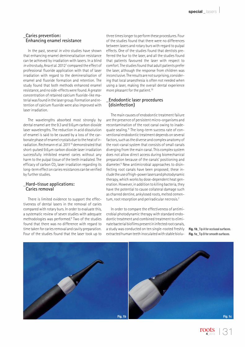
I 31
special _ lasers I
roots4_2013
_Caries prevention: Enhancing enamel resistance
In the past, several in vitro studies have shownthat enhancing enamel demineralisation resistancecan be achieved by irradiation with lasers. In a blindin vitro study, Ana et al. 20121 compared the effect ofprofessional fluoride application with that of laser irradiation with regard to the demineralisation ofenamel and fluoride formation and retention. Thestudy found that both methods enhanced enamel resistance, and no side-effects were found. A greaterconcentration of retained calcium fluoride-like ma-terial was found in the laser group. Formation and re-tention of calcium fluoride were also improved withlaser irradiation.
The wavelengths absorbed most strongly by dental enamel are the 9.3 and 9.6µm carbon dioxidelaser wavelengths. The reduction in acid dissolutionof enamel is said to be caused by a loss of the car-bonate phase of enamel crystals due to the heat of ir-radiation. Rechmann et al. 201110 demonstrated thatshort-pulsed 9.6µm carbon dioxide laser irradiationsuccessfully inhibited enamel caries without anyharm to the pulpal tissue of the teeth irradiated. Theefficacy of carbon C02 laser irradiation regarding itslong-term effect on caries resistances can be verifiedby further studies.
_Hard-tissue applications: Caries removal
There is limited evidence to support the effec-tiveness of dental lasers in the removal of cariescompared with rotary burs. In order to evaluate this,a systematic review of seven studies with adequatemethodologies was performed.8 Two of the studiesfound that there was no difference with regard totime taken for caries removal and cavity preparation.Four of the studies found that the laser took up to
three times longer to perform these procedures. Fourof the studies found that there were no differencesbetween lasers and rotary burs with regard to pulpaleffects. One of the studies found that dentists pre-ferred the bur to the laser, and all the studies foundthat patients favoured the laser with respect to comfort. The studies found that adult patients preferthe laser, although the response from children wasinconclusive. The results are not surprising, consider-ing that local anaesthesia is often not needed whenusing a laser, making the overall dental experiencemore pleasant for the patient.10
_Endodontic laser procedures (disinfection)
The main causes of endodontic treatment failureare the presence of persistent micro-organisms andrecontamination of the root canal owing to inade-quate sealing.11 The long-term success rate of con-ventional endodontic treatment depends on severalfactors, such as the diverse and complex anatomy ofthe root-canal system that consists of small canalsdiverging from the main canal. This complex systemdoes not allow direct access during biomechanicalpreparation because of the canals’ positioning anddiameter.6 New antimicrobial approaches to disin-fecting root canals have been proposed; these in-cludethe use of high-power lasers and photodynamictherapy, which works by dose-dependent heat gen-eration. However, in addition to killing bacteria, theyhave the potential to cause collateral damage suchas charred dentine, ankylosed roots, melted cemen-tum, root resorption and periradicular necrosis.2
In order to compare the effectiveness of antimi-crobial photodynamic therapy with standard endo -dontic treatment and combined treatment to elimi-nate bacterial biofilms present in infected root canals,a study was conducted on ten single-rooted freshlyextracted human teeth inoculated with stable biolu-
Fig. 1b_Tip A for occlusal surfaces.
Fig. 1c_Tip B for smooth surfaces.
Fig. 1cFig. 1b
RO0413_30-33_Rodrigues 28.11.13 13:17 Seite 2
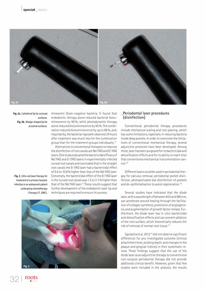
32 I
I special _ lasers
minescent Gram-negative bacteria. It found thatendodontic therapy alone reduced bacterial biolu-minescence by 90%, while photodynamic therapyalone reduced bioluminescence by 95%. The combi-nation reduced bioluminescence by up to 98%, and,importantly, the bacterial regrowth observed 24 hoursafter treatment was much less for the combinationgroup than for the treatment groups individually.12
Alternatives to conventional therapies to improvethe disinfection of root canals are Nd:YAG and Er:YAGlasers. One study evaluated the bactericidal efficacy ofNd:YAG and Er:YAG lasers in experimentally infectedcurved root canals and concluded that in the straightroot canals the Er:YAG laser had a bactericidal effectof 6.4 to 10.8% higher than that of the Nd:YAG laser.Conversely, the bactericidal effect of the Er:YAG laserin the curved root canals was 1.5 to 3.1% higher thanthat of the Nd:YAG laser.13 These results suggest thatfurther development of the endodontic laser tip andtechniques are required to ensure its success.
_Periodontal laser procedures (disinfection)
Conventional periodontal therapy procedures include mechanical scaling and root planing, whichhas some limitations, especially in reducing bacteriainside deep pockets. In order to overcome the limita-tions of conventional mechanical therapy, severaladjunctive protocols have been developed. Amongthese, laser has been proposed for its bactericidal anddetoxification effects and for its ability to reach sitesthat conventional mechanical instrumentation can-not.14
Different lasers could be used in periodontal ther-apy for calculus removal, periodontal pocket disin-fection, photoactivated dye disinfection of pocketsand de-epithelialisation to assist regeneration.15
Several studies have indicated that the diodelaser, with a wavelength of between 655 and 980nm,can accelerate wound healing through the facilita-tion of collagen synthesis, promotion of angiogene-sis, and augmentation of growth factor release. Fur-thermore, the diode laser has in vitro bactericidaland detoxification effects and can prevent ablationof the root surface, which theoretically reduces therisk of removal of normal root tissue.13
Sgolastra et al. 201214 did not observe significantdifferences for any investigated outcome (clinicalattachment level, probing depth, and changes in theplaque and gingival indices) in their systematic re-view. These findings suggest that the use of thediode laser as an adjunctive therapy to conventionalnon-surgical periodontal therapy did not provideadditional clinical benefit. However, given that fewstudies were included in the analysis, the results
roots4_2013
Fig. 2a
Fig. 3
Fig. 2b
Fig. 2a_Cylindrical tip for occlusal
surfaces.
Fig. 2b_Wedge-shaped tip for
proximal surfaces.
Fig. 3_Infra-red laser therapy for
treatment of a primary herpetic
infection in an adolescent patient
undergoing chemotherapy
(Therapy XT, DMC).
RO0413_30-33_Rodrigues 28.11.13 13:17 Seite 3

special _ lasers I
should be interpreted with caution. Important issuesthat remain to be clarified include the influence ofsmoking on clinical outcomes, the effectiveness ofthe adjunctive use of the diode laser on microbio-logical outcomes, and the effect of adverse events.Future studies are required to assess the effective-ness of the adjunctive use of the diode laser, as wellas the appropriate dosimetry and laser settings.
_Soft-tissue applications
There are numerous soft-tissue procedures thatcan be performed with laser. Two key advantages of this are reduced intra-operative bleeding and lesspost-operative pain compared with conventionaltechniques, such as electrosurgery. Certain proce-dures in patients with bleeding disorders are bettersuited to lasers with greater haemostatic capabili-ties.5
_Conclusion
Although the results of laser therapy are similar(in safety, efficacy and effectiveness) to those ob-
tained with conventional methods, new techniquesand devices have been developed. Laser could thus bean evidence-based and well-supported treatmentoption for the dentist in daily dental practice._
Editorial note: A list of references is available from the publisher.
Dr Jonas de Almeida RodriguesSchool of DentistryFederal University of Rio Grande do SulAv. Paulo Gama, 110Porto Alegre – RGS 90000-000Brazil
_contact roots
AD
RO0413_30-33_Rodrigues 28.11.13 13:17 Seite 4
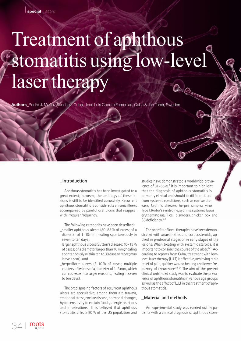
34 I roots4_2013
Treatment of aphthous stomatitis using low-levellaser therapyAuthors_Pedro J. Muñoz Sánchez, Cuba, José Luis Capote Femenias, Cuba & Jan Tunér, Sweden
[PICTURE: ©MATHAGRAPHICS]
I special _ lasers
_Introduction
Aphthous stomatitis has been investigated to agreat extent; however, the aetiology of these le-sions is still to be identified accurately. Recurrentaphthous stomatitis is considered a chronic illnessaccompanied by painful oral ulcers that reappearwith irregular frequency.
The following categories have been described:_smaller aphthous ulcers (80–85% of cases; of a
diameter of 1–10mm; healing spontaneously inseven to ten days);
_larger aphthous ulcers (Sutton’s disease; 10–15%of cases; of a diameter larger than 10mm; healingspontaneously within ten to 30 days or more; mayleave a scar); and
_herpetiform ulcers (5–10% of cases; multipleclusters of lesions of a diameter of 1–3mm, whichcan coalesce into larger erosions; healing in sevento ten days).1
The predisposing factors of recurrent aphthousulcers are speculative; among them are trauma,emotional stress, coeliac disease, hormonal changes,hypersensitivity to certain foods, allergic reactionsand intoxications.1 It is believed that aphthousstomatitis affects 20 % of the US population and
studies have demonstrated a worldwide preva-lence of 31–66%.2 It is important to highlightthat the diagnosis of aphthous stomatitis isprimarily clinical and should be differentiatedfrom systemic conditions, such as coeliac dis-ease, Crohn’s disease, herpes simplex virus Type I, Reiter’s syndrome, syphilis, systemic lupuserythematosus, T cell disorders, chicken pox and B6 deficiency.3–7
The benefits of local therapies have been demon-strated with anaesthetics and cortico steroids, ap-plied in prodromal stages or in early stages of the lesions. When treating with systemic steroids, it isimportant to consider the course of the ulcer.8–21 Ac-cording to reports from Cuba, treatment with low-level laser therapy (LLLT) is effective, achieving rapidrelief of pain, quicker wound healing and lower fre-quency of recurrence.22–24 The aim of the presentclinical unblinded study was to evaluate the preva-lence of aphthous stomatitis in various age groups,as well as the effect of LLLT in the treatment of aph-thous stomatitis.
_Material and methods
An experimental study was carried out in pa-tients with a clinical diagnosis of aphthous stom-
RO0413_34-36_Tuner 28.11.13 13:17 Seite 1
I 35
special _ lasers I
roots4_2013
atitis attending the Leonardo Fernández Sánchezdental clinic in Cienfuegos in Cuba between Sep-tember 2010 and March 2011. Among the 252 pa-tients registered, 208 attended the clinic until thelesions had healed completely. The study was ap-proved by the Scientific Council of the University ofMedical Sciences, Cienfuegos. All of the patientswere informed about the parameters of the studyand gave their informed consent.
LLLT was administered to 104 patients (studygroup) and the remaining 104 (control group) re-ceived conventional treatment, such as topicalanaesthetics (2 % lidocaine), dietary advice and oralpainkillers. Every second patient with the same typeof ulcer was allocated to either the study or the con-trol group. The two groups consisted of 56 malesand 148 females, with a great variation in age dis-tribution (Table 1).
All of the patients were seen daily and the pa-tients in the study group received LLLT every secondday unless their ulcer had already healed. The pa-tients were categorised with regard to age and ulcertype (Tables 1 & 2). Special diagnostic procedureswere performed in patients with large ulcers toconfirm a safe differential diagnosis. Pain was eval-uated, but since pain is subjective, it was decidedonly to register the time until wound closure (Fig. 1),which can be registered objectively. No patient inthe study group reported any negative effects ofthe laser irradiation.
The laser used was the Lasermed 670DL (Cubanmanufactured), 670nm, 40mW. Each ulcer under-went 40 seconds of irradiation at 1.6J, 2.04J/cm2
and 51mW/cm2 from a distance of about 0.5cm.The parameters used were based upon the success-ful application of these in a previous study on her-pes simplex virus Type I blisters.32
The study was unblinded. A 660nm laser emitsclear red light and, although a patient-blinded de-sign is possible, it is not possible to mask a placebolaser for the therapist.
The distribution of the age of the patients, of the types of ulcer according to age, and of the typesof ulcer in the study and control groups is shown inTables 1–3. This data corresponds with the litera-ture, where smaller aphthous ulcers represent80–85% of cases, larger ulcers 10–15% and her-petiform ulcers 5–10%.1
_Results
The main results of the study are presented inFigure 1. The results shown are for small ulcers,which were the dominant type of ulcers treated. Asfor the large ulcers in the study group, four scarred
RO0413_34-36_Tuner 28.11.13 13:17 Seite 2

36 I
I special _ lasers
between two and four days, and another fourscarred between five and seven days. In the controlgroup, eight large ulcers scarred within seven days.For ethical reasons, and because of the small num-ber of cases, the four herpetiform ulcers were alltreated with laser. They healed between two andfour days. According to the literature, the normalhealing time with no treatment is seven to ten daysfor small aphthous ulcers, ten to 30 days for largeaphthous ulcers, and seven to 14 days for herpeti-form ulcers.1, 3
_Discussion
The use of LLLT to treat aphthous stomatitis wassuggested in 1986 already by Quang-Hua25 using ahelium–neon laser and in 1987 by Von Alften26 us-ing a gallium arsenide laser. Recent studies, such asthe one by De Souza et al.27 have used a laser simi-lar to ours, 670nm, 50mW, 3J/cm2. It therefore ap-pears that the wavelength itself is not decisive. In-deed, Zand et al.28 report good results using a car-bon dioxide laser at non-thermal levels in combi-nation with a transparent gel to reduce beamabsorption. Surgical lasers have also been reportedto be effective, but the mechanism here ought to bedifferent, since it entails superficial tissue evapora-tion. Tezel et al.29 report fair results using an Nd:YAGlaser, while the erbium laser in addition can be used for surface modification of the lesion and painrelief.30
The biological mechanism behind the effect ofthe various lasers is still unknown. The power set-
ting used in our study was rather low and the effectof higher power settings needs to be investigated.Another aspect is the number of sessions. In ourstudy, the patients were treated every second dayuntil wound closure had been achieved. In the DeSouza study,27 patients were treated daily until anobvious result was observed. Only four patientswith a herpetiform ulcer took part in the currentstudy. Although these ulcers responded well to LLLT,no definite conclusions can be drawn, since all fourwere included in the study group.
Patients typically have an occasional aphthousulcer. The problem arises in patients with recurrentaphthous ulcers. The frequency of recurrence wasnot investigated in our study, but no presentlyavailable treatment has been able to reduce recur-rence.31 However, such a possibility is suggested insome sources.22–24 In a recently published study,32
our clinic found that LLLT reduced the recurrencefrequency in herpes simplex virus Type I patients.Although aphthous ulcers are different to herpessimplex virus Type I blisters, LLLT might reduce theirfrequency of recurrence too.
_Conclusion
LLLT appears to be a safe and effective option fortreating aphthous stomatitis. The distribution ofaphthous stomatitis in the various age groups wasfound to be in accordance with previous reports.33
Further studies are warranted to investigate theoptimal laser parameters and frequency of irradia-tion.
_Statement
The present study was financed by the authorsand their clinics. Pedro J. Muñoz Sánchez and JoséLuis Capote performed the clinical part. Jan Tunérserved as scientific advisor and authored the man-uscript._
Editorial note: A list of references is available from thepublisher.
roots4_2013
SexAge group (years)
Total0–9 10–19 20–39 40–59 60+
Male 4 4 16 24 4 52
Female 4 20 48 52 28 152
Total 8 24 64 76 32 204
Clinical typeAge group (years)
Total0–9 10–19 20–39 40–59 60+
Small ulcers 4 24 60 64 28 180
Large ulcers – – 8 12 – 20
Hepetiformulcers – – – – 4 4
Total 4 24 68 76 32 204
Clinical typeAge group (years)
TotalStudy group Control group
Small ulcers 92 92 184
Large ulcers 8 8 16
Hepetiformulcers 4 – 4
Total 104 100 204
Jan TunérSpjutvagen 11772 32 GrängesbergSweden
_contact roots
RO0413_34-36_Tuner 28.11.13 13:17 Seite 3

38 I
_The 16th European Society of Endodontology(ESE) biennial congress, which covered everything ofimportance to modern endodontic treatment, finishedin a middle of September. The event was hosted by thePortuguese Society of Endodontology from 12 to 14September and was a fantastic opportunity for endo -dontic specialists to meet friends and colleagues in thespectacular city of Lisbon. According to the organisers,the congress provided an exceptional scientific pro-gramme together with an exciting social programme.
The congress was aimed at the entire dental team,and general practitioners, postgraduate students, academics and hospital-based dentists in particular.The ESE aimed to attract not only specialists fromEurope but also those interested in endodontologyand the associated biological, biomaterial and clinicalsciences from the rest of the world.
Lectures in three halls were complemented byposter sessions and pre-congress courses. Topics cov-ered included just about everything of importance tomodern endodontic treatment: pulp regeneration andtherapy, irrigation and retreatment, canal anatomyand the use of CBCT; all these practical skills were con-sidered in depth. Further lectures dwelt on pure scienceand explored future techniques and technologies.
On the first day of the meeting, Prof. GottfriedSchmalz, the new President of the Pan-EuropeanRegion of the International Association for DentalResearch, presented the very interesting concept ofpulp regeneration and discussed a cell-based versuscell-free approach to the treatment. During a parallelsession, Dr Shanon Patel from King’s College Londonin the UK and Dr Sashi Nallapati from the NovaSoutheastern University in Florida in the USA focused
roots4_2013
Navigating canal systems—The 16th ESE biennial congressAuthor_ Magda Wojtkiewicz, DTI
I meetings _ ESE
RO0413_38-39_ESE 28.11.13 13:18 Seite 1

I 39
meetings _ ESE I
roots4_2013
on the advantages and disadvantages of using CBCTin the endodontics practice. In the afternoon, partic-ipants had the opportunity to learn more about suc-cessful bonding concepts for root-filling materialsand new instrumentation.
Friday and Saturday were completely filled withclinical and scientific sessions. Root-canal cleaningand shaping, safe irrigation, and disinfection werediscussed. In addition, endodontic surgery was apopular topic, with presentations by Prof. SyngcukKim, Chairman of the Department of Endodontics atthe University of Pennsylvania’s School of DentalMedicine in the USA, Prof. Christine Peters from theUniversity of the Pacific in the USA, and Dr Igor Tsesis,
coordinator of graduate endodontics at Tel Aviv Uni-versity in Israel. A well-known Italian endodonticschool was represented by Dr Arnaldo Castellucci,past president of the Italian Endodontic Society, and Prof. Gianluca Gambarini from the SapienzaUniversity of Rome.
The congress was supported by a number of major industry players, including DENTSPLY Maillefer,SybronEndo, VDW, SS White, ACTEON, MICRO-MEGAand Morita. DENTSPLY, SybronEndo and VDW alsoheld sponsored sessions.
The next ESE congress will take place in Barcelonafrom 16 to 19 September 2015._
RO0413_38-39_ESE 28.11.13 13:18 Seite 2

2014
Irish Endodontic Society ASM 201430–31 January 2014Dublin, Irelandwww.irishendodonticsociety.com
BES Spring Scientific Meeting: The Biomechanics of Root Canal Treatment8 March 2014London, UKwww.britishendodonticsociety.org.uk
Biannual symposium of AEDE14–15 March 2014Madrid, Spainwww.aede.info
AADR Annual Meeting19–22 March 2014Charlotte, USAwww.aadronline.org
AAE Annual Session30 April – 3 May, 2014Washington, USAwww.aae.org
DGET Spring Meeting9–10 May, 2014Witten, Germanywww.dget.de
Asia Pacific Dental Congress (APDC)Improving quality of life through better dental care17–19 June 2014Dubai, UAEwww.apdentalcongress.org
2014 AAE/AAP/ACP Join SymposiumTeeth for a life time: Interdisciplinary Evidencefor Clinical Success19–20 July 2014Chicago, USAwww.perio.org/meetings/joint-symposium2014.htm
FDI Annual World Dental Congress11–14 September 2014, New Delhi, Indiawww.fdi2014.org.in
Italian Academy of Endodontics (AIE)22nd National Congress2–4 October 2014Montecatini Terme, Italywww.accademiaitalianaendodonzia.it
155th ADA Annual Session9–12 October 2014San Antonio, USAwww.ada.org
BES: 2014 Regional Meeting14–15 November 2014 Manchester, UKwww.britishendodonticsociety.org.uk
International Events
40 I
I meetings _ events
roots4_2013
© David Burrows
RO0413_40_Events 29.11.13 11:33 Seite 1

submission guidelines:Please note that all the textual components of your submissionmust be combined into one MS Word document. Please do not submit multiple files for each of these items:
_the complete article;_all the image (tables, charts, photographs, etc.) captions;_the complete list of sources consulted; and_the author or contact information (biographical sketch, mailingaddress, e-mail address, etc.).
In addition, images must not be embedded into the MS Word document. All images must be submitted separately, and detailsabout such submission follow below under image requirements.
Text lengthArticle lengths can vary greatly—from 1,500 to 5,500 words— depending on the subject matter. Our approach is that if you need more or less words to do the topic justice, then please makethe article as long or as short as necessary.
We can run an unusually long article in multiple parts, but thisusually entails a topic for which each part can stand alone be-cause it contains so much information.
In short, we do not want to limit you in terms of article length, so please use the word count above as a general guideline and ifyou have specific questions, please do not hesitate to contact us.
Text formattingWe also ask that you forego any special formatting beyond theuse of italics and boldface. If you would like to emphasise certainwords within the text, please only use italics (do not use underli-ning or a larger font size). Boldface is reserved for article headers.Please do not use underlining.
Please use single spacing and make sure that the text is left jus -tified. Please do not centre text on the page. Do not indent para-graphs, rather place a blank line between paragraphs. Please donot add tab stops.
Should you require a special layout, please let the word processingprogramme you are using help you do this formatting automati-cally. Similarly, should you need to make a list, or add footnotes or endnotes, please let the word processing programme do it foryou automatically. There are menus in every programme that willenable you to do so. The fact is that no matter how carefully done,errors can creep in when you try to number footnotes yourself.
Any formatting contrary to stated above will require us to removesuch formatting before layout, which is very time-consuming.Please consider this when formatting your document.
Image requirementsPlease number images consecutively throughout the article by using a new number for each image. If it is imperative that certain images are grouped together, then use lowercase lettersto designate these in a group (for example, 2a, 2b, 2c).
Please place image references in your article wherever they are appropriate, whether in the middle or at the end of a sentence.If you do not directly refer to the image, place the reference at the end of the sentence to which it relates enclosed withinbrackets and before the period.
In addition, please note:
_We require images in TIF or JPEG format._These images must be no smaller than 6 x 6 cm in size at 300 DPI._These image files must be no smaller than 80 KB in size (or theywill print the size of a postage stamp!).
Larger image files are always better, and those approximately the size of 1 MB are best. Thus, do not size large image files downto meet our requirements but send us the largest files available.(The larger the starting image is in terms of bytes, the more lee-way the designer has for resizing the image in order to fill up morespace should there be room available.)
Also, please remember that images must not be embedded intothe body of the article submitted. Images must be submitted separately to the textual submission.
You may submit images via e-mail, via our FTP server or post a CD containing your images directly to us (please contact us for the mailing address, as this will depend upon the country fromwhich you will be mailing).
Please also send us a head shot of yourself that is in accordancewith the requirements stated above so that it can be printed withyour article.
AbstractsAn abstract of your article is not required.
Author or contact informationThe author’s contact information and a head shot of the authorare included at the end of every article. Please note the exact information you would like to appear in this section and for-mat it according to the requirements stated above. A short biographical sketch may precede the contact information if you provide us with the necessary information (60 words or less).
Questions?Magda Wojtkiewicz (Managing Editor)[email protected]
about the publisher _ submission guidelines I
I 41roots4_2013
RO0413_41_Submission 28.11.13 13:19 Seite 1

rootsinternational magazine of endodontology
I about the publisher _ imprint
Copyright Regulations _roots international magazine of endodontology is published by Oemus Media AG and will appear in 2013 with one issue every quarter. The magazine
and all articles and illustrations therein are protected by copyright. Any utilisation without the prior consent of editor and publisher is inadmissible and liableto prosecution. This applies in particular to duplicate copies, translations, microfilms, and storage and processing in electronic systems.
Reproductions, including extracts, may only be made with the permission of the publisher. Given no statement to the contrary, any submissions to theeditorial department are understood to be in agreement with a full or partial publishing of said submission. The editorial department reserves the right tocheck all submitted articles for formal errors and factual authority, and to make amendments if necessary. No responsibility shall be taken for unsolicitedbooks and manuscripts. Articles bearing symbols other than that of the editorial department, or which are distinguished by the name of the author, representthe opinion of the afore-mentioned, and do not have to comply with the views of Oemus Media AG.Responsibility for such articles shall be borne by the author.Responsibility for advertisements and other specially labeled items shall not be borne by the editorial department. Likewise, no responsibility shall be assumedfor information published about associations, companies and commercial markets. All cases of consequential liability arising from inaccurate or faulty representation are excluded. General terms and conditions apply, legal venue is Leipzig, Germany.
PublisherTorsten R. Oemus [email protected]
CEOIngolf Dö[email protected]
Members of the BoardJürgen [email protected]
Lutz V. [email protected]
Managing EditorMagda [email protected]
Executive ProducerGernot [email protected]
Designer Josephine [email protected]
Copy EditorsSabrina RaaffHans Motschmann
Published byOemus Media AGHolbeinstraße 2904229 Leipzig, GermanyTel.: +49 341 48474-0Fax: +49 341 [email protected]
Printed bySilber Druck oHGAm Waldstrauch 134266 Niestetal, Germany
Editorial BoardFernando Goldberg, ArgentinaMarkus Haapasalo, CanadaKen Serota, CanadaClemens Bargholz, GermanyMichael Baumann, GermanyBenjamin Briseno, GermanyAsgeir Sigurdsson, IcelandAdam Stabholz, IsraelHeike Steffen, GermanyGary Cheung, Hong KongUnni Endal, NorwayRoman Borczyk, PolandBartosz Cerkaski, PolandEsteban Brau, SpainJosé Pumarola, SpainKishor Gulabivala, United KingdomWilliam P. Saunders, United KingdomFred Barnett, USAL. Stephan Buchanan, USAJo Dovgan, USAVladimir Gorokhovsky, USAJames Gutmann, USABen Johnson, USAKenneth Koch, USASergio Kuttler, USAJohn Nusstein, USAOve Peters, USAJorge Vera, Mexico
Magda Wojtkiewicz, Managing Editor
42 I roots4_2013
RO0413_42_Impressum 28.11.13 13:19 Seite 1

rootsinternational magazine of endodontology
I would like to subscribe to roots for € 44 includingshipping and VAT for German customers, € 46 includingshipping and VAT for customers outside Germany, unlessa written cancellation is sent within 14 days of thereceipt of the trial subscription. The subscription willbe renewed automatically every year until a writtencancellation is sent to OEMUS MEDIA AG, Holbeinstr. 29,04229 Leipzig, Germany, six weeks prior to the renew-al date.
Last Name, First Name
Company
Street
ZIP/City/Country
E-mail Signature
Signature
OEMUS MEDIA AGHolbeinstraße 29, 04229 Leipzig, Germany, Tel.: +49 341 48474-0, Fax: +49 341 48474-290, E-mail: [email protected]
Reply via Fax to +49 341 48474-290 to OEMUS MEDIA AG or via E-mail to [email protected]
Notice of revocation: I am able to revoke the subscription within 14 days after my order by sen-ding a written cancellation to OEMUS MEDIA AG, Holbeinstr. 29, 04229 Leipzig, Germany.
roots 4
/13
" Subscribe now!
You can also subscribe via www.oemus.com/abo
roots_Abo_A4_engl_roots_ProbeAbo_A4 28.11.13 12:00 Seite 1

Produits Dentaires SA . Rue des Bosquets 18 . 1800 Vevey . Switzerland . www.pdsa.ch
PD MTA WhiteOptimized particle size Avoids bacterial migration Excellent marginal sealing capacityStimulates the formation of a dentine layer (pulp capping)2 sachets with 280 mg each
PD MTA WhiteLoad your MAP System