rostral agranular insular cortex lesion with motor cortex ... · rostral agranular insular cortex...
TRANSCRIPT

Research ArticleRostral Agranular Insular Cortex Lesion withMotor Cortex Stimulation Enhances Pain ModulationEffect on Neuropathic Pain Model
Hyun Ho Jung,1 Jaewoo Shin,1,2 Jinhyung Kim,1 Seung-Hee Ahn,3
Sung Eun Lee,3 Chin Su Koh,1,4 Jae Sung Cho,1 Chanho Kong,1 Hyung-Cheul Shin,4
Sung June Kim,3 and Jin Woo Chang1,2
1Department of Neurosurgery, Yonsei University College of Medicine, Seoul, Republic of Korea2Brain Korea 21 PLUS Project for Medical Science and Brain Research Institute, Yonsei University College of Medicine,Seoul, Republic of Korea3Department of Electrical and Computer Engineering, College of Engineering, Seoul National University, Seoul, Republic of Korea4Department of Physiology, Hallym University College of Medicine, Chuncheon, Republic of Korea
Correspondence should be addressed to Jin Woo Chang; [email protected]
Received 17 June 2016; Revised 29 August 2016; Accepted 26 September 2016
Academic Editor: Long-Jun Wu
Copyright © 2016 Hyun Ho Jung et al. This is an open access article distributed under the Creative Commons Attribution License,which permits unrestricted use, distribution, and reproduction in any medium, provided the original work is properly cited.
It is well known that the insular cortex is involved in the processing of painful input. The aim of this study was to evaluate the painmodulation role of the insular cortex during motor cortex stimulation (MCS). After inducing neuropathic pain (NP) rat modelsby the spared nerve injury method, we made a lesion on the rostral agranular insular cortex (RAIC) unilaterally and comparedbehaviorally determined pain threshold and latency in 2 groups: Group A (NP + MCS; 𝑛 = 7) and Group B (NP + RAIC lesion +MCS; 𝑛 = 7). Also, we simultaneously recorded neuronal activity (NP; 𝑛 = 9) in the thalamus of the ventral posterolateral nucleusand RAIC to evaluate electrophysiological changes fromMCS.The pain threshold and tolerance latency increased in Group A with“MCS on” and in Group B with or without “MCS on.” Moreover, its increase in Group B with “MCS on” was more than that ofGroup B without MCS or of Group A, suggesting that MCS and RAIC lesioning are involved in pain modulation. Compared withthe “MCS off” condition, the “MCS on” induced significant threshold changes in an electrophysiological study. Our data suggestthat the RAIC has its own pain modulation effect, which is influenced by MCS.
1. Introduction
Neuropathic pain is a neurodegenerative disease, caused bylesion or dysfunction of the central or peripheral nervoussystem. It is one of the most difficult types of pain to controlbecause it is a multidimensional clinical entity mediated bymany different pathophysiological mechanisms [1–4]. Drug-refractory neuropathic pain has been treated with invasivetreatments such as lesioning or electrical stimulation therapyin the central or peripheral nervous system. Because ofadvantages such as reversibility and adjustability, neuromod-ulation therapy has become more popular.
In 1991, Tsubokawa first reported the use of motor cortexstimulation (MCS) in a patient with chronic, drug-resistant
neuropathic pain [5]. MCS was initially applied to centralpain secondary to thalamic stroke, but, over time, its usageexpanded to various other types of neuropathic pain. Theclinical literature reveals that chronic MCS shows approx-imate 45 to 75% of pain control rate [6–10]. Thus, theMCS procedure was accepted as a promising therapy forpatients with severe drug-refractory pain. However, despitethe clinical use of MCS for pain modulation, the mechanismsunderlying its effects remain unclear.
Therewere several imaging studies and electrophysiologi-cal investigations performed to solve themechanism ofMCS,and they showed that many brain structures are activatedafter MCS [11–13]. MCS was found to attenuate hyperactivity
Hindawi Publishing CorporationNeural PlasticityVolume 2016, Article ID 3898924, 8 pageshttp://dx.doi.org/10.1155/2016/3898924

2 Neural Plasticity
of thalamic neurons [5]. We have previously reported thatMCS modulate pain-signaling pathways and suppress acti-vation of the ventral posterolateral nucleus (VPL) [14]. Theinsular cortex, although not yet extensively explored, alsoshowed clear involvement in pain perception through imag-ing studies using PET or fMRI. Within the insular cortex, inanimal studies, the rostral anterior insular cortex (RAIC) hasextensive reciprocal corticocortical connections which showsits involvement inmultiple aspects of pain behavior [15]. Alsoafter making a lesion in the RAIC, there were diminishedpain-related behaviors in neuropathic models without later-alization, which shows clear evidence of the pain modulationrole of RAIC [16].
The aim of this study was to evaluate the role of painmodulation in the RAIC during MCS.
2. Materials and Methods
2.1. Animals. All procedures were conducted according tothe guidelines of the Ethical Committee of the InternationalAssociation for the Study of Pain and approved by the Insti-tution Animal Care and Use Committee (IACUC) of YonseiUniversity [17]. Male Sprague-Dawley rats (𝑛 = 23) weighing180–200 g were used in this study. Three animals werehoused per laboratory cage with food and water available adlibitum. Light was controlled under a 12 h light/dark (light onbetween 07:00 am and 19:00 pm) cycle. The temperature wasmaintained at 22 ± 2∘C and relative humidity was at 55 ± 5%.Animals were allowed to acclimate for at least a week beforesurgery and behavioral testing. The behavior-based study oftheMCS effect was observed in two animal groups: Group A,a neuropathic pain group (𝑛 = 7), and Group B, neuropathicpain + RAIC lesion group (𝑛 = 7). Furthermore, neuronalactivity of MCS effect was measured electrophysiologically inthe neuropathic pain group (𝑛 = 9).
2.2. Surgical Procedures
2.2.1. Surgical Procedures for Pain Model. To induce neu-ropathic pain, we used the spared nerve injury (SNI)method [18]. Rats were deeply anesthetized with pentobarbi-tal sodium (50mg/kg, intraperitoneally), and the left sciaticnerve was exposed. Under a surgical microscope (Olympus,Tokyo, Japan), the three major divisions of the sciatic nervewere exposed, and the common peroneal and tibial nerveswere completely ligated and transected. Hemostasis wascompleted, and the cut was closed with muscle and skinsutures.
2.2.2. MCS Electrode Implant. For MCS, we used a custom-made liquid crystal polymer electrode [19]. One week afterestablishing the animal model for neuropathic pain, we mea-sured the pain threshold to determine whether the neuro-pathic pain had been effectively induced. A detailed descrip-tion of our behavior test for measuring pain threshold is inSection 2.3.1. After the behavior test, rats that did not exhibit aneuropathic pain response were excluded from this study. Toimplant the MCS electrode, rats were anesthetized with pen-tobarbital sodium (50mg/Kg, intraperitoneally) and fixed
with a stereotaxic frame (Narishige, Tokyo, Japan). The scalpwas opened and the skull was exposed. To place the electrodeon the left hindlimb area of the primarymotor cortex [20], wemade a rectangular hole (2.0mm × 2.0mm). The coordina-tion was from −0.2 to +1.8mm from the Bregma and from 0.5to 2.5mm from the midline. The electrode was placed in theepidural space, and the electrode was firmly fixed using boltsand glue.The scalp was secured with sutures after completingall procedures.
2.2.3. RAIC Lesion. In Group B, prior to implanting theMCSelectrode, we made a burr hole that allowed us to insert anelectrode in the target site (RAIC, AP: anteroposterior direc-tion: +1.0mm from the Bregma, ML: midline: +4.5mm rightside, lateral from midline, and DV: dorsoventral direction:−6.0mm from the duramater) [16]. After inserting electrodesin the target coordinates, we delivered an electrical pulseof 0.1mA for 10 seconds for the RAIC lesioning. Then, thelesioning electrode was removed and the MCS electrode wasimplanted.
2.3. Behavior Tests. The time table for SNI modeling andbehavioral test in the two groups is presented in Figure 1.
2.3.1. Measuring Tactile Threshold. Rats were placed insideacrylic cages (8×10×20 cm) on a wire mesh grid for measur-ing themechanical threshold. After 30minutes of adaptation,a series of von Frey filaments (0.4, 0.6, 1, 2, 4, 6, 8, and 15 gof bending force) were applied to the lateral edge of the lefthind paw. We calculated the tactile threshold by using theup and down method [21].
2.3.2. Measuring Response Latency. To measure the responselatency, rats were placed in the same acrylic cages. After30 minutes of adaptation, we applied painful stimulation tothe left hindlimb, using a Plantar test unit (model 37370,Ugo Basile Biological Instruments, Comerio, VA, Italy) whichmeasures the time by gradual application of strength auto-matically. When the rat initiated a withdrawal response, thePlantar test unit recorded the duration of resistance fromstimulation and the value of final force. We measured thelatency three times and used the average value for analysis.
2.3.3. Behavioral Test Schedule and MCS Parameters. After30min of adaptation in the acryl cages, MCS was turned on(biphasic pulses of 65Hz, 210𝜇s, 80 𝜇A, for 30min) using astimulator (Model 2100, A-M Systems, Sequim, WA, USA).Behavioral tests were conducted at the following time points:before stimulation, 30 minutes after the start of stimulation,immediately after ceasing stimulation, and 5 times every10min.
2.4. Electrophysiology. We simultaneously recorded neuronalactivity in the VPL of the thalamus and RAIC of NPmodel tocompare the changes before and afterMCS. Rats (𝑛 = 9), con-firmed NP models after behavioral tests, were anesthetizedwith urethane (1.3 g/kg), and a microelectrode (573220, A-MSystems, Sequim, WA, USA) was inserted into the VPL andRAIC to obtain extracellular recordings of single unit activity.

Neural Plasticity 3
SNI + lesion
0 1 2 43
Normal SNI
SNI
Pre-test &SNI modeling
SNI modelverification &
MCS implant with/ without lesion Behavior test
SNI + lesion
Behavior test
SNI Group A
Group B
(week)
Figure 1:The timetable of spared nerve injury (SNI) modeling and behavioral test in two groups (Group A: neuropathic pain + motor cortexstimulation and Group B: neuropathic pain + rostral agranular insular cortex lesion + motor cortex stimulation).
Bregma 1.08mm
S2
DCI GI
VCI DIAID
AIVDEn
3
2
Pir 1
Io
Figure 2: Histological verification of rostral agranular insular cortex (RAIC) lesions with fusing Mai atlas. Data from red dots (𝑛 = 7) wereanalyzed in this study. Blue dots were excluded from data analysis.
Two-channel array electrodes were positioned stereotacti-cally in the VPL (ML: +2.8mm; AP: −2.2mm; DV: −6.0mmfrom theBregma) and theRAIC (AP: +1.0mm;ML: +4.5mm;DV: −6.0mm from the Bregma).The neuronal activities wererecorded for 5 minutes. During acquisition of the neural sig-nal, mechanical stimulation, using 300 g of von Frey hair fila-ments, was applied to the rats’ left hind paw area. Signals fromthe microelectrode were amplified (amplifier model 1700, A-M Systems, Sequim,WA, USA), and the signal was convertedand transmitted to the recording system using an AD con-verter (Micro 1401, Cambridge Electronic Design Limited,Milton Road, Cambridge, UK). The data were stored bySpike 2 (Cambridge Electronic Design Limited, Milton Road,Cambridge, UK). Recorded waveforms were analyzed usingOffline Sorter (Plexon Inc., USA), NeuroExplorer (NeuroEx-plorer Inc., USA).
Signal analysis was obtained for 20 sec before and afterMCS. Because of firing differences in each region followingMCS, the interval between the signal analyses was regulated.
2.5. Histological Verification of RAIC Lesion. To verify theRAIC lesioning after completion of our experiments, ratswere intracardially perfused with normal saline and fixedwith 4% paraformaldehyde in PBS (pH = 7.4). The brainwas carefully removed and prepared for frozen sectioning.Coronal sections of 30 𝜇m thickness were obtained using amicrotome with deep freezer (Figure 2). The slices were dyedusing cresyl violet. Microscopy images were obtained using amicroscope (Olympus, Tokyo, Japan).
2.6. Statistical Analysis. Data are reported as mean ± SEM.Behavioral test data were analyzed using one-way and

4 Neural Plasticity
0
5
10
15
20
1 2 3 4
Mec
hani
cal t
hres
hold
s (g)
(Week)
Group AGroup B
∗∗∗∗∗∗
Figure 3:The change of mechanical thresholds was measured everyweek after pain modeling. Group B showed higher mechanicalthresholds compared to Group A at 3rd and 4th weeks, which wasstatistically significant [two-way analysis of variance (ANOVA)withBonferroni post hoc tests; ∗∗∗𝑝 < .0001].
two-way analysis of variance (ANOVA) with Bonferroni’spost hoc test. Electrophysiological data were evaluated usingthe Friedman test followed by Dunn’s post hoc test. The𝑝 values of <.05 were considered significant. All statisticalanalyses were performed using SPSS (Version 20, SPSS Inc.,Chicago, IL, USA).
3. Results
3.1. Changes of Mechanical Threshold in Groups A and B.One week after pain modeling, we measured mechanicalthreshold in these rats.The averagemechanical threshold wassignificantly decreased from 16.85±0.50 to 1.2±0.45 g (mean± SEM) in Group B and from 17.00 ± 0.43 to 1.22 ± 0.38 g inGroup A (Figure 3). After RAIC lesioning, in Group B, wemeasured the mechanical thresholds after the 2nd week. Theaverage mechanical threshold of Group B was increased to3.07 ± 0.53 g and this was also significantly higher (𝑝 < .001)than that of Group A (0.41 ± 0.09 g). At the 3rd week aftermodeling, the increased mechanical thresholds in Group Bwere maintained (2.75 ± 0.45 g), and the threshold was alsosignificantly higher than that in Group A (0.46 ± 0.09 g, 𝑝 <.001).
3.2. Changes inMechanicalThresholds in Groups A and B withMCS. To examine the effect of MCS over time in each group,we measured pain thresholds in both groups withMCS at the3rd week when the neuropathic pain model was established.Mechanical thresholds were measured in Groups A and B atregular time intervals. In both Groups A and B, the statisticalsignificant changes of threshold were observed only duringMCS on and immediate MCS off (Table 1). Compared toprestimulation value (Pre; 0.47 ± 0.90 g), mechanical thresh-old values were increased in Group A during MCS on (15minutes; 3.85 ± 0.69 g) and immediate MCS off (30 minutes;2.94±0.42 g) and 10minutes afterMCS off (40minutes; 2.27±0.32 g). Similarly, compared to prestimulation value (Pre;22.75 ± 0.45 g), mechanical threshold values were increasedin Group B during MCS on (15 minutes; 10.08 ± 1.95 g)
Table 1: Mechanical threshold measurement comparisons forGroups A and B at various time points: before (Pre), during (15 and30 minutes), and after (40, 50, 60, and 70 minutes) motor cortexstimulation. Motor cortex stimulation began at 15min time point.
Time (minute) Mechanical thresholds (gram)Group A Group B
Pre 0.470 ± 0.090 2.750 ± 0.456
15 3.859 ± 0.698∗∗∗ 10.080 ± 1.951∗∗
30 2.940 ± 0.423∗∗∗ 8.596 ± 2.454∗
40 2.226 ± 0.321∗∗∗ 8.428 ± 2.478∗
50 1.709 ± 0.360 5.015 ± 1.204
60 1.010 ± 0.339 3.646 ± 0.839
70 0.858 ± 0.218 3.045 ± 0.741
Comparisons among groups were made using repeated measures one-wayanalysis of variance (ANOVA). ∗∗∗𝑝 < .001, ∗∗𝑝 < .01, and ∗𝑝 < .05 forcomparisons.
0
5
10
15
Pre 15 30 40 50 60 70
Mec
hani
cal t
hres
hold
s (g)
Group AGroup B
MCS on
(min)
∗∗∗∗
∗∗
Figure 4: Changes of mechanical thresholds before, during, andafter electrical stimulation of motor cortex stimulation (MCS) inGroups A and B. Two groups showed the increment of mechanicalthresholds during MCS on, and these antinociceptive effects ofMCS lasted for more than 30 minutes even though the electricalstimulation was off state. And the difference between mechanicalthresholds of two groupswas statistically significant until 40minutes[two-way analysis of variance (ANOVA) with Bonferroni post hoctests; ∗∗𝑝 < .01].
and immediate MCS off (30 minutes; 8.59 ± 2.45 g) and 10minutes after MCS off (40 minutes; 8.42 ± 2.47 g).
In order to investigate the effect of RAIC lesion onmechanical thresholds over time, Groups A and B were com-pared at each time point. Statistically significant differences inthreshold values between Groups A and B were observed attime points of 15, 30, and 40 minutes (Figure 4).
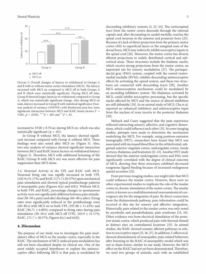
3.3. Latency. We measured pain response latency at 3 weeksafter SNI modeling in both Groups A and B. In Group A,the baseline mechanical latency was 9.44 ± 0.37 sec, andthe latency was significantly increased by MCS on, 15.37 ±0.89 sec, which was statistically significant (𝑝 < .05). InGroup B, the baseline latency was 15.96 ± 0.68 sec, and it
Neural Plasticity 5
0
10
20
30
Group A Group B
Late
ncy
to w
ithdr
aw (s
)
MCS offMCS on
∗∗
∗∗∗
∗∗∗
∗∗∗
Figure 5: Overall changes of latency to withdrawal in Groups Aand B with or without motor cortex stimulation (MCS).The latencyincreased with MCS on compared to MCS off in both Groups Aand B which were statistically significant. During MCS off state,Group B showed longer latencies to withdrawal compared to GroupA, which was statistically significant change. Also during MCS onstate, latency increased in Group B with statistical significance [two-way analysis of variance (ANOVA) with Bonferroni post hoc tests;significant interaction between MCS and RAIC lesion factors 𝐹 =5.081, 𝑝 = .0336]. ∗∗∗𝑝 < .001 and ∗∗𝑝 < .01.
increased to 19.00 ± 0.70 sec during MCS on, which was alsostatistically significant (𝑝 < .05).
In Group B without MCS, the latency showed signifi-cant increase compared with Group A without MCS. Thesefindings were also noted after MCS on (Figure 5). Also,two-way analysis of variance showed significant interactionbetweenMCS and RAIC lesion factors on withdrawal latency(Figure 5). Therefore, MCS with additional lesioning of theRAIC (Group B with MCS on) was more effective for painsuppression than MCS alone.
3.4. Neuronal Activity in the VPL and RAIC with MCS.Neuronal firing rate was rapidly increased in both VPL(245.9±51.17%) andRAIC (171.7±20.57%) uponmechanicalpain stimulation and showed typical postdischarge patternsof neuropathic pain (Figures 6(a) and 6(b)). Without MCS,in both VPL and RAIC, percentage changes in spontaneousactivity were not significantly different between pain stimula-tion (30–50 s; pain) and after discharge (60–80 s; after). Firingrates were significantly reduced in the postdischarge zone(60–80 s) with MCS on in both VPL (107.00 ± 11.43%) andRAIC (96.71 ± 6.00%) compared to firing rates during painstimulation (30–50 s) with MCS off (VPL: 245.9 ± 51.17%;RAIC: 171.7 ± 20.57%; Figures 6(c) and 6(d)).
4. Discussion
The purpose of our study was to investigate the pain mod-ulatory effect of MCS on the insular cortex, especially in theRAIC.Themechanism ofMCS-induced painmodulation hasstill not been elucidated despite its clinical use. One of themost widely accepted hypotheses to explain the antinoci-ceptive effect following MCS is that pain is modulated by
descending inhibitory systems [2, 22–24]. The corticospinaltract from the motor cortex descends through the internalcapsule and, after decussating in caudal medulla, reaches thespinal cord neurons in the anterior and posterior horn [25].Because of a lack of direct projection from the primarymotorcortex (M1) to superficial layers or the marginal zone of thedorsal horn,MCSmay indirectly inhibit nociceptive inputs inthe spinal cord [26]. Moreover, the motor cortex has diverseefferent projections to widely distributed cortical and sub-cortical areas. These structures include the thalamic nuclei,which receive strong projections from the motor cortex, animportant site for sensory modulation [27]. The periaque-ductal gray (PAG) system, coupled with the rostral ventro-medial medulla (RVM), exhibits descending antinociceptiveeffects by activating the opioid system, and these two struc-tures are connected with descending tracts [28]. AnotherMCS antinociception mechanism could be modulated byan ascending inhibitory system. The thalamus, activated byMCS, could inhibit nociceptive processing, but the specificnuclei affected by MCS and the source of altered inhibitionare still debatable [29]. In an animal study of MCS, Cha et al.reported an enhanced inhibitory and antinociceptive inputfrom the nucleus of zona incerta to the posterior thalamus[30].
Melzack and Casey suggested that the pain experiencereflected interacting sensory, affective, and cognitive dimen-sions, which could influence each other [31]. In some imagingstudies, attempts were made to determine the mechanismunderlying the MCS. For example, using positron-emissiontomography (PET), researchers have found that MCS wasassociatedwith increased blood flow in the orbitofrontal, sub-genual anterior cingulate cortex, midcingulate cortex, insulacortices, thalamus, and brainstem [11–13]. Another PET studyshowed that the anterior midcingulate cortex and PAG weresignificantly correlated with the degree of clinical outcomeof MCS, showing that these structures exhibited decreasedexogenous ligand binding because of increased endogenousopioid secretion [32].
Fromprevious imaging studies, onemight infer thatMCScould influence the insular cortex. However, there were noother experimental studies to implicate the role of the insularcortex in chronic stimulation of themotor cortex.The insularcortex is known as amultidimensional neuroanatomical con-vergence site for the integration of pain. By direct connectionfrom the thalamoinsula pathway, pain information could bereceived at this site for sensory and affective integration.Historically, pain related to the insular cortex was only notedby asymbolia and pseudothalamic pain syndrome [33, 34].Other evidence was from electrical stimulation of the poste-rior insula cortex, which produced pain with thermal stimuliin distinct sites on contralateral locations [35]. In animalstudies, the RAIC showed somatic afferent pathways in rela-tion to nociceptive input [15, 36, 37]. In addition, Coffeen et al.showed diminishment of neuropathic pain-related behaviorsafter lesioning in the RAIC of neuropathic model which wasnot in sham lesion, similar to our study. However, the MCSeffect on the RAIC had not yet been demonstrated.Therefore,we used two groups of animals, each with an established

6 Neural Plasticity
0
50
100
150
200
250
300
350
Pain After After + MCS
% ch
ange
in sp
onta
neou
s act
ivity
VPL
(c)
NS∗
∗∗
0
50
100
150
200
250
300
350
Pain After After + MCS
% ch
ange
in sp
onta
neou
s act
ivity
RAIC
(d)
NSNS
∗∗
(a)
(b)
0 10 20 30 40 50 60 70 80
(s)
200𝜇s
200𝜇s
Pain stimulation After discharge
MCSstimulation
artifact
MCSstimulation
artifact
Figure 6: Electrophysiological recordings showing firing rate changes upon mechanical stimulation with 300 g von Frey filament and motorcortex stimulation. Firing rate at the ventral posterolateral nucleus (VPL) markedly increased upon pain stimulation and the trend persistedafter discharge, but the postdischarge firing rate decreased when MCS was applied (a). Similarly, firing rate at the rostral agranular insularcortex (RAIC) markedly increased upon pain stimulation and the trend persisted after discharge, but the postdischarge firing rate decreasedwhen MCS was applied (b). Each VPL and each RAIC wave form are presented on the right. Percentage changes in spontaneous neuronalactivity, recorded from VPL (c) and RAIC (d), were decreased after MCS on state. And these changes were statistically significant comparedwith mechanical stimulation without MCS (𝑝 < .05). Statistical analysis was made using Friedman test followed by post hoc Dunn’s multiplecomparison test. ∗∗𝑝 < .01; ∗𝑝 < .05; NS = not statistically significant.
neuropathic pain model, either without RAIC lesioning(Group A) or with lesioning (Group B), and comparedtheir pain thresholds behaviorally and electrophysiologically.Upon mechanical stimulation, pain thresholds were signifi-cantly lower in Group A, which was what we expected fromprevious research. However, when we added MCS to bothgroups, Group B showed significant increase in thresholdcompared to Group A. These findings were also noted inpain latency from paw withdrawal tests. Therefore, we couldassume that RAIC has its own pain modulation effect, and
when adding MCS, the additional pain modulation effectcould be shown. But, in our electrophysiological study,the percentages of changes in spontaneous activity wereincreased in both VPL and RAIC after mechanical stimula-tion with 300 g von Frey filament. Compared to the MCS offstate, the changes of percentages after MCS on were notedin both regions (VPL and RAIC), which means the RAIC isalso influenced by MCS. However, the quantitative influenceof MCS on insular cortex is limited from our results. Andthough the RAIC has related to not only pain behavior but

Neural Plasticity 7
also anxiety behavior, we couldmake conclusion cautiously asabovewhenwe coupled it with our electrophysiological study.
In human studies, direct electrical stimulation of insularcortex during depth stereotactic EEG showed some degreeof somatotopic organization [38]. But, in animal studies, thesomatotopy related to pain process is not fully understood.Jasmin et al. showed a unique property of RAIC in that itcould respond as both analgesic and hyperalgesic responsesby selective modulation of GABA receptors [39]. The RAIChas multiple reciprocal connections with pain structures,such as orbital, infralimbic, and anterior cingulate cortices,rostroventral medulla, and periaqueductal gray matter [15].In addition, caudal granular insular cortex (CGIC), about4mm caudal to RAIC, is also known as having a role in longterm alleviation of allodynic pain modulation [40]. In thisstudy, we did not compare the MCS effect on both the RAICand the CGIC, so our findings have limitations on the infor-mation about MCS effect on the whole insular cortex. Addi-tionally, the centromedian/parafascicular (CM/Pf) nuclei,which receive dense projection from the motor cortex, wereinhibited byMCS, and these nuclei have interconnectionwiththe limbic system. Therefore, our electrophysiological resultscould be the result of direct response toMCS or from indirectthrough CM/pf nuclei [41]. To clarify the source of the effectof MCS on RAIC, we will need to block the CM/Pf effect,which could also demonstrate the amount of MCS effect onRAIC.
5. Conclusions
The results in this work suggest that the RAIC is influencedby MCS and that lesioning RAIC could produce more painreduction. Along with previous data, our findings may con-tribute to a better understanding ofMCS effect and the role ofRAIC in pain modulation.
Competing Interests
The authors report no conflict of interests.
Authors’ Contributions
Hyun Ho Jung and Jaewoo Shin contributed equally to thiswork as co-first authors.
Acknowledgments
This studywas supported by the grant fromCABMC(Controlof Animal Brain Using MEMS Chip) funded by DefenseAcquisition Program Administration (UD140069ID).
References
[1] C. J. Woolf and R. J. Mannion, “Neuropathic pain: aetiology,symptoms, mechanisms, and management,” The Lancet, vol.353, no. 9168, pp. 1959–1964, 1999.
[2] C. Benedetti, “Intraspinal analgesia: an historical overview,”Acta anaesthesiologica Scandinavica. Supplementum, vol. 85, pp.17–24, 1987.
[3] D. Bouhassira, N. Attal, J. Fermanian et al., “Development andvalidation of the neuropathic pain symptom inventory,” Pain,vol. 108, no. 3, pp. 248–257, 2004.
[4] N. B. Finnerup, M. Otto, H. J. McQuay, T. S. Jensen, and S. H.Sindrup, “Algorithm for neuropathic pain treatment: an evi-dence based proposal,” Pain, vol. 118, no. 3, pp. 289–305, 2005.
[5] T. Tsubokawa, Y. Katayama, T. Yamamoto, T. Hirayama, and S.Koyama, “Chronic motor cortex stimulation for the treatmentof central pain,” Acta Neurochirurgica Supplement (Wien), vol.52, pp. 137–139, 1991.
[6] D. Rasche, M. Ruppolt, C. Stippich, A. Unterberg, and V. M.Tronnier, “Motor cortex stimulation for long-term relief ofchronic neuropathic pain: a 10 year experience,” Pain, vol. 121,no. 1-2, pp. 43–52, 2006.
[7] G. Cruccu, T. Z. Aziz, L. Garcia-Larrea et al., “EFNS guidelineson neurostimulation therapy for neuropathic pain,” EuropeanJournal of Neurology, vol. 14, no. 9, pp. 952–970, 2007.
[8] Y. Lazorthes, J. C. Sol, S. Fowo, F. E. Roux, and J. C. Verdie,“Motor cortex stimulation for neuropathic pain,” Acta Neu-rochirurgica Supplement, vol. 97, part 2, pp. 37–44, 2007.
[9] Y. Saitoh and T. Yoshimine, “Stimulation of primary motor cor-tex for intractable deafferentation pain,” Acta Neurochirurgica,Supplementum, vol. 97, no. 2, pp. 51–56, 2007.
[10] J.-P.Nguyen, F.Velasco, P. Brugieres et al., “Treatment of chronicneuropathic pain by motor cortex stimulation: results of abicentric controlled crossover trial,”Brain Stimulation, vol. 1, no.2, pp. 89–96, 2008.
[11] L. Garcıa-Larrea, R. Peyron, P. Mertens et al., “Electrical stim-ulation of motor cortex for pain control: a combined PET-scanand electrophysiological study,” Pain, vol. 83, no. 2, pp. 259–273,1999.
[12] R. Peyron, B. Laurent, and L. Garcıa-Larrea, “Functional imag-ing of brain responses to pain. A review and meta-analysis(2000),” Neurophysiologie Clinique, vol. 30, no. 5, pp. 263–288,2000.
[13] R. Peyron, I. Faillenot, P. Mertens, B. Laurent, and L. Garcia-Larrea, “Motor cortex stimulation in neuropathic pain. Corre-lations between analgesic effect and hemodynamic changes inthe brain. A PET study,” NeuroImage, vol. 34, no. 1, pp. 310–321,2007.
[14] J. Kim, S. B. Ryu, S. E. Lee et al., “Motor cortex stimulationand neuropathic pain: how doesmotor cortex stimulation affectpain-signaling pathways?” Journal of Neurosurgery, vol. 124, no.3, pp. 866–876, 2016.
[15] L. Jasmin, A. Granato, and P. T. Ohara, “Rostral agranularinsular cortex and pain areas of the central nervous system: atract-tracing study in the rat,” Journal of ComparativeNeurology,vol. 468, no. 3, pp. 425–440, 2004.
[16] U. Coffeen, J. Manuel Ortega-Legaspi, F. J. Lopez-Munoz, K.Simon-Arceo, O. Jaimes, and F. Pellicer, “Insular cortex lesiondiminishes neuropathic and inflammatory pain-like behav-iours,” European Journal of Pain, vol. 15, no. 2, pp. 132–138, 2011.
[17] M. Zimmermann, “Ethical guidelines for investigations ofexperimental pain in conscious animals,” Pain, vol. 16, no. 2, pp.109–110, 1983.
[18] I. Decosterd and C. J. Woolf, “Spared nerve injury: an animalmodel of persistent peripheral neuropathic pain,” Pain, vol. 87,no. 2, pp. 149–158, 2000.
[19] S. E. Lee, S. B. Jun, H. J. Lee et al., “A flexible depth probeusing liquid crystal polymer,” IEEE Transactions on BiomedicalEngineering, vol. 59, no. 7, pp. 2085–2094, 2012.

8 Neural Plasticity
[20] K. S. Min, C. J. Lee, S. B. Jun et al., “A liquid crystal polymer-based neuromodulation system: an application on animalmodel of neuropathic pain,”Neuromodulation, vol. 17, no. 2, pp.160–169, 2014.
[21] S. R. Chaplan, F. W. Bach, J. W. Pogrel, J. M. Chung, and T. L.Yaksh, “Quantitative assessment of tactile allodynia in the ratpaw,” Journal of Neuroscience Methods, vol. 53, no. 1, pp. 55–63,1994.
[22] J. C. Yeung and T. A. Rudy, “Multiplicative interaction betweennarcotic agonisms expressed at spinal and supraspinal sites ofantinociceptive action as revealed by concurrent intrathecaland intracerebroventricular injections of morphine,” Journal ofPharmacology and ExperimentalTherapeutics, vol. 215, no. 3, pp.633–642, 1980.
[23] F. Porreca, H. I. Mosberg, J. R. Omnaas, T. F. Burks, and A.Cowan, “Supraspinal and spinal potency of selective opioid ago-nists in the mouse writhing test,” Journal of Pharmacology andExperimental Therapeutics, vol. 240, no. 3, pp. 890–894, 1987.
[24] J. Lipp, “Possible mechanisms of morphine analgesia,” ClinicalNeuropharmacology, vol. 14, no. 2, pp. 131–147, 1991.
[25] A. K. Senapati, P. J. Huntington, and Y. B. Peng, “Spinaldorsal horn neuron response tomechanical stimuli is decreasedby electrical stimulation of the primary motor cortex,” BrainResearch, vol. 1036, no. 1-2, pp. 173–179, 2005.
[26] S. K. Stanley, A. J. Ghanayem, L. I. Voronov et al., “Flexion-extension response of the thoracolumbar spine under compres-sive follower preload,” Spine, vol. 29, no. 22, pp. E510–E514, 2004.
[27] K. McAlonan and V. J. Brown, “The thalamic reticular nucleus:more than a sensory nucleus?” Neuroscientist, vol. 8, no. 4, pp.302–305, 2002.
[28] E. T. Fonoff, C. S. Dale, R. L. Pagano et al., “Antinociceptioninduced by epidural motor cortex stimulation in naive con-scious rats is mediated by the opioid system,” Behavioural BrainResearch, vol. 196, no. 1, pp. 63–70, 2009.
[29] R. Peyron, L. Garcia-Larrea, M. P. Deiber et al., “Electricalstimulation of precentral cortical area in the treatment of centralpain: electrophysiological and PET study,” Pain, vol. 62, no. 3,pp. 275–286, 1995.
[30] M. Cha, Y. Ji, and R. Masri, “Motor cortex stimulation activatesthe incertothalamic pathway in an animal model of spinal cordinjury,”The Journal of Pain, vol. 14, no. 3, pp. 260–269, 2013.
[31] R. Melzack and K. L. Casey, Sensory, Motivational, and CentralControl Determinants of Pain: A New Conceptual Model, 1968.
[32] J. Maarrawi, R. Peyron, P. Mertens et al., “Motor cortex stimula-tion for pain control induces changes in the endogenous opioidsystem,” Neurology, vol. 69, no. 9, pp. 827–834, 2007.
[33] J. D. Schmahmann andD. Leifer, “Parietal pseudothalamic painsyndrome. Clinical features and anatomic correlates,” Archivesof Neurology, vol. 49, no. 10, pp. 1032–1037, 1992.
[34] J. R. Augustine, “Circuitry and functional aspects of the insularlobe in primates including humans,” Brain Research Reviews,vol. 22, no. 3, pp. 229–244, 1996.
[35] K. Ostrowsky, M. Magnin, P. Ryvlin, J. Isnard, M. Guenot, andF. Mauguiere, “Representation of pain and somatic sensationin the human insula: a study of responses to direct electricalcortical stimulation,”Cerebral Cortex, vol. 12, no. 4, pp. 376–385,2002.
[36] U. Coffeen, A. Lopez-Avila, J. M. Ortega-Legaspi, R. del Angel,F. J. Lopez-Munoz, and F. Pellicer, “Dopamine receptors in theanterior insular cortex modulate long-term nociception in therat,” European Journal of Pain, vol. 12, no. 5, pp. 535–543, 2008.
[37] P. Alvarez, W. Dieb, A. Hafidi, D. L. Voisin, and R. Dallel,“Insular cortex representation of dynamicmechanical allodyniain trigeminal neuropathic rats,”Neurobiology of Disease, vol. 33,no. 1, pp. 89–95, 2009.
[38] L. Mazzola, J. Isnard, R. Peyron, M. Guenot, and F. Mauguiere,“Somatotopic organization of pain responses to direct electricalstimulation of the human insular cortex,” Pain, vol. 146, no. 1-2,pp. 99–104, 2009.
[39] L. Jasmin, S. D. Rabkin, A. Granato, A. Boudah, and P. T. Ohara,“Analgesia and hyperalgesia from GABA-mediated modulationof the cerebral cortex,” Nature, vol. 424, no. 6946, pp. 316–320,2003.
[40] A. M. Benison, S. Chumachenko, J. A. Harrison et al., “Caudalgranular insular cortex is sufficient and necessary for the long-term maintenance of allodynic behavior in the rat attributableto mononeuropathy,”The Journal of Neuroscience, vol. 31, no. 17,pp. 6317–6328, 2011.
[41] R. L. Pagano, E. T. Fonoff, C. S. Dale, G. Ballester, M. J. Teixeira,and L. R. G. Britto, “Motor cortex stimulation inhibits thalamicsensory neurons and enhances activity of PAG neurons: pos-sible pathways for antinociception,” Pain, vol. 153, no. 12, pp.2359–2369, 2012.