s deficiency type i: identification of point mutations in
TRANSCRIPT

Protein S Deficiency Type I: Identification of Point Mutations in 9 of l 0 Families
By Stefan Mustafa, Ingrid Pabinger, and Christine Mannhalter
We identified potentially causative mutations in the active protein S gene (PROS 1) by direct sequencing of PROS 1- specific polymerase chain reaction (PCR) products of all 15 exons, including exon-intron boundaries in 10 families with hereditary protein S deficiency type 1. Seven different muta- tions were found in 9 of 10 families, including one frame shift mutation, a previously published splice site mutation (both occurring in two unrelated families), four missense mutations, and a stop codon at the beginning of exon 12. In family studies, cosegregation of the mutation with the disease could be demonstrated for five mutations; for two
P ROTEIN C and its cofactor, protein S, play an important role in the anticoagulant system."' Protein C, a vitamin
K-dependent glycoprotein, is activated by the thrombin- thrombomodulin complex to activated protein C (APC), which, in the presence of its cofactor, protein S, selectively inhibits the procoagulant cofactors factor VIIIa and factor Va, thereby downregulating the coagulation cascade.' Pro- tein S, also a vitamin K-dependent glycoprotein, is synthe- sized in the liver: endothelial cells,s and a number of other tissues. Its mRNA is also found in platelets.6 Protein S circu- lates in plasma in part as complex (60%) with C4b-binding protein (C4BP) and in part as unbound, free form (40%).7 It has been shown that only free protein S is functionally active in the protein C anticoagulant pathway.'
The concentration of protein S in plasma is usually deter- mined by measurements of antigen (free or total protein S) and/or activity. Most cases of protein S-deficiency are classified as type I deficiency with reduced levels of antigen and activity.' Few reports describe cases of type I1 (normal total and free antigen, reduced activity)'0,'' and type 111 (nor- mal total antigen, reduced free antigen and activity)I2 defects (type attribution according to SSC Subcommittee Meeting, Munich, Germany, 1993).
Homozygous protein C or protein S deficiency can lead to life-threatening thrombotic complications shortly after birth.I3 Heterozygous individuals have a significant risk to develop venous thrombosis, but at a later age and usually with milder Using the currently available laboratory test systems, it is sometimes difficult to identify
From the Department of Clinical Chemistry and Laboratory Medi- cine, Molecular Biology Division, and Internal Medicine I , Depart- ment of Hematology and Hemostaseology, University Vienna Medi- cal School, Vienna, Austria.
Submitted February 21, 1995; accepted June 29, 1995. Supported by Grant No. PO9812-MED from the "Fond zur Forde-
rung der Wissenschaftlichen Forschung in Osterreich. " Address reprint requests to Christine Mannhalter, PhD, KIMCL-
AKH, Leitstelle 5H, Abteilung Molekularbiologie, Wahringergiirtel 18-20, A-1090 Wien, Austria.
The publication costs of this article were defrayed in part by page charge payment. This article must therefore be hereby marked "advertisement" in accordance with 18 U.S.C. section 1734 solely to indicate this fact. 0 1995 by The American Society of Hematology. 0006-4971/95/8609-0013$3.00/0
3444
missense mutations, this was not possible due to limited family data. All seven mutations were the only abnormalities identified in the respective index patients and were absent in 44 to 62 normal individuals. Therefore, they most likely represent the causal gene defects. For five mutations, analy- sis of ectopic RNA could be performed. Mutant transcripts were present in the case of the frame shift and three of the missense mutations, while no mutant RNA could be de- tected in the case of the stop codon. 0 1995 by The American Society of Hematology.
heterozygous protein S-deficient individuals. All test systems show a considerable overlap between protein S plasma levels found in normal individuals and in deficient heterozygotes. Especially in the case of oral anticoagulation, the diagnostic uncertainty with conventional test systems is very high. Only a genetic approach that identifies the causative mutations in protein S-deficient individuals can bypass these diagnostic pitfalls.
Protein S is encoded by the active PROS 1 gene on chro- mosome 3, and an inactive pseudogene, PROS 2, also exists on chromosome 3.lh.I7 PROS 2, which is not transcribed, shows a homology of 97% with PROS 1, but differs by the absence of exon 1 and the presence of stop codons within the gene."-'" It can be expected that mutations causing protein S defects will be located within the PROS 1 gene. To date, two major deletions2'.'' and 21 point mutations".23-2h in the PROS 1 gene have been reported as putatively causal muta- tions in protein S deficiency. Analysis of PROS 1 mRNA was shown to be unsatisfactory to search for mutations in protein S-deficient families, as gene defects interfering with mRNA synthesis and/or processing remained undetected.' In contrast, the screening of the 15 individual exons and the exonhntron boundaries of the PROS 1 gene allows identifi- cation of all genomic mutations in the coding region and the splice junction sites.
Using this approach, we identified mutations in 9 of I O individuals with protein S deficiency. The nine individuals shared seven different mutations; two mutations occurred in two subjects each. Interestingly, one of these mutations (a splice site mutation at position +5 of the donor splice site of intron 10) has previously been reported in two unrelated, protein S-deficient patients of Dutch origin.27 Whether this splice site mutation represents a mutational hot spot remains to be determined.
PATIENTS AND METHODS
Patients. The protein S-deficient patients selected for the study were identified during the investigation of individuals with a history of objectively documented thrombosis and/or pulmonary embolism. Individuals who had developed thrombosis early in life (before 45 years of age) or who had a positive family history (around 30% of all investigated patients) were screened for protein S deficiency by determination of free protein S antigen (Table I). In patients found to he protein S-deficient, as well as in their relatives, free and total protein S values were measured. Between 1985 and 1994, 19 families with protein S deficiency type I were identified. From the 19 index
Blood, Vol 86, No 9 (November l), 1995: pp 3444-3451
For personal use only.on April 11, 2019. by guest www.bloodjournal.orgFrom

POINT MUTATIONS IN PROTEIN S DEFICIENCY 3445
Table 1. Clinical Characteristics and Protein S Values of Protein S-Deficient Individuals and Their Relatives
Age at Last
Patient Free Protein Total Protein Heterozygous Age at First Recurrent Examination Code No. S (%) S (%l OAC Mutation Thrombosist Thrombosis (yr) Thrombosis (yr)
1-111 15 40 + + 22 + 68
1-11/2 14 37 + + 21 + 40
-
1-11/1 86 - 86 - - - - 46 -
1 -l 113 130 - 118 - - - 43 45
1-11/4* 7 26 + + + 29 + 39 1-111/1 90 - 17
1-111/2 15 40 - + 15
1-11113 76 88 12
2-111 2 29 + + + 21 + 53 2-112 7 48 - + + 19 + 44 2-1/3* 3 31 + + + 37 - 51
2-1111 108 - 90 31
2-1112 10 42 - + 20 2-1113 74 80 - 26 3-111 * 4 81 + + 20 + 44
- 24
- 80 - - -
- - -
- - - - -
- - - - - - -
- - - - -
3-1111 7 54 - + + 27 4-111 * 2 8 + + + 32 + 54 4-1111 48 107 - - - - - 23 5-111 * 1 18 + ? + 18 + 64 6-111 * 2 30 + + + 25 + 43 6-112 84 90 - - 6-11/1 16 64 - + -
6-1112 18 56 - + -
- - - 40 11 14
7-111 * 30 86 - + + 18 + 55 7-1111 38 59 - + - 22 8-111 16 59 - + + 21 + 75
- -
- -
- -
8-1111 11 57 - + + 46 - 54 8-1112 100 92 - - 57 8-1113 9 47 - + + 20 + 52 84lI/l* 2 26 + + + 16 + 23
- - -
8-111/2 20 61 - + + 19 - 24 9-111 * 2 26 + + + 21 + 40 9-112 14 84 - + + 26 - 34
10" 4 29 + + + 23 + 69 10-1/2* 4 23 + + + 57 + 67 10-11/1 100 96 - - - - - 46 1 0-1 1/2 122 116 - - - - - 44 1 0-1 113 23 49 - + - - - 37
Abbreviation: ?, mutation not identified. * Index patient. t Thromboses observed in patients were pulmonary embolism and/or deep venous thrombosis, one cerebral venous thrombosis (14/4), one
mesenteric vein thrombosis (9-l11), and one cava vein thrombosis (5.111).
patients, a panel of 10 probands was selected for the molecular biologic investigations. All index patients had a positive family his- tory for thromboembolism. Except patient 9 4 2 , who is an Argentin- ean of European origin, and patient 5-I/1, who is German, all index patients are Austrians.
Protein S determination and diagnostic criteria. Free and total protein S antigen were measured by a commercial enzyme-linked immunosorbent assay (Boehringer-Mannheim, Mannheim, Ger- many). For determination of free protein S, bound protein S was precipitated with polyethyleneglycol (PEG 8000; 3.75%) according to the method of Comp et al,'* and free protein S was determined in the supernatant. The amount of free protein S in PEG-precipitated pooled normal plasma was defined as 100%.
The lower limit of the normal range for free protein S was 64% in patients without oral anticoagulant (OAC) treatment and 19% for patients on OAC. For total protein S, the lower limit of the normal
range was 65% in patients without and 35% in patients with OAC treatment.
The diagnosis of hereditary protein S deficiency type I was consid- ered as established when decreased free and total protein S values were repeatedly found in a proband and when protein S deficiency was detected in at least one further relative, irrespective of the throm- botic history in that individual. Using the above-mentioned cut-off limits, in three families (families 3, 7, and 9). individuals with de- creased free but normal total protein S values (patients 3-U1, 7-I/1, and 9 4 2 ) were found in addition to the typical type I-deficient patients. Families in which all deficient family members had type 111 deficiency were not included in our study.
Genomic DNA analyses. Genomic DNA was extracted from EDTA or citrated blood samples (5 mL) according to standard proce- dures. For the identification of mutations, we concentrated on the analysis of the transcribed regions of the PROS 1 gene. Polymerase
For personal use only.on April 11, 2019. by guest www.bloodjournal.orgFrom

7
8
9
10
11
12
13
14
15A
3446 MUSTAFA, PABINGER, AND MANNHALTER
Table 2. Nuclsotide Sequences of the Primers Used for PCR Amplification and Sequencing
Exon Primer Primer Sequence (5'-3') Location*
1 PS 0/4t§ 5"GAA GAA GGA TGT CTC AGC AGT-3'
2 PS 211 t§ 5"CAT ATA AAC TGA TTG TTT CC-?
3 PS 4/4t 5 5 " m AGG TTT GCT AAG ATA TG-3' PS 4/5t§ PS 411 t § 5"ACC TCT TGG GAC AGT TCC TA-3' PS 5/2t PS 611 t § 5 " W TAT TTT TCC ATG ACA TGA GA-3' PS 6/25
PS 1/4t§ -181 to -161
5"TCT AGG AGC TGC AGC TCT AG-3' +81 to +l00
PS 311 t 5 -25 to -6
5"TAG TTG TAT GTC TAG ATG TG-3' +30 to +49 -41 to -22 +96 to +l15 -83 to -64 +35 to +57 -56 to -34 -82 to -68
5"TCC CAA GGA TAA TGA AAT TA-3' 4
5"GTA CTT TAC CTA CAG AGT TTT TG-3' 5 and 6
5"CCA GCT CAT CAG GAT-3' PS 7/2t 5"AAC TGG GAT TAT TCT CAC AT-3' PS 8/6t §
PS 812 t § 5"GAT CAG TAA TGA TAC CAC CA-3' PS 9/8t §
PS 9/9t§ 5"CTC ATA ACC TGC TTA AAG CTA-3' PS 9/4t §
PS 10/2t§ 5"CCT TAT CTG CTT AAC CTC TA-3' PS 21/ l t PS 2211 t 5 5"TCC ATT TTG G l T TGG TAT CA-3' PS 10/1* PS 22/2t§ 5"GAC ATT CCA AAT GAG TTG TAA-3' PS 12/2t PS 12/1* 5"GAG TTC ACT TTC CAC TTT CC-3' e1441 to 1460 PS 17/ l t 5"CCT ATA CTC ATA ATC GAG CC-3' -69 to -50
-59 to -45
+37 to +56 5"TAG ATT TAA TGT TTT TGG TCC A-3' -145 to -124
+ l 0 to +29 5"TAG TTG ATG TCA TAG TAT TCT T-3' -139 to -118
+l14 to +l34 5"CAT TAG TAA CCA AAC AAA AAT G-3' -100 to +79
+30 to +49 5"GCT TTC TGT ATT TCT TAC TC-3' -49 to -30
+74 to +93 5"AGA AAT CAG CAG ATT TTC AG-3' 1100 to 1119
-158 to -138 5"GAT CAT TTC AAG TTG TCA CTC-3' +l13 to +l33
PS 17/25 5"TAA TCG AGC CAC TGT-3' PS 13/l t PS 24St 5"GAT CAT TGA GAA AGG GAA TGG-Y PS 14/ l t§
5"TGG GCA CAC AGT AGA TAC TC-3' +91 to +l10 -135 to -115
5"TGC TGG AGA TTG TGC CAA AC-3' + l21 to +l40 PS EXIV-It§ 5"AAA AAC TCA AAA GTC ACT C-3' PS EXIV-2t§
-75 to -57 5"AAT AAA TGT CGG TAC TAG CC-3' + l31 to +l50
PS e14t 5"TAC TGT AAT ATA TCG GAT-3' e1814 to 1833 PS 18/3* 5"ATA GAA ACC ATC TCC CAT GA-3' e1920 to 1939 PS 19/l t§ 5"GGA TTA GAA TTT GGT TGG AAA C-3' - 106 to -85 PS 16/25 5"AGA GCT CAC TCA TGT-3' e2130 to 2144 PS 1611 5 5"GTC AAG GAA AAA AAT TCT GTT GTG AT-3' e2318 to 2343 PS 2011 5 5"CAC TAT TCT TAG ATA GCA AGA GAA GT-3' e2368 to 2393 PS 20125 5"GAT ATC TCA TCC TGA-3' e2827 to 2841 PS 20136 5"TTG TGT GGC TTC ACA-3' e2720 to 2734 PS-PlBtt 5"TGC TGC TCT CAG GAA AAT A-3' e2777 to 2795 PS 25ASt5 5"GTG GCA ATC TTA CCT CCT TA-3' e3230 to 3149
Locations of primers are indicated by the numbers of the first and the last nucleotide; the - or + placed in front gives locations upstream or downstream of the respective exon. Numbers following an e are cDNA positions according to Hoskins et a134 characterizing nucleotides located within exons.
t Primer for amplification of genomic sequences. * Primer for cDNA amplification. § Primer for sequencing.
chain reaction (PCR)" products were sequenced directly by the method of Sanger?' Primers chosen for PCR amplification (of exons, including exon-intron boundaries; Table 2) were constructed from genomic sequences using differences between the PROS 1 and the PROS 2 genes."-" They were positioned such that the 3' end of at least one of the two primers was only complementary to the PROS I gene. As no sequence differences between PROS 1 and PROS 2 were known for exon 3, we amplified this exon and large parts of boundering introns with primers presumed to be common to both genes. The PCR product (340 bp for PROS 1) was sequenced, and the differences found between the known PROS 1 gene sequence and the pseudogene sequence were used to design PROS l-specific primers PS-414 and P S 4 5 .
All amplification products included at least 10 nucleotides of 5'
and 3' flanking intron sequences, except for only five nucleotides at the 5' splice junction of exon 2 and only nine nucleotides at the 3' splice junction of exon 7. Amplifications were performed in a DNA Thermal Cycler (Perkin-Elmer Cetus, Norwalk, CT) using Taq- DNA-polymerase (Roche Molecular Systems, Inc. Branchburg, NJ). The final concentrations of ingredients (for exons 2 to 15) were 0.2 pmoVL each primer; 0.2 mmol/L each deoxynucleotide triphosphate (W); 10 mmol/L Tris-HC1 (pH 8.3 at 25°C); 50 mmol/L KCl; MgC12 at 1.5 mmollL (for exons 2, 4, 5 + 6, 11, 13 + 14, 14). 2.0 mmol/L (for exons 9 and 13), 2.5 mmol/L (for exons 7, 8, 10, 15), or 3.0 mmol/L (for exons 3 and 12); 1.25 U Taq-DNA-polymerase; and approximately 500 ng of genomic DNA.
Exon I was amplified in a buffer system containing dimethylsulf- oxide (DMSO) and (NH4)2S04?3 After a denaturation step of 3
For personal use only.on April 11, 2019. by guest www.bloodjournal.orgFrom

POINT MUTATIONS IN PROTEIN S DEFICIENCY 3447
Table 3. Molecular Defects Identified in Nine Patients With Type I Protein S Deficiency
No. of [affected] cDNA Family Memberdin Mutant Restriction
Mutation Position* Generations Cosegregation RNA Site Comment
mut-l: ACAAAG + ACAG 2166, 2167 fam-1: 8I4l/3 Yes Yes 2-bp deletion fam-4 2[11/2 Change of reading frame 3 codons before
stop codon, use of new stop codon. The last 3 amino acids (KNS) are replaced by 23 amino acids (QFLRHLFSAYNTFSLCVIILMFQ)
mut-2: GTACGT + GTACAT lntronic fam-2: 6[41/2 Yes N D Maell Splice site mutation in splice donor of intron 10
fam-3: 2[21/2 mut-3: GTGGCC + GTGCCC 1992 fam-6: 4[31/2 Yes Yes Eae I Ala 575 + Pro mut-4 AATCTG 4 AATCAG 1897 fam-7: 2[21/2 - Yes Bpm I Leu 543+Gln mut-5: CTGTTA + CTGTCA 1801 fam-8: 6[51/3 Yes Yes Leu 51 1 + Ser mut-6 TCCTGA + TCCCGA 1198 fam-9: 2[21/1 - N D Leu 310 + Pro mut-7: ATACGA + ATATGA 1497 fam-10: 5[31/2 Yes No Acc I Arg 410 4 STOP also present in the
protein S pseudogene (PROS 2)
Abbreviations: -, unknown; ND, not done. * cDNA positions according to Hoskins et al."
minutes at 93T , the 50-pL reaction mixtures were exposed to 35 cycles of 1 minute denaturing at 93°C; 1 minute annealing at 45°C (exons 8, 12, and 14), 50°C (exons 2,3, 10, and 13), or 55°C (exons 1, 4, 5 + 6, 7, 9, 11, and 15); and 2 minutes extension at 72°C. A final extension step at 72°C for 10 minutes assured completion of synthesis. With primers PS-24s and PS-EXIV-2 (exon 13 + 14). 10 cycles with an annealing temperature of 60°C were followed by 30 cycles at 50°C.
Because the pseudogene sequence at the 5' end of intron 14 was not available, and intron 13 sequences were not appropriate to choose a PROS 1 gene-specific primer, 0.5-pL PCR products (containing parts of intron 12, exon 13, intron 13, exon 14, and parts of intron 14) obtained with primers PS-24s and PS-EXIV-2 were submitted to 12 cycles of seminested amplification using primers PS-EXIV-1
Purification of PCR products was performed on low melting point agarose. Gel slices were melted (70Q shock-frozen (-7O"C), and centrifuged (12,OOOg) after thawing.29 Two to five microliters of the supernatant and 10 to 30 pmol primer were used for sequencing with Sequenase v 2.0 (US Biochemical, Cleveland, OH) and [a- '5S]-deoxyadenosine triphosphate (dATP; Amersham, Amersham, UK).
Index patients were sequenced in all exons. Any detected mutation was confirmed by analysis of the respective exon in key relatives.
cDNA preparation. Total RNA was isolated from 2 mL whole blood using a commercial kit (Fastube RNA Extraction System, ViennaLab, Vienna, Austria), based on a guanidinium phenol
and PS-EXIV-2.
method.30 Half the RNA obtained by one isolation was used for random-hexamer primed cDNA synthesis with M-MLV reverse tran- scriptase."
Preparation of PCR products for digestion with Acc l, Bpm l, BstXI, and Eae I. Primers (Table 2) for amplification of cDNA were constructed from exonic sequences of the PROS l gene. The primer pairs were located in adjacent exons, separated by an intron, to rule out amplification of genomic sequences.
Amplifications of cDNA with primers PS-18/3 and PS-P1B (1 pL cDNA, 40 cycles, annealing at 59°C) as well as of genomic DNA with PS-19/1 and PS-P1B (500 ng DNA, 35 cycles, annealing at 55°C) were performed with 0.8 pmoVL each primer, 0.4 mmoVL each d m , 5 mmoVL Tris-HC1 (pH 8.0 at 37°C). 50 mmoVL NaCI, 2.5 mmoVL MgCI2, 1 mmoVL 2-mercaptoethanol, and 1.25 U Taq- DNA-polymerase.
The buffer used for amplification of cDNA with primers 10/1 and 1511 (1 pL cDNA, seven cycles annealing at 62°C followed by 33 cycles at 50°C; 0.4 p m o m each primer, 0.4 mmoVL each dNTP, 2.5 U Taq-DNA-polymerase) as well as of genomic DNA with prim- ers PS-el4 and PS-EXIV-2 and primers PS-13/1 and PS-17/1 (500 ng DNA, 35 cycles, annealing at 55°C; 0.2 pmoVL each primer, 0.2 mmom each dNTP, 1.25 U Taq-DNA-polymerase) contained 16.5 mmol/L Tris-acetate (pH 7.9 at 37°C). 33 mmoVL K-acetate, 5 mmoVL Mg-acetate, and 0.5 mmoVL dithiotreitol. The respective enzyme (ACC I, 10 U; BstXI, 5 U; Eae I, 10 U [all Boehringer Mannheim, Mannheim, Germany]; and Bpm I, 4 U [New England BioLabs, Beverly, MA]) was added to 10 pL of unpurified PCR
. . . . : : ... 9 3 .. . 10 M 11 H 12 H 13 H 14 H 15
: .
+ + + + I 8 2 7 5 4 8 1
Fig 1. Diagram of the second half of the PROS 1 gene. Exons are symbolized by rectangles in scale, introns by connecting lines. The upper part of the graph shows the position of the primers used for amplification of cDNA. Bold numbers in the lower part of the figure give the location of the respective mutation.
For personal use only.on April 11, 2019. by guest www.bloodjournal.orgFrom
3448 MUSTAFA, PABINGER, AND MANNHALTER
I TI.
fam- I fam-2
fam-3 fam-4
fam-7
I1 = fam-6
I 2
fam-l0
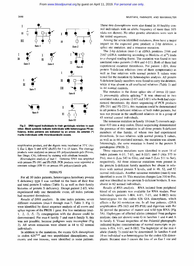
Fig 2. DNA-typed individuals in their geniologic relation to each other. Black symbols indicate individuals with heterozygous PS-de- ficiency. index patients are indicated by an arrow. An asterisk (*l marks individuals with thromboembolic events.
amplification product, and the digests were incubated at 37°C (Acc I, Eae I, Bpm I) and 45°C (BsrXI) for 3 to 15 hours. The cleavage products were analyzed on precast 6% polyacrylamide gels (Novex, San Diego, CA), followed by staining with ethidium bromide.
Heteroduplex analysis of mut-I. Genomic DNA was amplified with primers PS-19/1 and PS-P1B. PCR products were separated at constant voltage (100 V) on precast 6% polyacrylamide gels.
RESULTS
For all 10 index patients, heterozygous hereditary protein S deficiency type I was verified on the basis of their free and total protein S values (Table l), as well as their family histories of protein S deficiency. Except patient 2 4 3 , who experienced only one thrombotic event, all index patients had recurrent thrombosis.
Results of DNA analysis. In nine index patients, seven different mutations (mut-l through mut-7; Table 3, Fig l ) were identified by direct sequence analysis of all exons and splice regions of the PROS 1 gene. For five mutations (mut- 1, -2, -3, -5, -7), cosegregation with the disease could be demonstrated. For mut-4 family 7 and mut-6 family 9, this was not possible, because pedigrees were incomplete (Fig 2). All seven mutations were absent in 44 to 62 normal individuals.
In addition to the mutations, the exonic G/A dimorphism in codon 62632,33 and two unpublished dimorphisms, one exonic and one intronic, were identified in some patients.
These two dimorphisms were also found in 30 healthy con- trol individuals with an allelic frequency of more than 10% (data not shown). No other genetic alterations were seen in the tested sequences.
Among the seven identified mutations, three have a major impact on the expected gene product: a 2-bp deletion. a splice site mutation, and a nonsense mutation.
The 2-bp deletion (mut-l) at cDNA positions 2166 and 2167 (cDNA numbering according to Hoskins et al”) leads to a changed reading frame. The mutation was found in two unrelated index patients (1-1V4 and 4-VI). Both of them had experienced recurrent thrombosis. For patient 1 -W4, three protein S-deficient relatives (two of them symptomatic) as well as four relatives with normal protein S values were tested for the mutation by heteroduplex analysis. All protein S-deficient family members were found to carry the deletion, while it was absent in all unaffected relatives (Table 1) and in 44 normal controls.
The mutation in the donor splice site of intron 10 (mut- 2) presumably affects splicing.” It was observed in two unrelated index patients (2-1/3 and 3 4 1 ) who both had expe- rienced thrombosis. By direct sequencing of PCR products (PS-21/1 and PS-22/1), this mutation could be demonstrated in all protein S-deficient relatives of both index patients, but was not present in the unaffected relatives or in a group of 45 normal control individuals.
The nonsense mutation in family I O (mut-7) converts argi- nine-410 into a stop codon. Direct sequencing demonstrated the presence of this mutation in all three protein S-deficient members of this family, of whom two had experienced thrombosis. In two relatives with normal protein S values, as well as in 44 unrelated normals, the mutation was absent. Interestingly, the same mutation is found in the protein S pseudogene (PROS 2).
Three missense mutations were identified in exon 14 of index patients 6-V1, 7-1/1, and 8-IIVI: mut-3 (Ala 575 to Pro), mut-4 (Leu 543 to Gln), and mut-5 (Leu 51 1 to Ser), respectively. All three missense mutations were present in the protein S-deficient family members but absent in rela- tives with normal protein S levels, and in 46, 52, and 52 normal individuals. Another missense mutation (mut-6) was identified in exon 10. This mutation changes Leu 310 to Pro, and was identified in two protein S-deficient brothers. It was absent in 62 normal individuals.
Results of RNA analysis. RNA isolated from peripheral blood of six patients was available for RNA studies. Four individuals (patients LW4, 4-Y1, 7 4 1 , and 8-IIV2) were heterozygous for the codon 626 G/A dimorphism, which affects a Bst X1 restriction site. In all four patients, cDNA amplification (PS-18/3 and PS-PlB) and digestion with Bst XI proved the presence of mutant and wild type RNA (Fig 3A). Haplotypes of affected alleles (obtained from pedigree analysis; data not shown) were G in families I and 4 and A in family 8. Visual inspection of the Polaroid photographs indicated higher concentrations of normal transcripts in pa- tients l -11/4, 4-1/1, and 8-111/2. The haplotype of the mut-4 allele (family 7) could not be determined. In families 6 and 10 no individual was heterozygous for the codon 626 dimor- phism. Because mut-3 causes the loss of an Eue I site and
For personal use only.on April 11, 2019. by guest www.bloodjournal.orgFrom

POINT MUTATIONS IN PROTEIN S DEFICIENCY 3449
Fig 3. (A) Ratio of normal and mutant protein S transcripts in four deficient patients. BsrXl digests of cDNA amplification products (PS-18/3 and PS-PlB), containing the codon 626 GIA-dimorphism. Normal and mutant alleles are present in patient 1-11/4 (lane l), patient 441 (lane 2). patient 7 4 1 (lane 3). and patient 8-11112 (lane 4). The smaller band in patients 1-11/4 and 4 4 1 and the larger band in patient 8-111/2 represent the normal allele (haplotypes constructed from family studies). In all three patients the normal allele prevailed. For 7 4 1 the haplotype is unknown. The left and the right lanes show pBR322-Msp I marker bands at 622 bp and 527 bp. IBI RNA analysis in patient 6-1/1. Digestion of cDNA PCR products, obtained with primers PS-18/3 and PS-PlB, by Eae I gives a band at 876 bp in case of the wild-type sequence and a band at 803 bp in the presence of mut-3. Middle lane, patient 6-111 with bands at 876 and 803 bp. Right lane, normal control. Left lane, pBR322-Msp I marker. (C) RNA analysis in patient 10-1/2. Acc I digestion of cDNA PCR products generated with primers PS-10/1 and PS-15AS. Bands at 412, 322, and 73 bp represent the normal transcript. Mut-7 would result in a loss of an Acc I cleavage site and the generation of bands at 485 and 322 bp. Left lane, patient 10-1/2. Right lane, pBR322-Msp I marker. All digests were analyzed on 6% precast polyacrylamide gels.
mut-7, the loss of an Acc I site, restriction enzyme cleavage mented family history of protein S deficiency and thrombo- could be used for the detection of mutant transcripts. After embolism. Seven different, putatively causal mutations were digestion with &e 1. uncleaved (876 bp) mutant and cleaved identified in 9 of 10 unrelated patients with hereditary protein (803 bp + 73 bp) wild type Sequences could be Seen in equal S deficiency. Major evidence for pathogenicity of these mu- amounts in the cDNA PCR product of patient 6-1/1 (Fig 3R). tations was their at-,sence i n a group of normal controls (44 Cleavage or the respective cDNA PCR product (ps-I0/1 and to 62 individuals). ,411 mutations were also the only genetic PS-15-AS) from patient 10-1/2 with Act* l resu1ted i n the abnormalities found in the investigated sequences of the pro- generation of three bands with 412, 322, and 73 bp, repre- senting wild type. No mutant transcript (expected bands at 485, 322 bp) was present (Fig 3C).
tein S-deficient individuals. Pedigree analysis was performed to distinguish these putatively causal mutations from DNA alterations not linked to protein S deficiency. For five muta- tions (mut-l. -2. -3. -5. and -7). the transmission within
We established a useful protocol for the detection of gene sufficiently large families could be studied, and all five muta-
DISCUSSION
alterations in the PROS I gene in patients with a well-docu- tions cosegregated with the disease.
For personal use only.on April 11, 2019. by guest www.bloodjournal.orgFrom

3450 MUSTAFA, PABINGER, AND MANNHALTER
The causality of the 2-bp deletion (mut-l) and the splice site mutation (mut-2) for protein S deficiency is supported by the occurrence of each mutation in two different, reportedly unrelated protein S-deficient families. Mut-2 was also identi- fied in two of eight genetically investigated Dutch families with protein S deficiency.” Whether this high frequency of mut-2 is due to the presence of a mutational hot spot remains to be determined.
The deletion of two adenines in codon 633 leads to the use of a new reading frame. The normal stop codon is skipped and a new stop codon 62 bases downstream comes in frame. The new sequence codes for 23 amino acids (QFLRHLFSAYNTFSLCVIILMFQ), including one cys- teine, instead of only three amino acids (KNS) in individuals with normal protein S. As we could demonstrate transcrip- tion of mutant RNA, it is feasible that an aberrant protein is translated from this mutant RNA. The secretion of this protein would presumably be impaired due to interference with proper folding of one unpaired cysteine contained in the extra amino acid ~equence.’~~’~
Mutation mut-7 introduces a stop codon at the beginning of exon 12. Aberrant RNA is absent from ectopic transcripts in blood cells, and a truncated gene product in protein S synthesizing cells is unlikely.
Mutations mut-3 and mut-6 lead to the replacement of alanine-575 and leucine-3 10, respectively, by proline, which is known to interrupt the formation of any regular repeating structure. This could destabilize the conformation of protein S, making it vulnerable against degradation or retaining it from secretion."^" In a secondary structure model of protein S reported by Schmidel et all8 and calculated according to Chou and Fasman,” proline-575 would lie in the region of a conversion of an a-helical into an irregular structure. Using the same model, proline-310 would disrupt a region of pre- dicted p sheet structure and would, therefore, significantly impair the secondary structure.
Common to mut-4 and mut-5 is the change of leucine, a hydrophobic amino acid, into a polar amino acid: glutamine and serine, respectively. Leucine-543 (mut-4) is conserved in human, bovine,38 mouse,’’ and rabbit4’ protein S amino acid sequences. At position S1 1 (mut-S), all three nonhuman protein S sequences carry hydrophobic valine. Apparently, there is a demand for branched, aliphatic amino acids at position 543 and 5 1 1, as evidenced by the high conservation during evolution.
We could detect transcripts of the mutant allele in four of five different mutations when we analyzed cDNA generated from total RNA. No mutant transcript was found in the case of the stop codon. In the presence of the 2-bp deletion and the Leu-51 1 to Ser mutation, the concentration of mutant transcripts was lower than of wild type. Our results indicate that RNA analysis is not suitable for the detection of all mutations in the PROS 1 gene, such as Arg-410 to stop, and only DNA analysis is adequate.6
Interestingly, a high frequency of missense mutations (four of seven) was observed. Similar results have already been reported for gene alterations in hereditary protein C deficiency, in which more than half of all mutations are also missense mutations.4’
The identification of mutations in the PROS I gene not only contributes to the understanding of pathogenic muta- tions, but helps to clarify diagnostic uncertainties. I t is well known that an overlap of free and total protein S values exists in deficient patients and normal controls. We observed slightly reduced free and normal total protein S values in individual 4-1UI (daughter of a protein S-deficient father), and her protein S deficiency status was unclear. Gene analy- sis could redress uncertainties by demonstrating absence of the PROS 1 gene mutation in this individual. Thus, our study also shows the value of gene analysis for diagnostic applications.
REFERENCES
1. Davie EW, Fujikawa K, Kiesil W: The coagulation cascade: Initiation, maintenance, and regulation. Biochemistry 30: 10363, 1991
2. Esmon CT: The role of protein C and thrombomodulin in the regulation of blood coagulation. J Biol Chem 264:4743, 1989
3. Esmon CT: The regulation of natural anticoagulant pathways. Science 235:1348, 1987
4. Fair DS, Marlar RA: Biosynthesis and secretion of factor VIII. protein C, protein S, and the protein C inhibitor from a human hepatoma cell line. Blood 67:64, 1989
S . Stem D, Brett J, Harris K, Nawroth P: Participation of endothe- lial cells in the protein C protein S anticoagulant pathway: The synthesis and release of protein S. J Cell Biol 102:1971, 1989
6. Ploos van Amstel HK, Diepstraten CM, Reitsma PH, Bertina RM: Analysis of platelet protein S mRNA suggests silent alleles as frequent cause of hereditary protein S deficiency type 1. Thromb Haemost 65:808, 1991 (abstr)
7. Dahlback B, Stenflo J: High molecular weight complex in human plasma between vitamin K dependent protein S and comple- ment component C4-binding protein. Proc Natl Acad Sci USA 78:2512, 1981
8. Comp PC, Nixon RR, Cooper MR, Esmon CT: Familial protein S deficiency is associated with recurrent thrombosis. J Clin Invest 74:2082, 1984
9. Pabinger I, Briicker S, Kyrle PA, Schneider B, Kominger HC, Niessner H, Lechner K: Hereditary deficiency of antithrombin 111, protein C and protein S: Prevalence in patients with a history of venous thrombosis and criteria for rational patient screening. Blood Coagul Fibrinolysis 3:547, 1992
I O . Mannucci PM, Valsecchi C, Krachmalnicoff A, Faioni EM, Tripodi A: Familial dysfunction of protein S. Thromb Haemost 62:763, 1989
1 l , Hayashi T, Nishioka I, Shigekiyo T, Saito S, Suzuki K: Pro- tein S Tokushima: Abnormal molecule with substitution of glu for lys-155 in the second epidermal growth factor-like domain of protein S. Blood 83:683, 1994
12. Comp PC, Doray D, Patton D, Esmon CT: An abnormal plasma distribution of protein S occurs in functional protein S defi- ciency. Blood 67504, 1986
13. Pegelow CH, Ledford M, Joung JN, Zilleruello G: Severe protein S deficiency in a newborn. Pediatrics 89:674, 1992
14. Schwarz HP, Fischer M, Hopmeier P, Batard MA, Griffin JD: Plasma protein S deficiency in familial thrombotic disease. Blood 64: 1297, 1984
15. Pabinger 1, Kyrle PA, Heistinger M, Eichinger S, Wittmann E, Lechner K: The risk of thromboembolism in asymptomatic pa- tients with protein C and protein S deficiency: A prospective cohort study. Thromb Haemost 71:441, 1994
16. Ploos van Amstel JK, van der Zanden AL, Bakker E, Reitsma
For personal use only.on April 11, 2019. by guest www.bloodjournal.orgFrom

POINT MUTATIONS IN PROTEIN S DEFICIENCY 3451
PH, Bertina RM: Two genes homologous with human protein S cDNA are located on chromosome 3. Thromb Haemost 58:982,1987
17. Ploos van Amstel HK, Reitsma PH, Bertina RM: The human protein S locus: Identification of the PS alpha gene as a site of liver protein S messenger RNA synthesis. Biochem Biophys Res Commun 157:1033, 1988
18. Schmidel DK, Tatro AV, Phelps LG, Tomczak JA, Long GL: Organization of the human protein S genes. Biochemistry 29:7845, 1990
19. Ploos van Amstel HK, Reitsma PH, van der Logt CP, Bertina RM: Intron-exon organization of the active human protein S gene PS alpha and its pseudogene PS beta: Duplication and silencing during primate evolution. Biochemistry 29:7853, 1990
20. Edenbrandt CM, Lundwall A, Wydro R, Stenflo J: Molecular analysis of the gene for vitamin K dependent protein S and its pseudogene. Cloning and partial gene organization. Biochemistry 29:7861, 1990
21. Ploos van Amstel HK, Huisman MV, Reitsma PH, Wouter ten Cate J, Bertina RM: Partial protein S gene deletion in a family with hereditary thrombophilia. Blood 73:479, 1989
22. Schmidel DK, Nelson RM, Broxson EH Jr, Comp PC, Marlar RA, Long GL: A 5.3-kb deletion including exon XI11 of the protein S alpha gene occurs in two protein S-deficient families. Blood 77551, 1991
23. Reitsma PH, Ploos van Amstel HK, Bertina RM: Three novel mutations in five unrelated subjects with hereditary protein S defi- ciency type I. J Clin Invest 93:486, 1994
24. Gomez E, Ledford MR, Pegelow CH, Reitsma PH, Bertina RM: Homozygous protein S deficiency due to a one base pair dele- tion that leads to a stop codon in exon I11 of the protein S gene. Thromb Haemost 71:723, 1994
25. Borgel D, Gandrille S, Gouault-Heilmann M, Aiach M: First frame shift mutation in the active protein S gene associated with a quantitative hereditary deficiency. Blood Coagul Fibrinolysis 5593, I994
26. Gandrille S, Borgel D, Eschwege-Gufflet V, Aillaud Mf, Dreyfus M, Matheron C, Gaussem P, Abgrall JF, Jude B, Sie P, Toulon P, Aiach M: Identification of 15 different candidate causal point mutations and three polymorphisms in 19 patients with protein S deficiency using a scanning method for the analysis of the protein S active gene. Blood 85130, 1995
27. Saiki RK, Schaf SJ, Faloona F, Mullis KB, Horn GT, Ehrlich HA, Arnheim N: Enzymatic amplification of beta-globulin genomic sequence and restriction site analysis for diagnosis of sickle cell anemia. Science 230:1350, 1985
28. Sanger F: Determination of nucleotide sequence in DNA. Science 214:1205, 1981
29. Qian L, Wilkinson M: DNA fragment purification: Removal of agarose IO minutes after electrophoresis. Biotechniques 10:736, 1991
30. Chomczynski P, Sacchi N: Single-step method of RNA isola- tion by acid guanidinium thiocyanat-phenol-chloroform extraction. Anal Biochem 162:156, 1987
31. Maurer J, Janssen JWG, Thiel E, van Denderen J, Ludwig W-D, Aydemir U , Heinze B, Fonatsch Ch, Harbott J, Reiter A, Riehm H, Hoelzer D, Bartram CR: Detection of chimeric BCR-ABL genes in acute lymphoblastic leukemia by the polymerase chain reaction. Lancet 337:1055, 1991
32. Diepstraten CM, Ploos van Amstel HK, Reitsma PH, Bertina RM: A CCNCCG neutral dimorphism in the codon for Pro 626 of the human protein S gene PS-alpha (PROS l). Nucleic Acids Res 19:5091, 1991
33. Marchetti G , Legnani C, Patracchini P, Gemmati D, Ferrati M, Palareti G, Coccheri S, Bernardi F Study of a protein S polymor- phism at DNA and mRNA level in a family with symptomatic protein S deficiency. Br J Haematol 85:173, 1993
34. Hoskins J, Norman DK, Beckmann RJ, Long GL: Cloning and characterization of human liver cDNA encoding a protein S precursor. Proc Natl Acad Sci USA 84:349, 1987
35. Kassenbrock CK, Garcia PD, Walter P, Kelly RB: Heavy- chain binding protein recognizes aberrant polypeptides translocated in vitro. Nature 333:90, 1988
36. Gethring M-J, Sambrook J: Protein folding in the cell. Nature 355:33, 1992
37. Chou PY, Fasman GD: Prediction of the secondary structure of proteins from their amino acid sequence. Adv Enzymol Relat Areas Mol Biol 47:45, 1978
38. Dahlback B, Lundwall A, Stenflo J: Primary structure of bo- vine vitamin K-dependent protein S. Proc Natl Acad Sci USA 83:4199, 1986
39. Lu D, Schmidel DK, Long GL: Structure of mouse protein S as determined by PCR amplification and DNA sequencing of cDNA. Thromb Res 74:135, 1994
40. He X, Dahlback B: Molecular cloning, expression and func- tional characterization of rabbit anticoagulant vitamin-K-dependent protein S. Eur J Biochem 2175357, 1993
41. Reitsma PH, Poort SR, Bernardi F, Gandrille S, Long GL, Sala N, Cooper DN: Protein C deficiency: A database of mutations. Thromb Haemost 69:77, 1993
For personal use only.on April 11, 2019. by guest www.bloodjournal.orgFrom

1995 86: 3444-3451
S Mustafa, I Pabinger and C Mannhalter familiesProtein S deficiency type I: identification of point mutations in 9 of 10
http://www.bloodjournal.org/content/86/9/3444.full.htmlUpdated information and services can be found at:
Articles on similar topics can be found in the following Blood collections
http://www.bloodjournal.org/site/misc/rights.xhtml#repub_requestsInformation about reproducing this article in parts or in its entirety may be found online at:
http://www.bloodjournal.org/site/misc/rights.xhtml#reprintsInformation about ordering reprints may be found online at:
http://www.bloodjournal.org/site/subscriptions/index.xhtmlInformation about subscriptions and ASH membership may be found online at:
Copyright 2011 by The American Society of Hematology; all rights reserved.Society of Hematology, 2021 L St, NW, Suite 900, Washington DC 20036.Blood (print ISSN 0006-4971, online ISSN 1528-0020), is published weekly by the American
For personal use only.on April 11, 2019. by guest www.bloodjournal.orgFrom