safety assessment focus on: growth...
TRANSCRIPT
Safety Assessment
Focus On: Growth Hormone TOXICOLOGIC PATHOLOGY, vol. 26, no. 2, pp. 185-200. 1998 Copyright 8 1998 by the Society of Toxicologic Pathologists
Pharmacological and Toxicological Effects of Chronic Porcine Growth Hormone Administration in Dogs*
SRINIVASA PRAHALADA,' LEA G. STABINSKI,' HOWARD Y. CHEN? RICHARD E. MORRISSEY,'
GEORGES DE BURLET? DAN HOLDER: DARRYL H. PATRICK,' CHENNEKATU I? PETER,' AND MAITHEW J. VAN ZWIETEN~
Departments of 'Safety Assessnierit. 2Biotrietrics, 3Biocliernist~ arid Pliysiology, Merck Research Laboratories (MRL), West Point. Petirisylvatiia 19486, and Rahway, New Jersey 07065, USA, and 4Riorn, Fratice, arid
SSckeririg-Ploirgh, Lafayette, New Jersey 07848, USA
ABSTRACT
The purpose of this study was to evaluate the pharmacological and toxicological effects of exogenous GH administration in normal adult dogs. Because porcine GH (pGH) is structurally identical to canine GH, pGH was selected for a 14-wk study in dogs. Thirty- two dogs (<2 yr) were randomized to 4 groups (4 dogslsexlgroup); 1 group was treated with the vehicle and 3 groups received pGH at 0.025. 0.1, or 1.0 IU/kg/day subcutaneously. Daily clinical signs and weekly body weights were recorded. Hematology, serum biochemistry, urinalyses, electrocardiograms, and ophthalmoscopic examinations were done. Serum GH, insulin-like growth factor-1 (IGF- I), insulin, thyroxine (T4), triiodothyronine (T3). and cortisol levels werc determined. Necropsies were performed, organs weighed, and tissues were fixed and processed for light microscopic examination. Porcine GH caused increased body weight gain (p 5 0.05) through the mid dose; the mean weight gains at study termination in mid- and high-dose groups were 2.8 kg and 4.7 kg. respectively, compared to 0.4 kg and 0.8 kg in control and low-dose groups, respectively. Dose-related increased weights of liver, kidney, thyroid, pituitary gland, skeletal muscle, and adrenal gland were noted. In pGH-treated dogs, increased skin thickness seen grossly correlated histologically with increased dermal collagen. There was no gross or histomorphological evidence of edema. There were dose-related increased serum IGF-1 levels (=2-10-fold; p 5 0.05) that correlated with the elevated serum GH levels in pGH- treated dogs. Also, increased serum insulin levels 0, 5 0.05) through the mid dose were seen throughout the study. In high-dose dogs. the insulin levels remained elevated over 24 hr postdose. The serum glucose levels in fasted dogs remained within the control range and there was no chronic hyperglycemia based on glycosylated hemoglobin levels. Renal glomerular changes, significant polyuria with decreas'ed urine specific gravity, and increased serum insulin levels suggested that the dogs had early insulin-resistant diabetes. There was minimal or no biologically significant effect of pGH on serum T3. T4, and cortisol levels in dogs. Other serum biochemical changes in pGH-treated dogs included decreased urea nitrogen and creatinine, and increased potassium, cholesterol, and triglycerides. Significant increases in serum calcium and phosphorous levels and alkaline phosphatase activity (bone isozyme) correlated with the histological changes in bone. In pGH-trcated dogs, there was a dose-related normochromic. normocytic, nonregenerative anemia. The changes described above, except for the anemia, are related to either anabolic or catabolic effects of high doses of GH. Based on this study, it is concluded that the dog is a good model in which to evaluate the safety of GH secretagogues as well as compounds with GH-like activity.
Ke~~rords. Insulin-like growth factor-1 (IGF-I) levels; insulin levels: insulin resistance; body weight change; erythroid depletion; hepatic change; anabolic effects; catabolic effects
INTRODUCTION Growth hormone (GH) plays an important role in me-
tabolism in addition to its well-known effect on body growth. In children with GH deficiency, beneficial effects of chronic GH administration have been well established. Exogenous GH administration results in high circulating
* Address correspondence to: Dr. Srinivasa Prahalada, Merck Re- search Laboratories, Merck and Co.. WP 45-232, Sumneytown Pike, West Point, Pennsylvania 19486, USA.
levels of GH in contrast to the pulsatile pattern release of endogenous GH; this could potentially lead to exagger- ated indirect (anabolic) and direct (catabolic) GH effects. Therefore, newer approaches are being considered to facil- itate release of endogenous GH using GH secretagogues to avoid exposure to high bolus doses of exogenous GH. GH secretagogues are a class of peptidyl or nonpeptidyl com- pounds that are being developed as oral compounds for the treatment of conditions in man that may benefit from aug- mentation of the GH axis (2, 3, 13, 14, 24).
185 0192-6233/98$3.00+$0.00
by guest on December 24, 2014tpx.sagepub.comDownloaded from
186 PRAHALADA ET AL TOXICOLOGIC PATHOLOGY
In order to evaluate adequately the safety of a GH secretagogue, it is important to define the pharmacolog- ical effects of increased circulating GH levels as well as to evaluate the potential direct toxicity of the GH secre- tagogue itself. Based on available data of GH effects in dogs (7). it appears that the dog is an appropriate model in which to evaluate the pharmacological and toxicolog- ical effects of GH secretagogues. It is well known that chronic elevation of GH (spontaneous or experimental) levels leads to an acromegalic-like condition and is dia- betogenic in dogs (6, 7, 8,23, 26). In most of these stud- ies, increased circulating progesterone (endogenous or exogenous) levels were the underlying cause of GH el- evation in dogs. There are no detailed published studies evaluating the direct systemic effects of GH elevation in dogs. The objective of this study was to determine the pharmacological and toxicological effects of chronic por- cine GH (pGH) administration in dogs. It has been shown that canine GH and pGH have identical protein structures (1); therefore, pGH was selected for the study in dogs.
MATERIALS AND METHODS Aniinals and Animal Husbandry. Sixteen male and 16
female beagle dogs, 54-69 wk old (6.9-15.7 kg at study start), from Hazleton Research Products, Inc. (Kalama- zoo, MI) and Marshall Farms (North Rose, NY) were used. Dogs were assigned to dosage groups with dogs from both vendors represented within a dosage group and litter mates prohibited. Dogs were selected for study based on acceptable pretest hematologic, serum biochem- ical and urinalysis values, ophthalmoscopic and electro- cardiographic evaluations, and general physical appear- ance.
Dogs were housed in individual stainless steel pens, in environmentally controlled rooms with a 12-hr light cy- cle, and were individually identified with tattoos. Ap- proximately 350 g of Purina Certified Canine Chow bis- cuits was provided once daily until week 10 of the study, when the ration was increased to 550 g daily for all dogs; drinking water was available ad libitwn. Food was with- drawn overnight prior to scheduled venipunctures for he- matology and serum biochemical sample collections (ex- cept serum hormone samples) and necropsy. Dogs were weighed pretest and once weekly throughout the study; food consumption generally was measured 4 days per week. All procedures were approved by the Institutional Animal Care and Use Committee at Merck Research Lab- oratories (West Point, PA).
Conipoiind Adniiitistration. There were 4 dogs/sex/ group. Porcine GH was obtained from Dr. Parlow (Re- search and Education Institute, Inc., Torrance, CA). Por- cine GH was administered subcutaneously daily for 14 wk as a sterile solution of 0.03 M sodium bicarbonate (NaHCO,) in 0.15 M sodium chloride (NaCl), final pH 9.5. The doses of pGH were 0.025 IU/kg/day (0.014 mg/kg/day), 0.1 IU/kg/day (0.056 mg/kg/day), and 1.0 IU/kg/day (0.56 mg/kg/day). The lowest dose (0.025 IU/kg/day) of pGH selected is roughly equivalent to the therapeutic dose of recombinant human GH and the mid and high dose represent multiples of human dose. The subcutaneous injection was rotated among 7 sites on the
back of each dog identified by clipping hair. Each site was injected once per week. The control group of dogs received 0.2 mVkg of the vehicle: 0.03 M NaHCO, in 0.15 M NaCl, adjusted to a final pH of 9.5.
Ophthalmoscopic Exainiiintions and Electrocardio- grams. Ophthalmoscopic examinations were conducted during the pretest period, and in study weeks 5 and 13, on all dogs on study. Similarly, electrocardiograms were recorded from all dogs during the pretest period and in study weeks 6 and 12, in lateral recumbency.
Hematology, Serion Biochentistry, and Urine Analyses. Hematologic and serum biochemical parameters were de- termined from samples collected from all animals in the pretest period and in study weeks 3, 7, and 11.
Hematologic examinations were done on blood with- drawn from jugular veins into vacutainers (with EDTA), and the following parameters were determined: erythro- cyte count, hemoglobin, hematocrit, mean corpuscular volume, leukocyte count, mean corpuscular hemoglobin, differential leukocyte count, mean corpuscular hemoglo- bin concentration, prothrombin time, activated partial thromboplastin time, and platelet count. In addition, a 3- ml blood sample (EDTA tube) was collected in study week 12 from all dogs for determination of glycosylated hemoglobin concentration.
Serum biochemical determinations performed were to- tal protein, albumin, glucose, urea nitrogen, creatinine, albumirdglobulin ratio, alkaline phosphatase, cholesterol, triglycerides, sodium, potassium, chloride, phosphorus, calcium, aspartate transaminase, and alanine transami- nase. In addition, blood samples from the pretest period and study week 12 were tested for circulating pGH an- tibodies.
Urine samples were collected from all animals over- night (study weeks 3, 7, 11) and analyzed for protein, bilirubin, glucose, volume, occult blood, pH, specific gravity, ketones, urobilinogen, and microscopic exami- nation of sediment.
Serum Hormone Level Detenniiiations. A 5-ml blood sample was collected immediately prior to dosing (0 hr) and at 0.5, 1, 2, 6, 8, and 24 hr postdose from all dogs during pretest and in study weeks 4, 8, and 12 for the determination of serum GH, insulin-like growth factor-1 (IGF-1), cortisol, insulin, triiodothyronine (T,), and thy- roxine (T4) levels. Dogs were not fasted overnight for blood sampling.
These samples were separated into aliquots and frozen. The samples were analyzed for IGF-1 by a specific ra- dioimmunoassay (4). The pGH radioimmunoassay (RIA) kit was purchased from Dr. Parlow. The procedure for RIA has been previously described (17).
Serum cortisol concentrations were assayed with a kit for human use (Coat-A-Count@ Cortisol from Diagnostic Product Corporation, Los Angeles, CA) with modifica- tions recommended by the manufacturer for its veterinary application. Modifications consisted of doubling of the incubation time and using a modified calibration curve to accommodate the lower values found in dogs. The stan- dards from the kit were diluted with the zero calibrator in order to obtain a standard curve ranging from 0.125 to 12.5 pg/dl (7 standard points). All assays were run in
by guest on December 24, 2014tpx.sagepub.comDownloaded from
Vol. 26, No. 2, 1998 GROWTH HORMONE EFFECTS IN DOG 187
6o T c
I
D c 0
+I
0 2 3 4 5 6 7 8 9 1 0 1 1 1 2 1 3 1 4 Study Week
FIG. 1.-Body weight gain over the course of the study in vehicle control and drug-treated dogs (male and female combined). The in- creased body weight gain was significant (p I 0.05) -through the mid- dose group in both sexes. The data are expressed as a percent change (mean 5 SD) in body weight compared to pretest body weight.
duplicate. Standards and unknown samples (100 pl each) were pipetted into test tubes coated with anticortisol an- tibodies. One milliliter of '251-cortisol was added to each tube. After a 90-min incubation at 37°C. the liquid was decanted and the activity remaining in the tube was de- termined using a gamma counter. Unknown values were interpolated from the standard curve. To validate the as-' say, the parallelism of serial dilutions of canine sera to the standard curve was verified. The recovery obtained after adding various amounts of standard cortisol to ca-
.
nine serum samples was satisfactory. Intra-assay and in- terassay precision, expressed as coefficient of variation, were determined at 3 different levels and were comprised between 4.2% and 7.5%, and 7.5% and 9.3%. respec- tively. Mean control values for Beagle dogs bled at 6 different time points (28 males and 28 females per time point) between 8 am and 5 pm were in the range 1.1-2.3 pg/dl.
Canine-specific kits were used for insulin T4 and T3 determinations by RIA without any modification (Coat- A-Count@ kits from Diagnostic Product Corporation).
Bone Lnbefing. In order to facilitate histomorphomet- ric analysis of various bones at study termination, bones were labeled by intravenous injections of the following agents: oxytetracycline hydrochloride (15 m a g body weight; Oxytet-50, Vedco, Inc., lot #9304, Anthony Prod- ucts, Arcadia, CA) was injected at a rate of 1 mumin into male dogs on study days 72 and 74, and into female dogs on study days 73 and 75; calcein (15 m a g ) in 1 M sodium citrate solution (Sigma C-0875, lot #123H0594) was injected at a rate of 1 mVmin into male dogs on study days 87 and 88, and into females on study days 88 and 89. The bone samples are being evaluated and will be the subject of a separate paper.
Necropsy. A detailed necropsy was performed on all dogs at scheduled termination (week 14). Weights of brains, pituitaries, spleens, hearts, kidneys, livers, adre- nals, thyroids, testes/ovaries, and prostates were recorded. Samples of most tissues were fixed in 10% neutral buf- fered formalin. Testes were fixed in Bouin's solution, eyes in Zenker's acetic fixative, and bone marrow smears were made.
Histopatlzofogy. The following tissues from all ani- mals were paraffin embedded, stained with hematoxylin and eosin (H&E), and examined by light microscopy:
TABLE L-Changes in hematologic parameters in dogs (n =. 8 dogs/group).
Hematology paramelen (mean ? SD. sexes combined)
RBC Hemoglobin Hematocrit Platelets A m Group Study week (milliodmrn') (dl00 ml) (a) (1 .000/mrn3) (see)
Vehicle control Pretest 7.4 t 0.4 17.0 4 1.0 46 t 3 ' 286 t 21 11.0 f 0.5 Week 3 6.8 2 0.4 16.3 rt 1.5 42 t 3 315 t 36 10.9 f 0.6 Week 7 7.0 2 0.4 16.4 t 1.2 4 4 2 3 308 2 70 10.3 f 0.6 Week 11 7.6 2 0.3 17.2 f 1.4 47 2 3 291 t 44 10.8 f 0.6 Week 14 7.1 t 0.6 16.6 f 1.5 4 4 4 3 329 2 45 -C
pGH (IUlkglday) 0.025
0.1
1 .o
Pretest Week 3 Week 7 Week I1 Week 14
Pretest Week 3
. Week 7 Week 11 Week 14
Pretest Week 3 Week 7 Week 11 Week 14
7.2 t 0.4 6.9 2 0.3ssa 6.7 2 0.2ss 7.1 t 0.9s 6.3 5 0.4s
7.4 t 0.8 6.8 2 0.75s 6.2 t 0.65 6.1 t 0.65 5.8 t 0.6s
7.3 t 0.6 6.2 2 0 9 5.3 t 0.4s 4.8 t 0.35 4.8 t 0.4s
16.3 t 1.0 16.4 t 0.9 15.4 4 O.grs 16.2 4 1.SX5 14.6 4 l . l s
16.9 4 1.3 16.2 rt 1SSs 14.6 4 1.25 14.4 4 1.25 13.6 f 1.2s
16.5 4 1.1 14.7 f O.Ss 12.3 4 0.6s 11.4 4 0.7s 11.5 4 0.9s
4 4 2 3 43 t 3 42 t 2ss 44 5 5s 39 2 35
46 t 4 42 t 4ss 38 t 35 39 t 35 37 t 35
4 4 2 3 38 t 2s 33 2 25 31 t 2s 31 2 3 s
316 t 90 308 t 62 291 2 6 3 275 2 88 326 t 82Ss
269 t 67 296 t 53 312 2 53s5 296 t 74ss 402 5 116s
251 t 39 348 2 76s 415 2 S3s 456 t 99s 566 2 99s
10.7 2 0.4 10.4 f 0.3 10.0 -c 0.2 10.4 2 0.4b
10.7 f 0.6 10.3 f 0.5
-c
9.6 2 0.4s 9.9 f O S b
10.8 f 0.6 10.5 2 0.4s5 9.6 2 0.S 9.6 f 0.4s
2
-c
* NS = trend not statistically significant through indicated dose (p > 0.05); S = trend statistically significant through indicated dose (p 5 0.05). b p 5 0.05 (only in males).
Not determined.
by guest on December 24, 2014tpx.sagepub.comDownloaded from
188 PRAHALADA ET AL TOXICOLOGIC PATHOLOGY
TABLE 11.-Serum biochemical changes in dogs following pGH treatment (mean f SD, sex combined).
Porcine Gtf (IU/kg/day)
Vehicle control study week 0.025 at study week
Paramelers Pretest 3 7 1 1 Pretest 3 7 I 1
Fasted glucose (mgldl) 102 2 7 91 f 15 98 rtr 13 95 f 10 92 t 3 89 f loySb 91 2 6 88 f 5 Urea nitrogena 13 rtr 3 12 f 2 14 rtr 2 39 f 31 13 f 2 12 f 2 13 f 2ss 32 f 17ss Creatinine (mg/dl) 0.9 f 0.1 0.9 f 0.1 0.9 f 0.1 1.7 t 1.0 0.9 2 0.1 0.9 t 0.1 0.9 f 0.1 1.4 f 0.4"s
ALP ( U L ) Optimized 129 f 67 195 f 176 173 f 130 176 2 75 90 f 25 108 ? 30 113 2 35 133 rtr 32 Liver 75 2 56 133 ? 170 106 2 117 121 f 67 42 2 14 57 f 25 61 f 27 92 f 26 Bone 50 f 17 54 f 19 61 f 21 48 f 19 46 f 12 49 f 8 48 t 12 41 f 9
Calcium (rngldl) 10.0 f 0.3 10.5 f 0.2 10.3 f 0.2 10.8 f 0.2 10.2 f 0.3 10.6 f 0.5sc 10.6 f 0.4ss 10.8 f 0.3ss Phosphorus (rng/dl) 3.8 f 0.6 3.9 2 0.4 4.1 f 0.5 5.0 f 1.3 4.1 2 0.6 4.4 f 0.4ss 4.5 t 0.6"s 4.8 rtr 0.5% Potassium (meqL) 4.3 f 0.2 4.4 f 0.3 4.4 2 0.3 4.5 f 0.4 4.6 f 0.3 4.6 f 0.3 4.5 f 0.3 4.6 f 0.3 Cholesterol (mgldl) 150 2 27 182 -C 59 188 t 58 230 ? 109 147 f 46 175 f 3lKS 199 2 32sc 273 f S F Triglycerides (rngdl) 38 f 12 39 f 15 40 f 13 40 2 12 34 2 10 46 2 21 45 2 13 43 f 12
a Increased BUN values in DW 11 are related to injection of oxytetracycline as it bone marker (18).
< p 5 0.05 only for male or female. NS = trend not statistically significant through indicated dose (JJ > 0.05); S = trend statistically significant through indicated dose 0, 5 0.05).
stomach, liver, gallbladder, pancreas, pituitary, thyroid, parathyroid, kidneys, ovaries, injection sites, skin, spleen, bone, bone marrow, and peripheral nerve. In addition, the following tissues were also examined from control and high-dose (1 .O IU/kg/day) dogs: salivary gland, small in- testine, large intestine, adrenal, urinary bladder. testis, uterus/prostate, skin, mammary gland, lung, heart, lymph node, thymus, skeletal muscle, brain, spinal cord, eye, and optic nerve.
In addition to routine skeletal muscle sampling, both cranial tibialis muscles were removed, weighed, and sam- ples fixed for light and/or transmission electron micros- copy or frozen for immunohistochemistry. The results of the findings in the skeletal muscle are reported in detail in the accompanying paper (20). A portion of the pitui- tary was fixed in 4F1G for transmission electron micros- copy. The results of the findings in the pituitary are the subject of the accompanying paper (16).
Statistical Analysis. The organ weights were analyzed (males and females separately) for normality using the Wilk-Shapiro W-statistic (22) and for homogeneity of variance using Levene's test (18). Serum hormone data were summarized by calculating the area under the curve (AUC) for 0-8 hr postdose, using the trapezoidal rule. Missing observations were estimated using least-squares methodology (25). For insulin, the value 2.5 pIU/ml was used for measurements below the 5 pIU/ml lower limit of quantitation, and the value 400 pIU/ml was used for measurements above the 400-pIU/ml upper limit of quan- titation. The logarithm of AUC was used in all statistical tests.
The NOSTASOT (no statistical significance of trend) sequential trend test (27) was used to assess a trend in dose of pGH. In this procedure, trend was defined as the progressiveness of response with increasing dose. Anal- ysis of variance was used for organ weights. Separate analyses of covariance, with covariate pretest AUC, were used for each serum hormone variable, drug week, and sex. Due to the large spacing between doses, the trend test was performed only on the ordinal dose scale. The
'
p-values were two-sided and testing stopped when the NOSTASOT dose (p > 0.05) was reached.
RESULTS
For most parameters evaluated, the results were similar in both male and female dogs; therefore, the results were combined for purposes of discussion unless specified.
Physical Signs and Body Weight Changes
The pGH injections were well tolerated by dogs with no physical signs of local imitation or systemic toxicity. In pGH-treated dogs, increased skin thickness as evi- denced by heavily folded skin was noted, especially on the head (details discussed later). During the course of the study, there was no evidence of local or generalized edema. All dogs survived to the scheduled necropsy in study week 14. There was a dose-related increase in body weight gain in pGH-treated dogs that was statistically sig- nificant (p 5 0.05) through the mid dose (0.1 IU/kg/day) in both sexes. At study termination, the magnitude of mean increases in body weight gains were 0.8, 2.8, and 4:7 kg at 0.025, 0.1, and 1 IU/kg/day dose, respectively, compared to 0.4 kg in controls. As shown in Fig. 1, the magnitude of mean weight gain expressed as a percent change from pretest weight was approximately 20% and 4096, in mid- and high-dose groups, respectively. The GH-related weight gain was evident within 2-3 wk after initiation of treatment; however, the rate of weight gain began to plateau beginning with week 6 of the study. Therefore, the food ration was increased from 350 g/day to 550 dday beginning week 10 of the study. Associated with an increased food consumption (400-550 dday in- stead of 350 g), there were further increases in body weight gains in mid- and high-dose dogs. A minimal or no increase in food consumption was evident following increase in food ration to low-dose and control groups in week 10 of the study. The reason for increasing the food ration is discussed under hematologic findings below. There were no drug-related ophthalmologic or electro- cardiographic changes in dogs.
by guest on December 24, 2014tpx.sagepub.comDownloaded from
Vol. 26, No. 2, 1998 GROWTH HORMONE EFFECTS IN DOG 189
TABLE 11.-Extended.
Porcine GH (IUkcldw) ~
0.1 at study week 1.0 at study week
Pretest 3 7 1 1 Pretest 3 7 1 1
91 t 4 94 2 10s 97 5 7 96 t 5 94 t 7 94 t 11s 92 t loxs 10.4 2 2SSs 14 t 2 12 t 2x5 12 2 35 19 r?: 65 16 t 2 8 t 25 11 2 3 5 14 5 3‘
0.9 t 0.1 0.9 5 O.lKs 0.9 2 0.1 1.0 t 0.25 1.0 5 0.1 0.8 t 0.0’ 0.8 t 0.15 0.9 2 O.Is
92 t 25 45 t 21 47 5 12
10.3 t 0.3 3.9 t 0.6 4.4 t 0.3 145 t 30 29 t 7
150 -t 57s5 99 t 5455
11.2 t 0.3s 4.9 t 0.75 4.6 t 0.2s5 234 t 425 75 2 38’
142 2 54“‘s 88 2 51K5
11.2 2 0.45 4.8 2 0.7s 4.6 2 0.2“5 309 2 S F 78 2 345
49 r?: 11”’s 54 12x5
160 t 54s5 112 t 55x5 47 r?: 13ss
11.4 2 0.S 5.0 t 0.75~ 4.6 t 0.3sr 353 r?: 71’ 70 t 2g5
104 t 68 52 r?: 39 51 t 31
10.4 t 0.3 4.2 t 0.5 4.6 t 0.3 166 t 38 32 t 7
259 t 1555 192 ? 137ss 62 5 29s5
11.2 t 0.35
5.0 t 0.45 352 t 8 S 84 t 165
5.8 t 0.85
202 t 1245 124 t 9555 76 r?: 3 S
11.3 t 0.3’ 5.9 t 0.7s 5.0 t 0.3s 480 r?: 1175 118 2 545
200 2 131’ 112 5 88’5 85 2 565
11.2 2 0.35 6.6 2 O.SSc 5.3 5 0.25
445 2 755 91 2 32s
Heniatologic Cliaitges Administration of high doses of pGH resulted in nor-
mochromic, normocytic anemia in dogs. As shown in Ta- ble I, in dogs treated with the highest dose (1 IU/kg/day) of pGH, an approximate 10% decrement in mean eryth- rocyte count, hematocrit, and hemoglobin values when
4,500
4,000
+ 3,500 e 2 3.000
d 2,500
W v)
h
r 00
A
5 2,000 I
p 1,500 v
2 1,000 a
500
0
3,000
%! 2.500
2 + h
a 2,000
2 s a 1,000 z 0 3 a 500
c
-I 1,500
I
0
Females - GH p 5 0.05
*
7
compared to control values were observed by week 3 of the study. The anemia became more pronounced with continued pGH treatment. By week 11 of the study, the decrease in erythrocyte count was statistically significant (p 5 0.05) through the lowest dose (0.025 IU/kg/day) group. The magnitude of decrease at the lowest dose was
*
O.llU/kg/day 0 1.OIUkglday r
*
A *
T
A c
Males-GH *
*
DW4
*
* T
I
DWB
- T
DW12
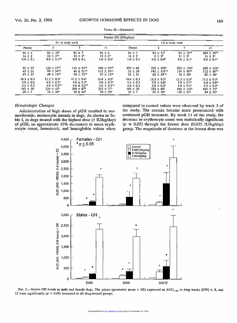
FIG. 2.--Serum GH levels in male and female dogs. The values (geometric mean r?: SE) expressed as AUC,.,, in drug weeks (DW) 4. 8. and 12 were significantly (p < 0.05) increased in all drug-treated’groups.
by guest on December 24, 2014tpx.sagepub.comDownloaded from

190 PRAHALADA ET AL TOXICOLOGIC PATHOLOGY
not considered biologically significant. The anemia was nonregenerative, and the results of the bone marrow (dis- cussed later) evaluation are consistent with the hemato- logical findings.
As shown in Table I, a dose- and time-dependent in- crease (p I 0.05) in mean platelet count relative to con- trol group was seen in high-dose (1 IU/kg/day) dogs; the mean percent increase over control value was 1096, 3596, 57%, and 72% in study weeks 3, 7, 11, and 14, respec- tively. In the mid-dose group of dogs, the increase in platelet count was significantly (p 5 0.05) increased only in study week 14. Platelet count in the low-dose group was similar to control group. In addition, the activated partial thromboplastin time (APTT; sec) was decreased in a dose-related manner beginning study week 7 in mid- and high-dose dogs. The decrease at the lowest dose (0.025 mg/kg/day) was significant (p 5 0.05) in week 11 in males only (Table I). There were no other pGH-related hematological changes in dogs.
Serum Biocheniical Cliaizges A spectrum of serum biochemical changes was ob-
served in dogs given high doses (20.1 IU/kg/day) of pGH. Table I1 has a summary of all drug-related changes. There was a minimal (<lo%) increase in fasted serum glucose levels in some mid- and high-dose pGH-treated dogs and was significant (p 5 0.05) only in study week 3. In study week 11, 2 dogs had high fasted serum glu- cose values (135 and 151 mg/dl). However, the plasma glycosylated hemoglobin values (week 12) in all dogs were within the normal range (mean values ranged fro-m 2.2% to 2.4%), indicating no evidence of chronic hyper- glycemia. Blood urea nitrogen (BUN) levels were slightly decreased (21-39%; p 5 0.05) throughout the study com- pared to concurrent controls in the high-dose group of dogs and in study weeks 7 and 11 in the mid-dose group. The serum creatinine values in the high-dose group also tended to be slightly lower (p < 0.05) in weeks 3 and 7 of the study. Serum alkaline phosphatase (ALP) activity was slightly higher in high-dose dogs (14-33%) com- pared to controls. Determination of bone ALP activity showed that there was a significant (p I 0.05) increase throughout the study (15-77%) in high-dose dogs. Al- though not significant (p > 0.05), the increase in liver ALP activity (17-44%) also contributed to the overall increase in serum ALP activity. Adrenal cortical ALP ac- tivity was unchanged (mean values ranged from 1 to 7 UL).
Administration of pGH resulted in dose-related in- creases in serum calcium and phosphorous levels. The magnitude of increase in serum calcium levels was min- imal (mean value 4-10%) and was significant (p 5 0.05) through the mid dose. Similarly, the elevation in serum phosphorous levels (mean values 10-49%) was signifi- cant through the mid-dose group. The serum potassium levels increased (p 5 0.05) by 14-18’36 in the high-dose group throughout the study relative to controls.
Dose-related increased serum total cholesterol and tri- glycerides were seen throughout the study. The magni- tude of increase (mean values 29-155’36) in serum cho- lesterol levels was significant (p 5 0.05) through mid
dose. Associated with elevated serum cholesterol levels, significant (p 5 0.05) dose-related increased serum tri- glyceride levels were observed through the mid dose. The magnitude of increases in serum cholesterol and triglyc- eride levels was minimal and not significant (p I 0.05) at the lowest dose.
Urinnlysis Administration of high doses (1 IUkglday) of pGH
resulted in polyuria in most dogs throughout the study; the observed increased urine volume (-2-fold) was sig- nificantly (p I 0.05) greater in weeks 3 and 7 of the study. Associated with increased urine volume, there was a significant decrease (p 5 0.05) in the mean specific gravity of urine when compared with the pretest values (1.034 vs 1.012, 1.020, and 1.020, pretest and weeks 3, 7, and 11, respectively). The other urinary parameters evaluated were within the control ranges.
Senun Honnoiie Levels Clear dose-related increases in serum GH, IGF-1, and
insulin levels were observed in pGH-treated dogs. There was a slight decrease in serum T., levels in high-dose dogs. Serum cortisol and T3 levels were unaffected (data not shown).
GH Levels. As shown in Fig. 2, GH levels expressed as AUC were significantly (p 5 0.05) increased in both sexes given exogenous GH. The GH AUCs (0-8 hr geo- metric means) in week 4 were approximately 2-, 8-, and 70-fold greater in the low-, mid-, and high-dose groups, respectively, than in controls. . GH values in the pretest period were generally consis- tent (except for one outlier among 224 values) over the 24-hr interval; the absolute values ranged between 0.1 ng/ml to 11.1 ng/ml; the peak values ranged between 2.8 ng/ml to 11.1 ng/ml. In week 4, the 0-hr (predose) values were comparable to pretest values, except in the high- dose group, where the peak value was 39.2 ng/ml com- pared to 7.2 ng/ml in controls. Following dosing in week 4, there was a marked elevation in serum GH levels in pGH-treated groups (up to 479 ng/ml in high dose). In subsequent study weeks (8 and 12), there was a slight increase in GH noted prior to dosing (0 hr) in all pGH groups when compared to vehicle controls. There was no neutralizing antibody to pGH following 14 wk of daily subcutaneous injections with high doses of pGH. ZGF-1 Levels. As shown in Fig. 3, serum IGF-1 levels
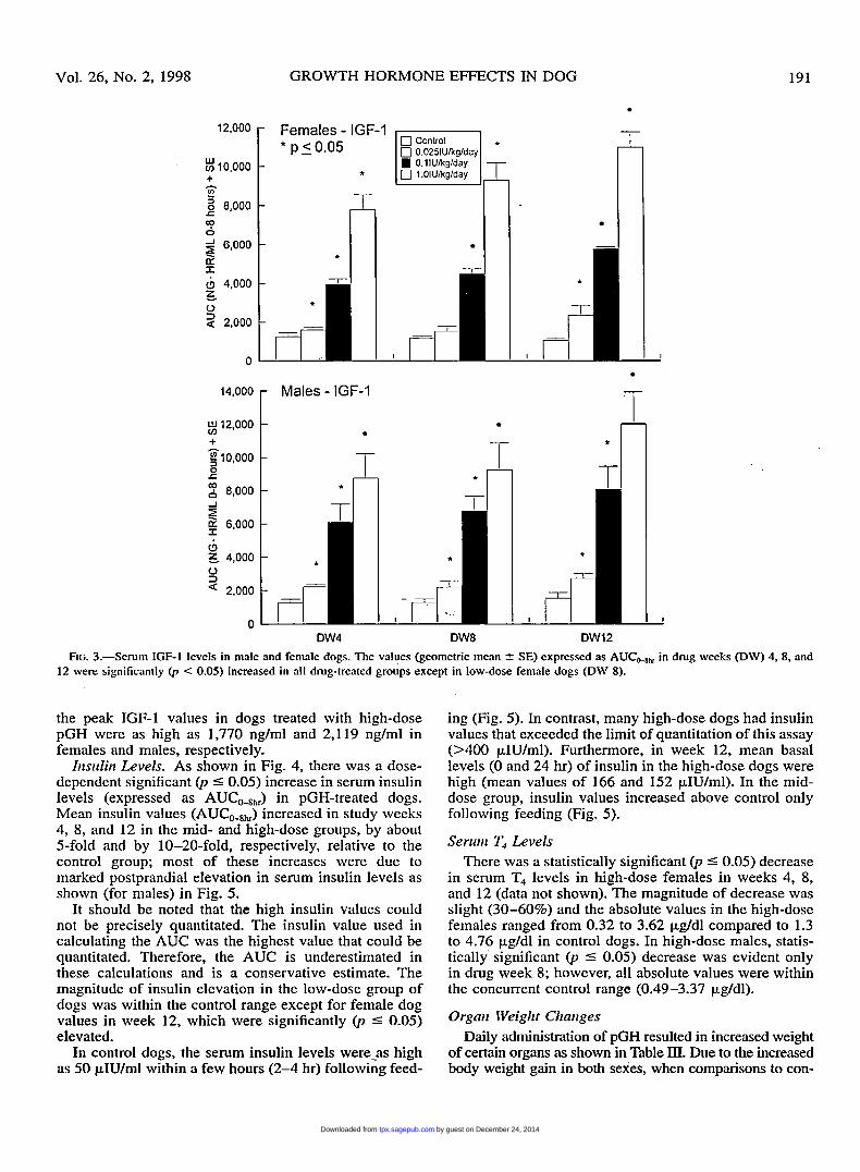
expressed as AUC were significantly ( ~ 1 5 0 . 0 5 ) increased in pGH-treated dogs. The mean serum IGF-1 AUC(O-Bhr) values in the mid-dose group were 3-5-fold greater than in control and in the high-dose group the values were 6- 10-fold higher than controls; in these 2 groups there was a tendency for the AUC values to increase with continued treatment with pGH. The magnitude of increase in serum IGF-1 levels in the low-dose group of dogs was minimal (25-100%) when compared to controls.
In general, the serum IGF-1 levels showed minimal variation over a 24-hr period in a given dog during the pretest period as well as in study weeks 4, 8, and 12. The pretest IGF-1 peak values ranged from 59 to 265 ng/rnl in females and 104 to 344 ng/ml in males. In contrast,
by guest on December 24, 2014tpx.sagepub.comDownloaded from
Vol. 26, NO. 2, 1998 GROWTH HORMONE EFFECTS IN DOG
$12,000
g 10,000 + n
0 r
2 8,000
$ 6,000
Z, 4,000 0 3
-I
&
a 2,000
0
191
T 'i -
-
-
T
-
-
- - -
121000 Females - IGF-1 * p 5 0.05 + F $10,000 *
n v,
0 0.0251Ukglday 0.1IUkglday
0 1.OIUkglday
-
14,000 r Males - IGF-1
I
-
T
I
FIG. 3 . S e r u m IGF-I levels in male and female dogs. The values (geometric mean -+ SE) expressed as AUCo4b in drug weeks (DW) 4. 8, and 12 were significantly @ < 0.05) increased in all drug-treated groups except in low-dose female dogs (DW 8).
the peak IGF-1 values in dogs treated with high-dose pGH were as high as 1,770 ng/ml and 2,119 ng/ml in females and males, respectively.
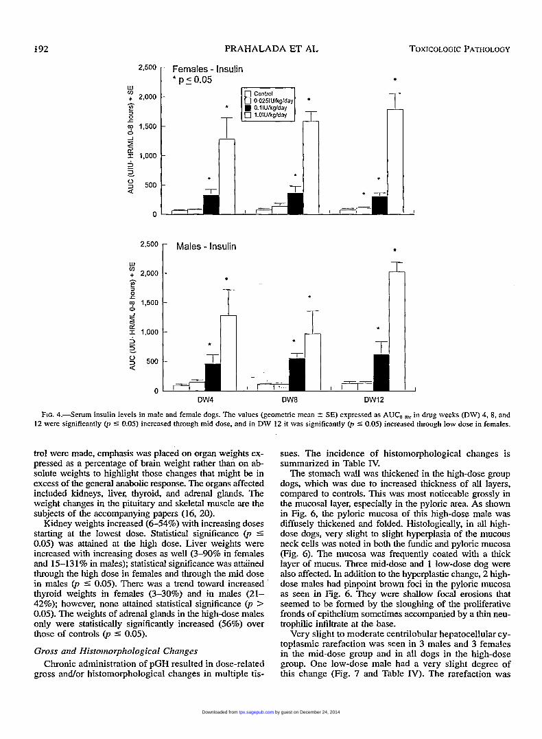
Iitsiilirt Levels. As shown in Fig. 4, there was a dose- dependent significant 0, I 0.05) increase in serum insulin levels (expressed as AUC,-,,,) in pGH-treated dogs. Mean insulin values (AUC,-,,) increased in study weeks 4, 8, and 12 in the mid- and high-dose groups, by about 5-fold and by 10-20-fold, respectively, relative to the control group; most of these increases were due to marked postprandial elevation in serum insulin levels as shown (for males) in Fig. 5.
It should be noted that the high insulin values could not be precisely quantitated. The insulin value used in calculating the AUC was the highest value that could be quantitated. Therefore, the AUC is underestimated in these calculations and is a conservative estimate. The magnitude of insulin elevation in the low-dose group of dogs was within the control range except for female dog values in week 12, which were significantly (p 5 0.05) elevated.
In control dogs, the serum insulin levels were-as high as 50 pIU/ml within ;I few hours (2-4 hr) following feed-
ing (Fig. 5). In contrast, many high-dose dogs had insulin values that exceeded the limit of quantitation of this assay (>400 pIU/ml). Furthermore, in week 12, mean basal levels (0 and 24 hr) of insulin in the high-dose dogs were high (mean values of 166 and 152 pIU/ml). In the mid- dose group, insulin values increased above control only following feeding (Fig. 5) .
Senan T4 Levels There was a statistically significant (p 5 0.05) decrease
in serum T4 levels in high-dose females in weeks 4, 8, and 12 (data not shown). The magnitude of decrease was slight (30-60%) and the absolute values in the high-dose females ranged from 0.32 to 3.62 pg/dl compared to 1.3 to 4.76 pg/dl in control dogs. In high-dose males, statis- tically significant (p I 0.05) decrease was evident only in drug week 8; however, all absolute values were within the concurrent control range (0.49-3.37 pg/dl).
Organ Weight Changes Daily administration of pGH resulted in increased weight
of certain organs as shown in Table III. Due to the increased body weight gain in both sexes, when comparisons to con-
by guest on December 24, 2014tpx.sagepub.comDownloaded from
192
*
PRAHALADA ET AL TOXICOLOGIC PATHOLOGY
215~0 - Females - Insulin * p z 0.05
0 0.025IUkglday
0 1.OlUkglday
w v) + 2,000 - - E? 3
r
0 -I
I 1,000 -
2 -
rT f
2*500 r Males - Insulin
I- W v) + 2,000
e
7 1,500
3 0 r
0 -J
5 I 1,000
2 2 2 500 a
0
7 * I I T * I I
DW4 DW8 OW12
FIG. 4.--Serum insulin levels in male and female dogs. The values (geometric mean t SE) expressed as AUCO.l,,, in drug weeks (DW) 4, 8, and 12 were significantly (p 5 0.05) increased through mid dose, and in DW 12 it was significantly 0, 5 0.05) increased through low dose in females.
trol were made, emphasis was placed on organ weights ex- pressed as a percentage of brain weight rather than on ab- solute weights to highlight those changes that might be in excess of the general anabolic response. The organs affected included kidneys, liver, thyroid, and adrenal glands. The weight changes in the pituitary and skeletal muscle are the subjects of the accompanying papers (16, 20).
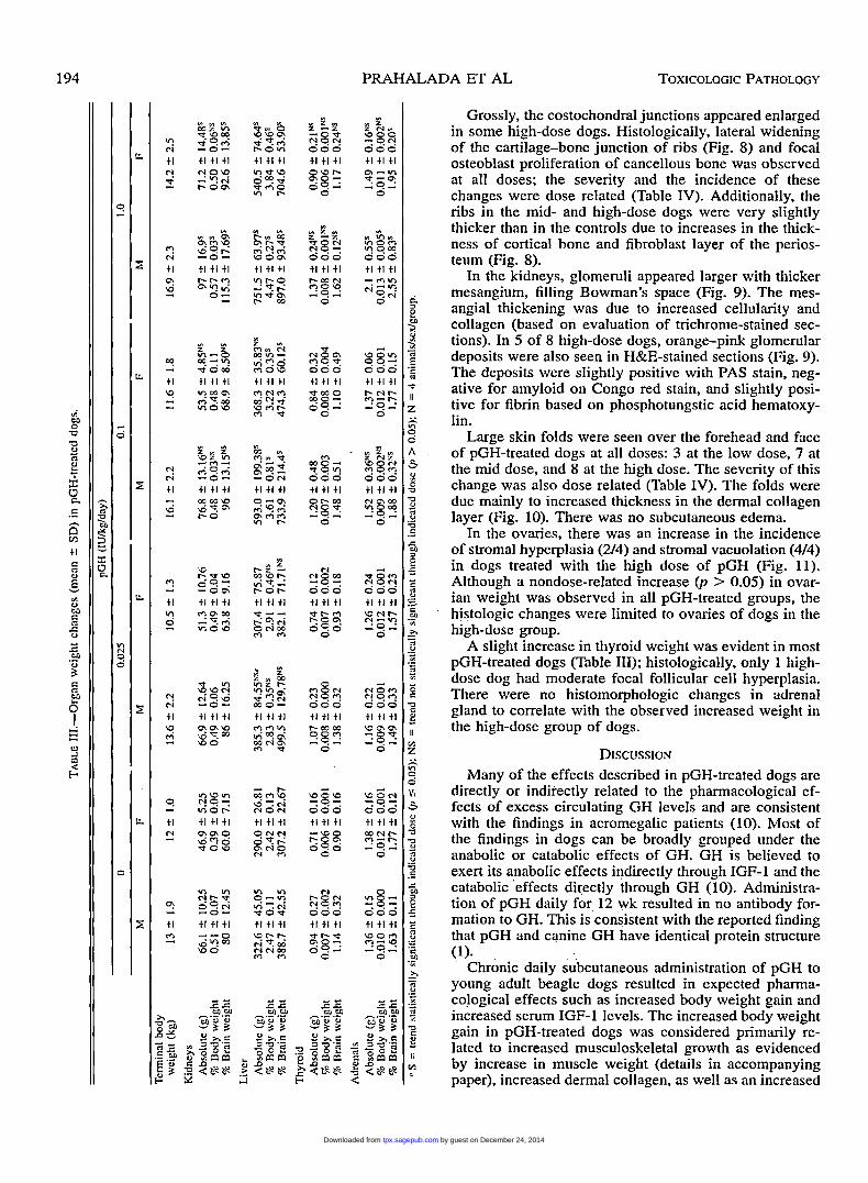
Kidney weights increased (654%) with increasing doses starting at the lowest dose. Statistical significance (p 5 0.05) was attained at the high dose. Liver weights were increased with increasing doses as well (340% in females and 15-131% in males); statistical significance was attriined through the high dose in females and through the mid dose in males (p 5 0.05). There was a trend toward increased thyroid weights in females (3-30%) and in males (21- 42%); however, none attained statistical significance (p > 0.05). The weights of adrenal glands in the high-dose males only were statistically significantly increased (56%) over those of controls (p 5 0.05).
Gross mid Histoiiiorphologicnl Cliaiiges Chronic administration of pGH resulted in dose-related
gross and/or histomorphological changes in multiple tis-
sues. The incidence of histomorphological changes is summarized in Table IV.
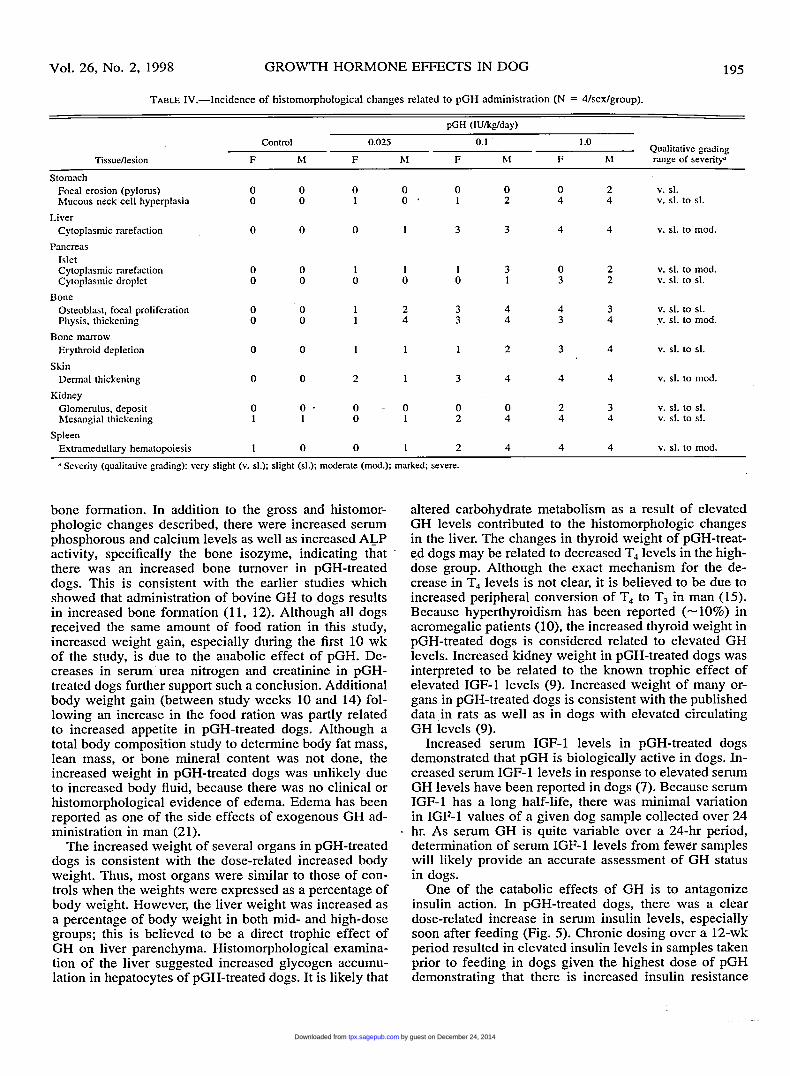
The stomach wall was thickened in the high-dose group dogs, which was due to increased thickness of all layers, compared to controls. This was most noticeable grossly in the mucosal layer, especially in the pyloric area. As shown in Fig. 6, the pyloric mucosa of this high-dose male was diffusely thickened and folded. Histologically, in all high- dose dogs, very slight to slight hyperplasia of the mucous neck cells was noted in both the fundic and pyloric mucosa (Fig. 6). The mucosa was frequently coated with a thick layer of mucus. Three mid-dose and 1 low-dose dog were also affected. In addition to the hyperplastic change, 2 high- dose males had pinpoint brown foci in the pyloric mucosa as seen in Fig. 6. They were shallow focal erosions that seemed to be formed by the sloughing of the proliferative fronds of epithelium sometimes accompanied by a thin neu- trophilic infiltrate at the base.
Very slight to moderate centrilobular hepatocellular cy- toplasmic rarefaction was seen in 3 males and 3 females in the mid-dose group and in all dogs in the high-dose group. One low-dose male had a very slight degree of this change (Fig. 7 and Table IV). The rarefaction was
by guest on December 24, 2014tpx.sagepub.comDownloaded from

Vol. 26, No. 2, 1998 GROWTH HORMONE EFFECTS IN DOG 193
W v) + -1
2 2
2
3 c Q
500 DW4: Insulin 450 1 Males T 400 350
300 250 200 150 100
50 0
300 r DW8: Insulin
+- 250 200 i W v) 1
400
c 150
2 100
50
0 0 0.5 1 6 8 24 Hrs 2 , r
Fed
FIG. 5.-Serum insulin levels (mean 2 SE) in male dogs. Data represent insulin values in serum samples collectcd at multiple time points in drug weeks 4. 8, and 12. The zero time point is the time prior to dosing the dogs with pGH. All dogs were fed approximately 4 hr postdose. Similar insulin profiles were observed in female dogs.
characterized by poorly delineated cytoplasmic clear ar- eas. Livers from control and treated dogs stained intense- ly for glycogen even though all were fasted overnight before necropsy. This was determined by positive peri- odic acid-Schiff (PAS) staining of the cytoplasm; these rarefied areas of the cytoplasm were almost empty and contained very little PAS-positive material after diastase digestion. By transmission electron microscopy (TEM), the large clear spaces in the cytoplasm seen in the cen- trilobular hepatocytes, when stained by H&E, were filled with fine grains of glycogen. Organelles were pushed aside by the glycogen, but otherwise, the hepatocytes had a normal appearance by TEM.
Cytoplasmic rarefaction of the pancreatic islets was seen in a dose-related pattern. This was characterized by ballooned cells with sparse, dispersed cytoplasm (Table IV and Fig. 7). Less frequently, in the mid- and high- dose groups, some islet cells had cytoplasmic droplets,
usually 1 per cell, characterized by a dense pink core surrounded by a clear halo. These structures stained in- tensely with an immumohistochemical stain for insulin. In general, the islets in the pancreases from control dogs stained more homogeneously for insulin than did those from the pGH-treated dogs.
In the marrow of bone sections and in bone marrow smears there was depletion of the erythroid cell series in 7 of 8 high-dose and 2 of 8 mid- and low-dose dogs (Table IV). In general, although the bone marrow was still quite cellular, there appeared to be a shift toward a more a immature, mainly erythroid. cell type. Very slight to moderate extramedullary hematopoiesis was observed in spleens of mid- and high-dose dogs (Table IV). In addition to the diffuse changes in the bone marrow, many high-dose dogs with the large costochondral junctions had discrete patches of marrow atrophy adjacent to the expanding cartilage-bone interface.
by guest on December 24, 2014tpx.sagepub.comDownloaded from
PRAI 4LADA ET AL TOXICOLOGIC PATHOLOGY
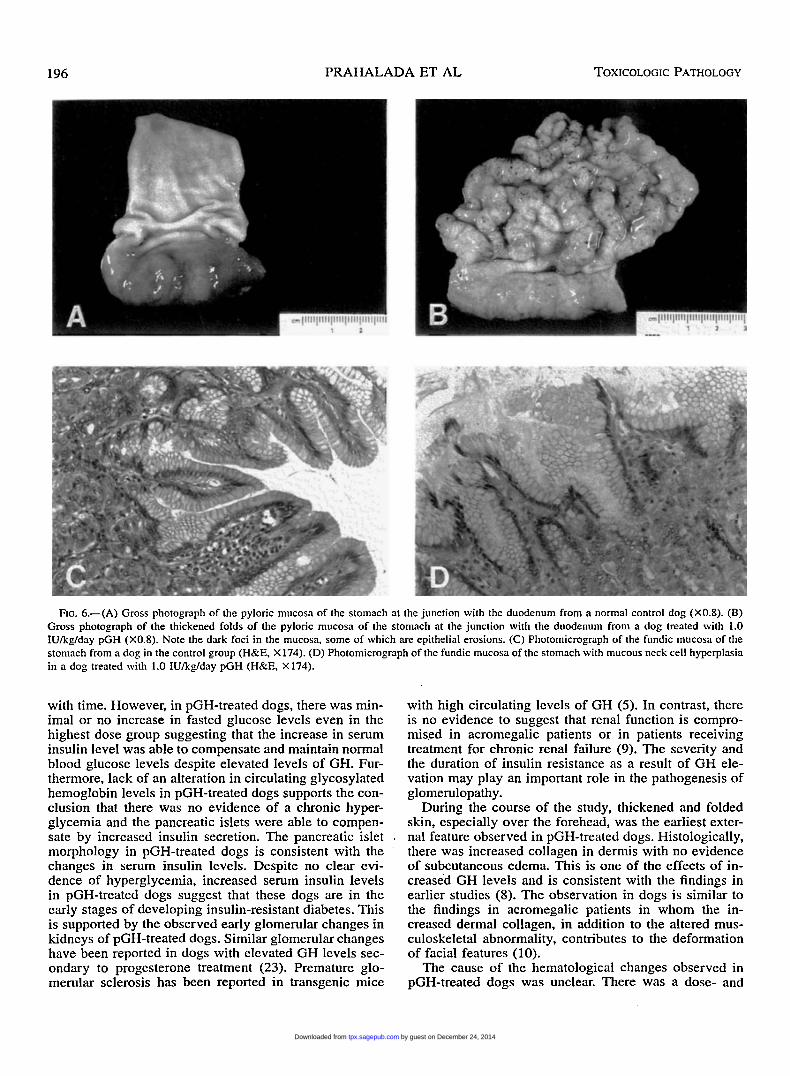
Grossly, the costochondral junctions appeared enlarged in some high-dose dogs. Histologically, lateral widening of the cartilage-bone junction of ribs (Fig. 8) and focal osteoblast proliferation of cancellous bone was observed at all doses; the seventy and the incidence of these changes were dose related (Table IV). Additionally, the ribs in the mid- and high-dose dogs were very slightly thicker than in the controls due to increases in the thick- ness of cortical bone and fibroblast layer of the perios- teum (Fig. 8).
In the kidneys, glomeruli appeared larger with thicker mesangium, filling Bowman's space (Fig. 9). The mes- angial thickening was due to increased cellularity and collagen (based on evaluation of trichrome-stained sec- tions). In 5 of 8 high-dose dogs, orange-pink glomerular deposits were also seen in H&E-stained sections (Fig. 9). The deposits were slightly positive with PAS stain, neg- ative for amyloid on Congo red stain, and slightly posi- tive for fibrin based on phosphotungstic acid hematoxy- lin.
Large skin folds were seen over the forehead and face of pGH-treated dogs at all doses: 3 at the low dose, 7 at the mid dose, and 8 at the high dose. The seventy of this change was also dose related (Table IV). The folds were due mainly to increased thickness in the dermal collagen layer (Fig. 10). There was no subcutaneous edema.
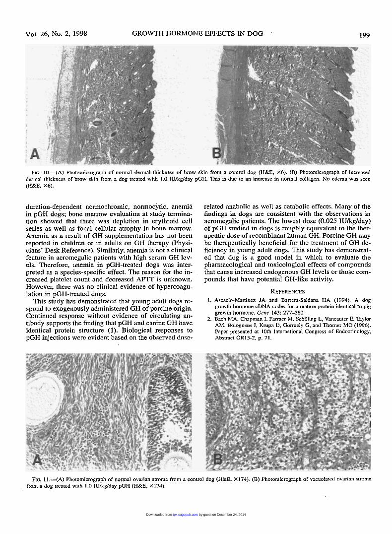
In the ovaries, there was an increase in the incidence of stromal hyperplasia (2/4) and stromal vacuolation (4/4) in dogs treated with the high dose of pGH (Fig. 11). Although a nondose-related increase (p > 0.05) in ovar- ian weight was observed in all pGH-treated groups, the histologic changes were limited to ovaries of dogs in the high-dose group.
A slight increase in thyroid weight was evident in most pGH-treated dogs (Table 111); histologically, only 1 high- dose dog had moderate focal follicular cell hyperplasia. There were no histomorphologic changes in adrenal gland to correlate with the observed increased weight in the high-dose group of dogs.
DISCUSSION Many of the effects described in pGH-treated dogs are
directly or indirectly related to the pharmacological ef- fects of excess circulating GH levels and are consistent with the findings in acromegalic patients (10). Most of the findings in dogs can be broadly grouped under the anabolic or catabolic effects of GH. GH is believed to exert its anabolic effects indirectly through IGF-1 and the catabolic effects directly through GH (10). Administra- tion of pGH daily for 12 wk resulted in no antibody for- mation to GH. This is consistent with the reported finding that pGH and canine GH have identical protein structure (1).
Chronic daily subcutaneous administration of pGH to young adult beagle dogs resulted in expected pharma- cological effects such as increased body weight gain and increased serum IGF-1 levels. The increased body weight gain in pGH-treated dogs was considered primarily re- lated to increased musculoskeletal growth as evidenced by increase in muscle weight (details in accompanying paper), increased dermal collagen, as well as an increased
by guest on December 24, 2014tpx.sagepub.comDownloaded from
Vol. 26, No. 2, 1998 GROWTH HORMONE EFFECTS IN DOG 195
TABLE 1V.-Incidence of histomorphological changes related to pGH administration (N = 4/sex/group).
pGH (IU/kg/day) Control 0.025 0.1 1 .o
Qualitative grading range of severity” Tissuenesion F hl F hI F hl F hl
Stomach Focal erosion (pyloms) 0 0 0 0 0 0 0 2 v. sl. hlucous neck cell hyperplasia 0 0 1 0 . 1 2 4 4 v. sl. to sl.
Liver Cytoplasmic rarefaction
Pancreas Islet Cytoplasmic rarefaction Cytoplasmic droplet
0 0 0 1 3 3 4 4 v. sl. to mod.
0 0 1 1 1 3 0 2 v. sl. to mod. 0 0 0 0 0 1 3 2 v. sl. to sl.
Bone Osteoblast, focal proliferation 0 0 1 2 3 4 4 3 v. sl. to sl. Physis, thickening 0 0 1 4 3 4 3 4 v. sl. to mod.
Bone marrow
Skin Erythroid depletion
Dermal thickening
Glomerulus, deposit hlesangial thickening
Kidney
0 0 1 1 1 2 3 4 v. sl. to sl.
0 0 2 1 3 4 4 4 v. sl. to mod.
0 0 . 1 1
0 . o 0 0 2 3 v. sl. to sl. 0 1 2 4 4 4 v. sl. to sl.
Spleen Extramedullary hematopoiesis 1 0 0 1 2 4 4 4 v. sl. to mod.
* Severity (qualimtive grading): very slight (v. sl.); slight (sl.); moderate (mod.); marked; severe.
bone formation. In addition to the gross and histomor- phologic changes described, there were increased serum phosphorous and calcium levels as well as increased ALP activity, specifically the bone isozyme, indicating that there was an increased bone turnover in pGH-treated dogs. This is consistent with the earlier studies which showed that administration of bovine GH to dogs results in increased bone formation (11, 12). Although all dogs received the same amount of food ration in this study, increased weight gain, especially during the first 10 wk of the study, is due to the anabolic effect of pGH. De- creases in serum urea nitrogen and creatinine in pGH- treated dogs further support such a conclusion. Additional body weight gain (between study weeks 10 and 14) fol- lowing an increase in the food ration was partly related to increased appetite in pGH-treated dogs. Although a total body composition study to determine body fat mass, lean mass, or bone mineral content was not done, the increased weight in pGH-treated dogs was unlikely due to increased body fluid, because there was no clinical or histomorphological evidence of edema. Edema has been reported as one of the side effects of exogenous GH ad- ministration in man (21).
The increased weight of several organs in pGH-treated dogs is consistent with the dose-related increased body weight. Thus, most organs were similar to those of con- trols when the weights were expressed as a percentage of body weight. However, the liver weight was increased as a percentage of body weight in both mid- and high-dose groups; this is believed to be a direct trophic effect of GH on liver parenchyma. Histomorphological examina- tion of the liver suggested increased glycogen accumu- lation in hepatocytes of pGH-treated dogs. It is likely that
altered carbohydrate metabolism as a result of elevated GH levels contributed to the histomorphologic changes in the liver. The changes in thyroid weight of pGH-treat- ed dogs may be related to decreased T4 levels in the high- dose group. Although the exact mechanism for the de- crease in T4 levels is not clear, it is believed to be due to increased peripheral conversion of T4 to T3 in man (15). Because hyperthyroidism has been reported (- 10%) in acromegalic patients (lo), the increased thyroid weight in pGH-treated dogs is considered related to elevated GH levels. Increased kidney weight in pGH-treated dogs was interpreted to be related to the known trophic effect of elevated IGF-1 levels (9). Increased weight of many or- gans in pGH-treated dogs is consistent with the published data in rats as well as in dogs with elevated circulating GH levels (9).
Increased serum IGF-1 levels in pGH-treated dogs demonstrated that pGH is biologically active in dogs. In- creased serum IGF- 1 levels in response to elevated serum GH levels have been reported in dogs (7). Because serum IGF-1 has a long half-life, there was minimal variation in IGF-1 values of a given dog sample collected over 24 hr. As serum GH is quite variable over a 24-hr period, determination of serum IGF-1 levels from fewer samples will likely provide an accurate assessment of GH status in dogs.
One of the catabolic effects of GH is to antagonize insulin action. In pGH-treated dogs, there was a clear dose-related increase in serum insulin levels, especially soon after feeding (Fig. 5). Chronic dosing over a 12-wk period resulted in elevated insulin levels in samples taken prior to feeding in dogs given the highest dose of pGH demonstrating that there is increased insulin resistance
by guest on December 24, 2014tpx.sagepub.comDownloaded from
196 PRAHALADA ET AL TOXICOLOGIC PATHOLOGY
FIG. 6 4 A ) Gross photograph of the pyloric mucosa of the stomach at the junction with the duodenum from a normal control dog (X0.8). (B) Gross photograph of the thickened folds of the pyloric mucosa of the stomach at the junction with the duodenum from a dog treated with 1.0 IUkg/day pGH (X0.8). Note the dark foci in the mucosa, some of which are epithelial erosions. (C) Photomicrograph of the fundic mucosa of the stomach from a dog in the control group (H&E, X 174). (D) Photomicrograph of the fundic mucosa of the stomach with mucous neck cell hyperplosia in a dog treated with 1.0 IU/kg/day pGH (H&E, X174).
with time. However, in pGH-treated dogs, there was min- imal or no increase in fasted glucose levels even in the highest dose group suggesting that the increase in serum insulin level was able to compensate and maintain normal blood glucose levels despite elevated levels of GH. Fur- thermore, lack of an alteration in circulating glycosylated hemoglobin levels in pGH-treated dogs supports the con- clusion that there was no evidence of a chronic hyper- glycemia and the pancreatic islets were able to compen- sate by increased insulin secretion. The pancreatic islet morphology in pGH-treated dogs is consistent with the changes in serum insulin levels. Despite no clear evi- dence of hyperglycemia, increased serum insulin levels in pGH-treated dogs suggest that these dogs are in the early stages of developing insulin-resistant diabetes. This is supported by the observed early glomerular changes in kidneys of pGH-treated dogs. Similar glomerular changes have been reported in dogs with elevated GH levels sec- ondary to progesterone treatment (23). Premature glo- merular sclerosis has been reported in transgenic mice
with high circulating levels of GH (5). In contrast, there is no evidence to suggest that renal function is compro- mis,ed in acromegalic patients or in patients receiving treatment for chronic renal failure (9). The seventy and the duration of insulin resistance as a result of GH ele- vation may play an important role in the pathogenesis of glomerulopathy.
During the course of the study, thickened and folded skin, especially over the forehead, was the earliest exter- nal feature observed in pGH-treated dogs. Histologically, there was increased collagen in dermis with no evidence of subcutaneous edema. This is one of the effects of in- creased GH levels and is consistent with the findings in earlier studies (8). The observation in dogs is similar to the findings in acromegalic patients in whom the in- creased dermal collagen, in addition to the altered mus- culoskeletal abnormality, contributes to the deformation of facial features (10).
The cause of the hematological changes observed in pGH-treated dogs was unclear. There was a dose- and
by guest on December 24, 2014tpx.sagepub.comDownloaded from

Vol. 26, No. 2, 1998 GROWTH HORMONE EFFECTS IN DOG 197
FIG. 7.-(A) Photomicrograph of the cytoplasmic appearance of hepatocytes from a dog in the control group (H&E, X 174). (B) Photomicrograph of the rarefied cytoplasm of hepatocytes seen in the centrilobular area from a dog treated with 1 .O IUkdday pGH (H&E, X 174). (C) Photomicrograph of the normal cytoplasmic appearance of pancreatic islets from 8 dog in the control group (H&E, X349). (D) Photomicrograph of the cytoplasmic rarefaction and intensified cytoplasmic droplets in the pancreatic islets from a dog treated with 1.0 IU/kg/day pGH (H&E, X349). (E) Photomicro- graph of the normal cytoplasmic appearance of pancreatic islets from a dog in the control group (immunostain for insulin, X349). (F) Photomicro- graph of the cytoplasmic rarefaction and intensified cytoplasmic droplets in the pancreatic islets from a dog treated with 1.0 IU/kg/day pGH (immunostain for insulin, X349).
by guest on December 24, 2014tpx.sagepub.comDownloaded from
198 PRAHALADA ET AL TOXICOLOGIC PATHOLOGY
FIG. &--(A) Photomicrograph of a normal costochondral junction of a rib from a control dog. (H&E, XS). (B) Photomicrograph of an abnormally thickened costochondral junction of a rib from a dog treated with 1.0 IUkg/day pGH (H&E, X8). (C) Photomicrograph of normal cancellous bone in the rib of a dog in the control group, showing a normal amount of osteoblastic activity (arrow) and normal bone marrow (H&E, X349). (D) Photomicrograph of cancellous bone in the rib of a dog treated with 1.0 IUkg/day pGH, showing proliferation of osteoblasts and depletion of the bone marrow (H&E, X349).
FIG. 9 4 A ) Photomicrograph of a glomerulus in kidney of a control dog (H&E, X349). (B) Photomicrograph of an enlarged glomerulus in the kidney of a pGH-treated ( 1 IUkglday) dog. Mesangial thickening. increased cellularity as well as fibrinous deposits are evident (H&E, X349).
by guest on December 24, 2014tpx.sagepub.comDownloaded from
Vol. 26, No. 2, 1998 GROWTH HORMONE EFFECTS IN DOG 199
FIG. lO.-(A) Photomicrograph of normal dermal thickness of brow skin from a control dog (H&E, X6). (B) Photomicrograph of increased dermal thickness of brow skin from a dog treated with 1.0 IUkglday pGH. This is due to an increase in normal collagen. No edema was seen (H&E, X6).
duration-dependent normochromic, normocytic, anemia in pGH dogs; bone marrow evaluation at study termina- tion showed that there was depletion in erythroid cell series as well as focal cellular atrophy in bone marrow. Anemia as a result of GH supplementation has not been reported in children or in adults on GH therapy (Physi- cians’ Desk Reference). Similarly, anemia is not a clinical feature in acromegalic patients with high serum GH lev- els. Therefore, anemia in pGH-treated dogs was inter- preted as a species-specific effect. The reason for the in- creased platelet count and decreased APTT is unknown. However, there was no clinical evidence of hypercoagu- lation in pGH-treated dogs.
This study has demonstrated that young adult dogs re- spond to exogenously administered GH of porcine origin. Continued response without evidence of circulating an- tibody supports the finding that pGH and canine GH have identical protein structure (1). Biological responses to pGH injections were evident based on the observed dose-
related anabolic as well as catabolic effects. Many of the findings in dogs are consistent with the observations in acromegalic patients. The lowest dose (0.025 IU/kg/day) of pGH studied in dogs is roughly equivalent to the ther- apeutic dose of recombinant human GH. Porcine GH may be therapeutically beneficial for the treatment of GH de- ficiency in young adult dogs. This study has demonstrat- ed that dog is a good model in which to evaluate the pharmacological and toxicological effects of compounds that cause increased endogenous GH levels or those com- pounds that have potential GH-like activity.
REFERENCES 1. Ascacio-Martinez JA and Barrera-Saldana HA (1993). A dog
growth hormone cDNA codes for a mature protein identical to pig growth hormone. Gene 143: 277-280.
2. Bach MA, Chapman I, Farmer M, Schilling L, Vancauter E, Taylor AM, Bolognese J, Krupa D, Gormely G, and Thorner MO (1996). Paper presented at 10th International Congress of Endocrinology, Abstract OR15-2, p. 71.
FIG. 1 l . - (A) Photomicrograph of normal ovarian stroma from a control dog (H&E, X 174). (B) Photomicrograph of vacuolated ovarian stroma from a dog treated with 1.0 IUkglday pGH (H&E. X174).
by guest on December 24, 2014tpx.sagepub.comDownloaded from
200 PRAHALADA ET AL TOXICOLOGIC PATHOLOGY
3. Bowers CY (1996). Xenobiotic growth hormone secretagogues: Growth hormone releasing peptides. In: Growth Hormone Secre- ragogues, BB Bercu and RF Walker (eds). Springer-Verlag, New York, pp. 9-28.
4. Cahill M, Satiritz S . Patrick D, and Keuler J (1995). Insulin-like growth factor 1 (IGF-I) by extraction and radioimmunoassay in rodent and canine species. Clin. CIzenz. 41: S161 (abstract 566).
5. Doi T, Striker U. Quaife C, Conti FG, Palmiter R, Behringer R, Brinster R. and Striker GE (1988). Progressive glomerulosclerosis develops in transgenic mice chronically expressing growth hor- mone and growth hormone releasing factor but not in those ex- pressing insulin-like growth factor-I. Am. J. Pathol. 13 1: 398-403.
6. Eigenmann JE (1984). Acromegaly in the dog. Vet. Clin. N. Am. Sinall Anitti. Pract. 14: 827-836.
7. Eigenmann JE (1985). Growth hormone and insulin-like growth factor in the dog: Clinical and experimental investigations. Dornest. Anini. Endocrinol. 2: 1-16.
8. Eigenmann JE and Venker-van Haagen AJ (1981). Progestagen- induced and spontaneous canine acromegaly due to reversible growth hormone overproduction: Clinical picture and pathogenesis. J. Am. Aniiii. Hosp. Assoc. 17: 813-822.
9. Feld S and Hirschberg R (1996). Growth hormone, the insulin-like growth factor system, and the kidney. Endocrinol. Rev. 17: 423- 480.
10. Harris AG (1996). Clinical features of acromega1y:In: Acroniegaly arid Its hfanagenienr, AF Daly (ed). Lippincott-Raven Publishers, New York, pp. 24-37.
11. Hams WH, Heaney RP, Jowsey J, Cockin J, Akin SC. Graham J, and Weinberg EH (1972). Gross hormone: The effect of skeletal renewal in the adult dog. I. Morphometric studies. Calcif: Tiss. Res.
12. Heaney RP. Harris WH, Cockin J, and Weinberg EH (1972). Growth hormone: The effect on skeletal renewal in the adult dog. 11. Mineral kinetic studies. Calcif: Tiss. Res. 10: 14-22.
13. Hickey G. Jacks T, Schleim K-D. Frazier E. Chen H, Krup3-D; Fenney W, Nargund R, Patchett A, and Smith RG (1997). Repeat '
administration of growth hormone secretagogue MK-0677 increas- es and maintains elevated IGF-1 levels in beagles. J. Endocrinol.
14. Jacks T. Smith R, Judith E Schleim K-D, Frazier E, Chen H, Krupa D, Hora D Jr, Nargund R, Patchett A, and Hickey G (1996). MK- 0677, a potent, novel, orally-active growth hormone (GH) secre-
10: 1-13.
152: 183-192.
tagogue: GH, IGF-I and other hormonal responses in beagles. En- docrinology 137: 5284-5289.
15. Jorgensen JOL, Moller J, Skakkebak NE, Weeke J, and Christiansen JS (1992). Thyroid function during growth hormone therapy. Honn. Res. 38 (suppl.): 63-67.
16. Laroque P, Molon-Noblot S, Prahalada S, Stabinski LG, Hoe C-M, Peter CP, Duprat P. and van Zwieten MJ (1998). Morphological changes in the pituitary gland of dogs chronically exposed to ex- ogenous growth hormone. Toxicol. Patliol. 26: 201-206.
17. Leung FC. Jones B, Steelman SL, Rosenblum CL, and Kopchick JJ (1986). Purification and physiochemical properties of 3 recom- binant bovine growth hormone produced by cultured murine fibro- blasts. Endocrinology 119: 1489-1496.
18. Levene H (1960). In: Contributions to Probability and Statistics, Stanford University Press, Stanford, California, p. 278.
19. Moalli MR, Dysko RC, Rush GH. Chrisp CE, Decoster JL. Sweet KA, and Goldstein SA (1996). Oxytetracycline-induced nephrotox- icosis in dogs after intravenous administration for experimental bone labeling. Lnb Aniin. Sci. 46: 497-502.
20. Molon-Noblot S. Laroque P, Prahalada S, Stabinski LG. Hoe C-M. Peter CR Duprat P, and van Zwieten MJ (1998). Effect of chronic growth hormone administration on skeletal muscle in dogs. Toxicol. Paflzol. 26: 207-212.
21. Pratt JH, Peacock M, and Henry DP (1993). Effect of recombinant human growth hormone on adreno-cortical function, and on sodium and potassium homeostasis. Plzaniiacology 47: 36-42.
22. Shapiro SS and Wilk MB (1965). An analysis of variance test for normality (complete samples). Biornetrika 52: 591-61 1.
23. Sloan JM and Oliver IM (1975). Progestagen-induced diabetes in the dog. Diabetes 24: 337-344.
24. Smith RG, Chang K, Pong S-S. Leonard R, Cohen CJ. Arena JP. Hickey GJ, Chang CH, Jacks T, Drisko J, Robinson ICAE Dickson SL, and Leng G (1996). Mechanism of action of GHRP-6 and nonpeptidyl growth hormone secretagogues. In: Groirtli Hormone Secreragogues, BB Bercu and RF Walker (eds). Springer-Verlag. New York, pp. 147-163.
25. Snedecor G and Cochran W (1980). Stntistical hfetlzods. 7th ed. Iowa State University Press, Ames, Iowa, pp. 276-279.
26. Tucker MJ (1971). Some effects of prolonged administration of a progestagen to dogs. In: The Correlation of Adr-erse Effects in hfnn with Obsenations in Anitnals, SB De and C Baker (eds). Excerpta Medica, Amsterdam, pp. 228-238.
27. Tukey JW, Ciminera JL, and Heyse JF (1985). Testing the statistical certainty of a response to increasing doses of a drug. Bionzetrics 41: 295-301.
by guest on December 24, 2014tpx.sagepub.comDownloaded from