safety reports series · pdf filesafety reports series no. 17 lessons learned from accidental...
TRANSCRIPT
SafetyReports Series
No. 17


LESSONS LEARNED FROMACCIDENTAL EXPOSURES
IN RADIOTHERAPY

The following States are Members of the International Atomic Energy Agency.
AFGHANISTANALBANIAALGERIAANGOLAARGENTINAARMENIAAUSTRALIAAUSTRIABANGLADESHBELARUSBELGIUMBENINBOLIVIABOSNIA AND HERZEGOVINABRAZILBULGARIABURKINA FASOCAMBODIACAMEROONCANADACHILECHINACOLOMBIACOSTA RICACOTE D'lVOIRECROATIACUBACYPRUSCZECH REPUBLICDEMOCRATIC REPUBLIC
OF THE CONGODENMARKDOMINICAN REPUBLICECUADOREGYPTEL SALVADORESTONIAETHIOPIAFINLANDFRANCEGABONGEORGIAGERMANYGHANAGREECE
GUATEMALAHAITIHOLY SEEHUNGARYICELANDINDIAINDONESIAIRAN, ISLAMIC REPUBLIC OFIRAQIRELANDISRAELITALYJAMAICAJAPANJORDANKAZAKHSTANKENYAKOREA, REPUBLIC OFKUWAITLATVIALEBANONLIBERIALIBYAN ARAB JAMAfflRIYALIECHTENSTEINLITHUANIALUXEMBOURGMADAGASCARMALAYSIAMALIMALTAMARSHALL ISLANDSMAURITIUSMEXICOMONACOMONGOLIAMOROCCOMYANMARNAMIBIANETHERLANDSNEW ZEALANDNICARAGUANIGERNIGERIANORWAYPAKISTAN
PANAMAPARAGUAYPERUPHILIPPINESPOLANDPORTUGALQATARREPUBLIC OF MOLDOVAROMANIARUSSIAN FEDERATIONSAUDI ARABIASENEGALSIERRA LEONESINGAPORESLOVAKIASLOVENIASOUTH AFRICASPAINSRI LANKASUDANSWEDENSWITZERLANDSYRIAN ARAB REPUBLICTHAILANDTHE FORMER YUGOSLAV
REPUBLIC OF MACEDONIATUNISIATURKEYUGANDAUKRAINEUNITED ARAB EMIRATESUNITED KINGDOM OF
GREAT BRITAIN ANDNORTHERN IRELAND
UNITED REPUBLICOF TANZANIA
UNITED STATES OF AMERICAURUGUAYUZBEKISTANVENEZUELAVIETNAMYEMENYUGOSLAVIAZAMBIAZIMBABWE
The Agency's Statute was approved on 23 October 1956 by the Conference on the Statute of theIAEA held at United Nations Headquarters, New York; it entered into force on 29 July 1957 TheHeadquarters of the Agency are situated in Vienna Its principal objective is "to accelerate and enlarge thecontribution of atomic energy to peace, health and prosperity throughout the world"
© IAEA, 2000
Permission to reproduce or translate the information contained in this publication may beobtained by writing to the International Atomic Energy Agency, Wagramer Strasse 5, P.O Box 100,A-1400 Vienna, Austria
Printed by the IAEA in AustriaFebruary 2000STI/PUB/1084

SAFETY REPORTS SERIES No. 17
LESSONS LEARNED FROMACCIDENTAL EXPOSURES
IN RADIOTHERAPY
INTERNATIONAL ATOMIC ENERGY AGENCYVIENNA, 2000

VIC Library Cataloguing in Publication Data
Lessons learned from accidental exposures in radiotherapy — Vienna :International Atomic Energy Agency, 2000.
p , 24 cm — (Safety reports series, ISSN 1020-6450; no 17)STI/PUB/1084ISBN 92-0-100200-9Includes bibliographical references.
1. Radiotherapy—Exposure 2. Radiotherapy—Safety measures.I. International Atomic Energy Agency II Series
VICL 00-00236

FOREWORD
The medical use of radiation is unique in that patients are intentionally exposedto radiation. The aim in radiation therapy is twofold: to deliver a dose and dosedistribution that is adequate for tumour control, but which also minimizescomplications in normal tissues. In therapeutic applications, the doses are high and adeviation from the prescribed dose may have severe or even fatal consequences. Thereis therefore a great need to ensure adequate radiation protection and safety inradiotherapy by verifying that all personnel involved are appropriately trained fortheir duties, that the equipment used meets relevant international specifications forradiation safety and that safety culture is embedded in routine activities inradiotherapy departments.
Many individuals must interact and work together on highly technicalmeasurements and calculations, and therefore the potential for mistakes is great.A review of the mistakes shows that most are due to human error. The InternationalBasic Safety Standards for Protection against Ionizing Radiation and the Safety ofRadiation Sources (IAEA Safety Series No. 115) require that a prompt investigationbe conducted whenever an accidental medical exposure of patients occurs. The reportof the investigation is to be disseminated to the appropriate parties so that lessons canbe learned to prevent similar accidents or mitigate their consequences in the future.
This Safety Report is a collection of a large number of events that may serve asa checklist against which to test the vulnerability of a facility to potential accidents,and to provide a basis for improving safety in the use of radiation in medicalapplications. A further purpose of this report is to encourage readers to develop aquestioning and learning attitude, adopt measures for the prevention of accidents, andprepare for mitigation of the consequences of accidents if they occur.
The IAEA officer responsible for preparation of this report was M. Oresegun ofthe Division of Radiation and Waste Safety, Department of Nuclear Safety.

EDITORIAL NOTE
Although great care has been taken to maintain the accuracy of information containedin this publication, neither the IAEA nor its Member States assume any responsibility forconsequences which may arise from its use.
The use of particular designations of countries or territories does not imply anyjudgement by the publisher, the IAEA, as to the legal status of such countries or territories, oftheir authorities and institutions or of the delimitation of their boundaries.
The mention of names of specific companies or products (whether or not indicated asregistered) does not imply any intention to infringe proprietary rights, nor should it beconstrued as an endorsement or recommendation on the part of the IAEA
Reference to standards of other organizations is not to be construed as an endorsementon the part of the IAEA
CONTENTS
1. INTRODUCTION 1
1.1. Background 11.2. Objective 21.3. Scope 21.4. Structure 2
2. REVIEW OF ACCIDENTS 3
2.1. Definitions 32.2. Methodology for the review 52.3. Clinical considerations related to quality assurance 62.4. Radiation measurement systems 72.5. External beam therapy: Machine commissioning
and calibration 102.6. External beam therapy: Treatment planning,
patient setup and treatment 222.7. Decommissioning of teletherapy equipment 362.8. Mechanical and electrical malfunctions 392.9. Brachytherapy: Low dose rate sources and applicators 402.10. Brachytherapy (high dose rate) 592.11. Unsealed sources 61
3. CLASSIFICATION OF ACCIDENTS 66
3.1. Classification of causes based on radiotherapy techniques 663.2. External beam therapy 673.3. Low dose rate brachytherapy 693.4. High dose rate brachytherapy 703.5. Use of unsealed sources 70
4. LESSONS LEARNED AND MEASURES FOR PREVENTIONOF ACCIDENTS 71
4.1. Resources: Personnel and equipment 714.2. Human factors 714.3. Training 73
4.4. Communication 744.5. Equipment 754.6. Human-machine problems 784.7. Improper decommissioning of equipment and
unsafe storage of radioactive sources 794.8. Documentation 804.9. Integration of safety and quality assurance 814.10. Safety assessment 824.11. Regulatory control 86
APPENDIX: APPENDIX II OF THE BSS — MEDICAL EXPOSURE:OPTIMIZATION OF PROTECTIONFOR MEDICAL EXPOSURES 87
REFERENCES 90
CONTRIBUTORS TO DRAFTING AND REVIEW 91
RELATED IAEA PUBLICATIONS 93

1. INTRODUCTION
1.1. BACKGROUND
This Safety Report provides a review of events that constitute incidents oraccidents related to the therapeutic use of ionizing radiation. The events concernexternal beam therapy, brachytherapy and nuclear medicine. Most of the casesinvolve patients, although some examples illustrate accidental exposure of medicalpersonnel or the general public. Most events involve radiation exposure; however, afew examples are included that illustrate injuries resulting from mechanical orelectrical failure of equipment.
The International Basic Safety Standards for Protection against IonizingRadiation and for the Safety of Radiation Sources (BSS) state that the regulatoryauthority should require all parties to develop a safety culture that includes measuresto encourage a questioning and learning attitude and discourage complacency withrespect to safety [1]. Information on unusual events during operation of the facilitieswhich led or might have led to accidents provides background material that can beused to prevent accidents when such information is disseminated to regulators,manufacturers, users and radiation safety specialists.
With regard to patients, the BSS require prompt investigation by licenseesin the event of an accidental medical exposure, defined by the BSS (p. 55) asfollows:
"(a) any therapeutic treatment delivered to either the wrong patient or the wrongtissue, or using the wrong radiopharmaceutical, or with a dose or dosefractionation differing substantially from the values prescribed by the medicalpractitioner or which may lead to undue secondary effects;
(c) any equipment failure, accident, error, mishap or other unusual occurrence withthe potential for causing a patient exposure significantly different from the oneintended."
Following an investigation, the BSS requires the licensee to:
"(a) calculate or estimate the doses received and their distribution within thepatient;
(b) indicate the corrective measures required to prevent recurrence of such anincident;

(c) implement all the corrective measures that are under [the local] responsibility;(d) submit to the Regulatory Authority, as soon as possible after the investigation
or as otherwise specified by the Regulatory Authority, a written report whichstates the cause of the incident and includes the information specified in (a)to (c), as relevant, and any other information required by the RegulatoryAuthority; and
(e) inform the patient and his or her doctor about the incident."
1.2. OBJECTIVE
This Safety Report is a collection of a large number of events that may serve asa checklist to test the vulnerability of a facility to potential accidents and to providea basis for improving safety in the use of radiation. The plain presentation of manyfacts may permit a reader to ask whether events that took place at other institutionsmight also occur at his/her own institution. A further purpose of this report is toencourage readers to develop a questioning and learning attitude, adopt measures forthe prevention of accidents and prepare for mitigation of the consequences ofaccidents if they occur.
1.3. SCOPE
The events presented here have been reported to regulatory authorities andprofessional associations, published in scientific journals, or otherwise becomeknown through publication. No individuals, institutions or countries are identified inthis report. The rationale behind this anonymous method of presentation is toencourage individuals and institutes to report events of misadministration or accidentswithout the fear of being professionally prosecuted. This report is not intended to bean exhaustive analysis of individual accidents.
1.4. STRUCTURE
Section 2 contains descriptions of the events. Each description consists of ashort account of what happened, followed by the initiating event and the relevantcontributing factors, as well as the remedial action taken, if known.
Section 3 discusses lessons learned from the events, with references to specificevents described in Section 2. Section 4 presents general principles for the preventionof rnisadministrations and accidents. Section 5 discusses recommendations forimplementing safety at a radiotherapy facility.
2. REVIEW OF ACCIDENTS
2.1. DEFINITIONS
For the purposes of this report, the terms below are defined as follows, in linewith the BSS:
—Potential exposure is exposure that may result from an accident due to an eventor sequence of events of a probabilistic nature; the probability, while notnegligible, is significantly less than one.
— Normal exposure is exposure which is expected to be received under normaloperating conditions, including minor mishaps or errors whose probability ofoccurring is not significantly less than one.
The preceding two definitions encompass the full range of exposures from aradiation source and apply to occupational, public and medical exposure.
— Medical exposures are exposures incurred by individuals in the course ofdiagnostic examinations or treatment and exposures (other than occupational)endured knowingly and willingly by individuals helping in the support andcomfort of patients undergoing diagnosis and treatment. Medical exposure alsoincludes exposures incurred by volunteers participating in programmes ofbiomedical research.
— Accident refers to any unintended event, including operating errors, equipmentfailures or other mishaps, the consequences or potential consequences of whichare not negligible from the point of view of protection or safety.
Potential exposure is concerned with the potential for accidents in radiotherapyor involving radiotherapy sources, the consequences of which are relevant to safety.However, when an accident actually occurs it is no longer potential. It is real.
There are aspects of potential exposure that are unique to the medical use ofradiation sources since patients are intentionally exposed to direct radiation beamsand radiation sources are incorporated in their body as part of the diagnosis andtreatment. In therapeutic applications, the doses are extremely high, and a smalldeparture from the prescribed dose may have severe or even fatal consequences.Potential exposure, in the context of the treatment of patients, concerns not only dosessignificantly above the intended dose but also doses significantly below the intendeddose, since these too can have severe consequences.
The expression 'substantially differing from the prescription' deserves furtherconsideration to establish a common understanding in this review of accidental

medical exposures. The ultimate goal of radiotherapy is to deliver a specifiedradiation dose to the prescribed target volume with the least dose to healthy tissues.The demands for precision and accuracy are high, because any deviation in theradiation dose will increase the probability of a detrimental effect to the patient [2],as explained in the following paragraph.
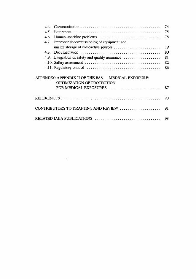
The dose and dose distribution within the patient are aimed at maximizing thelocal tumour control probability (TCP) while minimizing the normal tissuecomplications probability (NTCP). The margin for the overall error is very small(ideally below 5% overall deviation). A simplified example of this statement isillustrated in Fig. 1, where the TCP and NTCP curves are presented in relation to thedose to the planning target volume. The probability of local tumour control withoutcomplications has a maximum value, which provides the basis for the justificationand optimization of the radiotherapy treatment (dose, dose fractionation and dosedistribution).
It is worth taking a more detailed look at Fig. 1: the slope of the sigmoid curvesis usually steep. In the simplified example, if the maximum is situated at a dose wherethe slopes of the sigmoids are near 2, a deviation of 15% means a decrease in TCP oran increase in NTCP of 30%, which may jeopardize the justification of the decisionmade. However, this is only one example and the slope of the curves varies depending
100 r
Q.0 75
oOLO
50
25
Tumour -
Normal tissue —-f
Dose
FIG. 1. Tumour control probability (TCP) and the probability of normal tissue injury as afunction of radiation dose, in a hypothetical case. If normal tissue injury is to be avoidedaltogether, the radiation dose cannot exceed A; the TCP is low. By accepting a relatively smallprobability of normal tissue injury, the radiation dose can be increased to B, and the TCP issignificantly improved. A further increase in the radiation dose above C results in an increasedcomplication rate with very little improvement in TCP.

on the types of tumour and normal tissue as well as on the separation between theTCP and NTCP. Therefore, there is no single and sharp limit for the deviation onwhich the decision that a deviation is 'substantial' enough to constitute an accidentalexposure from the point of view of the outcome can be based; nevertheless, forpractical reasons, the generic level of 20% has been chosen by some nationalauthorities to make an investigation mandatory (10% if the prescribed number offractions is equal to or less than three).
On the basis of these considerations, it is understandable that mistakes andequipment faults have an immediate influence on the outcome of the treatment, andthat most of the accidents in this report involve patients, in some cases with serious,even fatal, consequences.
In addition to concern for patients undergoing radiotherapy treatment, potentialexposure from radiotherapy sources includes medical exposures to persons helping inthe comfort of patients, occupational exposures to staff and exposures to members ofthe public.
Accidental exposures may occur to visitors helping in the support ofbrachytherapy patients if rules are misunderstood or violated or if a radiation sourcebecomes accidentally exposed (i.e. accidentally falls out of the patient).
Potential exposures to staff may result from equipment failure such as a stucksource in a teletherapy machine or a remote control after-loading device, or when abrachytherapy source is accidentally removed from a patient. Accidents with severeconsequences have also occurred during maintenance or source exchanges.
With regard to potential exposures to the public, the most important case ofexposure is due to loss of control or abandonment of sources. Very seriousconsequences have also occurred with teletherapy units that are not well controlled orare improperly decommissioned. A patient discharged from the hospital withbrachytherapy sources mistakenly left inside his or her body may not only risk death,but also expose members of the public. A particular problem is posed by old 226Rabrachytherapy sources introduced in many countries several decades ago before anyregulatory control existed. The owners may have died, and the sources may remainwith members of the public who have no knowledge of the potential hazards theyentail.
2.2. METHODOLOGY FOR THE REVIEW
The description of an accidental exposure consists of a short account of theaccident itself, followed by the initiating events and the relevant contributing factors.Consistent with its objectives, the emphasis in this Safety Report is on the collectionand collation of a large selection of events and contributing factors, rather than on anexhaustive analysis of individual accidents.

Care has been taken to avoid making judgements. Rather, the plain presentationof a collection of facts was considered to be more effective towards increasingawareness, contributing to a questioning and learning attitude and adopting measuresfor the prevention of accidents and mitigation of their consequences.
In some accident scenarios where the dosimetric units are relevant to theprimary cause of the accident, SI units have not been used. Instead the old units suchas millicurie (mCi) and milligram radium-equivalent (mg Ra-eq) were quoted asused.
An initiating event is the action which triggers the chain of events (or eventsequence) which results in an accident. A contributing factor is any circumstance,condition, action or omission which plays a part in the precipitation of anaccident.
Frequent contributors to accidents were: insufficient education in radiotherapyphysics; a lack of a set of procedures and protocols integrated into a comprehensivequality assurance programme; and/or the lack of supervision over compliance withthe programme. Another finding was that training generally addresses only normalsituations and does not prepare radiotherapy staff for unusual situations, resulting ina lack of a 'safety culture'.
The review focuses on pure facts from which general lessons can be drawn andintegrated into the oncology service as part of a system for safety and quality.Contributing factors have been indicated by a general statement (which is common toa number of accidents), followed by detailed information specific to that particularevent. As an example, for Event No. 26, the general statement: "lack of or ineffectiveprocedures, protocols and documentation" is followed by the more specificinformation: "incorrect tables were accepted for use".
However, overly general statements such as "lack of overall quality assurance"have been avoided, since they would apply to most of the accidents and would conveyvery little information. The reader will be able to conclude that quality assurance waslacking whenever procedures were not complied with (or did not exist at all), orwhenever safety provisions to prevent accidents (such as independent checks forsafety critical issues) were non-existent.
2.3. CLINICAL CONSIDERATIONS RELATED TO QUALITY ASSURANCE
In clinical institutions and hospitals where accuracy and precision are notmaintained within a narrow range, there can be a tendency to avoid normal tissuecomplications by lowering the prescribed dose. The consequence is a drastic decreasein the probability of tumour control. The only acceptable solution is improvement ofaccuracy and precision so that the right doses can be prescribed. This practice ofprescribing lower doses than desirable cannot be considered as a potential exposure

or accident since the problem is associated with the prescription itself. This is an issueof ethics and is therefore not included in the methodology dealing with potentialexposures.
2.4. RADIATION MEASUREMENT SYSTEMS
The output of a therapy machine is measured by an ionization chamber andelectrometer with a calibration by, or traceable to, an authorized standard dosimetrylaboratory. Malfunction or improper use of this basic equipment can affect manypatients. Consistency among different sets of equipment requires frequent inter-comparison. Constancy checks verify that equipment performs correctly over time.
Event No. 1: Dosimeter calibration report used incorrectly
The institution had its ionization chamber and electrometer calibrated for 60Coat a standards dosimetry laboratory. The calibration certificate was in terms of doseto water, but was interpreted as specifying dose in air. The resulting error caused anoverdose to patients of approximately 11% for at least one year.
Initiating event— Calibration error: Incorrect use of a calibration certificate.
Contributing factors— Insufficient education, training or expertise: The physicist did not seem to
understand the calibration certificate.— Insufficient safety provisions (defence in depth): There was no independent
beam calibration by another physicist.
Event No. 2: Incorrect use of a plane parallel chamber
A new physicist at a hospital used a pancake chamber to calibrate severalelectron beams. A label on the chamber, placed by the previous physicist, indicatedthe side on which the beam should be incident. Although the previous physicist hadused the chamber correctly, his labelling was incorrect and the new physicist used thechamber upside down in the beam, resulting in the following errors in dose delivery,with the electron beams having the indicated energies:
6 MeV: 20% overdose,9 MeV: 10% overdose,
12 MeV: 8% overdose,

16 MeV: Correct dose,20MeV: 1% underdose.
Mailed thermoluminescence dosimeters (TLDs) from an independentlaboratory were used routinely by the institution for quality assurance of radiationbeam calibration. These dosimeters revealed the above discrepancies, whichprompted the physicist to investigate the cause. By the time the error was corrected,the incorrect beam outputs had been used for part of the treatments of only a fewpatients.
Initiating event— Incorrect calibration: Incorrect use of the plane parallel chamber to measure the
output of the electron beams.
Contributing factors— Insufficient education, training or expertise: The physicist used a chamber
unfamiliar to him without verifying the proper technique.— The previous physicist had labelled the chamber incorrectly.— Deficient communication/transfer of essential information: Either there was
miscommunication, or a faulty method was transferred from one physicist toanother.
Remedial action— The calibration measurements were repeated, with the chamber properly
oriented in the beam. The label on the chamber was corrected.
Event No. 3: Error in correction for atmospheric pressure
An institution did not own a barometer and the physicist relied on the localairport for measurement of atmospheric pressure. Pressure reported by the airport wascorrected to sea level, but the physicist interpreted the pressure report as appropriatefor the elevation of the site, about 1000 m. The use of the incorrect pressure resultedin errors in the measurement of machine outputs, which caused a 13% overdose topatients. The error affected the calibration of all machines at this institution.
Initiating event— Calibration error: The use of incorrect pressure values to correct for
atmospheric pressure resulted in an incorrect beam output.
Contributing factors— Shortage of instruments: The institution did not have its own barometer.
— Insufficient awareness:• Physicists relied on pressure data from an airport without knowing that the
data referred to sea level (requesting both station pressure and pressurecorrected to sea level should eliminate ambiguity).
• Physicists did not consider altitude as having a bearing on atmosphericpressure.
Remedial action— The institution purchased its own barometer which measured local pressure.
Event No. 4: Error in correction for atmospheric pressure
Atmospheric pressure was determined using data from nearby weather stations.The physicists concerned did not realize that these data were actually corrected to sealevel. This happened at four institutions, and six physicists were involved.
In at least two of the treatment centres the same incorrect pressure wasobtained redundantly. At three of the centres the overdose was 13-14% and at onecentre the overdose was 21%. Many patients were treated at each institute. The21% overdose by a 60Co unit continued for about ten months until a differentphysicist calibrated the machine and informed the radiation oncologist of themiscalibration. The radiation oncologist chose to continue with the older andincorrect calibration.
Initiating event— Incorrect calibration: The use of incorrect pressure values to correct for
atmospheric pressure resulted in an incorrect beam output.
Contributing factors— Shortage of instruments: The institutions did not have their own barometers.—Insufficient awareness:
• Physicists relied on pressure data from an airport without knowing that thedata referred to sea level (requesting both station pressure and pressurecorrected to sea level should eliminate ambiguity).
• Physicists did not consider altitude as having a bearing on atmosphericpressure.
Event No. 5: Lack of consistency between dosimetry at affiliated institutions
Three radiation oncologists and two physicists worked at three affiliatedhospitals. Each person worked in at least two, sometimes three, of the hospitals. Therewere two sets of calibration equipment (ionization chambers and electrometers),
usually assigned to a particular hospital. The two sets of equipment were neverintercompared although they were used by both physicists.
A review by an outside physicist showed a discrepancy of up to 15% betweenmeasurements made with the two sets of equipment. After discussion with thephysicists and radiation oncologists, one physician stated that he had noticed adifference in clinical results at two hospitals and had allowed for the difference byprescribing 70 Gy to the prostate in one hospital and 60 Gy in the other hospital foran equivalent treatment.
Initiating event— Incorrect calibration: An invalid calibration factor was used.
Contributing factors— Lack of or ineffective procedures: Different sets of equipment, which differed
in calibration, were used, without intercomparison, at two hospitals served bythe same radiation oncologists and physicists.
— Ineffective communication or transfer of essential information: Physiciansfailed to inform physicists that apparently there were differences in clinicalresults at two hospitals presumed to be using the same dosimetry techniques.Rather than discuss the problem with the physicists, at least one cliniciandealt with the discrepancy by using a 'clinical prescription correctionfactor'.
2.5. EXTERNAL BEAM THERAPY: MACHINE COMMISSIONINGAND CALIBRATION
Event No. 6: Incorrect calibration procedures
A new physicist adopted the calibration procedures used by the previousphysicist. The previous physicist had calibrated all beams for a linear acceleratorusing 200 monitor units, with the exception of one beam, which he calibrated at300 monitor units. For the latter beam, he used a 'conversion factor' to convert from300 to 200 monitor units. The new physicist calibrated all beams at 200 monitor units;however, he applied the previous physicist's conversion factor to one beam althoughit was now unnecessary. The miscalibration resulted in an overdose of 50% forpatients treated with that beam.
Initiating event—Incorrect calibration: Use of an inappropriate correction factor.
10
Contributing factors— Lack of or ineffective procedures and protocols: A physicist used data and
procedures established by a previous physicist without understanding them.— Ineffective communication or transfer of essential information: Poor handing
over of procedures from one physicist to another.— Lack of safety provisions (defence in depth): There was no independent beam
calibration by another physicist.
Event No. 7: Incorrect calibration of machine output
An institution had a new 60Co teletherapy source installed. The installer left acertificate specifying the exposure in rontgen per minute (R/min) at 1 m for the largestfield available. The institution used the installer's output, with corrections for inversesquare and decay, as the output in centigray per minute for all field sizes. When fieldsize dependence, backscatter, rontgen to centigray conversion, timer, etc., wereconsidered, the correct calibration revealed that patients received doses that wereapproximately 15% too low; the magnitude of the error depended on the field size.
Initiating event— Incorrect calibration of the beam: The machine 'calibration' was based on the
manufacturer's certificate of source strength for conditions inappropriate fortreatment.
Contributing factors—Lack of education in radiotherapy physics: There was poor understanding of the
factors needed to convert from the installer's statement of source strength to thedata used clinically.
— Lack of procedures and protocols: The physicist did not measure the machineoutput.
— Lack of safety provisions (defence in depth): There was no independent beamcalibration by another physicist.
Event No. 8: Calibration error after a source change in a 60Co unit
After a source change in a 60Co unit, the physicist incorrectly calibrated theoutput. An incorrect time of 30 s instead of 18 s (0.3 min) was used to calculate theabsorbed dose. This resulted in a 166% overestimation of the exposure time. Over aperiod of about one month, 115 patients were treated and received overdosage,resulting in severe health effects including fatalities. Inconsistencies in the dosimetrywere detected by the IAEA/WHO postal dose quality audit and by an external expert,but this did not trigger any significant improvements in the procedures.
11
Initiating event— Incorrect calibration of the beam: The dose rate used clinically was incorrect
because of a mistake in the exposure time during calibration.Contributing factors— Insufficient education on radiotherapy physics.— Lack of procedures, protocols and documentation: The absence of documented
procedures allowed the mistake to go unnoticed.— Lack of safety provisions (defence in depth):
• There was no independent calibration by another physicist of a new beam(after a source change).
• There was no comparison of the calibration for compatibility with the sourcemanufacturer's certificate of source strength or output.
• The treatment times were not reviewed for consistency with previoustreatments.
— Lack of awareness of physicians and of the person in charge of the dosimetry:Inconsistencies previously detected and notified were not investigated.
Event No. 9: Incorrect calibration of machine output
Electron beams of 7 and 11 MeV were calibrated incorrectly, resulting inunderdosage of 17-18%. On the same machine, a photon beam was calibratedincorrectly, resulting in overdosage of 5%. In addition, there was a drift in the beamoutput over time, up to 7%. Mailed TLDs indicated a potential problem, and a reviewby an independent, outside physicist revealed that the calibrations were incorrect.Apparently, the incorrect calibrations had been used clinically for at least 11 months.During that time, there was no record of quality control performed by the institution'sphysicist. The cause of the incorrect calibrations was unknown.
Initiating event— Incorrect calibration of the beam: The cause was unknown.
Contributing factors— Insufficient safety provisions (defence in depth): There was no independent
beam calibration by another physicist.— Lack of or insufficient quality control: Over an 11 month period no checks were
carried out on the beams.
Remedial action— Calibrations were corrected as a result of an external review by an independent
physicist. At the time of this report, it is not known whether the deviation inbeam output has been corrected.
12

Event No. 10: Calibration error after changing a ̂ Co teletherapy source
Physics staff calibrated the output of a new source installed in a 60Co unit.About three and a half months later, nursing staff began to notice that the skinreactions of some of the patients treated on the machine were not healing as rapidlyas would be expected. These concerns were communicated to the physics staff, whoreviewed the output tables in clinical use as well as the original calibration; theyreported that the data were correct. There was no explanation for the slow healing ofthe patients, which continued to be observed.
An intercomparison exercise, organized by a national association of medicalphysics, led to the discovery of a miscalculation in the original calibration. Over thefive month period, the 207 patients treated on the 60Co machine had received a dosethat was 25% higher than prescribed.
Initiating event— Incorrect calibration of a beam (the specific reason for the miscalibration was
not reported).
Contributing factors— Lack of safety provisions (defence in depth):
• There was no independent beam calibration by another physicist.• There was no comparison of the calibration for compatibility with the source
manufacturer's certificate of source strength or output.— Inefficient quality control:
• A verification of the calculations did not reveal the error. It is possiblethat the time for a calibration measurement was recorded in-correctly.
• Subsequent reviews of the calculations by the local staff were ineffective,despite indications of clinical problems.
— Insufficient investigation of unusual patient response: There was no persistentsearch for the causes of the slowly healing skin effects.
Event No. 11: Incorrect calibration of a machine with asymmetric jaws
A linear accelerator with asymmetric jaws was calibrated with the detectorpositioned in the penumbra region and did not represent the dose in the centre of thefield. The result was that patients received an overdose of 27%.
Initiating event— Incorrect calibration of the beam: The calibration was made in the
penumbra.
13

Contributing factors— Insufficient education and training in radiotherapy physics: There seemed to be
a lack of understanding of the features of an asymmetric beam.— Insufficient defence in depth: There was no independent beam calibration by
another physicist.— Lack of or inefficient procedures and protocols: Procedures for the
commissioning of a new machine were not appropriate.— Insufficient awareness and alertness.
Remedial action—The machine was calibrated correctly and new data placed in clinical use.
Event No. 12: Incorrect decay of a 60Co source and fabrication of records
A 60Co unit was calibrated correctly initially, but subsequently corrected fordecay in the wrong way and no re-measurement of the output was made. The overdoseto patients increased progressively over a period of 22 months, reaching 10% in5.5 months and as much as 50% in the subsequent 16.5 months. During the latterperiod, 426 patients were treated. Of the 183 patients who survived beyond one year,34% had severe complications in various sites, including the brain, spinal cord, skin,oropharyngeal mucosa, colon and rectum, which led to the death of some patients.
Initially the incorrect machine output was attributed to a faulty measuring system.The physicist at the institution produced ten calibration documents, which turned outto be fabncated, that showed that the machine output used clinically was correct.
When some patients showed symptoms of overexposure, the hospital askedconsultant physicists to investigate the dosimetry. Their findings were that:
(a) Both the 60Co unit and the measuring system apparently had been functioningcorrectly during the 22 month period.
(b) The hospital physicist had fabricated all but one of the reports of measurementof machine output. The output used clinically had been calculated, without acheck by measurement, and the calculation was incorrect.
Initiating event— Incorrect calculation of output: The output was based on an incorrect decay of
source output.
Contributing factors— Apparently, insufficient staff resources were devoted to dosimetry and quality
control: The beam output was not checked over a 22 month period, since thephysicist was assigned to a new accelerator.
14

— Insufficient defence in depth: There was no independent check of decay curves.—The physicist falsified records in an attempt to justify his calculations.
Remark— The number of staff and training needs should be reviewed whenever the
workload increases, a new machine is purchased or a new technology ortreatment technique is introduced.
Event No. 13: Use of incorrect tables for decay of a 60Co source
A physicist generated tables of decay factors to correct the output of a 60Coirradiator. The tables were generated by a computer, with the year incorrectly enteredas one year earlier. Consequently, for a three month period, patients received a doseapproximately 12% less than that prescribed. The error was detected during a reviewof dosimetry by an outside physicist.
Initiating event— Incorrect calculation of output (it was based on incorrect decay of source output).
Contributing factors—Insufficient safety provisions (defence in depth):
• There was no independent check of decay curves.• The treatment times were not reviewed for consistency with previous
treatments.
Event No. 14: Lack of communication regarding units of outputof a treatment machine
A physicist calibrated a 60Co unit in terms of rontgen/mmute at a 5 cm depth ina Masonite phantom. The radiation oncologist, who performed all treatment planningand dosimetry calculations, assumed that the output was in rad/minute to aminiphantom (muscle) at the depth of maximum dose (0.5 cm). The magnitude of theerror, depending on the field size, can reach an overdose of 20-25%.
Initiating event— Incorrect clinical use of a beam calibration.
Contributing factors—Lack of or ambiguous assignment of responsibilities: The entire dosimetry
system, from calibration of machine output to calculation of treatment time,was not the responsibility of one person (preferably the physicist).
15

— Ineffective communication/transfer of essential information: There was a lackof communication between the physicist who calibrated the machine and thephysician who performed treatment planning.
—Lack of or inefficient procedures, protocols and documentation: Thetechnique used by the physicist for calibrating the machine output, in terms ofrontgen in a Masonite phantom, was unusual and does not follow acceptedprotocols.
Remedial action— A review of dosimetry by an outside physicist revealed the misunderstanding
concerning the machine outputs and corrections were made.
Event No. 15: Radiation leakage from the collimator of a linear accelerator
A patient who had been treated for prostatic cancer was seen a few days afterhe had finished his treatment course. The patient had developed a severe skin reactionon the ventral part of the right arm, the right part of the thorax and the right part ofthe head. In addition, he had lost some hair from his head. The skin reaction wasinvestigated further, leading to the diagnosis of radiation dermatitis caused by anestimated dose of 10-20 Gy. The treatment had consisted of a course of 25 x 2 Gywith 25 MV photons, using a three beam technique (anterior plus lateral-obliquebeams), produced by a 14 year old accelerator.
During the radiotherapy treatment period of this patient, the accelerator showeda slow start at 'radiation on' and radiation was sometimes delivered at a dose rate thatwas less stable than normal, especially at some gantry angles; sometimes the startingprocedure failed completely. Qualified hospital and manufacturer's technicians madea thorough investigation of the origin of these problems, resulting in some additionalmaintenance and an increase in the frequency of quality control checks. Thefunctioning of the accelerator was monitored almost daily. No deviations from thenormal beam properties was observed. The interlock system had never been set offduring patient treatment.
Detailed observations of the skin reaction of the patient revealed that a skinreaction was not seen behind the right ear and that the oral mucosa did not show anyreaction. It was therefore concluded that the skin reaction could have been causedonly by low energy radiation.
The most probable position and direction of the apparent source of radiationleakage was reconstructed from the region of the skin reactions in relation to thegantry and collimator position of the accelerator under the treatment setup. Fromthis reconstruction it was concluded that the source of radiation leakage was locatedat the upper end of the beam guidance tube close to the last part of the bendingsystem.
16

It was thereby demonstrated that, despite the relatively high degree of controland the presence of safety systems, this old accelerator could be prone to producinga large amount of radiation leakage which was harmful to patients.
Initiating event— Unknown, but thought to be a misadjustment of an accelerator parameter which
made the accelerator operate in a very unusual mode.
Contributing factor— Possibly, advanced age of the installation. The only unusual symptom was
beam instability. Operating procedures called for this to be investigated but didnot recognize it as a reason to stop operation of the machine when the beamproperties appeared normal.
Event No. 16: Treatment with an accelerator operated in the PHYSICALmode
During clinical use of a linear accelerator, a problem arose with the selection ofelectron and X ray energies. A technician from the manufacturer reported that theproblem was associated with a microswitch required to select the scattering foil forelectron beams or the beam flattening cone for photon beams. About one month later,on two consecutive days, the same problem reappeared briefly. The machine was notrepaired at this time. Four days later the problem returned permanently and themachine could be operated only at two X ray energies and one electron energy(10 MeV). Two weeks later, it was impossible to start the accelerator in theMEDICAL mode. The electronics engineer informed the chief radiation oncologistabout the problem. The radiation oncologist asked the electronics engineer to alter themachine so that patients could be treated in the PHYSICAL mode.
The electronics engineer switched the linear accelerator to the PHYSICALmode and made the following provisions for treatment:
(1) He covered the push buttons that are operative in the MEDICAL mode withpaper;
(2) In co-operation with the technician, he measured the absorbed dose understandard conditions for all beams;
(3) He prepared a written report describing procedures for the treatment ofpatients under the PHYSICAL mode and fixed it to the console of the linearaccelerator;
(4) He instructed two radiographers regarding operation of the linear acceleratorfor treatment in the PHYSICAL mode and observed the treatment of the firstfour patients.
17

Thirteen patients were treated with apparently no problems, the last patientbeing treated with a 10 MeV electron beam. Data for the next patient were set on theconsole for treatment with 20 MV X rays, dose rate 300 MU/min, without anyproblem. The treatment started but was terminated by a timer after 21 s; the timer wasthe third independent safety system designed to switch off the beam if two lonizationchamber monitors failed. (According to the radiographer, the audible dose rateindicator beeped with a very low frequency.) When the treatment was terminated,only a few monitor units were displayed on both monitors; the radiographer thereforeimmediately called the electronics engineer to check the machine. When the patientwas removed from the treatment room, the radiographer observed a skin reaction onthe patient, which indicated a high degree of overexposure. Treatment wasimmediately suspended.
The electronics engineer used an ionization chamber to measure the acceleratoroutput under the same operating conditions used for treatment. He found that therewas an extremely high dose in the centre of the field, caused by a failure to deploythe X ray target, monitor chamber and beam flattening cone, leaving the 10 MeVelectron scattering foil and (disconnected) electron monitor chamber in position. Thismalfunction was due to the failure of a power supply, which supplied the interlocksystems, that should have been active in the non-clinical mode. Hence, no indicationof the wrong positioning of the essential elements in the beam line appeared on thecontrol panel. Operation in the PHYSICAL mode had disabled interlocks that couldhave detected this dangerous condition.
Initiating event— Treatment in non-clinical, PHYSICAL mode (decision by the radiation
oncologist after several failures with interruption of treatments).
Contributing factors— Equipment failure: The accelerator failed to operate in the clinical (MEDICAL)
mode.— Maintenance problems: Intermittent, unresolved equipment failures, the cause
of which was not identified.
Event No. 17: Mishandling of equipment failure
Staff operating a linear accelerator noted that the machine would not producean electron beam. They reported the fault to a maintenance technician from themanufacturer, who was working on the 60Co unit located in an adjacent room. Thetechnician performed corrective maintenance but, after the repair, the analog displayof electron energy permanently indicated 36 MeV regardless of the energy selected.
18

The energy selection keys correctly indicated the energy selected. The analogindication of the 36 MeV was interpreted as a faulty meter displaying only 36 MeV.
Patients were treated under the above conditions for ten days before theproblem with the energy display was reported to the Department of Physics andRadiation Protection. Treatments were then terminated. Physicians began to correlatethe poor tolerance and severe reactions observed in some patients with themalfunction of the machine.
A dosimetric check performed the next day showed that the energy of theelectrons was 36 MeV, regardless of the energy (7, 10 or 13 MeV) selected onthe console. The manufacturer was informed and sent technicians to repair themachine.
The failure to select the electron beam energy was due to a short circuit of thesystem that selects the trajectory of the electron beam. The path curvature is afunction of both the electron energy and the intensity of the magnetic field generatedby the coils. Instead of correcting the basic cause (the wrong coil current resultingfrom the short circuit), the energy of the electrons was manually adjusted to match thewrong current. The correct trajectory was obtained for the electron beam, but at thewrong energy (permanently 36 MeV). The analog indicator of the energy (whichindicates the current of the bending coil and depends on the selected energy) waspermanently indicating 36 MeV, even between treatments, owing to the short circuitin the control of this current.
When the maintenance technician had selected the manual mode of energyadjustment, he effectively put the machine into a non-clinical mode in which it wouldignore the energy selected on the console. Therefore, the energy of the electrons wasalways 36 MeV, with the beam concentrated in the centre of the field. The result wastreatment with electrons that delivered a dose several times higher and irradiated avolume deeper than prescribed. The malfunction affected 27 patients.
Initiating event— Incorrect maintenance: Misadjustment of the beam energy.
Contributing factors— Ineffective communication/transfer of essential information and non-
compliance with procedures: Physicists were not notified immediately aboutthe malfunction of the machine (treatments were resumed without a check ofthe beam output). Thus procedures for transferring the machine to and from themaintenance technician were not followed.
— Incorrect interpretation of conflicting signals: The reading on the analog meterindicating the actual energy was ignored.
•—It was possible to operate the accelerator with the energy selection disabled(equivalent to 'non-clinical mode').
19
— Shortage of instruments for quality control: Instruments for quick (daily)constancy checks of beam output and energy, compatible with patientworkload, were not available.
Event No. 18: Dose on central axis incorrect because of a loosewedge mounting mechanism
Wedge factors were measured on a 60Co unit with the beam in the verticalposition. When the machine gantry was rotated 90° for treatments with a horizontalbeam, a loose wedge mounting mechanism allowed wedge filters to shift so that thedose distribution and the central axis wedge factor were incorrect. The error was aslarge as 8%, with the dose to the patient too high across the beam for one horizontalmachine position and too low for the other horizontal beam position.
Initiating event— Deficiency in the design of accessories of the machine: Mechanical deficiency
related to the wedge holder.
Contributing factor— Lack of or ineffective procedures for commissioning: Wedge factors were not
measured with a horizontal beam at the time of commissioning of the machine(the use of accepted protocols with documentation of the data would avoidomitting measurements).
Event No. 19: Holder for port film left in beam during treatment
A lucite plate 1.25 cm (0.5 in) thick used to hold port films was left in the beamduring treatment with a linear accelerator. There was no interlock system to detect thepresence of the plate. An independent check of beam output with TLDs indicated thefollowing per cent relationship between the measured and expected output:
_ , . , Institution s expected dose/Radiation beams , ., —,*,„*
dose measured by TLD (%)
Photons: 10 MV 90Electrons: 6 MeV 10
9 MeV 2612 MeV 3016 MeV 4620 MeV 57
20

Further investigation by the institution's physicist revealed that the outputmeasured by the TLDs was correct when the plate was in the beam.
Initiating event— Mistake in the treatment setup: A holder for portal films was left in the beam
during treatment.
Contributing factor— Equipment design deficiency: There was no interlock to detect the presence of
the portal film holder.
Remedial action—The linear accelerator was retrofitted with an interlock to detect the presence of
the portal film holder.
Event No. 20: Design error in accelerator control software
A linear accelerator equipped with computer controlled electron and photonbeams was in clinical operation. Several serious incidents led to the recognition of aproblem whenever an initial instruction for 25 MV X rays was changed quickly to anelectron beam instruction. The computer process for setting and verifying themachine parameters required about 20 s to complete after the X ray mode wasselected. If an electron mode was selected before the computer process for X rays wascompleted, the monitor displayed electron mode but operated from hybridinstructions. The resulting beam consisted of 25 MeV electrons with high beamcurrent appropriate for X rays but without beam scanning, with no X ray target andno X ray beam flattener in the beam. The resulting electron dose at the position ofmaximum dose was 160-180 Gy before the beam cut off automatically, in one secondor less.
When the above sequence of events first occurred it was not recognized as afault by the manufacturer of the accelerator. One month later the same sequenceoccurred at another hospital and was attributed to microswitch failure. Six monthslater a third hospital reported a similar experience with yet another patient. Themanufacturer made some modifications in response to the assumed microswitchfailure.
Three months later, following a fourth occurrence at another hospital, thepossibility of a malfunction was rejected by the manufacturer. One month later afurther accident in the same department led to an investigation by the hospitalphysicist. The fault was demonstrated and software errors were suggested as thecause. The physicist verified that other machines of the same type functioned in thesame way. One additional accident occurred before all accelerators of that type were
21

withdrawn from clinical use. Of six patients treated during the machine malfunction,two died, apparently of overexposure.
Initiating event— Equipment malfunction due to operation in extreme conditions (it operated on
hybrid instructions after the operator changed too quickly from the X ray modeto electron mode).
Contributing factors— Insufficient factory testing: Computer controlled radiation equipment was not
factory tested for extreme or unusual operating conditions that could occur inpractice.
— Staff operating the linear accelerator requested the electron mode beforethe computer had completed processing of the previously requested X raymode.
— Insufficient follow-up by the manufacturer: The reported incidents weremisinterpreted or dismissed by the manufacturer until, after similar incidents insix different hospitals, a hospital physicist identified and demonstrated thenature of the fault.
2.6. EXTERNAL BEAM THERAPY: TREATMENT PLANNING,PATIENT SETUP AND TREATMENT
Event No. 21: Inconsistent sets of basic machine data
For one machine, an institution had two sets of data for output factors, depthdose and other basic parameters. The two sets of data differed by 10%, with one setbeing correct. The data were used interchangeably for a period of time, with the resultthat some patients were underdosed by 10%.
Initiating event— Incorrect data for patient dose calculations
Contributing factors— Lack of or ineffective procedures, protocols and documentation: The institution
had two sets of data that were inconsistent.— Insufficient safety provisions (defence in depth): An incorrect table of values
was used without verification.
22

Event No. 22: Incorrect data for tissue maximum ratios
Tables of tissue maximum ratios were generated for a 10 MV photon beam,based on the institution's measurements. Inspection of the tables during a review ofdosimetry by an outside physicist after several months of use revealed aninconsistency in the trend of data with increasing depth and field size. Checks againstthe original measurements showed that at least 15 entries in the table were incorrect,some by as much as 13%.
Initiating event— Incorrect data for patient dose calculations.
Contributing factors— Insufficient safety provisions (defence in depth): Tables of measured tissue
maximum ratios were not verified against published values before clinicaluse.
Remedial action— The tables were corrected. Charts of patients affected by the incorrect data were
reviewed and the calculations corrected.
Event No. 23: Insufficient understanding of the treatment planning system(TPS) algorithm
A treatment planning computer was used to calculate 60Co treatment plansinvolving wedges. The technologist and dosimetrist were not sure whether thecomputer calculation included the wedge factor. They asked the consultantphysicist and were told that the wedge factor was not included in the computercalculation and should be accounted for manually. Three patients were treatedin this way. While preparing to treat an additional patient following thesame treatment protocol, a hand calculation of the treatment time indicated alarge discrepancy with the computer generated value. The computer alreadyincluded the wedge factor, so that this factor had been applied twice. A review ofcases revealed misadministration for three other patients, who receivedoverexposures of close to 11, 12 and 14% for prescribed doses of 62, 65 and 50 Gy,respectively.
Initiating event— Incorrect data for patient dose calculations (the wedge factor was applied
twice, once by hand and once by the treatment planning computer).
23

Contributing factors— Lack of or ineffective procedures, protocols and documentation:
Commissioning with incomplete validation of the computer TPS.— Insufficient safety provisions (defence in depth): There was no regular manual
checking of computer calculations.
Event No. 24: Incorrect basic data in a TPS
Basic data used in a TPS differed from measured data for a particular linearaccelerator; the inconsistency was not detected during commissioning of the planningsystem. The result was that patients received a 15% overdosage. The data had beenentered into the computer by the hospital's previous physicist and the reason for theerrors was unknown.
Initiating event— Incorrect data for patient dose calculations (incorrect basic data used in a TPS).
Contributing factors— Lack of or ineffective procedures, protocols and documentation:
• Inadequate commissioning (TPS).• Inadequate transfer of responsibilities and/or taking over by newly appointed
staff, in this case the physicist.— Insufficient safety provisions (defence in depth): No manual checks of the
treatment plans were made.
Remedial action— The data in the TPS were corrected.
Event No. 25: Incorrect depth dose data
During installation of a linear accelerator, an institution contracted the servicesof the manufacturer to measure depth dose data. The institution's physicist laterchecked the data and found an 8% discrepancy between his measurements and those ofthe manufacturer for some field sizes and depths. He concluded that the manufacturer'sdata were correct and used them clinically. A review by an outside consultantphysicist revealed that the physicist's measurements were correct. During a period ofseveral months, some patients received doses that were 8% lower than prescribed.
Initiating event— Incorrect data for patient dose calculations: The manufacturer provided basic
data that were incorrect for some field sizes and depths.
24

Contributing factors—Lack of or ineffective procedures, protocols and documentation: Incorrect
commissioning (data generated by the manufacturer were accepted fortreatments, although the institution's physicist found an 8% discrepancy whenhe compared these data with his measurements).
— Insufficient education, training or expertise: A known discrepancy was notresolved.
— Lack of or ineffective assignment of responsibilities: The physicist did not takeresponsibility for all aspects of dosimetry.
Event No. 26: Incorrect calculation of treatment times
Tables of treatment times used clinically were incorrect; the reason for theerrors was unknown. Thirteen patients who were prescribed 60Co teletherapytreatments received doses that were from 15 to 40% lower than the doses intended.
Initiating event— Incorrect data for patient dose calculations: Basic data (tables of treatment
times) were incorrect.
Contributing factors—Lack of or ineffective procedures, protocols and documentation: Incorrect
tables were accepted for use.—Insufficient safety provisions (defence in depth):
• There was no independent verification of basic data (time charts used tocalculate treatment times).
• There was no independent check of treatment times calculated for individualpatients.
Remedial action— A new departmental policy required a check by an independent person of daily
treatment times for each patient. The check was performed on a weekly basisor whenever a treatment time was changed.
Event No. 27: Misapplication of distance correction
An institution treated most patients with a constant source-skin distance (SSD)technique, although some patients were treated with a constant source-axis distance(SAD) or isocentric technique. For an isocentric treatment plan, the dosimetristassumed that it was necessary to use a correction factor to convert from SSD to SAD.The physicist checked the method. However, the computer program for isocentric
25

techniques had already allowed for this factor. This practice of manually correcting forisocentric treatments continued for nine years, until the systematic error was discovered.
Over the nine year period, all patients treated isocentrically received incorrectdoses due to the double application of the inverse square correction. Although thesepatients were a small percentage of all patients, at least 1000 patients received dosesless than prescnbed. The magnitude of the error depended upon the depth of theisocentre, but for some patients the error may have been 30%.
Initiating event— Incorrect patient dose calculations: The SSD was entered manually into a
computer treatment planning program for isocentnc techniques, although theprogram allowed for that already.
Contributing factors— Insufficient education, training or expertise: The training should include the
particular piece of equipment TPS.— Lack of or ineffective procedures, protocols and documentation: Incorrect
commissioning of the treatment planning computer, with poor understanding ofthe computer algorithm.
— Insufficient safety provisions (defence in depth): Lack of independent checkingand verification procedures.
Event No. 28: Incorrect calculation using the inverse square law
A patient was prescribed a 60Co teletherapy treatment of 20 fractions of 2 Gyeach. The prescribed SSD was 70 cm; the SSD used for most patients was 80 cm. Thephysicist who calculated the dose used an incorrect inverse square correction factorthat led to a given dose which was 70% too high. Consequently, the patient received3.4 Gy per fraction instead of the intended 2 Gy for the first eight fractions. Thecalculations were not checked until after the eighth fraction, when a junior physicistdiscovered the mistake. The dose for the remaining fractions was reduced tocompensate for the excess dose delivered during the earlier treatments.
Initiating event— Incorrect data for patient dose calculations (an incorrect inverse square
conversion factor was used).
Contributing factors— Insufficient awareness: The treatment was unusual (the treating distance was
unusual, 70 cm rather than 80 cm SSD), without there being sufficientawareness of the difference.
26

— Insufficient safety provisions (defence in depth): Lack of an independent checkof the treatment plan.
— Lack of or ineffective procedures, protocols and documentation: Treatmenttimes were not checked until the patient received eight fractions of histreatment.
Remedial action—A procedure was introduced which required that calculations be checked before
the third fraction of treatments consisting of more than three fractions. Fortreatments requiring three or fewer fractions, calculations were to be checkedby the first fraction.
Event No. 29: Incorrect calculation of treatment times for openand wedged beams
One field was treated with a combination of open and wedged beams on a60Co unit. The treatment times were reversed when calculating the dose distribu-tion for the total treatment, with the result that the patient received an overdoseof 10%.
Initiating event— Incorrect patient dose calculations (treatment times were reversed for open and
wedged beams).
Contributing factor— Insufficient safety provisions (defence in depth): Lack of an independent check
of the treatment plan.
Event No. 30: Error in the wedge factor
During a review of past treatment data, 12 wrong treatments were discovered.Some patients were found to have received doses 10-18% higher and some 10-27%lower than the prescribed doses. The overdoses resulted from erroneous informationin the treatment planning computer reference data. All these treatments, with oneexception, involved the use of wedge filters.
The treatment discrepancies were first discovered when a therapy technologistexamined the files of previously treated patients in order to practice hand calculateddosimetry for an upcoming board certification test. The radiation oncology staff thenbegan recalculation of doses to all patients who had received wedge relatedtreatments since the introduction of that type of treatment two years earlier. The staffalso re-ran the original computer calculations, which showed that administered doses
27
had deviated significantly from prescnbed doses. In 11 of 12 wrong treatments theplanning staff failed to use a computer program 'wedge normalization factor' inmaking initial dose calculations. The use of this wedge normalization factor isdescribed in the manufacturer's computer instruction manual. Instead of using thisgenerated factor, the planning staff used different measured wedge factors.
Initiating event— An incorrect procedure was used for computerized treatment planning
involving wedges.
Contributing factors— Lack of or ineffective procedures, accepted protocols and documentation.— Insufficient safety provisions (defence in depth):
• Lack of an independent check (of dosimetry involving the use ofwedges).
• Computer calculations were also not manually checked.
Event No. 31: Wedge factors used twice in the calculation of treatment times
A physicist began employment at a new institution, which had the same TPS hehad used in his previous place of employment. At the new institution, the wedgefactors were included in the computer calculations; at the previous institution, thewedge factors were applied manually for each patient. The physicist used, in error, theTPS in the same way as he had used it in his earlier position. In other words, heapplied the wedge factor at the new institution manually for each patient, whichmeant that this factor was used twice for the calculation of treatment times. For onepatient the result was a 53% overdose for a boost (wedged) field, resulting in a totaldose that was 17% higher than prescribed.
Initiating event— Incorrect patient dose calculations (a wedge factor was applied twice in the
calculation of the treatment time).
Contributing events— Insufficient education, training or expertise: Lack of understanding of the TPS
operation.— Insufficient safety provisions (defence in depth):
• Application without verification of a calculation adapted from anotherhospital.
• The computer calculations were not manually checked.
28
Event No. 32: Failure to include intended wedges in the treatment setup
A patient was prescribed a 60Co teletherapy treatment of 16.6 Gy. The treat-ment required two fields using 30° wedges. The physicist failed to record the wedgesin the setup portion of the patient's chart and the wedges were not used duringtreatment. When the error was detected, the patient had received a dose of 23.0 Gyto the reference point on the central axes instead of the prescribed 16.6 Gy, anoverdose of almost 40%. In addition to the overdose at the reference point, the dosedistribution was non-uniform, with the hottest region receiving a much largeroverdose.
Initiating event— Incorrect patient dose calculations: The physicist failed to specify wedges in the
setup instructions.
Contributing factors— Insufficient safety provisions (defence in depth):
• There was no independent verification of the treatment parameters.• There was no inspection of the resulting isodose distribution.
Event No. 33: Misunderstanding of a complex treatment plan given verbally
A patient was prescribed a 60Co teletherapy treatment to two different treatmentsites. The two sites were to be treated daily, with the first site receiving 2.4 Gy eachday for 20 days and the second site receiving 2.5 Gy each day for 10 days. The twotechnologists involved in this procedure misunderstood the physician's verbalinstruction concerning the treatment plan and failed to recognize the different numberof treatment fractions for the two sites. Therefore, the second site received anadditional four days of treatment before the error was detected. The patient received10 Gy more than was prescribed, an overdose of 40%.
Initiating event— Incorrect treatment setup.
Contributing factors— Lack of or ineffective procedures, protocols and documentation:
• There were no written procedures for treatment prescription.• Unclear verbal prescription.
— Unusually complex treatment involving two sites that were to receive differentdoses in different fractions.
29

Remedial action— A new protocol required that a 'stop' mark be placed in each patient's
daily treatment log, indicating the projected final day of treatment for eachsite.
Event No. 34: Incorrect identification of patient
The radiation technologist called the patient's name but did not confirm hisidentity by looking at his photograph in the record. The radiation technologist had notseen the patient and, therefore, did not realize that another patient had responded.When he was being positioned for therapy, freckles on his back were mistaken fortreatment positioning tattoos and were used for the setup. The radiation technologistdid not recognize the absence of treatment tattoos in the patient's lumbar/sacral spinearea despite a photograph. The patient indicated that his setup was not correct and theradiation technologist called the oncology physician to venfy that the treatment wascorrect. The physician verified that the treatment was correct on the chart but did notspeak to or examine the patient. The patient was then administered 2.5 Gy from ateletherapy unit to the lumbar/sacral spine, i.e. the wrong site.
Initiating event— Incorrect treatment setup: A patient responded when another patient's name
was called.
Contributing factors— Non-compliance with procedures:
• For patient identification: There was no check of the patient's photograph oridentity bracelet, if there was one.
• For site identification: There was no confirmation of anatomical marks forbeam location.
— Lack of awareness: No one heeded the patient's warning and objection to beingtreated at the wrong site.
Event No. 35: Treatment setup based on another patient's chart
A patient was prescnbed a 60Co teletherapy dose to the brain but received anunintended dose of 0.45 Gy to the lung. For instructions regarding setup, thetechnologist picked up the chart of another patient, who was prescnbed a lungtreatment. The technologist positioned the patient without confirming his identityagainst the chart and proceeded with the lung treatment. During the treatment, asecond technologist noted the discrepancy between the patient's name and the chartand terminated the treatment.
30
Initiating event— Incorrect treatment setup: The technologist used another patient's chart to set
up the treatment.
Contributing factors— Inattention or lack of awareness.— Non-compliance with procedures for patient identification.
Event No. 36: Calculation error after a change of treatment regimen
A 60Co teletherapy treatment was prescribed to the larynx, a total of 76.8 Gyin 64 fractions of 1.2 Gy each, two fractions per day. After 18 treatments, theoncologist revised the treatment plan. He reduced the field size and moved theradiation field off the spinal cord. The technologist made a mistake in thecalculation of the new treatment time and the patient received twice the prescribeddose for the next 22 fractions. Instead of the intended 26.4 Gy for the second partof the treatment, the patient received a dose of 52.8 Gy. The physicist reviewedthe treatment plan and patient chart on a weekly basis, but failed to notice themistake until the third review. The treatment was immediately suspended. By then,40 fractions had been administered. It was estimated that the patient received a totaldose of about 75 Gy in 40 fractions rather than the prescribed dose of 76.8 Gy in64 fractions.
Initiating event— Incorrect patient dose calculation: The technologist miscalculated the treatment
time for the revised treatment plan.
Contributing factors— Insufficient safety provisions (defence in depth): Lack of an independent check
of the treatment plan (treatment times).— Procedures followed mechanically without sufficient awareness: Ineffective
weekly checking of the patient's chart.
Event No. 37: Confusion of fractional dose and total dose
A child was prescribed 60Co teletherapy treatment to the brain. The totalprescribed dose was 3 Gy, to be delivered in two fractions of 1.5 Gy each. Thedosimetrist calculated the dose for 3 Gy per treatment. Three other individualsreviewed the calculations before the treatment started and failed to notice the error.As a result, the patient received a total dose of 6 Gy. The error was discovered by astudent therapy technologist who reviewed the treatment plan.
31

Initiating event— Incorrect patient dose calculation: The dosimetrist misunderstood the total dose
for the fractional dose in a two fraction treatment.
Contributing factors— Ineffective communication/transfer of essential information.— Procedures followed mechanically without sufficient awareness: Three persons
failed to detect the error.— The nature of the treatment combined with insufficient awareness favoured the
mistake.• The treatment was unusual (fractionation).• The treatment was very short (two sessions), which increased the risk of not
detecting the error before treatment was completed.
Event No. 38: Incorrect positioning of treatment beams
A patient was prescribed 60Co teletherapy to both the right hip and thethoracolumbar spine, to be completed on different days. After the patient completedthe thoracolumbar spine treatment, the technologist mistakenly continued treatmentto the same region for a total of 12 Gy. The staff technologist involved in thistreatment failed to notice the statement in the patient's chart that the treatment to thespinal area had been completed. Six more treatments were given. The mistake was notdetected during the routine chart review by a physicist.
Initiating event— Incorrect treatment setup: The technologist did not check the patient's chart
carefully before setting up a field and administering radiation.
Contributing factor— Inattention or insufficient awareness.— Insufficient safety provisions (defence in depth): The patient setup was not
checked by another person (the technologist worked alone).
Event No. 39: Wrong tattoo used in treatment setup
A patient was prescribed a 60Co teletherapy treatment to the lumbar spine butreceived an unintended dose of 2.5 Gy to the thoracic spine. The patient hadpreviously received teletherapy treatments over the thoracic spine, and the tattoo(anatomical marker) was still visible. The technologist setting up the treatment failedto review the patient's chart, which indicated the proper treatment, and used the tattoofrom the previous treatment.
32

Initiating event— Incorrect treatment setup: The technologist used a tattoo from a previous
treatment to set up a new series of treatments.
Contributing factors— Ineffective procedures: The patient had two tattoos, one of which was from a
previous treatment that was near the site of the tattoo for the current treatment— Inattention or insufficient awareness: The technologist did not carefully review
the patient's chart for the location of the treatment site.
Remedial action— After this incident, technologists were instructed to have a physician or
dosimetrist present during patient setup.
Event No. 40: Treatment of the wrong patient and lack of communicationbecause of language problems
A patient was scheduled to receive treatment to the head and neck area using alinear accelerator. The patient spoke little English and the oncologist did not speak thepatient's language. The oncologist asked the patient which area of the body was beingtreated and the patient pointed to his head. Without reviewing the patient's chart, thephysician assumed that he was scheduled for treatment with a 90Sr plaque andadministered a dose of 10 Gy to the surface of the right eye.
Initiating event— Incorrect treatment setup: Incorrect patient identification.
Contributing factors— Non-compliance with procedures for patient identification: The oncologist did
not check the patient's chart.— Inattention or insufficient awareness: The radiation oncologist relied on the
patient to locate the treatment site.— Ineffective communication/transfer of essential information: The patient and
the oncologist did not speak the same language.
Event No. 41: Incorrect labelling of a simulator film
A patient was to receive a 60Co treatment of 2.16 Gy to the left posterior neck.However, the treatment was delivered to the right posterior neck. Because thetreatment simulation was performed in the prone position rather than in the routinesupine position, the right side of the simulator film was marked as the left and the
33

patient was set up incorrectly. In reviewing the simulator film, the oncologist failedto notice that the treatment simulation was to the wrong side of the patient's neck.
Initiating event— Incorrect simulation/localization: Incorrect labelling of the localization film.
Contributing factors— The treatment was simulated in an unusual position.— Insufficient safety provisions (defence in depth): There was no check of the
orientation of the anatomical site relative to the film.
Event No. 42: Field improperly located on patient
A patient was prescribed a treatment of 3 Gy to the cervical spine. Thetechnologist looked at the patient's chart but still mistakenly set up the treatment fieldon the thoracic spine. The patient received 2.87 Gy to the thoracic spine instead of theprescribed 3 Gy to the cervical spine The patient had previously received treatmentto the thoracic spine, leading the technologist to assume that the thoracic treatmentwas continuing.
Initiating event— Incorrect treatment setup: Failure to adequately examine the patient's chart
before administering treatment.
Contributing factor— Insufficient safety provisions (defence in depth): There was no independent
check of the chart and the setup.
Event No. 43: Field improperly located on patient
A patient was prescribed 60Co teletherapy of 2 Gy per fraction to themediastinum. The technologist marked the beam setup incorrectly as a brain treatmentand the patient received ten fractions to the brain before the error was detected,even though the technologist was to have been supervised by qualified staff members.
Initiating event— A technologist set up a new patient and marked the wrong beam position.
Contributing factors— Ineffective communication/transfer of essential information: The prescription
of the oncologist was misread, by the technologist, ten different times.
34
— Inattention or insufficient awareness: Checks by three staff members failed todetect the setup mistake.
Event No. 44: Incorrect positioning of patient
A patient was to receive 60Co treatments of 10 fractions, 3 Gy each, to the rightscapula. After the second fraction, the oncologist reviewed the port film and noticedthat 80% of the intended target area had been missed. A chart review indicated thatduring simulation of the treatment, the oncologist had placed a mark on the patient'schest as indicated by the ceiling laser. However, for treatment the back pointer on theteletherapy unit was used to position the beam. Since the back pointer and ceilinglaser locate the radiation beam at different angles, the tissue volume treated wasmedial to the intended site.
Initiating event— Incorrect treatment setup.
Contributing factor— Ineffective communication/transfer of essential information:
• Poor communication between the radiation oncologist and radiationtechnologist during simulation.
• Procedures were misunderstood: Procedures for simulation wereunsuccessful in ensuring that the technologist understood the treatment planof the oncologist.
Remedial action— The oncologist changed the original prescription to include two additional
treatment fractions in order to bring the total treatment dose to 30 Gy.
Event No. 45: Improper positioning of the patient for treatment
A patient with metastatic lung disease received a bone scan in the prone position(face down). A metastatic lesion was found in the left hip, for which the patient was toreceive 27 Gy. For radiation therapy, the patient was positioned in the supine position(face up). The orientation of the bone scan was misinterpreted and the patient wastreated on the right hip rather than the left hip. The treatment continued for two weeks,until a resident oncologist discovered the error while reviewing the patient's chart.
Initiating event— A nuclear medicine bone scan was misinterpreted with regard to laterality of a
hip lesion.
35

Contributing factors— Ineffective procedures, protocols and documentation:
• Different imaging conventions (for X ray and nuclear medicine images).• The radiotherapy staff was not familiar with imaging conventions in the
nuclear medicine department and did not review the report of the bone scan.• The prescnption for radiotherapy did not indicate clearly which hip was to
be treated.
Event No. 46: Machine incorrectly set for rotation rather thanfor a stationary field
A patient was prescribed a stationary field treatment of 40 Gy to the pelvis, in20 fractions of 2 Gy each For the 14th fraction, the technologist, who was workingalone, performed the setup but did not notice that the machine was still set for therotational technique used for the previous patient. She also failed to visually check theoperation of the machine after the beam-on switch was activated. Therefore, thepatient received 0 8 to 1.0 Gy and 0.6 to 0.7 Gy to the left and right sides of the pelvis,respectively, both of which were outside the region of the intended treatment.
Initiating event— Incorrect treatment setup (machine settings).
Contributing factors— Design deficiency. The machine design allowed the use of parameters from the
previous treatment.— Insufficient safety provisions (defence in depth): There was no independent
check of the machine settings.— Ineffective procedures or non-compliance with procedures: The technologist
did not observe the patient during irradiation.
2.7. DECOMMISSIONING OF TELETHERAPY EQUIPMENT
Event No. 47: Abandonment of a 137Cs machine
A private radiotherapy institute moved to new premises, leaving behind a 137Csteletherapy unit without notifying the licensing authority. Because of partialdemolition of the building, the teletherapy unit became totally unsecured. Two peopleentered the building and removed the source assembly from the radiation head. Theytried to dismantle the source assembly at home and in the attempt the source capsulewas ruptured. Because the radioactive source was in the form of caesium chloride,
36
which is highly soluble and easily dispensed, there was considerable contaminationof the environment, resulting in external irradiation and internal contamination ofseveral persons.
After the source capsule was ruptured, remnants of the source assembly weresold for scrap to a junkyard owner. He noticed that the source material glowed bluein the dark. Several persons were fascinated by this and over a period of daysfriends and relatives came to witness the phenomenon. Fragments of the source thesize of rice grains were distributed to several families. Five days later, a number ofpersons showed gastrointestinal symptoms that were not recognized initially asbeing due to exposure to radiation. However, one of the persons irradiatedconnected the illnesses with the source capsule and took the remnants to the publichealth department. This action started a chain of events that led to investigation ofthe accident.
Many individuals received external and internal radiation exposure exceedingacceptable limits. In total, some 112 000 persons were monitored; 249 persons werecontaminated either internally or externally. Some suffered very high internal andexternal contamination owing to the way they had handled the caesium chloridepowder, such as daubing their skin, eating with contaminated hands, and handlingvarious objects. Four persons died within four weeks of admission to hospital, havingreceived total body doses of at least 5 Gy.
Initial radiation surveys were conducted on foot over the contaminated areas.Seven main sites of contamination were identified, including the junkyardsconcerned, some of them with dose rates of up to 2 Sv-h~I(~200 R/h) at 1 m. Aerialsurveys were conducted over 67 km2. Of 159 houses monitored, 42 requireddecontamination. The final volume of waste stored was 3500 m3 or 275 lorryloads. The radioactivity accounted for in the decontamination was estimated at44.4 TBq, compared with the known activity of the source before the accident of50.9 TBq.
Initiating event— The abandoned machine was stolen and dismantled; the source capsule was
broken.
Contributing factors— Lack of compliance with regulations:
• There was no safe, formal and complete decommissioning of the facility.• The 137Cs teletherapy unit, no longer in use, was abandoned and
unsecured.— The source was not recognized by the general public as being dangerous.— The chemical form of the source facilitated the spread of radioactive
contamination.
37

Event No. 48: Illegal import, storage and disposal of a teletherapy unit
A teletherapy unit with a 37 TBq 60Co source was purchased and importedwithout complying with all the existing import requirements. It was stored in awarehouse for six years, when its scrap value attracted the attention of a maintenancetechnician, who dismantled the head of the unit and removed the cylinder containingthe 60Co.
The technician removed the cylinder and other metal parts from the unit, loadedthem into a pickup truck, drove to a junkyard and sold the parts as scrap. Beforearriving at the junkyard, he deliberately ruptured the cylinder containing the source.The pickup truck thus contained a large quantity of radioactive material from thesource.
The source consisted of about 6000 tiny pellets (1 mm x 1 mm in diameter) of60Co, a metal with magnetic properties and low mechanical resistance. When thecylinder ruptured, several pellets were dispersed and remained in the truck when theheavy parts were unloaded at the junkyard.
When the ruptured cylinder was moved by cranes, together with the other metalpieces, the 60Co pellets were spread over the junkyard, attracted by the crane'smagnetic field, and mixed with the other metal materials. Consequently, pellets andpellet fragments were also transferred to the vehicles used for transporting scrap tovarious foundries. The main purchaser of the scrap was a firm that manufacturedconstruction reinforcing rods and connecting rods for motor vehicles. Owing tomechanical defects, the truck that was contaminated with 60Co pellets remainedparked on the street for 40 days. The truck was then moved to another street where itstood for a further ten days.
It was later found that scrap contaminated with 60Co had been used by steelproduction plants to manufacture reinforcing rods and metal table bases. A lorrytransporting contaminated rods passed near a nuclear laboratory where radiationdetectors were used to monitor removal of radioactive material. The detectors notonly indicated the presence of radioactivity, but also activated a camera thatphotographed the contaminated vehicle. Two days later the authorities ascertained theorigin of the contaminated rods.
An extensive investigation showed that 30 000 table bases and 6600 t ofreinforcing rods had been made from the contaminated material. Aerial surveys of anarea of 470 km2 were conducted, resulting in the recovery of 27 cobalt pellets. Visitswere made to 17 636 buildings to determine whether contaminated material was usedin their construction Acceptable radiation levels had been exceeded in 814 buildings,that were then partly or completely demolished.
The accident exposed approximately 4000 people to radiation, about 80 ofwhom received doses greater than 250 mSv. Apparently, five people received doses of3-7 Sv over a two month period.
38

Initiating event— A person dismantled an insecurely stored head of a 60Co teletherapy unit and
broke the source capsule.
Contributing factors— Non-compliance with regulations:
• A radiotherapy unit was illegally imported and transported.• The unit remained in storage for six years under unsafe and unsecured
conditions.— A technician did not recognize the potentially dangerous situation.— Radioactive parts of the unit were sold as scrap for industrial processing.
2.8. MECHANICAL AND ELECTRICAL MALFUNCTIONS
Event No. 49: Mechanical failure of a 60Co unit
A spider gear in the drive unit of a vertically mounted 60Co unit was notfully engaged. As a result, the head of the unit fell on the patient, causing a fatalinjury.
Initiating event— Failure of equipment (fault in spider gear in the drive unit).
Event No. 50: Accidental fall of lead blocks
During treatment with a linear accelerator, a lead block weighing 1.4 kg fellfrom a height of 0.5 m, striking the patient, after the therapist accidentally knocked itoff the accessory mount.
Initiating event— Inadequate setup of accessories (lead block accidentally knocked off the
mount).
Contributing factors— Inadequate securing of block.—Positioning of lead block, which had the potential of falling and hitting patients
if it fell.
39
Event No. 51: Electrical failure of a simulator
Failure of a diode on the limit switch allowed the image intensifier on asimulator to over-travel. The power cables to the intensifier were subsequentlypinched by the unit covers, causing an electrical shock to the patient.
Initiating event— Equipment failure.
Contributing factor— Radiotherapy machines involve other risks (electrical, mechanical, chemi-
cal) to patients and staff in addition to those arising from radiation.These should always be recognized and appropriate inspection should beensured.
Event No. 52: Mechanical failure of cassette holder assembly
When the gantry on a simulator was being rotated, the cassette holder assembly,weighing approximately 9 kg, detached and fell to the floor, glancing off the head ofthe patient. The accident was caused by two sheared cassette holder screws whichwere to have been replaced by the manufacturer.
Initiating event:— Inadequate setup of accessories.
Contributing factor— Delayed maintenance of the sheared cassette holder screws.
2.9. BRACHYTHERAPY: LOW DOSE RATE SOURCESAND APPLICATORS
Event No. 53: Incorrect dose calculations due to the use of incorrectsource strength units
To calculate brachytherapy dose distributions, a physicist used a treatmentplanning computer that required input of the source strengths in terms of exposurerate at a distance in special units of R-cm"2^"1 He specified the source strength in SIunits, reference air kerma rate in (j.Gy-m~2-h~'. Therefore, five patients received doses14% higher than prescribed.
40
Initiating event— Incorrect patient dose calculations: The physicist specified the source activities
in units which were inconsistent with the manufacturer's calibration.
Contributing factors— Lack of harmonization of source strength units: Different units were used by
the manufacturer of the TPS and by the physicist.— Insufficient training/understanding of TPS software.— Insufficient safety provisions (defence in depth): There was no independent
check of the computer calculations either by another person or by the use of amanual method.
Event No. 54: Inconsistent units of source activity
A patient received a brachytherapy implant of the prostate gland using I92Irseeds encased in nylon ribbon. The prescribed dose to the prostate was 32.6 Gy. Twomonths later, while reviewing the shipping documents associated with the implant,the dosimetrist noted a discrepancy in the units of activity between what she hadordered and what she had received. The hospital had ordered ribbons containing0.79 mCi per seed. This is equivalent to 29 MBq. However, the vendor had deliveredbrachytherapy ribbons containing 0.79 milligram radium-equivalent (mg Ra-eq)1 perribbon. This is equivalent to 60 MBq. When the shipment was received, thedosimetrist had checked the prescription order against the activity received and notedthat the numbers (0.79) matched; she failed to notice that the units differed.Therefore, the dose to the prostate was 56.7 Gy rather than the prescribed 32.6 Gy, anoverdose of 74%.
Initiating event—Incorrect data provided by the vendor for dose calculation.
Contributing factors— Lack of harmonization of source strength units: Different units were used by
the manufacturer of the sources and by the physicist.— Insufficient safety provisions (defence in depth):
• The dosimetrist did not verify consistency between the activity of thesources ordered and those received.
• The strength of the source was not measured at the hospital.
1 mg Ra-eq = 2.06 mCi.
41

Event No. 55: Sources of incorrect activity
A patient was prescribed a brachytherapy dose of 28.8 Gy to the prostate, usinga transpenneal interstitial implant containing 70 192Ir seeds encased in ten nylonribbons. The hospital ordered 192Ir brachytherapy ribbons containing seven seeds perribbon with an activity of 0.79 mCi (29 MBq) per seed. However, the vendordelivered ribbons containing seven seeds each but with an activity of 0.79 mg Ra-eq(50 MBq) per seed. Ten catheters containing the sources were implanted in thedesired treatment site.
During the course of the treatment two catheters became displaced and wereremoved by the physician. As the treatment progressed the patient became confusedand removed four additional catheters, which were recovered by an attending nurse.The treatment was discontinued prior to the planned time of removal. The error in thestrength of the sources was discovered during a recalculation procedure after removalof the catheters. Because of the higher strength of the sources and the early removalof the catheters, the administered dose was estimated to be 35.2 Gy rather than28 8 Gy, an overdose of 22%. The overdose was enhanced by the fact that theradiation was delivered in a time shorter than that prescribed. In addition, removal ofsources at different times altered the dose distribution.
Initiating event— Incorrect data provided by the vendor for dose calculation.
Contributing factors— Lack of harmonization of source strength units: Different units were used by
the manufacturer of the sources and by the hospital.— Insufficient safety provisions (defence in depth):
• The dosimetrist did not venfy the consistency between the activity of thesources ordered and those received (the number 0.79 was checked but notthe unit of activity).
• The strength of the source was not measured at the hospital.
Event No. 56: Implant of the wrong 137Cs source
A patient was prescribed brachytherapy of the cervix, using two 137Cs sources.The prescnption required source strengths of 20 and 25 mg Ra-eq for 26 h. After thetreatment was completed and the sources had been returned to the storage facility,it was discovered that a 5 mg Ra-eq source had been used instead of the prescribed25 mg Ra-eq source. Therefore, the patient received about 56% of the intendeddose.
42
It was reported that the 137Cs sources were appropriately colour coded and thatthe storage drawers in the safe were clearly labelled with the strength as well as thecolour code of the sources. Nevertheless, one drawer contained sources of twodifferent strengths. It was concluded that the mix-up was due to human error, leadingto the selection of a source of the wrong strength.
Initiating event— Use of the wrong source.
Contributing factors— Lack of or ineffective procedures, protocols and documentation:
• The labelling of the sources was ineffective.• A storage drawer contained sources of different activities.• There was no verification of source activity prior to treatment.
Event No. 57: Use of the wrong 137Cs sources
A patient was prescribed a gynaecological brachytherapy treatment of 27 Gyusing five 137Cs sources with a total activity of 4322 MBq. The individual loadingthe applicators selected two sources with incorrect activity owing to faded colourcoding on the sources. The total activity used was 5937 MBq. Consequently, thepatient received an overdose. The error in the selection of sources was not detectedby the individual responsible for performing an independent verification of sourceactivity.
Initiating event— Use of the wrong source.
Contributing factors—Lack of or ineffective procedures, protocols and documentation: The colour
coding for source identification was faded.— Insufficient awareness: The sources were verified prior to the implant, but the
error was not detected.
Event No. 58: Treatment time based on the wrong isotope
A patient was prescribed a brachytherapy treatment of 32.5 Gy using a 137Cslow dose rate afterloading unit. The physicist calculated the treatment time using thecharacteristics of 192Ir rather than data for 137Cs. The patient received a dose of 22 Gy,an underdose of about 30%. The error resulted because the console button whichactivated calculation of the treatment plan was selected by mistake.
43

Initiating event— Incorrect patient dose calculation (calculation of the treatment time was based
on 192Ir rather than 137Cs).
Contributing factor— Insufficient safety provisions (defence in depth): There were no independent
checks of the treatment plan and calculation of treatment time.
Remedial action— The hospital modified the isotope selection system to require more than one
step when selecting an isotope and instituted independent verification of alltreatment plans and calculations.
Event No. 59: Treatment planning based on the wrong isotope
A patient was treated with combined interstitial brachytherapy andhyperthermia. Both 125I and 192Ir seeds were discussed as potential brachytherapysources with respect to cost and availability. The decision was taken to use 125I.However, for the final dose calculations, the dosimetrist used 192Ir instead of 125I.Therefore, the patient received 5 Gy instead of the prescribed 25 Gy, or 20% of theintended dose. The error was discovered during a review of the patient's record at aroutine brachytherapy conference.
Initiating event— Incorrect patient dose calculation: The dosimetrist based the treatment plan on
the wrong radionuclide.
Contributing factors— Ineffective communication/transfer of essential information: There was a
misunderstanding between the radiation oncologist and the dosimetrist.— Insufficient safety provisions (defence in depth): There was no effective
independent check of the treatment plan.
Event No. 60: Error in the calculation of dose from an eye plaque
A patient was prescribed brachytherapy for ocular melanoma by means of acustom made eye plaque containing 125I seeds. The prescription was for 300 Gy to beadministered to the base and 143 Gy to the apex of the tumour.
The physicist designed the eye plaque and calculated the dose to the prescribedpoints. However, before implantation he modified the plaque to have a differentradius of curvature. He changed the co-ordinates for placement of the 125I seeds in the
44

plaque but failed to change the associated points for calculation of the dose to thepoints of interest.
Four days after application of the eye plaque, while he was planning the treatmentof another patient, the physicist suspected that he had made a mistake in the earlier case.He retrieved the relevant data from the computer, reviewed the calculations, andconfirmed that an error had been made. The patient's eye plaque was then removed.As a result, the patient received a dose of about 590 Gy (an overdose of 97%) tothe base of the tumour and 195 Gy (an overdose of 36%) to the apex of the tumour.
Initiating event— Incorrect patient dose calculation: The physicist altered the eye plaque without
changing the dose calculations.
Contributing factors— Insufficient safety provisions (defence in depth): The dose calculation was not
verified by a second person.— Lack of or ineffective procedures, protocols and documentation: The procedure
for the calculation was not well defined.
Event No. 61: Incorrect source strength
Intracavitary radium tubes that were 35 years old, were stored in the nuclearmedicine hot laboratory. No certificates of activity were available. The sources wereput back into clinical use, despite the fact that a note attached to the door of the safestated that they were not to be used for therapy. The sources were identifiedincorrectly, with activities interchanged, so that the 10 mg source was assumed to be15 mg and the 15 mg source was used as 10 mg. In addition, the filter was assumedto be 0.5 mm Pt when it was actually 1 mm Pt. The interchange of sources couldresult in either over- or under-dosage for an individual patient, as well as in anincorrect dose distribution. The error in regard to filter thickness resulted in a dose tothe patient that was approximately 7% lower than prescribed.
Initiating event— Incorrect source identification: The sources had been labelled with incorrect
activities.
Contributing factors— Non-compliance with procedures:
• Sources removed from use were used clinically, contrary to instructions.• Sources were used for which there were no manufacturer's certificates of
activity.
45
— Insufficient safety provisions (defence in depth): Source activities were notverified by a measurement.
Event No. 62: Error in calculating the time of removal
A patient was prescribed a brachytherapy dose of 40 Gy, using a 137Cs implantfor a period of 50 h. However, an error was made in calculating the time of removaland the sources were left for an additional 22 h. The resulting overdose wasapproximately 44%.
Initiating event— Incorrect patient dose calculation. There was a miscalculation of the time for
the removal of the implant.
Contributing factor— Insufficient safety provisions (defence in depth): There was no independent
check of the time of removal.
Event No. 63: Wrong treatment distance in dose calculation
A patient was prescribed endobronchial brachy therapy treatment using 192Irseeds to a dose of 30 Gy to the treatment area. The physicist performed calculationsfor a treating distance of 1 cm instead of the prescribed distance of 1.5 cm.The brachytherapy calculation was based on an old calculation form with analgorithm that was incorrect for this procedure. There was only a manualcalculation of dose to one point, without a full isodose distribution. The patientthus received a dose of 20.5 Gy at 1.5 cm, approximately 32% less than theprescribed dose.
Initiating event— Incorrect patient dose calculation: The physicist used a form and a calculation
method that were obsolete.
Contributing factors— Lack of or ineffective procedures, protocols and documentation:
• Copies of the obsolete form had not been withdrawn and were still availablefor clinical use
• The documentation of the dosimetry calculations did not include the treatingdistance.
— Insufficient safety provisions (defence in depth): There was no independentcheck of the calculations.
46

Event No. 64: Treatment based on an obsolete treatment plan
The radiation oncologist prescribed a dose of 30 Gy from 137Cs sources bymeans of a brachytherapy afterloading device. Before initiating treatment, thedosimetrist revised the original treatment plan and the oncologist approved therevision. The physicist who loaded the 137Cs sources into the applicator used theoriginal instead of the revised treatment plan. As a result, the patient received 14.6 Gyrather than 30 Gy, or 49% of the prescribed dose.
Initiating event— Incorrect patient dose calculation: The patient's treatment was based on a plan
that was later revised.
Contributing factor—Insufficient safety provisions (defence in depth): There was poor
communication between the oncologist, dosimetrist and physicist.
Event No. 65: Failure to implant all sources as planned
A patient was prescribed a brachytherapy treatment of 24 Gy using an implantthat combined 192Ir and 137Cs sources. The licensed radiation oncologist was notpresent during insertion of the sources. The oncology resident who performed theimplant inserted only the 192Ir sources and left the 137Cs sources in the transportcontainer. Therefore, the patient received a dose to the treated area of 10 Gy insteadof 24 Gy, or 42% of the prescribed dose.
Initiating event— Incomplete source setup: The oncology resident failed to carry out the
treatment as prescribed.
Contributing factors— Insufficient education, training or expertise: The oncology resident performing
the procedure was inadequately trained and was not given proper supervision.— Lack of or ineffective procedures, protocols and documentation: There were no
procedures to account for the total activity in the implant or the activityremaining in the carrier.
— Ineffective communication/transfer of essential information: The oncologist didnot ensure that the prescription was clearly understood.
— The implant was unusual in that it combined two different isotopes.
47

Event No. 66: Sources with incorrect activity
A review of source activity by an independent consulting physicist showed thatsome 137Cs sources which had been in clinical use for several years had a lowermeasured strength than the stated strength used to calculate treatment times. Thenominal, stated and measured source strengths were as follows:
Source strength (mg Ra-eq)a
Nominal activity(mg Ra-eq)
51010102025
Stated activity(mg Ra-eq)
4.89.19.19.5
17622.7
Measured activity(mg Ra-eq)
3.48.48.69.4
16421.1
Measured/stated(mg Ra-eq) (%)
719295999393
a 1 mg Ra-eq = 76 MBq
Upon investigation, the hospital's physicist found that when the institutionswitched from radium to 137Cs, a conversion factor was derived for use with a sourcecalibrator that was based on an incorrect assumption regarding the filtration of theradium sources. As a result of this and possibly other errors, sources were used thathad a measured activity lower than the stated activity, many by more than 5% andsome by as much as 29%. Furthermore, the activity of the nominal 10 mg sourcesdiffered among themselves to such an extent that they should not have been usedinterchangeably.
Initiating event— Incorrect supplier data for patient dose calculations.
Contributing factors— Incorrect calibration: An incorrect factor was used in checking the source
strength in a calibrator.— Insufficient safety provisions (defence in depth):
• Sources grouped in a batch of the same nominal strength (10 mg) were notchecked for consistency.
• Sources were used clinically without correct assessment of theirstrengths.
48

Remedial action— The stated strengths of sources used clinically were corrected. Sources of the
same nominal strength were batched and those that deviated more than 2%from the average were withdrawn from clinical use.
Event No. 67: Treatment based on incorrect source activity
Three new 137Cs sources were ordered, to contain a nominal strength of 20 mgRa-eq each. When they were received, the manufacturer's certificates stated that thestrength of each was 17.5 mg Ra-eq. After a period of clinical use, measurement ofthe source activities showed that two sources contained 15.7 and 20.7 mg Ra-eq,respectively. The sources had been used interchangeably, with treatment times basedon a nominal activity of 20 mg Ra-eq each. Therefore, one source resulted in anoverdose of 4% and the other caused an underdose of 22%.
Initiating event—Incorrect supplier data for patient dose calculations: Sources used clini-
cally had large and variable differences between nominal and measuredactivities.
Contributing factors— Lack of or ineffective procedures, protocols and documentation:
• Sources were accepted from the manufacturer although their stated strengthswere 12.5% lower than the strength ordered.
• Sources were accepted from the manufacturer although their activities didnot meet the criterion for interchangeable use.
• Sources with a wide range of activities were not individually identified andlabelled.
• Source strengths used clinically were as ordered and not as stated on themanufacturer's certificates.
Event No. 68: Clinical use of a source with essentially no activity
An external review of a large inventory of 137Cs sources showed agreementwithin 1% between the measured and stated strengths, with the exception of onesource. A source with a stated strength of 20.8 nig Ra-eq was found to have a strengthof only 0.20 mg Ra-eq. The source strengths had not been checked upon receipt,although the sources had been used clinically for three months. It was not possible todetermine for which patients the low strength source had been used. However,depending upon the total activity and geometry of an implant, this source would causea lower dose than prescribed and a distorted dose distribution.
49

Initiating event— Incorrect supplier data for patient dose calculations: One 137Cs source of
essentially no activity was used clinically.
Contributing factor— Lack of or ineffective procedures, protocols and documentation: There was no
verification of source activity prior to accepting the source from themanufacturer.
Remedial action— The defective source was returned to the manufacturer.
Event No. 69: Loss of a 137Cs brachytherapy source
When preparing for a brachytherapy procedure, the medical physicist noted thatthere were only two 137Cs sources in the source storage drawer instead of theexpected three. The activity of the missing source was 1960 MBq. The radiationsafety officer and the physicist made a physical search and radiation survey with aGeiger-Miiller counter and a scintillation detector but did not locate the source. Areview of the brachytherapy source inventory records revealed that the source had notbeen returned to the storage safe after completion of a brachytherapy procedure twomonths earlier.
At this facility, all sealed sources are colour coded according to their nominalactivity; the 20 mg Ra-eq sources are colour coded m white. The licensee speculatedthat the 20 mg source may have been mistaken for a cut piece of white nylon spacerand, therefore, inadvertently placed in the trash can. Personnel monitonng deviceshad not indicated unusual levels of radiation exposure, although one technologist whohad been involved in the earlier procedure did not wear a ring badge. It was notpossible to determine whether there was unusually high exposure to personnel. It wasconcluded that the source probably had been disposed of in a local landfill and wasburied at a depth of approximately 8-13 m below the surface, and that no radiationsurvey of the patient had been conducted following removal of the sources; thepossibility that a lost source remained in the patient could not be eliminated.
Initiating event— The staff did not return all brachytherapy sources to the source storage area
promptly after removal from the patient.
Contributing factors— Lack of or ineffective procedures, protocols and documentation: The
procedures to account for the return of sources to the safe were inadequate.
50

— Insufficient safety provisions (defence in depth):• Monitoring of the patient and the treatment area after removal of the
applicator was not performed or was ineffective.• No radiation survey of disposable waste material was performed after
sources were removed from a patient.
Event No. 70: Inaccurate implantation and loss of sources
A patient received a permanent implant of 125I seeds to the prostate. Thephysician localized the sources by means of a transrectal ultrasound guided probe.The prescribed dose was 120 Gy from 58 seeds, each with a nominal activity ofUMBq.
Shortly after the implant, radiographs and computerized axial tomographyscans revealed that two of the seeds had been excreted in the urine. In addition, 21 ofthe 56 remaining seeds were located in tissue surrounding the prostate rather than theprostate gland. Calculations showed that the tumour received a dose of 50 Gy insteadof 120 Gy, 42% of the intended dose.
Initiating event— Incorrectly performed medical implant.
Contributing factors— Either the equipment used to implant the seeds did not work properly or the
operator was not sufficiently trained in its use.— Insufficient safety provisions (defence in depth): Precautions for the recovery
of lost sources were insufficient.
Event No. 71: Loss of 192Ir sources
Seven 192Ir seeds, each of a strength of 266 MBq, were obtained for thetreatment of a patient with lung cancer. It was decided that only five of the sevenseeds would be needed to deliver the prescribed dose. Therefore, the nylon ribboncontaining the seven seeds was cut into two pieces. The two ribbons, one containingtwo seeds and the other containing five seeds, were placed in a transport container andtaken to the patient's room. The five seed ribbon was implanted into the patient andremoved 10 h later. The two seed ribbon was left in the storage container in thepatient's room during the 10 h treatment period. At the time of source removal, theradiation oncologist counted the seeds removed from the patient and verified that theymatched the number of seeds implanted. After removal, a radiation survey of thepatient and the patient's room was conducted and showed no detectable radiationabove background.
51

Three weeks later, an inventory of seeds, carried out in preparation for theirreturn to the supplier, revealed that the ribbon containing the two seeds was missing.A search revealed two seeds in a crack between the carpet and the wall in the patient'sroom where the brachytherapy procedure had taken place. It is assumed that the seedswere pushed into the crack when the room was vacuumed after the patient wasreleased. If so, the seeds remained in the room for 22 d before being recovered. Theroom had remained empty for three weeks, after which time another patient wasadmitted to the room; this patient and his wife stayed in the room for the next 15 h,at which time the sources were found and removed
Initiating event— Source not returned to the safe: two 192Ir seeds were left unsecured and were
subsequently lost.
Contributing factors— More sources were sent to the patient's room than were required for the
implant.— An implant was performed in a patient's room rather than in an operating suite
or treatment room.— Insufficient safety provisions (defence in depth):
• Sources removed from the patient were accounted for, but not the totalnumber of sources sent to the room.
• There was no monitoring of the room after the patient completedtreatment and left the area.
Event No. 72: Use of a leaking 125I source
A 333 MBq source of 125I was used in an implant to the brain. After theimplant, it was discovered that the source was leaking and a review of recordsindicated that it was one of 36 seeds that had been used in previous treatments. It waspossible that the source had been ruptured during removal from an earlier implant,although the manufacturer had specified that the seeds could be reused aftertemporary implants. The accident was discovered because personnel had becomeaware of the potential for such an event through a notice describing a similar event inanother hospital.
Initiating event— A leaking 125I source was re-used for therapy.
Contributing factors— Insufficient safety provisions (defence in depth):
52

• The leakage of the source was not detected before implantation.• Available information on a similar incident that had occurred elsewhere had
not triggered a checking procedure, although personnel were alert to thepossibility after the implant.
Event No. 73: Physical sizes of sources and applicators incompatible
A patient was prescribed a treatment with 137Cs tubes. The technologistmistakenly used sources that were physically too small to fit the applicator. In fact,because of their size, the sources had been withdrawn from clinical use. As a result,the sources slipped out of the applicator and irradiated normal tissue. The error wasdiscovered at the midpoint of the treatment, at which time the incorrect sources werereplaced by proper sources and the treatment continued. The dose to normal tissuewas estimated at 4-5 Gy.
Investigation showed that the technologist who loaded the applicator had neverperformed the procedure before and the supervising technologist had not loaded anapplicator for eight years.
Initiating event— Incorrect treatment setup: Sources were used that were not suitable for the
applicator and, therefore, could not be properly secured.
Contributing factors— Insufficient education, training or expertise: The personnel handling sources
and applicators lacked proper training.— Lack of or ineffective procedures, protocols and documentation: Sources
withdrawn from clinical use were not sent elsewhere for disposal nor properlylabelled and stored.
Remedial action—The hospital arranged for safe, remote storage of sources no longer in use,
posted a map of the source storage vault indicating the type of source at eachlocation and enhanced source accountability practices.
Event No. 74: Sources became dislodged before the calculatedtime of removal
A patient received an endobronchial implant with 192Ir seeds. During the night,a ribbon containing 25 seeds became dislodged, although the endotracheal catheterremained in place. At about midnight, a nurse noticed the dislodged ribbon but tookno action. At about 02:00 on the same night, the same nurse returned and taped the
53
ribbon to the side of the patient's face. At approximately 04:15, another nurse calledthe radiation safety officer, who removed the ribbon using a remote handling tool.
Dose estimates showed that the side of the patient's face receivedapproximately 10 Gy, with 2.8 Gy to the eyes. The scalp received 3.5 Gy, since thepatient at one point folded the ribbon into her hair The nurse who handled the ribbonreceived an estimated 0.2 Gy to her fingers. The tumour received a much lower dosethan prescnbed, although no quantitative estimate was possible.
A few days later the patient was readmitted to the hospital complaining ofburning eyes and visual sensitivity to light. An ophthalmologist diagnosed hercondition as keratoconjunctivitis, probably radiation induced.
Initiating event— Sources dislodged from the catheter in the patient.
Contributing factors— Insufficient education, training or expertise: Owing to the lack of training for
nurses caring for brachytherapy patients, a nurse taped the sources to thepatient's face.
— Ineffective communication/transfer of essential information: Neither the patientnor the nurse understood the nature of the treatment; they did not recognize thatthe seeds were radioactive.
— The event occurred at night, when there was less bedside nursing care; theribbon taped to the face might otherwise have been noticed earlier.
Event No. 75: Improper handling of dislodged sources
A patient was implanted with a total of 1776 MBq of 192Ir in two nylon ribbons.The ribbons were inserted into two catheters that extended from the patient'sabdomen into the common bile duct. The procedure was scheduled to last 20-23 hand deliver 15-20 Gy to a colon tumour obstructing the common bile duct.
After the 192Ir ribbons were inserted into the catheters, the implant site wasdressed and the nursing personnel were instructed not to change the dressing. Nursingstaff on the next shift were not aware of these instructions since they were not writtenon the patient's chart. Therefore, owing to excessive drainage of bile at the implantsite, the dressing was changed and reinforced with additional absorbent several timesduring the evening and early the next morning.
At about 04:00, the nurse on duty noted that the dressing was completelydisplaced and replaced the dressing. The nurse noticed the two nbbons of iridiumseeds but, not knowing what they were, coiled the ribbons in her hand and taped themto the patient's abdomen. A routine radiograph of the patient late next morningshowed that the seeds were no longer in the catheters. The ribbons were removed
54

from the surface of the patient's abdomen by a physician at approximately noon onthe same day.
It was estimated that the patient received approximately 11.5 Gy to the targettumour site, between 1.7 and 10 Gy to the skin of the abdomen, 20 Gy to the liver andsmall bowel, 0.1 Gy to the kidney, 0.5 Gy to the colon and 0.03 Gy to the testes. Thenurse who coiled the ribbons and taped them to the patient's abdomen receivedapproximately 0.08 Gy to her hands.
Initiating event— Inadequate instructions to brachytherapy nurses: Sources were displaced by the
nurses from their implanted position.
Contributing factors— Lack of formal training in radiation safety for workers in the nursing unit.— Ineffective communication/transfer of essential information:
• Instructions to nursing staff were not in writing.• Oral instructions to nurses on duty were not transmitted to the next shift.• A suitable nurses' manual for implants was lacking.• Neither the patient nor the nurse understood the nature of the treatment; they
did not recognize that the seeds were radioactive.
Event No. 76: Source dislodged before implant was terminated
For treatment of the uterine cervix, a patient was to receive a 42 h insertion ofapplicators loaded with 137Cs sources. The tandem applicator contained three sources(15, 10 and 10 mg Ra-eq) and the two ovoids each contained a 15 mg Ra-eq source.After the sources were inserted, the attending nurse discovered a 15 mg Ra-eq sourcein the patient's bed. The patient later developed an ulceration beneath her nght thigh.Investigation showed that the source had apparently fallen out of the applicator as itwas inserted; the reason was not known. The patient became uncooperative andrefused further treatment.
Initiating event— Inadequate source application: A 137Cs source fell out of an applicator during
treatment.
Contributing factors— The sources were not well packed and secured in the patient during insertion of
the applicators.— Training of nurses in charge of brachytherapy patients was deficient.— Ineffective procedures.
55

— Routine nursing care of the patient failed to detect a loose source in her bed.
Remedial action— Members of the nursing staff were given refresher training in radiation safety.
Procedures were changed to require the presence of two individuals duringthe insertion of brachytherapy sources. In addition, all tandem and ovoidapplicators were to be taped securely with tamper resistant tape.
Event No. 77: Source removed prematurely by patient
For cancer of the utenne cervix, a patient was treated with applicatorscontaining radium sources, which were to remain in place for a total of three days.The radiation oncologist and physicist came to the ward to remove the applicators andfound them hanging on an intravenous stand next to the patient's bed. The patientreported that she had removed them sometime during the previous day because theywere uncomfortable. The nursing staff had not noticed that the applicators had beenremoved. It was not possible to accurately estimate the underdosage, but the patientprobably received a dose close to 30% lower than prescribed.
Initiating event— The patient removed the applicators containing the radioactive sources.
Contributing factors— Inadequate training of nursing staff.— Failure to communicate the nature of the treatment to the patient.— Failure of nursing staff to notice that the applicators had been removed by the
patient.
Remedial action— Nursing staff received written instructions concerning their responsibility to
check that applicators are in place in a patient.
Event No. 78: Misadministration due to patient interference
A patient with cancer of the uterine cervix was treated with an applicatorcontaining 137Cs sources. During a routine check, the nursing staff found that thepatient had removed the applicator, causing unintended exposure of her legs. Thelicensee estimated that the treatment area received 17% less than the prescribed doseand the patient's legs received some 6 Gy.
56

Initiating event— The patient removed the applicators containing the radioactive sources.
Contributing factors—The patient's removal of the applicators was not anticipated by the nursing staff.— Ineffective communication: Failure to communicate the nature of the treatment
to the patient.
Event No. 79: Brachytherapy equipment failure
A patient was prescribed 137Cs brachytherapy treatment for uterine cancer, anda remote afterloading device was used for this purpose. During the procedure, amalfunction of the device disconnected the tube used to transfer the source from theshielded storage unit to the patient. The disconnection resulted in a 137Cs source beinglocated near the upper part of the patient's leg, rather than properly inserted into thepatient. Estimates of the dose to the leg were highly uncertain, ranging from 0.23 to237 Gy, as the length of time of the exposure was unknown. Later examinations ofthe patient revealed no physical signs of radiation damage, indicating that theexposure was at the lower end of the range.
Initiating event— Equipment failure (disconnecting the source from the drive mechanism).
Contributing factors—Possibly poor design and construction.— Inefficient procedures: Staff failed to check key parts, such as the tube
connector, prior to treatment.
Event No. 80: Mishandling of source ribbons
A patient was scheduled to have an endobronchial implant of two ribbonscontaining 35 192Ir seeds with a total of 2516 MBq. One ribbon contained 20 seedsand the other 15. During the insertion the physicist mistakenly gave the attendingphysician the inactive end of the 15 seed ribbon (the portion that did not includeradioactive sources) and the physician inserted the inactive end into the patient. Theother ribbon, containing 20 seeds, was inserted correctly. The physician cut off theremaining lengths of the ribbons and gave them to the physicist. The physicist,assuming that these pieces of ribbon contained no radioactive material, coiled themand held them in her hands. One of these pieces contained 15 seeds. After completion
57

of the procedure, the physicist discarded the unused pieces of nbbon in a wastebasketlocated in a waiting room across from the patient's room The dose rate in the waitingroom was approximately 0.63 mSv per hour, a radiation level well above theregulatory limit of 0.02 mSv in any one hour in unrestncted areas.
The implant was performed at 14:30 and the patient was scheduled to have atumour dose of 15 Gy. The physician decided to remove the ribbons from the patientearlier than planned because the dose rate was higher than he normally administered.The ribbons were removed at 20.30. Neither the physicist nor the hospital's radiationprotection officer was present during the removal procedure.
The following morning at approximately 08:30, the medical physicistinventoried the sources removed from the patient and found that one of the ribbonscontained no seeds. She immediately informed the radiation protection officer, whoconducted a search for the missing radioactive material At approximately 11 '00 inthe morning two pieces of nbbon were found in the wastebasket. It was determinedthat the dose to the medical physicist's hands was approximately 2.7 Gy, assumingthat she held the ribbon for about 5 mm. The physician stated that the patient receivedapproximately half the prescnbed dose
Initiating event— Incorrect treatment setup (the physicist failed to correctly identify the active
end of one ribbon).
Contributing factors— Lack of or ineffective procedures, protocols and documentation.
• There was no survey of all radioactive sources before implanting them intothe patient.
• There was no radiograph of the implant after insertion of the ribbons.— Insufficient safety provisions (defence in depth):
• Unused parts of nbbons or other material potentially containing radio-active sources were not checked for radioactivity before being discarded aswaste.
• Neither a radiation protection officer nor a physicist was present during theremoval of the sources from the patient.
• Accounting for radioactive materials was delayed until it was of littlevalue.
Event No. 81: Mechanical failure of brachytherapy equipment
Upon removal of an intracavitary applicator from a patient, it was noted that theweld of a pivot point had broken. This left a sharp, semicircular piece of stainless steelin the patient, which had to be removed surgically
58

Initiating event— Equipment failure: An applicator broke owing to mechanical failure of a pivot
point.
Contributing factor— Lack of or ineffective procedures, protocols and documentation: The applicator
was not properly inspected during cleaning and sterilization.
2.10. BRACHYTHERAPY (HIGH DOSE RATE)
Event No. 82: Delivery of tumour dose to the wrong site becauseof a defective catheter
A patient was prescribed a brachytherapy dose of 35 Gy to one lung using 192Irseeds. A kink in the catheter used to insert the seeds caused the seeds to be positioned26 cm from the target site. The error was not discovered until the end of the treatmenttime. Because of the error, the hypopharynx received 35 Gy and the prescribed targetsite received only 0.1 Gy.
Initiating event— Inadequate treatment setup: A kink in the catheter prevented sources from
moving to the prescribed treatment site.
Contributing factor—Lack of or ineffective procedures, protocols and documentation:
• There was no quality control of the catheter, such as physical inspection,before its use.
• There was no radiographic verification of the location of the source, whichcould have detected the error at the beginning of treatment.
Remedial action— For future implants, dummy seeds were used to test the proper movement of
sources through the catheter and radiographs were taken to ensure the correctlocation of the active sources after insertion.
Event No. 83: Using a high dose rate prescription for the wrong patient
A patient was undergoing a series of five treatments for cancer of the nasalseptum, using a high dose rate 192Ir afterloading unit. The first four treatments werecompleted without incident. However, prior to the fifth treatment the operating
59

physicist picked up a patient's chart located next to the control panel without realizingthat it was the chart for another patient. As a result, he entered programmeinformation for the wrong patient. While the treatment was under way, a studenttechnologist inquired about the length of time to complete the treatment. Theprescribing physician and the operating physicist responded with different times. Thephysician, realizing there was an error, terminated the treatment immediately. Theerroneous information had led to the 192Ir source being positioned so that the patient'slips were exposed for about 1 min, for a dose of approximately 0.75 Gy. The plannedtreatment would have delivered 0.25 Gy to the lips. (In this case, the overexposure ofnormal tissue was not serious, however, this type of error has the potential for harmto the patient.)
Initiating event— Deficient patient identification: The physicist entered treatment information for
the wrong patient.
Contributing factors— Poor housekeeping' The treatment chart for the wrong patient was placed next
to the machine console.— Insufficient safety provisions (defence in depth): There was no independent
check of the information used for the treatment.
Event No. 84: Malfunction of high dose rate equipment
A patient was to be treated with a high dose rate unit containing 159 GBq ofI92jr jne prescribed dose was 18 Gy in three treatments. Five catheters were placedin the tumour and the source was stepped through the preprogrammed positions ineach catheter. During the first treatment, the physician experienced difficulty placingthe source wire into the fifth catheter. Personnel disregarded the alarm from an arearadiation monitor because the unit console indicated 'safe'. The staff assumed that thearea radiation monitor was malfunctioning. In fact, the source wire had been brokenand the source had remained in the patient.
The patient, with the source still in the catheter, was transported back to thenursing home. The source remained in the patient for almost four days, at which timethe catheter containing the source fell out. Because of this misadministration, thepatient received 16 000 Gy at 1 cm from the source instead of the prescribed 18 Gy.The nursing home staff placed the catheter in an area used to store non-radioactivemedical waste. The waste was removed later by an incinerator company. The sourcewas discovered when it tripped a radiation monitor located at the incinerator.
The patient died shortly after removal of the source; it was clear that radiationexposure had been a major contributing cause of death. The loss of the source resulted
60

in radiation exposure to 94 individuals, including persons at the cancer clinic and thenursing home, ambulance staff, and workers at the waste disposal company.
Initiating event— Equipment failure: While it was inside a catheter in the patient, the source broke
free from the source wire.
Contributing factors— Staff ignored a monitor alarm and assumed that a 'safe' indication on the
console was correct (previous false alarms of the external monitor contributedto this assumption).
— Insufficient safety provisions (defence in depth):• There was no spare or backup portable monitoring equipment to verify the
alarm.• The patient and work area were not monitored before and after treat-
ment.
2.11. UNSEALED SOURCES
Event No. 85: Treatment of the wrong patient because two patientshad the same name
A therapeutic dose of 370 MBq of 131I was prescribed to a patient for treatmentof hyperthyroidism. The physician who was familiar with the patient was notavailable and asked another physician to administer the isotope. In arranging fortransportation, a porter noted that the patient was listed as being assigned to a bed thatshe believed was occupied by another patient. The porter asked the nuclear medicinesecretary to check the discrepancy. The secretary referred to a list for the patient'sname, obtained the bed assignment area from a computer file and changed the requestform. The secretary did not know that there were two patients in the hospital with thesame first and last names, one for iodine administration and the other for treatment ofa lung disease. In addition, the secretary did not know that the computer program thatgenerated the patient list did not print (and in fact deleted) duplicate entries: the nameof the patient who was to undergo treatment for hyperthyroidism was thus not printedon the list. The physician who administered the dose picked up the request form andthe 131I dosage and went to the nursing station on the floor of the patient with the lungproblem. The physician did not inform the nursing staff that he was about toadminister a therapeutic dose to one of their patients and went to the patient's room.There, he asked the patient's name and verified the name on the wristband, but didnot cross-check the patient's identification number on the wristband with the number
61

on the request form. The physician completed the request form and returned thepatient's folder to the nurse's station.
Within five minutes of the administration of 131I, the nurses discovered the errorand informed the physician and the radiation protection officer. As a remedy, a doseof 1000 mg of potassium iodide was administered immediately, followed by threesubsequent doses of 1000 mg each at four hour intervals. The estimated radiation doseto the patient's thyroid was between 1 2 and 1 4 Gy.
Initiating events— Incorrect patient identification.
Contributing factors— Two patients in the hospital had the same first and last names.— Computer software, designed to avoid duplicate entries, deleted the name of
one patient— Administration of 131I treatments was not restricted to designated rooms or
wards where the nursing staff had specialized training.— Deficient procedures or non-compliance with procedures:
• The physician did not communicate with the nursing staff at the ward, butwent directly to the patient's room to administer the iodine.
• Hospital protocol for the identification of patients was not followed withregard to verifying the patient's hospital number.
Event No. 86: 131I treatment of the wrong patient
A therapy dose of 333 MBq of 131I was given inadvertently to the wrongpatient (patient A instead of patient B). Patient A was to receive 740 MBq of"Tcm for a diagnostic bone scan; the 99Tcm was administered and the patientwas seated in the waiting room Patient B, who was scheduled to receive a 131Ihyperthyroidism treatment, arrived, completed an interview, signed a consentform and sat in the waiting room pending the iodine treatment. The techno-logist prepared a dose of 333 MBq of 131I and called patient B; however,patient A responded. The technologist explained the 131I treatment, scheduleda follow-up appointment, and administered the dose to patient A. The patientthen questioned the technologist, and it became evident that the wrong patienthad been treated. Patient A was immediately informed of the error and hisstomach was pumped, retrieving 118 MBq of the material. The patient wasgiven potassium perchlorate and Lugol's solution to release 131I trapped in thethyroid and to block further uptake. Patient A's dose to the thyroid was estimatedat 8.2 Gy.
62

Initiating event— Incorrect patient identification.
Contributing factors— Ineffective communication: One patient responded to another patient'
name.—Non-compliance with procedures: Patient identification procedures were not
followed.
Event No. 87: Wrong dose of 131I
A 60 year old woman was referred to the nuclear medicine department forthyroid ablation following a thyroidectomy for cancer. The physician prescribed6475 MBq of 131I to be administered orally.
The hospital received from the distributor the patient's prescribed amount of131I in one vial, together with a second vial containing 5180 MBq of 131I. Atechnologist assayed both vials and placed them together in a fume hood located inthe nuclear pharmacy. Both vials were in their original lead shields with the contentscorrectly labelled.
When the physician was ready to administer the 131I, the technologist who hadassayed the vials was not available and another technologist went to the pharmacy toobtain the radiopharmaceutical. The administering technologist picked up both vialsand, without reviewing the labels, assumed that both vials were required for theproper dose. The technologist did not consider the use of two vials for oneadministration to be unusual since this was a common occurrence at this facility. Afterreviewing the dosage record, the physician instructed the technologist to administerthe 131I. Without reviewing the labels on the containers, the physician assumed thatthe use of two vials was correct.
The mistake was discovered the next day when the nuclear pharmacist receiveda request for 925 MBq of 131I and could not find the second vial. The resultinginvestigation determined that the vial had been used the previous day.
Initiating event— Two unsealed sources were administered in place of one.
Contributing factors— Ineffective communication: Improper identification of radionuclides, starting
with receipt of the material, through the preparation of doses, to adminis-tration.
— Insufficient safety provisions (defence in depth): There was no independentcheck of the material to be administered.
63

Event No. 88: Radiopharmacy error
A patient was referred for treatment of Graves' disease with 555 MBq of 13II.The radiopharmacist assumed that the dosage to be delivered was 1073 MBq ratherthan 555 MBq, since a 1073 MBq dose was routinely used for Graves' disease inthat hospital. Therefore, he requested a 1073 MBq dose from a commercialradiopharmacy. The dose received was 1058 MBq, labelled as such. When theradiopharmacist logged the dosage into the computer after it had been measured by adose calibrator, he failed to take note of the dose of 550 MBq in the referringphysician's prescription. In addition, the physician who administered the isotope didnot check the prescription. As a result, the patient's thyroid received about 319 Gyinstead of the intended 167 Gy, an overdose of 91%.
Initiating event— The wrong source was administered: Incorrect activity of 131I.
Contributing factors— Insufficient awareness/alertness: The radiopharmacist assumed that the
prescribed dose was the usual dose, without verification.— Lack of or ineffective procedures, protocols and documentation:
• The radiopharmacist failed to verify the dose received against thatprescribed.
• The nuclear medicine physician failed to verify the activity before headministered the isotope.
Event No. 89: Confusion regarding the activity of 131I
A patient was prescribed 370 MBq of 131I for a thyroid treatment. A capsulecontaining 370 MBq was ordered, but the distributor shipped a capsule containing444 MBq. Personnel receiving the capsule did not note the discrepancy. Prior toadministration, the capsule was assayed in a dose calibrator. However, because thetechnician was expecting a reading of 370 MBq, he misread 444 MBq as 370 MBq.The administration of 444 MBq resulted in an overdosage of 20%.
Initiating event— Incorrect source for patient treatment: The supplier shipped a capsule with an
activity higher than had been ordered.
Contributing factors— Lack of or ineffective procedures, protocols and documentation:
64

• Hospital staff did not check the activity stated by the supplier against theactivity ordered.
• The calibrator scale was misread.— Insufficient safety provisions (defence in depth): Lack of independent
verification of the 131I dose against the prescription.
Event No. 90: Dose to an ineligible patient
A patient was administered 180 MBq of 131I for a whole body scan. The scanindicated an unusually high breast uptake of 13!I and it was discovered that the patientwas a nursing mother. Before administering the dose, both the physician and nuclearmedicine technologist failed to confirm that the patient was not breastfeeding.Consequently, the infant received an estimated 300 Gy to the thyroid and 0.17 Gy tothe whole body and will require thyroid hormone medication for life to ensure normalgrowth and development.
Initiating event— A therapy dose of 131I was given to a nursing mother.
Contributing factors— Insufficient awareness: The technologist was distracted and forgot to ask the
patient a standard list of questions.— There was a staff shortage on the day of administration.
Event No. 91: Radioactive spills from a patient during resuscitation efforts
A therapy dose of 7400 MBq of 131I was administered to an 87 year old patientin an effort to relieve oesophageal compression caused by metastatic thyroidcarcinoma. The patient had a gastrostomy tube and a Foley catheter in place.Approximately 34 h after receiving the dose, the patient had a cardiopulmonaryarrest. Sixteen staff members attempted to resuscitate the patient; their effortsincluded insertion of a pacemaker. Blood and urine contaminated with radioactivitywere spilled but the clothing of those present was not checked for activity. Althoughcontamination was extensive, subsequent thyroid bioassays showed no uptakes by theinvolved staff. Monitoring of personnel showed that the highest reading was 0.3 mGyfor one of the nurses.
Initiating event— The patient experienced cardiac failure shortly after thyroid ablation therapy.
65
Contributing factors— There were no contingency procedures in place for emergency situations
involving patients with radioactivity.— There had been no training exercises simulating emergencies.— Shortage of instruments: Radiation monitoring instruments and deconta-
mination utensils were not available.
Event No. 92: Administration of a wrong dose of 131I
A patient was to be administered 259 MBq of 131I The isotope was in the formof two capsules, 130 MBq each, which were labelled correctly. Previous doses of thislevel had been administered in the form of one capsule. When the vial was invertedby the technologist, only one of the two capsules fell out and she assumed that thiswas the entire dose. Later, when disposing of the vial shield, the technologistdiscovered the other capsule. As a result, the patient received only 50% of theprescribed dose.
Initiating event— One of two 131I capsules remained stuck in the vial.
Contributing factors— Deficiencies in procedures, or procedures were not followed
• Staff failed to check the vial label to ascertain the number of vials and theprescribed dose.
• There was no measurement of the vials before and after administering thedose.
3. CLASSIFICATION OF ACCIDENTS
3.1 CLASSIFICATION OF CAUSES BASED ONRADIOTHERAPY TECHNIQUES
In order to gain an overview of the initiating events and contributing factors ofthe cases reviewed in this Safety Report, and to facilitate the practical application ofthe lessons to be learned, it is illustrative to identify the accidents following thechronological order of the treatment process. The following subsections attempt to dothis. The numbers given in parentheses after some of the steps refer to the eventsdescribed in Section 2.
66

3.2. EXTERNAL BEAM THERAPY
3.2.1. Events related to equipment, thus affecting many patients
Accidents related to problems with equipment, such as calibration of the beamoutput, involve all patients treated with the beam until the problem is discovered. Themost important events were those resulting in an error in the determination of doserate, and therefore wrong irradiation times for patients treated under these conditions.In the three worst cases, 115, 207 and 426 patients were involved, with dosedeviations of up to 60% and many deaths. In addition, there were two major accidentsrelated to maintenance of accelerators, one of them involving 27 patients (several ofwhom died as a direct result of radiation exposure).
Radiation measurement system: These include lonization chambers andelectrometers.
Step at which the event occurred (chronological order) Event Nos (see Sec 2)
Calibration of reference systemIntercomparison with secondary systemRoutine use
152,3,4
Treatment machine
Step at which the event occurred (chronological order) Event Nos (see Sec. 2)
Commissioning (acceptance)Calibration (annual)Constancy check (daily, weekly)Malfunction of machineIncorrect use
7,8,10,11,14,256, 9, 121315, 16, 17, 18,20,49,5119,20,23,24,48,50
Simulator
Step at which the event occurred (chronological order) Event Nos (see Sec. 2)
Malfunction 52, 53, 54 (malfunction)
Treatment planning system
Step at which the event occurred (chronological order) Event Nos (see Sec 2)
Commissioning and input of basic dataRoutine use
21, 22, 24, 25, 3023,26,27,28,29,31
67
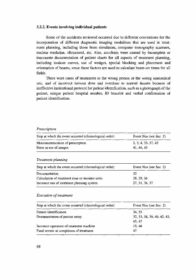
3.2.2. Events involving individual patients
Some of the accidents reviewed occurred due to different conventions for theincorporation of different diagnostic imaging modalities that are used in treat-ment planning, including those from simulators, computer tomography scanners,nuclear medicine, ultrasound, etc. Also, accidents were caused by incomplete orinaccurate documentation of patient charts for all aspects of treatment planning,including isodose curves, use of wedges, special blocking and placement andorientation of beams, since these factors are used to calculate beam-on times for allfields.
There were cases of treatments to the wrong person or the wrong anatomicalsite, and of incorrect tumour dose and overdose to normal tissues because ofineffective institutional protocol for patient identification, such as a photograph of thepatient, unique patient hospital number, ID bracelet and verbal confirmation ofpatient identification.
Prescription
Step at which the event occurred (chronological order) Event Nos (see Sec 2)
Miscommumcation of prescnptionError in use of images
2, 3, 4, 33, 37, 4541, 44, 45
Treatment planning
Step at which the event occurred (chronological order) Event Nos (see Sec 2)
DocumentationCalculation of treatment time or monitor unitsIncorrect use of treatment planning system
3228, 29, 3627, 31, 36, 37
Execution of treatment
Step at which the event occurred (chronological order) Event Nos (see Sec 2)
Patient identificationDocumentation of patient setup
Incorrect operation of treatment machineFinal review at completion of treatment
34,3532, 33, 38, 39, 40, 42, 43,45,4719,4647
68

3.3. LOW DOSE RATE BRACHYTHERAPY
3.3.1. Events related to equipment, thus affecting many patients
Cited in this review are accidents which occurred when source characteristics,such as source strength, isotope and filter thickness, are not verified by measurementupon receipt from a supplier and each time they are used, by identification of sourcemarking. Mistakes arose when strengths of sources were quoted in two non-equivalent units, e.g. mg Ra-eq or mCi, and these were used erroneously orinterchangeably, when users did not properly distinguish between nominal andmeasured source strength, and when suppliers provided source activities differentfrom those ordered. Other causes of accidental exposures were when sourceswithdrawn from clinical use were not clearly labelled as such and taken out ofcirculation, as well as the improper use of leaky sources.
Step at which the event occurred (chronological order) Event Nos (see Sec. 2)
Verification of activity 55, 56, 57, 58, 69, 70Verification of batch uniformity 68, 69, 70Verification of identification markers 58, 59Storage and inventory 58,71,73Performance of leakage test 74Applicators: commissioning and maintenance 75, 83
3.3.2. Events involving individual patients
Brachytherapy requires an individual treatment plan based on the geometry ofthe target site and prescribed dose with the treatment time based on the geometry ofthe implanted or inserted sources. Serious misadministrations occurred when thesource geometry or activities were not correct or the correct placement of sources wasnot maintained throughout the treatment time. Severe under-irradiation of the tumourand over-irradiation of normal tissues were also reported when sources weredislodged during treatment. In a few reported cases of accidents, nursing staff lackedthe required special training in radiation safety needed to perform nursing care withminimal exposure to staff and visitors and to deal with emergencies. Patients were notinformed that sources were radioactive and should not be removed or tampered withduring treatment.
69

Step at which the event occurred (chronological order) Event Nos (see Sec 2)
Prescription documentation and communication 67Planning of application including activity, geometry, 55, 56, 60, 61, 62, 65, 66
anatomical site, estimation of dose rate and treatment timeIdentification of patient 85Informing the patient 74, 79, 80Documentation 62, 65, 67Radiographic localization of sources 72, 82Sources correctly in place for duration 57, 72, 76, 77, 79, 80, 81,
83Source identification 58, 59, 63, 67Final calculation of dose and treatment time 64Correct delivery of prescription 78, 81, 82Correct treatment time 82Monitoring of patient after removal of sources 82
3.4. HIGH DOSE RATE BRACHYTHERAPY
3.4.1. Events related to equipment, thereby affecting many patients
High dose rate units, because they involve very high activity sources, have apotential for damage to patients as a result of misadmimstration Cited in this reportare cases of misadmimstration owing to malfunctioning equipment, such as cathetersthat do not allow full movement of sources Also cited are cases of inadequatelytrained personnel operating equipment, resulting in misadministration of treatmentBecause high dose rate brachytherapy is relatively new compared with low dose ratebrachytherapy, the total number of high dose rate events is lower.
Step at which the event occurred (chronological order) Event Nos (see Sec 2)
Training of personnel 77, 78Malfunction of equipment 81, 84, 86
3.5. USE OF UNSEALED SOURCES
Step at which the event occurred (chronological order) Event Nos (see Sec 2)
Planning and dispensing 89, 90, 91Administration 87, 88, 89, 92Safety and contamination 92
70

4. LESSONS LEARNED AND MEASURESFOR THE PREVENTION OF ACCIDENTS
The purpose of radiotherapy is to achieve maximum control of the tumour withminimum complications to normal tissue. This objective sets very high requirementson systematic accuracy and precision in relation to the total dose, dose distributionand dose fractionation. In order to satisfy these requirements, both systematic andrandom errors must be kept within a few percentage points during the entire process,from the time of prescnption to the completion of the radiation treatment, a chain ofevents that involves a number of persons.
A review of the initiating events and contributing factors described in Section 2confirms that most of the accidents could have been prevented by consistentapplication of the requirements cited in Appendix II (medical exposures) andAppendix IV (potential exposures) of the BSS. The lessons learned from theaccidents are discussed below.
4.1 RESOURCES: PERSONNEL AND EQUIPMENT
A radiotherapy department needs to have sufficient staff in relation to thenumber of patients and types of treatment modalities; guidelines have beenrecommended by national and international organizations. A shortage of staff usuallyleads to an increase in errors. In addition, a reassessment of staff and training has tobe undertaken when the workload increases (more patients) or new equipment ispurchased, or when a new technology or treatment modality is introduced. The failureto reassess staff workload led to a large scale accident in which a 60Co beam was usedfor a period of 22 months without quality control checks because the only medicalphysicist available was assigned to a new accelerator.
4.2. HUMAN FACTORS
Radiotherapy is very much dependent on human performance. A large numberof steps, tasks and subtasks have to be performed many times every day and differonly slightly from one patient to the next. Rarely are all steps in radiotherapyperformed by a single individual; many people work together, each contributing asmall part of the entire process. Many workers in several disciplines must interact inhighly technical measurements and calculations, and the potential for mistakes islarge if care is not taken. Even well trained and well qualified personnel make errors,owing to inattention to details, lack of alertness, or lack of awareness, especially if
71

they have to work in less than ideal conditions. In some of the accidents, even thereview of charts and calculations failed to detect a mistake.
4.2.1. Identification of safety critical steps and activities
A list of safety cntical activities and steps in the radiotherapy process is givenbelow. The list is not intended to be exhaustive and is derived from the examplesdiscussed in this report:
(a) Commissioning of radiotherapy machines and validation of treatment planningsystems,
(b) Periodic calibration and constancy checks of beams and sources,(c) Decommissioning of radiation sources and devices containing sources,(d) Identification of the patient,(e) Identification of the treatment target site ('localization'),(f) Preparation of the treatment plan,(g) Formal prescription of the treatment plan,(h) Simulation of the treatment and placement of body marks (tattoos),(i) First treatment session,(j) Daily patient positioning and selection of parameters and treatment accessories,(k) Accumulation of doses in the patient's chart,(1) Modifications during the course of treatment,(m) Radiopharmaceutical administration,(n) Maintenance of radiotherapy equipment and devices,(o) Removing sources from the patient after a brachytherapy treatment and
returning them to storage,(p) Identification of long lived brachytherapy sources kept in storage,(q) Measurement of activity (dose rate) of short lived brachytherapy sources
obtained for single treatments.
The assignment of functions and lines of authority and responsibility shouldensure that there are no gaps or ambiguities which leave safety critical tasks insufficient-ly covered. At this stage, the necessary degree of human redundancy is established.
4.2.1 1. Measures for the prevention and mitigation of consequences
Measures in this group are:
(a) Clear and concise written instructions to prepare staff for unusual situations, ingeneral as well as on a case by case basis. Clear job descriptions, assignment ofresponsibilities and lines of authority.
72
(b) Anticipation of unusual, unplanned situations following a review of allprocedures. A drill or rehearsal of measures to be taken may be appropriate forsome types of accident. A review of all procedures to identify those whichcould lead to an accident in an unusual or unplanned situation.
Special care needs to be taken when there are complex or unusual treatmentplans or when there is a change in procedures, such as:
(1) A change of supplier of radioactive material;(2) A non-typical dose, an unusual target area or a treatment with the patient in an
unusual position;(3) A complex treatment, for example, treatment of more than one anatomical site
on the same day.
4.3. TRAINING
Oncologists, physicists and other paramedical staff necessarily require formaltraining to effectively carry out their duties. In addition, opportunity for continuingtheir education by attending professional meetings and refresher courses is necessaryin a field as dynamic as radiotherapy. Acquisition of new equipment often requiresattendance at training sessions provided by manufacturers. Membership inprofessional organizations provides a valuable mechanism for communication withcolleagues through meetings, journals and other publications. These contacts promotefurther informal interaction, which is particularly important for a physicist workingalone.
Nurses and ancillary personnel who interact with patients have to be trained inradiation safety. They need to be cognizant of techniques for reducing exposure tothemselves, other workers and visitors. They have to be able to recognizeemergencies, such as unintended removal of radioactive sources, and know whichprocedures are appropriate in such an event.
Training has to cover not only normal situations, but also the identification ofabnormal situations (case histories) and unusual events, as well as measures foraccident prevention.
4.3.1. Training for special or unusual situations
Experience has shown that in an unusual situation, for example after anequipment failure or when conflicting signals appear, the behaviour of staff is oftenunsafe. Wrong assumptions are made, leading to accidental exposures. This may bebecause the staff member makes the interpretation that matches his or her experience,
73
expectation or convenience. Expectations are usually based on expenence gaineddunng normal operation of equipment or routine procedures. Additional specifictraining is needed to cover special or unusual situations, for example how to deal withconflicting signals on a console, a change of supplier, or complex types of treatment.The inclusion of unusual situations, the discussion of case histones and contingencyplans, and exercises in training coupled with penodic refresher training will help staffrecognize potential problems.
4.4 COMMUNICATION
Communication between staff members is essential for all aspects of treatment,since many persons with various responsibilities must interact. Mistakes may bemade owing to lack of adequate communication, incorrect information, or poorunderstanding of correct information.
The review of events reveals the following sources of error:
(a) Failure to transmit information.(b) Transmitting incorrect communication.(c) Communication to the wrong person.(d) Correction of a problem by an unqualified person without help or review.(e) Oral communication, either in person or by telephone, without written con-
firmation, resulting in misunderstanding.(f) Mistakes in reading or transferring information.(g) Unreadable or confusing handwritten communication, informal expressions or
use of jargon that is not understood by everyone in the same way.(h) Misunderstanding of communication in a foreign language: This may include
manufacturers' instructions for the use of equipment, as well as communicationbetween staff and between staff and patients
(i) Incomplete or poorly written instruction manuals for complex equip-ment such as treatment machines and treatment planning computers Ofparticular concern are instructions that do not cover unusual or specialapplications.
Not explicitly cited in this review of accidents, but often playing a role, are thefollowing circumstances:
(1) Interpersonal problems among staff;(2) Noisy working conditions, leading to distraction and loss of concentration;(3) Insufficient access to, or non-availability of, personnel in essential positions at
cntical times
74
4.4.1. Measures for the prevention and mitigation of consequences
In any institution, one person, usually a medical physicist, should have overallresponsibility for all aspects of the dosimetry system. It is his or her responsibility tosee that the lines of communication concerning dosimetry of patients are workingproperly. Other measures are:
(a) Identification and posting of all rules for communication critical to safety.(b) Clear assignment of responsibilities of the staff, including written job descriptions(c) An organizational structure that clearly identifies the 'chain of command', i.e.,
each worker's supervisor and the persons he/she supervises(d) Checklists that describe all steps of routine procedures as well as actions to be
taken when accidents occur.(e) Clear and concise written procedures for safety critical communications.(f) Signatures on documents critical to safety. For example, prescnptions are to be
written and signed by the physician. Basic data, treatment plans, daily logs ofpatient therapy, etc., are to be signed or initialled by the person performing orchecking that step in the process.
Some examples of critical communications relevant to safety are given in Table I.Once the safety critical communications have been identified, responsibilities
for these communications are assigned. Formal procedures are established for safetycritical communications, design of forms and checklists. Rules for clear, concise andcomplete communication and for developing protocols, drawings, charts, languageand clearances with signatures are drawn up.
4.5. EQUIPMENT
Equipment failure accounts for only a moderate percentage of all accidentscited in Section 2. However, the consequences can be severe and many patients maybe affected when equipment does malfunction. Contributing factors are:
(a) Insufficient redundancy in the design of equipment (single fault criterion,interlock failure);
(b) Software problems;(c) Hardware incompatibilities in equipment and accessories (wedge or shielding
block incompatible with coding system, or ionization chamber that does not fitan electrometer);
(d) Possibility of operating the equipment in a 'non-clinical mode' with the key inthe usual 'beam-on' position.
75
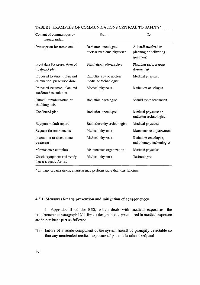
TABLE I. EXAMPLES OF COMMUNICATIONS CRITICAL TO SAFETY3
Content of communique ormemorandum
From To
Prescription for treatment
Input data for preparation oftreatment plan
Proposed treatment plan andcalculation, prescribed dose
Proposed treatment plan andconfirmed calculation
Patient immobilization orshielding aids
Confirmed plan
Equipment fault report
Request for maintenance
Instruction to discontinuetreatment
Maintenance complete
Check equipment and venfythat it is ready for use
Radiation oncologist,
nuclear medicine physician
Simulation radiographer
Radiotherapy or nuclearmedicine technologist
Medical physicist
Radiation oncologist
Radiation oncologist
Radiotheraphy technologist
Medical physicist
Medical physicist
Maintenance organization
Medical physicist
All staff involved m
planning or delivering
treatment
Planning radiographer,dosimetrist
Medical physicist
Radiation oncologist
Mould room technician
Medical physicist orradiation technologist
Medical physicist
Maintenance organization
Radiation oncologist,radiotherapy technologist
Medical physicist
Technologist
' In many organizations, a person may perform more than one function
4.5.1. Measures for the prevention and mitigation of consequences
In Appendix II of the BSS, which deals with medical exposures, the
requirements in paragraph II. 11 for the design of equipment used in medical exposure
are in pertinent part as follows:
"(a) failure of a single component of the system [must] be promptly detectable so
that any unintended medical exposure of patients is minimized; and
76

(b) the incidence of human error in the delivery of unintended medical exposure beminimized."
Methods for good engineering are well developed. One basic element is thesingle fault criterion, which leads to redundancy. In other words, failure of onecomponent should not cause an accident; an accident should be possible only iftwo or more systems or components fail simultaneously. A faulty componentshould be repaired immediately in order to maintain redundancy and preventaccidents [3, 4]:
(1) Analysis of common failures is used to ensure that one common failure wouldnot nullify the redundancy. For this purpose, redundancy is complemented byindependence.
(2) Components and systems have to be designed to be fail-safe, so that a failuredoes not lead to an unsafe situation. For example, the source driver of a gammateletherapy unit is designed so that the radioactive source returns to the shieldedposition if there is a cut-off of electrical power (see the BSS, Appendix II,paragraphs 11.11-13 and 15).
(3) Prototype tests need to include special or extreme conditions of equipmentoperation. Tests need to involve clinical personnel and others who useequipment routinely and not be limited to engineers and personnel who designand install equipment.
(4) Equipment is to be designed so that when it is in 'non-clinical mode' (reductionof interlocks), it is impossible to enter a 'beam-on' command from the regularkey in the control console.
(5) Validation of the equipment and its operation, under clinical conditions, has tobe made before initiating treatment of patients. Detailed documentationdescribes the tests performed and the personnel responsible. Commissioning ofequipment for clinical use is the responsibility of the institution's medicalphysicist.
(6) There is a need for manufacturers of radiation therapy equipment to promptlyand thoroughly investigate any reported incident or unusual event and notifyauthorities and other users of their findings and appropriate preventivemeasures. A formal system of reporting and disseminating information frommanufacturers to users is essential.
(7) Machine malfunctions are not to be corrected until the full dosimetncconsequences to patients who were treated during the period of the mal-function are known. Measurements while the machine is malfunctioningmay be necessary in order to estimate an incorrect dose delivered to apatient.
77
4.6 HUMAN-MACHINE PROBLEMS
4.6.1. Problems of human-machine interface
Personnel need to understand the functioning of equipment, including thesignificance of alarms, signals, and displays that indicate a malfunction.
4 6.1 1. Measures for the prevention and mitigation of consequences
(a) Inconsistent signals are investigated and the one indicating the more seriousconsequence is assumed to be correct until proven otherwise. For example, if aradiation monitor gives off an alarm indicating that a beam is on, but theconsole display shows that the beam is off, it is more prudent to assume that thebeam is on, not that the alarm is malfunctioning.
(b) The human-machine interface in the clinical environment is considered in thedesign and testing of equipment.
(c) Specific training to recognize and respond to abnormal signals and displayinterlocks, as well as contradictory signals.
4.6.2. Bypassing of interlocks and operation in a 'non-clinical mode'
Operation in a 'non-clinical mode' sometimes is the response to frequent orunresolved maintenance problems. However, circumventing interlocks and othersafety devices in order to continue with patient treatments increases the possibility ofaccidents, which can lead to serious or even fatal consequences.
4 6.2 1. Measures for the prevention and mitigation of consequences
(a) Plan and implement an efficient maintenance programme and avoidimprovisation as far as practicable.
(b) Never treat patients with equipment operating in the 'non-clinical mode'.(c) Never try for a 'quick-fix' or 'patch-job' in order to continue treatment of
patients.(d) Withdraw from clinical use equipment that is malfunctioning until formal
repair is complete and its operation is checked in a thorough and deliberateway.
(e) Design equipment so that it is possible to turn 'beam on' in the 'non-clinicalmode' only by using special tools or computer codes. It is only possible tooperate the machine in the non-clinical mode for maintenance or other non-chnical work (BSS, Appendix II, paragraphs 11.15 (c) and (d)).
78
4.6.3. Maintenance problems
Mistakes made in maintenance can and have affected many patients and haveled to severe, even fatal, consequences.
4.6.3.1. Measures for the prevention and mitigation of consequences
(a) Maintenance personnel have to be cognizant of the potential consequences ofany manipulation, alteration or adjustment of components of a machine for thesafety of patients and personnel who use the machine clinically.
(b) Before a machine is released for clinical use following maintenance or repair, acheck of the output, as well as any other operating parameters that could havebeen affected, has to be made. Formal procedures for handing over the machineafter such maintenance or repair to the physicist must be in place for necessarychecks before clinical use.
(c) Instructions for the use of a machine that has been altered must be clearly andunambiguously documented in writing. Machine functions are only to bealtered by or with the authorization of the manufacturer. Unauthorizedmodifications may compromise safety and may also nullify a manufacturer'swarranty obligations and product liability.
4.7. IMPROPER DECOMMISSIONING OF EQUIPMENTAND UNSAFE STORAGE OF RADIOACTIVE SOURCES
Unauthorized handling, dismantling or abandonment of teletherapy isotopeunits has led to large scale serious consequences, including fatalities and widespreadenvironmental contamination [5].
4.7.1. Measures for the prevention and mitigation of consequences
(a) Governmental licensing needs to regulate the importation, storage,commissioning, operation, temporary suspension, decommissioning anddisposal of radiation sources.
(b) There have to be clear, unambiguous and authorized procedures for thetemporary or indefinite secure storage of radioactive sources until safe disposal.Formal control has to be in place during this period of time.
(c) Equipment no longer in use has to be disabled in a way that prevents connectionto an electrical power supply.
(d) Sealed sources found to be damaged or leaking are to be stored in a way thatprevents their clinical use and contamination of the environment.
79

(e) Obsolete brachytherapy sources have to be disposed of safely, in accordancewith governmental regulations.
(f) Radioactive sources used in nuclear medicine are to be disposed of safely, inaccordance with governmental regulations.
(g) Empty source containers have to be clearly labelled and stored in a locationseparate from containers with sources.
48. DOCUMENTATION
Documentation is essential for high quality and safe radiotherapy and to fostercommunication among staff and continuity of practice over time [6, 7].
Some of the accidents reviewed have a strong component of misinterpretationof instructions, displays and interlocks as well as signals relating to unusual events.Many maintenance mistakes are related to a lack of comprehensive training andcontinuing education, especially in dealing with unusual situations.
4.8.1. Measures for the prevention and mitigation of consequences
A log book is needed for each major item of equipment, such as 60Co units,linear accelerators, TPSs, iomzation chambers and electrometers, in whichinformation concerning calibration, repairs, maintenance and checks of performanceare routinely recorded Each entry has to be dated and signed by the responsibleperson.
For individual patients, all aspects of treatment have to be documented as apermanent part of each patient's chart and retained for at least as long as specified bylocal laws and regulations. The prescription has to be written by a physician.Treatment plans, generated by manual or computer methods, need to include isodosedistributions, with placement of beams, any wedge filters or beam blocks. The basicdata and TPS used to generate the plan, including data such as depth dose, tissue-airratios, wedge factors, etc., have to be clearly written down. All details required for thesetup of the patient and machine time or monitor units for each field are to be clearlyshown. A diagram of the patient with location of the beams is essential, and aphotograph of the patient in the treatment position is useful for more complextreatments, particularly those involving blocked or irregular fields. Each patient'streatment is to be detailed in a daily log, with beam times or monitor units listed.
Original documentation concerning equipment or a patient's treatment isnever to be altered If errors are detected that require correction later, commentsmay be added to the record but the onginal documentation is not to be changed.Addenda to documentation must include the signature or initials of the personresponsible.
80

Comprehensive training is complemented by familiarization with themanufacturer's documents. In this respect, as indicated, the BSS recommends thatperformance specifications and operating and maintenance instructions be providedin a major world language understandable to the users, in compliance with therelevant International Electrotechnical Commission (IEC) and InternationalOrganization for Standardization (ISO) standards, and that this information betranslated into local languages whenever appropriate. In addition, the operatingterminology (or its abbreviations) and operating values must be displayed onoperating consoles in a major world language acceptable to the user. Therequirements imply that instructions be understandable, without ambiguity, to usersof all categories, such as radiation oncologists, technologists, medical physicists andmaintenance engineers.
It is emphasized that accompanying documents, according to the IECdefinition, should be a part of the equipment (see IEC-601-1, subclause 6.8 [3]) andthat equipment is incomplete if the accompanying documents are not delivered.According to IEC-601-1, the accompanying documents include at least:
(a) Instructions for use, including accessories, interlocks and displays, as well asany maintenance to be performed by the user;
(b) Technical descriptions, including, among other information, a statement thatthe supplier will make available on request circuit diagrams, component partlists, calibration instructions or other information that will assist appropriatelyqualified user personnel to repair those parts of equipment designated by themanufacturer as reparable;
In addition, it is strongly recommended that the user maintain:
(1) Records of repairs, revisions of the equipment and faults and lessons learned;(2) Manufacturer's newsletters;(3) Manufacturer's notes on investigations of malfunctions of the equipment,
accumulated experience, preventive recommendations and, most importantly,warning notes on incidents with similar machines in other facilities.
4.9. INTEGRATION OF SAFETY AND QUALITY ASSURANCE
Technical and managerial principles need to be applied to prevent unintentionalexposures and to mitigate the consequences, should they occur. These principles startwith a systematic assessment of both equipment and treatment procedures, followedby the application of 'defence in depth' measures and the integration of prescriptionand delivery of treatment in an overall approach. This approach involves the attitudes
81

of all the persons involved in radiation therapy — physicians, physicists,technologists, nurses, equipment maintenance engineers and clerical staff. All need todemonstrate a 'culture of safety'.
Independent checks, internal and external, are an important means of avoidingerrors or detecting them before they cause injury to patients.
External independent checks may be formal, such as those performed by anoutside physicist from an authorized reviewing organization, as required by clinicaltrials. External reviews also may be informal, where colleagues intercomparelonization chambers and other measuring systems. In addition, mailed dosimetersfrom reviewing organizations provide an inexpensive means of checking machineoutput and other basic machine parameters.
Within an institution, having a second person check data and repeat calculationsor measurements is an important way of finding mistakes before they result inincorrect doses to patients. Internal independent checks need to be part of the routineflow of work m a dosimetry system. Independent verification procedures can beeffective only if physicists and other staff work together as colleagues, in a spint ofgoodwill and co-operation. Staff at all levels need to feel free to challenge the workof any other person if they feel that an error has been made that would cause detrimentto a patient.
Independent checks are successful only if each person involved is free to point outerrors and discuss problems as well as accept correction of his or her own errors. It iscrucial that interaction be conducted in a professional manner, bearing in mind thebest interests of the patient. A professional attitude has to be maintained by all cate-gories of radiotherapy staff, independent of their vantage point in the review process
4.10. SAFETY ASSESSMENT
The mam requirements for a safety assessment — out in the BSS inAppendix IV, 'Potential Exposure: Safety of Sources' [1] — are also applicable toradiotherapy sources and installations.
A safety assessment requires a systematic approach to identify events that canlead to potential exposures and to estimate the probability and magnitude of suchexposures There are two approaches to the safety assessment: deterministic andprobabilistic methods.
The deterministic method is often used first to provide safety, with appropriatesafety margins, in systems where accident scenarios can be identified. Appropriatemeasures may include changes in equipment design or operating procedures, withrelevant training of personnel and formulation of contingency plans.
The probabilistic method is used to identify weaknesses in safety measures thatmay be overlooked by the deterministic approach, because they may not yet have
82

resulted in an actual accident. The method identifies and calculates the expectedfrequency of initiating events that may lead to potential exposure and calculates theexpected detriment due to initiating events for comparison with acceptance criteria.The method also establishes the relative importance of individual events andquantifies the probability of their leading to the detrimental effect.
Probabilistic and deterministic methods should not be viewed as being mutuallyexclusive, but rather as complementary techniques.
4.10.1. System for quality management
Integrated therapy systems involve both quality and safety. In fact, most of themeasures to monitor quality serve equally to detect any deviation concerning safetysince the parameters to be controlled are often the same. The quality control pro-gramme can be designed to combine both quality and safety. For example, test toolsthat perform constancy checks serve to monitor quality and safety at the same time.
To meet the requirements of radiotherapy, many centres are implementingtechniques used in other disciplines where the prevention of errors is an everydayissue. While the differences between industry and medicine should be allowed for,appropriate elements of these techniques known as the 'Quality Management System'and defined by ISO Standard 9000 can be incorporated into the planning andmanagement of radiotherapy services. The system includes the calibration of sources,clinical dosimetry, quality assurance of medical exposures and records, as well asregular and independent quality audits, as listed in Appendix II of the BSS. A detaileddescription of quality assurance programmes and quality control tests has been set outin many recent publications [8-13].
4.10.2. Monitoring effectiveness
The vulnerability of a facility to accidental or erroneous exposures needs tobe assessed in order to ensure that the probability of their occurrence remainslow and that remedial action would be effective. Some methods for assess-ment are:
(a) Reviewing records of normal conditions and minor mishaps to detect anyabnormal conditions or occurrences that may increase the probability ofaccidents;
(b) Checking the layers of the defence in depth system to detect if any are notfunctional, thereby making the system vulnerable;
(c) Developing a system of investigation and analysis of accidents and unusualevents, including initiating events and contributing factors, and disseminatinginformation on lessons learned;
83

(d) Testing the facility for vulnerability against initiating events and contributingfactors emerging from item (c);
(e) Internal and external audits and mtercomparisons to detect weaknesses andindicate corrective measures;
(f) Evaluation of mistakes made, with a review of steps for mitigation of theireffects and future avoidance;
(g) Evaluation of protocols for patient follow-up after therapy to detect abnormalradiation reactions that may suggest errors in exposure.
4.10.3. Defence in depth
The term defence in depth is defined in the BSS as "the application of morethan one single protective measure for a given safety objective such mat the objectiveis achieved even if one of the protective measures fails". There are layers ofoverlapping safety provisions, such as physical components, procedures andcombinations of both. A low overall probability of failure can be more readilyachieved by a combination of independent protective layers such that the probabilitiesare multiplicative.
Examples of defence in depth are:
(1) A treatment plan that is produced by a technologist, checked by the medicalphysicist and rechecked by in vivo dosimetry, and
(2) The output of a linear accelerator that is controlled by an lonization chamber,checked by a second ionization chamber preset to 7-10% higher dose, and abackup timer set to the prescribed time +15%.
4.10.4. Safety culture
An essential managerial principle for all individuals and organizations is toestablish a 'safety culture' encompassing both personal attitudes and habits of thoughtas well as organizational policies and priorities.
Good practices are essential but not sufficient. There is a need to go beyond theimplementation of a good practice, so that both personal attitudes and organizationalpolicies are onented towards the same goal: that all duties be carried out correctly,with alertness, full knowledge, sound judgment and a proper sense of accountability.Even some of the provisions for defence in depth, such as redundant reviews andchecks, may fail to detect a problem. The basic principles of safety culture are that:"all duties important to safety should be carried out correctly, with due thought andfull knowledge, sound judgment and a proper sense of accountability" [14].Management should provide the environment, training and provisions to maintain andexercise alertness and awareness of the staff.
84
Each person responsible for any aspect of patient treatment needs to have acritical view of his or her own and other people's work. Freedom to challenge thework of others in a spirit of goodwill points up potential accidents before theyhappen. It also discourages complacency amongst staff.
Furthermore, it implies a learning attitude by all concerned, whereby bothoperating experience and the results of new research are used to reassess and improvesafety.
4.10.5. Emergency preparedness plans
Despite preventive measures, accidents or unusual events may occur. The safetyassessment will identify possible accident scenarios and situations, andcountermeasures for mitigation are designed. Some of the actions in the emergencyresponse plan need to be taken immediately, without hesitation or mistakes. Toachieve this, personnel need to be trained and simulation drills carried out. A clearand concise list of actions and responsible officers is posted in relevant places. Thereis written documentation of rules for action and training, including simulatingexercises, for the emergency response plan. The development and periodic review ofthe plan is part of the system of protection for radiotherapy.
4.10.6. Investigation of accidental medical exposuresand dissemination of information on accidents and unusual events
In a facility where an accident occurs, corrective measures can be applied onlyif a prompt investigation is made. With regard to accidental medical exposure, theBSS promulgates specific investigative action, calculation of dose and correctivemeasures, as well as the requirement to report the incident to the RegulatoryAuthority and to inform the patient (BSS, Appendix II, paragraphs 29, 30).
In addition to the investigation of accidents, which leads to corrective andpreventive measures, the dissemination of operational experience can avoid accidentselsewhere. The number of reported events in a single country is usually insufficientto provide a significant number of lessons in a reasonable time. However, acompilation of accidents at the international level would allow all countries to benefitfrom the lessons from each of them. Moreover, the experience gained from unusualevents that did not culminate in accidents can contribute to the knowledge needed toavoid accidents. The dissemination of this information can be used to test thevulnerability of many facilities worldwide and improve preventive measures withoutwaiting until an accident occurs.
Therefore, a reporting system which allows for anonymous reports of cases thatare otherwise not published is necessary for such reports to reach a wider community.An international reporting system of unusual events and operational experience is in
85

place in other fields, such as nuclear power, and a similar approach can equally serveto prevent accidents in radiotherapy.
4.11. REGULATORY CONTROL
The assessment of safety, together with measures to reduce the likelihood ofunwarranted or unjustified radiation exposure, requires the incorporation of safetyinto the management system and the development of a safety culture as defined inSection 4.10.4. Furthermore, these measures need to be ensured by means ofappropriate regulatory control. This implies a regulatory framework involvingauthorization, inspection and enforcement. Radiotherapy is a practice that is subjectto licensing. Licensing requirements include mandatory safety assessment of allaspects of the acquisition, use and disposal of radiation sources, includingconstruction, importation, storage, commissioning and decommissioning, whethertemporary or permanent. In addition, plans for the prevention of mistakes and foremergency preparedness are to be assessed before licences are issued. In this regard,an IAEA technical document [15] gives detailed guidance on safety assessmentrequirements for the authorization and inspection of radiotherapy facilities.
86

Appendix
APPENDIX II OF THE BSS —MEDICAL EXPOSURE: OPTIMIZATION OF PROTECTION
FOR MEDICAL EXPOSURES
DESIGN CONSIDERATIONS
General
"11.11. The requirements for the safety of sources specified in other partsof the Standards shall also apply to sources used in medical exposure, whererelevant, and, in particular, equipment used in medical exposure shall be so designedthat:
fa) failure of a single component of the system be promptly detectable so that anyunplanned medical exposure of patients is minimized; and
(b) the incidence of human error in the delivery of unplanned medical exposure beminimized.
11.12. Registrants and licensees shall:
(a) taking into account information provided by suppliers, identify possibleequipment failures and human errors that could result in unplanned medicalexposures;
(b) take all reasonable measures to prevent failures and errors, including theselection of suitably qualified personnel, the establishment of adequateprocedures for the calibration, quality assurance and operation of diagnosticand therapeutic equipment, and the provision to personnel of appropriatetraining and periodic retraining in the procedures, including protection andsafety aspects;
(c) take all reasonable measures to minimize the consequences of failures anderrors that may occur; and
(d) develop appropriate contingency plans for responding to events that may occur,display plans prominently, and periodically conduct practice drills.
11.13. Registrants and licensees, in specific co-operation with suppliers, shallensure that, with regard to equipment consisting of radiation generators and thatcontaining sealed sources used for medical exposures:
87
(a) whether imported into or manufactured in the country where it is used, theequipment conform to applicable standards of the InternationalElectrotechmcal Commission (IEC) and the ISO or to equivalent nationalstandards;
(b) performance specifications and operating and maintenance instructions,including protection and safety instructions, be provided in a major worldlanguage understandable to the users and in compliance with the relevant EECor ISO standards with regard to 'accompanying documents', and that thisinformation be translated into local languages when appropriate;
(c) where practicable, the operating terminology (or its abbreviations) andoperating values be displayed on operating consoles in a major world languageacceptable to the user;
(d) radiation beam control mechanisms be provided, including devices that indicateclearly and in a fail-safe manner whether the beam is 'on' or 'off';
(e) as nearly as practicable, the exposure be limited to the area being examined ortreated by using collimating devices aligned with the radiation beam;
(f) the radiation field within the examination or treatment area without anyradiation beam modifiers (such as wedges) be as uniform as practicable and thenon-uniformity be stated by the supplier; and
(g) exposure rates outside the examination or treatment area due to radiationleakage or scattering be kept as low as reasonably achievable."
"Requirements for radiation generators and irradiation installations forradiotherapy
11.15. Registrants and licensees, in specific co-operation with suppliers, shallensure that:
(a) radiation generators and irradiation installations include provisions forselection, reliable indication and confirmation (when appropriate and to theextent feasible) of operational parameters such as type of radiation, indicationof energy, beam modifiers (such as filters), treatment distance, field size, beamorientation and either treatment time or present dose;
(b) irradiation installations using radioactive sources be fail-safe in the sense thatthe source will be automatically "shielded in the event of an interruption ofpower and will remain shielded until the beam control mechanism is reactivatedfrom the control panel;
(c) high energy radiotherapy equipment:(i) have at least two independent 'fail to safety' systems for terminating the
irradiation; and
(ii) be provided with safety interlocks or other means designed to prevent theclinical use of the machine in conditions other than those selected at thecontrol panel;
(d) the design of safety interlocks be such that operation of the installation duringmaintenance procedures, if interlocks are bypassed, could be performed onlyunder direct control of the maintenance personnel using appropriate devices,codes or keys;
(e) radioactive sources for either teletherapy or brachytherapy be so constructedthat they conform to the definition of a sealed source; and
(f) when appropriate, monitoring equipment be installed or be available to givewarning of an unusual situation in the use of radiation generators andradionuclide therapy equipment."
89

REFERENCES
[1] FOOD AND AGRICULTURE ORGANIZATION OF THE UNITED NATIONS,INTERNATIONAL ATOMIC ENERGY AGENCY, INTERNATIONAL LABOURORGANISATION, OECD NUCLEAR ENERGY AGENCY, PAN AMERICANHEALTH ORGANIZATION, WORLD HEALTH ORGANIZATION, InternationalBasic Safety Standards for Protection against Ionizing Radiation and for the Safety ofRadiation Sources, Safety Series No. 15, IAEA, Vienna (1996)
[2] PEREZ, C A , BRADY, L W, Principles and Practice of Radiation Oncology, 2nd edn,J B Lippmcott Company, Philadelphia and New York (1992)
[3] INTERNATIONAL ELECTROTECHNICAL COMMISSION, Safety of MedicalElectrical Equipment, IEC, Part 1, General Requirements, Publication 601-1, IEC,Geneva (1981)
[4] INTERNATIONAL ELECTROTECHNICAL COMMISSION, Medical ElectricalEquipment, Medical Electron Accelerators — Functional Performance Characteristics,Publication IEC-976, IEC, Geneva (1989).
[5] INTERNATIONAL ATOMIC ENERGY AGENCY, The Radiological Accident inGoiama, IAEA, Vienna (1988)
[6] INTERNATIONAL COMMISSION ON RADIATION UNITS AND MEASURE-MENTS, Dose and Volume Specification for Reporting Intracavitary Therapy inGynaecology, Report 38, ICRU, Bethesda, MD (1985)
[7] INTERNATIONAL COMMISSION ON RADIATION UNITS AND MEASURE-MENTS, Prescribing, Recording, and Reporting Photon Beam Therapy, Report 50,ICRU, Bethesda, MD (1993)
[8] BELLETI, S , et al, Quality assurance in radiotherapy The importance of medicalphysics staffing levels Recommendations from an ESTRO/EFOMP joint task group,Radiother Oncol 41 1 (1996) 89-94
[9] WORLD HEALTH ORGANIZATION, Quality Assurance in Radiotherapy, WHO,Geneva (1988).
[10] AMERICAN ASSOCIATION OF PHYSICISTS IN MEDICINE, Comprehensive QAfor radiation oncology Report of Radiation Therapy Committee Task Group 40, Med.Phys 204(1994)581-618
[11] FRENCH SOCIETY OF RADIATION ONCOLOGY AND FRENCH SOCIETY OFHOSPITAL PHYSICISTS, Recommendations for a Quality Assurance Programme inExternal Radiotherapy, ESTRO Physics for Clinical Radiotherapy Booklet No 2,Garaant (1995)
[12] DEPARTMENT OF HEALTH, A Quality Management System for Departments ofRadiotherapy, Publication PL/CMO 10, London (1991)
[13] AMERICAN ASSOCIATION OF PHYSICISTS IN MEDICINE, Medical AcceleratorSafety Considerations Report of the AAPM Radiation Therapy Committee, Task GroupNo 35, Med Phys 204(1994) 1261-1275
[ 14] INTERNATIONAL NUCLEAR SAFETY ADVISORY GROUP, Safety Culture, SafetySeries No 75-INSAG-4, IAEA, Vienna (1991)
90

[15] INTERNATIONAL ATOMIC ENERGY AGENCY, Safety Assessment Plans for
Authorization and Inspection of Radiation Sources, IAEA-TECDOC-1113, IAEA,
Vienna (1999).
91

CONTRIBUTORS TO DRAFTING AND REVIEW
Andreo, P International Atomic Energy Agency
Cohen, M McGill University, Canada
Gultresa, J Centre de Dosimetrfa, Spam
Haywood, J. South Cleveland Hospital, United Kingdom
Novotny, J Hospital Na Homolce, Czech Republic
Oresegun, M International Atomic Energy Agency
Ortiz-L6pez, P. International Atomic Energy Agency
Seng, D Nuclear Regulatory Commission, United States of America
Stovall, M University of Texas, United States of America
van de Geijn, J Consultant, United States of America
Vivanco, J. Hospital General de Astunas, Spain
Zufiiga-Bello, P Consejo Nacional de Ciencia y Tecnologia, Mexico
Technical Committee Meeting
Vienna, Austria- 17-21 March 1992
Consultants Meetings
Vienna, Austria 16-20 October 1993, 26 November-7 December 1995,
18-29 November 1996
92

RELATED IAEA PUBLICATIONS
Accidental Overexposure of Radiotherapy Patients in San Jose", Costa Rica (1998).
Lessons Learned from Accidents in Industrial Irradiation Facilities (1996).
Lessons Learned from Accidents in Industrial Radiography, Safety Reports SeriesNo. 7 (1998).
93

No. 9, June 1999
WHERE TO ORDER IAEA PUBLICATIONSIn the following countries IAEA publications may be purchased from the sources listed below, orfrom major local booksellers Payment may be made in local currency or with UNESCO coupons
Hunter Publications, 58A Gipps Street, Collingwood, Victoria 3066Telephone +61 394175361 'Fax +61 39419 7154• E-mail ipdavies&ozemail com auJean de Lannoy, avenue du Roi 202, 8-1190 Brussels • Telephone +32 2 538 43 08 • Fax +32 2 538 08 41E-mail jean de lannoy@tnfoboard be • Web site http //www/ean-de-lannoybe
Parry's Book Center Sdn Bhd, 60 Jalan Negara, Taman Melawati, 53100 Kuala Lumpur, MalaysiaTelephone +60 3 4079176, 4079179, 4087235, 4087528 • Fax +60 3 407 9180E-mail haja ©pops taring my • Web site http //www mol net my/~parrybook/parrys htm
IAEA Publications in Chinese China Nuclear Energy Industry Corporation, Translation Section, PO Box 2103, BeijingMunksgaard Subscription Service, N0rre S0gade 35, PO Box 2148, DK-1016 Kabenhavn KTelephone +45 33 12 85 70 -Fax +45 33 12 93 87E-mail subscription service©mail munksgaarddk'Web site http//wwwmunksgaard dkThe Middle East Observer, 41 Shenf Street, CairoTelephone +20 2 3939 732, 3926 919 • Fax +20 2 3939 732, 3606 804 • E-mail fouda@soficom com eg
Nucleon, Immeuble Platen, Pare les Algonthmes,F-91194 Gif-sur-Yvette, CedexTelephone +33 1 69 353636 • Fax +33 1 69 350099 • E-mail nucleon@wanadoo fr
UNO-Verlag, Vertnebs- und Verlags GmbH, Poppelsdorfer Allee 55, D-53115 BonnTelephone +49 228 94 90 20 • Fax +49 228 21 74 92Website http//www uno-verlag de • E-mail unover/ag@aolcom
Librotrade Ltd , Book Import, PO Box 126, H-1656 BudapestTelephone +36 1 257 7777 • Fax +36 1 257 7472 • E-mail books@librotrade huViva Books Private Limited, 4325/3, Ansan Road, Darya Ganj, New Delhi-110002Telephone +91 11 327 9280, 328 3121, 328 5874 • Fax +91 11 326 7224E-mail vinod viva iSgndel globalnet ems vsnl net in
YOZMOT Ltd , 3 Yohanan Hasandlar St, PO Box 56055, IL-61560 Tel AvivTelephone +972 3 5284851 • Fax +972 3 5285397
Librena Scientifica Dott Lucio di Biasio "AEIOU", Via Coronelli 6,1-20146 MilanTelephone +39 2 48 95 45 52 or 48 95 45 62 • Fax +39 2 48 95 45 48
Maruzen Company, Ltd, PO Box 5050,100-31 Tokyo InternationalTelephone +81 332727211 • Fax +81 332781937' E-mail yabe@maruzencoip' Website httpj/www maruzencojp
Parry's Book Center Sdn Bhd , 60 Jalan Negara, Taman Melawati, 53100 Kuala Lumpur,Telephone +60 3 4079176, 4079179, 4087235, 4087528 • Fax +60 3 407 9180E-mail haja@pop3iaringmy*Website http//wwwmol net my/~panybook/parrys htmMartmus Nijhotf International, PO Box 269, NL-2501 AX The HagueTelephone +31 793 684 400 • Fax +31 793 615698- E-mail mto@nijhorl nl • Website http//www nijhoff nl
Swets and Zeitlmger b v, PO Box 830, NL-2160 SZ LisseTelephone +31 252435111 -Fax +31 252 415 888 • E-mail mfoho@swets nl • Website nttpj/www swets nlForeign Trade Enterprise, Ars Polona, Book Import Dept, 7, Krakowskie Przedmies"cie Street, PL-00-950 WarsawTelephone +48 22 826 1201 ext 147,151,159 • Fax +48 22 826 6240E-mail ars_pol@bevyhsn com pi* Website http J/wwwarspolona com pt
Parry's Book Center Pte Ltd , 528 A MacPHERSON Road, Singapore 1336Telephone +65 744 8673 • Fax +65 744 8676E-mail haja@pop3 janng my • Website http/Avwwmol net my/~parrybook/parrys htmAlfa Press, s ro, Racianska 20, SQ 832 10 Bratislava • Telephone/Fax +421 7 566 0489
Diaz de Santos, Lagasca 95, E-28006 MadridTelephone +34 91 431 24 82 • Fax +34 91 575 55 63 • E-mail madnd@diazdesantos es
Diaz de Santos, Balmes 417-419, E-08022 BarcelonaTelephone +34 93 212 86 47 • Fax +34 93 211 49 91 • E-mail balmes@diazsantos com
General E-mail librenas@diazdesantos es • Web site httpj/www diazdesantos esThe Stationery Office Ltd, International Sales Agency, 51 Nine Elms Lane, London SW8 SDRTelephone +44 171 873 9090 • Fax +44 171 873 8463E-mail Orders to book orders@theso co uk • Enquiries to ipa enqwries@theso co ukWeb site http //www the-stationery-office co ukBernan Associates, 4611-F Assembly Drive, Lanham, MD 20706-4391, USATelephone 1-800-274-4447 (toll-free) • Fax (301) 459-0056 /1-800 865-3450 (toll-free)E-mail query@bernan com • Web site http//www bernan com
Orders and requests for information may also be addressed directly to:
Sales and Promotion Unit, International Atomic Energy AgencyWagramer Strasse 5, PO. Box 100, A-1400 Vienna, AustriaTelephone: +43 1 2600 22529 (or 22530) • Facsimile +43 1 2600 29302E-mail: sales.publlcatlons@!aea org • Web site: http://www.laea org/worldatom/publlcatlons
AUSTRALIA
BELGIUM
BRUNEI
CHINADENMARK
EGYPT
FRANCE
GERMANY
HUNGARY
INDIA
ISRAEL
ITALY
JAPAN
MALAYSIA
NETHERLANDS
POLAND
SINGAPORE
SLOVAKIASPAIN
UNITED KINGDOM
UNITED STATESOF AMERICA


INTERNATIONAL ATOMIC ENERGY AGENCYVIENNA
ISBN 92-0-100200-9ISSN 1020-6450