sages fls – intraoperative pathology examplesfls-online.org/fscommand/pathology.pdfsages fls –...
TRANSCRIPT
SAGES FLS – Intraoperative Pathology Examples
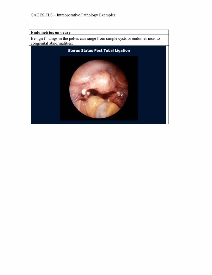
Endometrius on ovaryBenign findings in the pelvis can range from simple cysts or endometriosis to congenital abnormalities.

SAGES FLS – Intraoperative Pathology Examples
Dermoid cystA benign but complex lesion is the dermoid.When present in an ovary it can contain hair and other remnants.The dermoid may be removed laparoscopically with care to avoid intra-abdominal spillage.

SAGES FLS – Intraoperative Pathology Examples
Corpus luteumNon-hemorrhagic corpus luteum and follicular cysts are rarely greater than 2 cm in diameter and should be left alone.
SAGES FLS – Intraoperative Pathology Examples
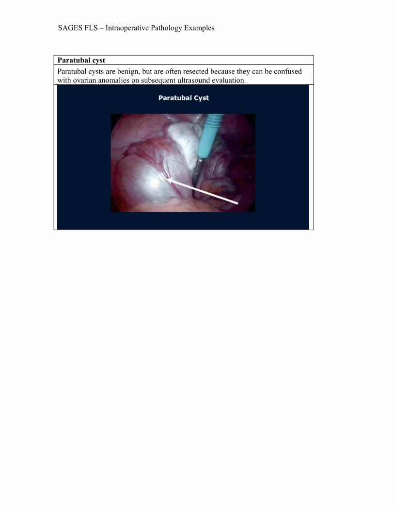
Paratubal cystParatubal cysts are benign, but are often resected because they can be confused with ovarian anomalies on subsequent ultrasound evaluation.

SAGES FLS – Intraoperative Pathology Examples
CystadenomasCystadenomas, while benign, do not generally resolve rapidly and thus often require resection in order to prove that cystadenocarcinoma is not present.

SAGES FLS – Intraoperative Pathology Examples
Ovarian torsionRight lower pain in women may be caused by ovarian torsion.The decision to resect or de-torse the ovary is based on viability and other gynecological factors.
SAGES FLS – Intraoperative Pathology Examples
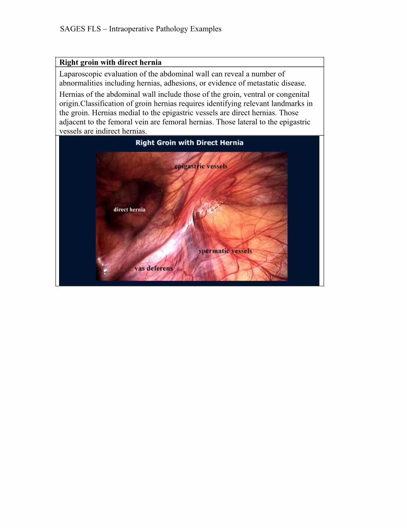
Right groin with direct herniaLaparoscopic evaluation of the abdominal wall can reveal a number of abnormalities including hernias, adhesions, or evidence of metastatic disease.Hernias of the abdominal wall include those of the groin, ventral or congenital origin.Classification of groin hernias requires identifying relevant landmarks in the groin. Hernias medial to the epigastric vessels are direct hernias. Those adjacent to the femoral vein are femoral hernias. Those lateral to the epigastric vessels are indirect hernias.
SAGES FLS – Intraoperative Pathology Examples
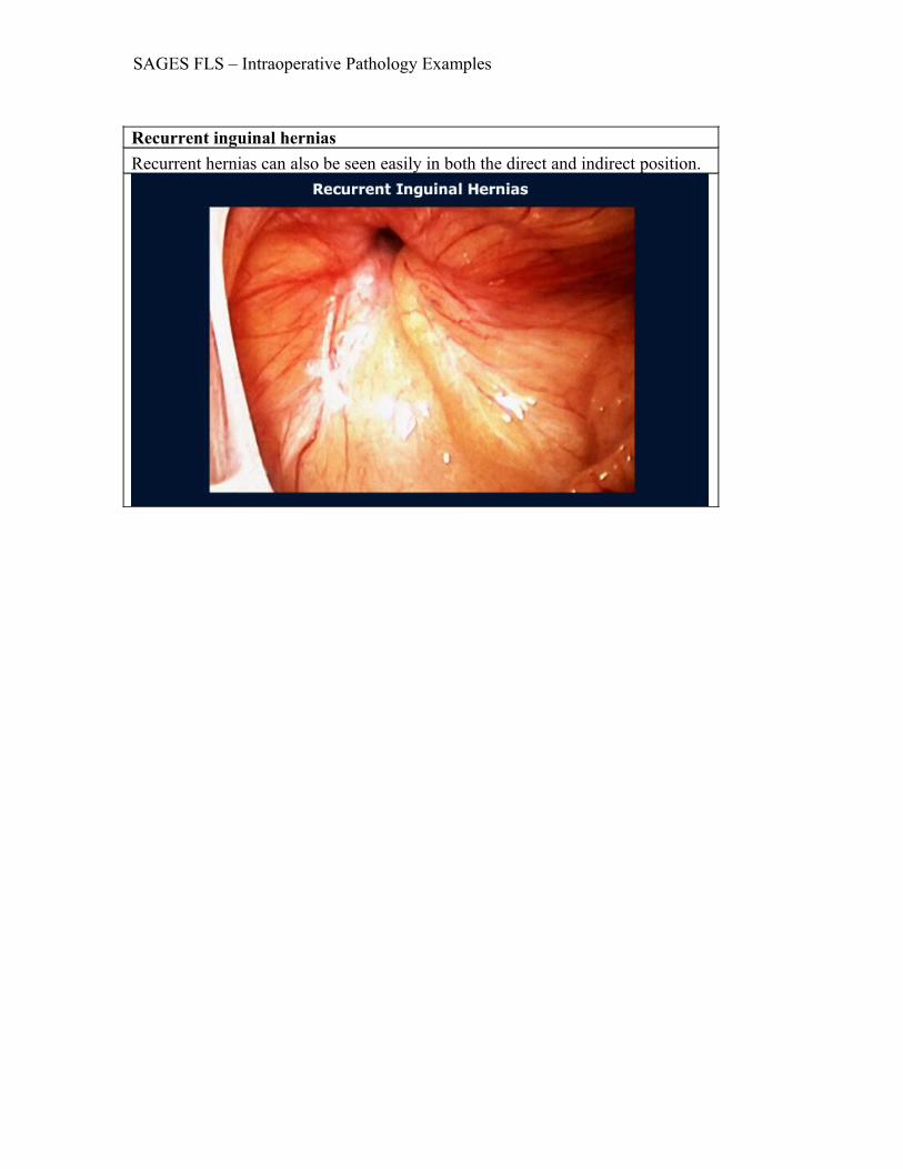
Recurrent inguinal herniasRecurrent hernias can also be seen easily in both the direct and indirect position.
SAGES FLS – Intraoperative Pathology Examples
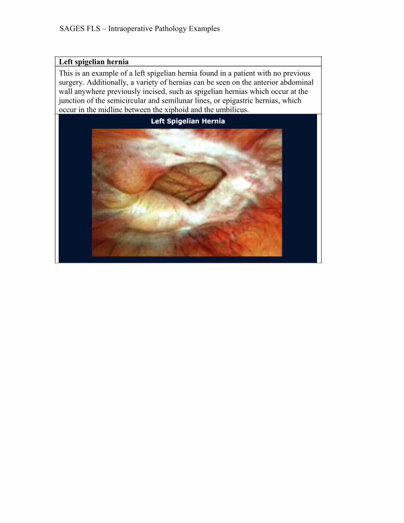
Left spigelian herniaThis is an example of a left spigelian hernia found in a patient with no previous surgery. Additionally, a variety of hernias can be seen on the anterior abdominal wall anywhere previously incised, such as spigelian hernias which occur at the junction of the semicircular and semilunar lines, or epigastric hernias, which occur in the midline between the xiphoid and the umbilicus.
SAGES FLS – Intraoperative Pathology Examples
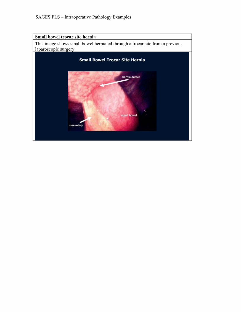
Small bowel trocar site herniaThis image shows small bowel herniated through a trocar site from a previous laparoscopic surgery
SAGES FLS – Intraoperative Pathology Examples
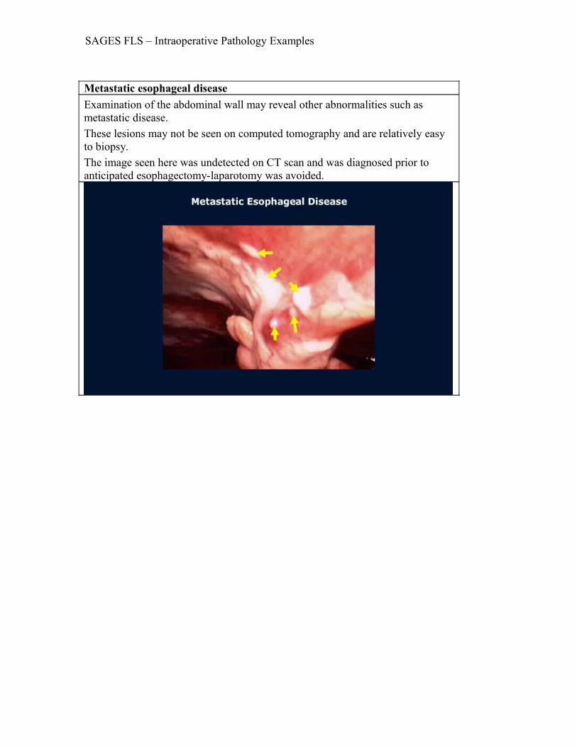
Metastatic esophageal diseaseExamination of the abdominal wall may reveal other abnormalities such as metastatic disease.These lesions may not be seen on computed tomography and are relatively easy to biopsy.The image seen here was undetected on CT scan and was diagnosed prior to anticipated esophagectomy-laparotomy was avoided.
SAGES FLS – Intraoperative Pathology Examples
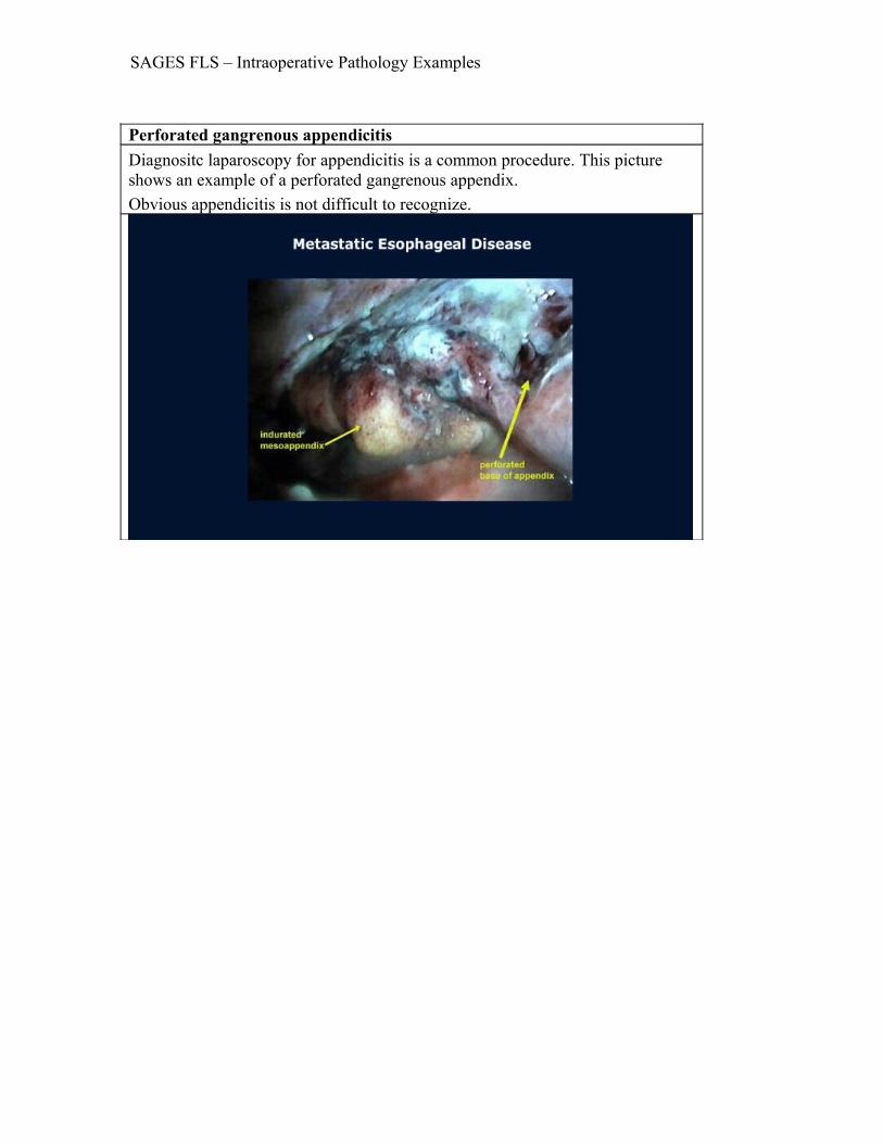
Perforated gangrenous appendicitisDiagnositc laparoscopy for appendicitis is a common procedure. This picture shows an example of a perforated gangrenous appendix. Obvious appendicitis is not difficult to recognize.

SAGES FLS – Intraoperative Pathology Examples
Cytomegalovirus colitis in cecumOther appendiceal disease, such as appendiceal neuroma, carcinoid, cytomegalovirus, or carcinoma as causes for right lower quadrant pain, may be more difficult to discern. The arrow indicates cytomegalovirus colitis presenting in the cecum.

SAGES FLS – Intraoperative Pathology Examples
Creeping fatMesenteric fat, creeping toward the antimesenteric border of the bowel wall, suggests a diagnosis of Crohn’s disease

SAGES FLS – Intraoperative Pathology Examples
Small intestinal arteriovenous malformationSmall intestinal arteriovenous malformation in the ileum (indicated by the arrow) in a patient where laparoscopic exploration was undertaken for suspected Meckel’s diverticulum.

SAGES FLS – Intraoperative Pathology Examples
Kaposi’s Sarcoma in ileumExamination of the intestines can lead to a number of findings and the laparoscope is valuable in examining the external anatomy of the small intestines. The image seen here is Kaposi’s Sarcoma in ileum. Atraumatic bowel forceps are recommended for handling the intestines.
SAGES FLS – Intraoperative Pathology Examples
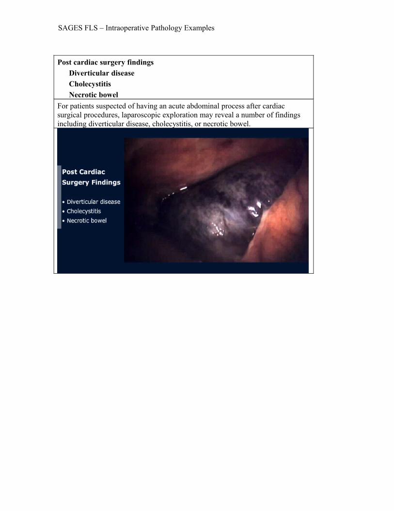
Post cardiac surgery findings Diverticular disease Cholecystitis Necrotic bowelFor patients suspected of having an acute abdominal process after cardiac surgical procedures, laparoscopic exploration may reveal a number of findings including diverticular disease, cholecystitis, or necrotic bowel.

SAGES FLS – Intraoperative Pathology Examples
Ovarian carcinomaThis is an example of ovarian carcinoma in a patient with non-diagnostic cytology.
SAGES FLS – Intraoperative Pathology Examples
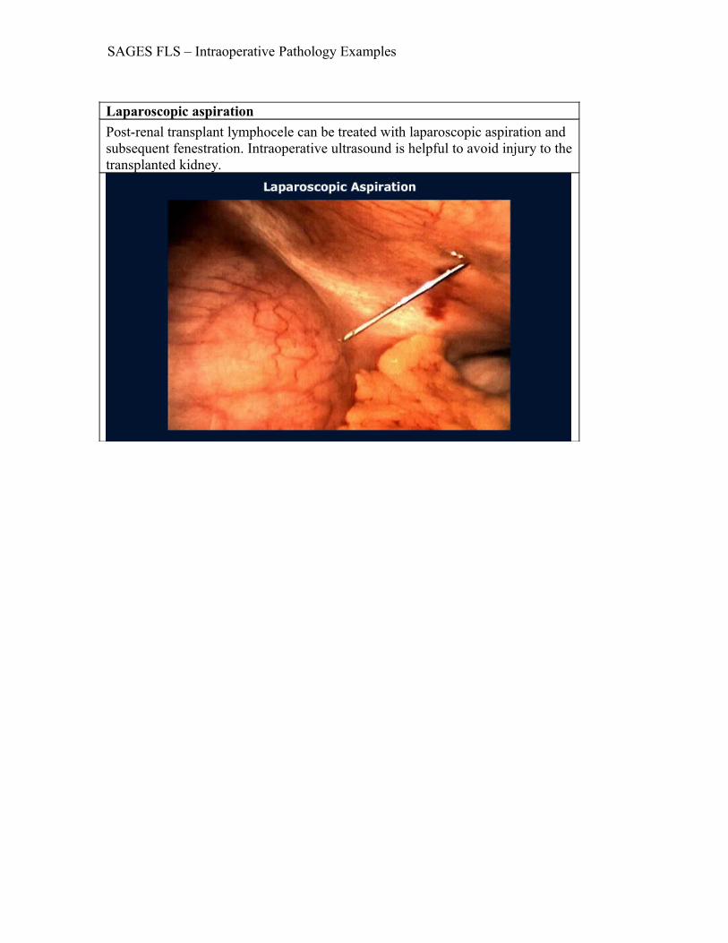
Laparoscopic aspirationPost-renal transplant lymphocele can be treated with laparoscopic aspiration and subsequent fenestration. Intraoperative ultrasound is helpful to avoid injury to the transplanted kidney.

SAGES FLS – Intraoperative Pathology Examples
Benign peritoneal mesotheliomaDiagnostic laparoscopy performed for a complex pelvic mass revealed this benign, peritoneal mesothelioma.

SAGES FLS – Intraoperative Pathology Examples
Large uterine fibroidOn occasion, the surgeon is confronted with an unexpected large mass. While this is cause for concern, many of these findings are benign, especially if the surface of the mass is smooth. Careful attention to anatomic detail will reveal the nature of these findings, which often include uterine fibroids, massive bladder enlargement, pregnancy (pre- or post-partum), or ovarian cyst. The image seen here is a large uterine fibroid (equivalent to 20-week pregnancy) discovered incidentally during cholecystectomy.

SAGES FLS – Intraoperative Pathology Examples
Mottled macronodular cirrhosisLaparoscopic examination of the liver is a useful aid in hepatic diagnosis. Biopsy of suspicious lesions can be performed with low morbidity. Biopsy of hemangiomas or livers with obvious increased portal pressures should be discouraged in order to avoid excessive bleeding. Metastatic cancer, cirrhosis, and hepatitis are usually recognized easily on visual inspection. The image seen here shows mottled macronodular appearance of the left lobe of the liver with cirrhosis of cardiac origin.

SAGES FLS – Intraoperative Pathology Examples
Hepatic hemangiomasHepatic Hemangiomas are generally easily identified visually. Biopsy may result in significant hemorrhage and should be avoided.