sarah jaar marah al-darawshehdoctor2017.jumedicine.com/wp-content/uploads/sites/7/...marah...
TRANSCRIPT

22
Sarah Jaar
Marah Al-Darawsheh
Faisal Mohammad

1 | P a g e
Receptors can be membrane proteins (for water-soluble hormones/ligands) or
intracellular (found in the cytosol or nucleus and bind to DNA, for lipid-soluble
hormones/ligands).
Receptors can also be classified as:
1) Ionotropic: coupled to ion channels (K+/Na+/Cl- …etc) that open/close as a result of
binding of the hormone to the receptor eg: acetylcholine. This opening/closing of
channels may alter the membrane potential or generate electrical signal that triggers
cellular response.
2) Metabotropic: G-protein receptors. G-proteins are transmembrane proteins. There
are 2 sides; an extracellular side which binds to ligands, and an intracellular side which
binds to GTP/GDP. G-proteins are heterotrimeric (made up of 3 subunits: alpha beta &
gamma). *G-protein refers to any protein that binds to GTP/GDP and acts as a signal
transducer. When bound to GDP they are inactive, but once a hormone/ligand binds to
the receptor, it’s activated (GDP is replaced by GTP). The alpha subunit bounded to GTP
dissociates and does its action. This can be changing the permeability of any channel
directly or activating a second messenger system; it activates another membrane
enzyme like Guanylate Cyclase or Adenylate Cyclase…etc.
2 | P a g e
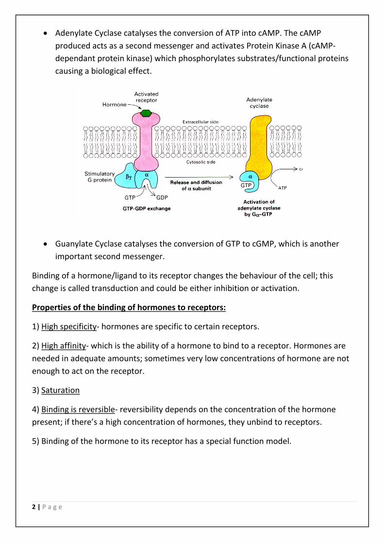
• Adenylate Cyclase catalyses the conversion of ATP into cAMP. The cAMP
produced acts as a second messenger and activates Protein Kinase A (cAMP-
dependant protein kinase) which phosphorylates substrates/functional proteins
causing a biological effect.
• Guanylate Cyclase catalyses the conversion of GTP to cGMP, which is another
important second messenger.
Binding of a hormone/ligand to its receptor changes the behaviour of the cell; this
change is called transduction and could be either inhibition or activation.
Properties of the binding of hormones to receptors:
1) High specificity- hormones are specific to certain receptors.
2) High affinity- which is the ability of a hormone to bind to a receptor. Hormones are
needed in adequate amounts; sometimes very low concentrations of hormone are not
enough to act on the receptor.
3) Saturation
4) Binding is reversible- reversibility depends on the concentration of the hormone
present; if there’s a high concentration of hormones, they unbind to receptors.
5) Binding of the hormone to its receptor has a special function model.

3 | P a g e
Types of receptors:
1) Channel-linked (ionotropic)
2) Enzyme-linked (involve neurotrophins and phosphorylation by protein kinases) eg:
tyrosine kinase
3) G-protein coupled (metabotropic)
4) Intracellular (involve activation by cell-permeant signals)
Hormones only bind to specific receptors. Sometimes a hormone can slightly interact
with a receptor for a hormone very similar to it in structure. Therefore, receptors
determine response; if there’s no receptor, there won’t be a response.
ionotropic
metabotropic

4 | P a g e
*G-protein coupled receptors’ signal cascade
Signal is passed from GPCR (G-protein coupled receptors) which are seven-helices
receptors. There are many different types of GPCRs (around 800 in humans). Binding of
a ligand may cause GPCRs to dimerize, forming oligomeric complexes across the
membrane which would affect the activity of the receptor.
GPCRs not only react with heterotrimeric G-proteins, but can also react with certain
proteins called GIPs (GPCR-interacting proteins). These GIPs modulate receptor
functions and may have several effects:
• Altered affinity for the ligand
• Receptor dimerization/oligomerization
• Control the localization of receptors, including their transfer or removal from
plasma membrane
• Promoting close association with other signal proteins
Activation of cAMP signaling (wasn’t explained in detail in the lecture; from slides):
• A G-protein that activates cAMP formation within a cell
is called a stimulatory G-protein Gs, with the alpha subunit Gsα.
• Gs is activated by receptors for epinephrine or glucagon.
• The GPCR for epinephrine is β2-adrenergic receptor.
• The G-protein is bound to the cytosolic surface of the
plasma membrane by lipid anchors from alpha & gamma
subunits of the G-protein.
• Adenylate Cyclase is a transmembrane protein with
cytosolic domains which form the catalytic site for the
conversion of ATP to cAMP and PPi2 (2 phosphate). It is
activated when a certain hormone (e.g: epinephrine) binds to its receptor on the
outer surface of the cell.
• cAMP acts as a second messenger
Sequence of events:
1) Initially, Gα is bounded to GDP and the alpha, beta and gamma subunits are
complexed together. Gβγ (the complex formed by beta and gamma subunits) inhibits Gα.
2) A hormone binds to the extracellular domain of a GPCR causing a change in the
conformation of the receptor. This change is transmitted to a heterotrimeric G-protein
N
N N
N
NH2
O
OHO
HH
H
H2C
HO
PO
O-
1'
3'
5' 4'
2'
cAMP

5 | P a g e
on the cytosolic side of the membrane. The nucleotide-binding site on Gsα becomes
more accessible to the cytosol. Inside the cytosol, the concentration of GTP is much
greater than that of GDP, so Gsα releases GDP and replaces it with GTP (GDP-GTP
exchange).
3) This substitution of GDP for GTP causes another conformational change in Gsα,
causing the Gsα-GTP complex to dissociate from the inhibitory Gβγ complex. Gsα-GTP
complex can now bind to Adenylate Cyclase and activate it.
4) Due to its activation, Adenylate Cyclase catalyses the synthesis of cAMP.
5) The increasing concentration of cAMP formed activates Protein Kinase A (cAMP-
dependant Protein Kinase), which phosphorylates intracellular substrates (e.g: enzymes
or serine/threonine residues of various cellular proteins), it modulates the activity of
enzymes present inside the cell and alters the metabolism of the cell
*The phosphorylation of enzymes could either activate or inactivate them (some
enzymes get activated when phosphorylated and others may get deactivated).

6 | P a g e
Turning off the signal:
1) Gsα hydrolyses GTP to GDP+Pi (acts as GTPase). The presence of GDP on Gsα causes it
to bind back to the beta and gamma inhibitory complex.
2) cAMP-dependant phosphodiesterase enzymes catalyse the conversion of cAMP to
AMP. This phosphodiesterase enzyme is activated by phosphorylation (which was
catalyzed by Protein Kinase A). We can see that cAMP stimulates its own degradation.
When there’s no more cAMP, there’s no more stimulation.
*Phosphodiesterases are very specific, and there are many different types
corresponding to different substrates.
*Enzyme-linked receptors
The best example is tyrosine kinase-linked receptors. They’re made up of alpha and beta
subunits, with the alpha subunits exposed to the extracellular side. They include
receptors for insulin and most growth factors (nerve growth factor NGF, epidermal
growth factor EGF, platelet-derived growth factor PDGF, vascular endothelial growth
factor VEGF…etc.). These receptors can either be single polypeptides or dimers (usually
dimers).
When an insulin ligand binds to this receptor via the alpha subunit, it causes the alpha
and beta subunits to dimerise (becoming 2 alpha & 2 beta subunits). The binding of
insulin and alpha subunits brings about autophosphorylation of the beta subunits. This
causes the structure to act as an enzyme (tyrosine kinase), when the activity of tyrosine
kinase increases, it phosphorylates intracellular substrates or it creates new specific
binding sites to which proteins can bind, transmitting intracellular signals.
Phosphorylation of intracellular substrates can open channels, or they may go to DNA to
increase protein synthesis, usually glucose transporter proteins. The glucose
transporters that are formed are translocated to the plasma membrane where they are
needed. They increase glucose uptake into the cell, therefore decrease concentration of
glucose in the plasma. As we can see, the binding of insulin lowers blood glucose.
Glucose that enters the cell is converted to glycogen, lipids…etc.

7 | P a g e
As we saw earlier, second messengers may target:
• Enzymes- they modulate phosphorylation which causes activation/inactivation of
the enzymes.
• Protein Kinases- they increase phosphorylation.
• Protein Phosphatases- they decrease phosphorylation (cause dephosphorylation).
Can be activated by Ca2+/calmodulin.
*Calmodulin (cal- from calcium & modulin- from modulation) is an intracellular
protein that is found in almost every cell, it binds Ca2+ and regulates cell activities.
The equivalent to calmodulin that’s found only in muscles is called troponin.
Once the concentration of intracellular Ca2+ increases, it binds to calmodulin. Each
calmodulin molecule binds 4 Ca2+. This binding exhibits the positive co-operativity
phenomenon, where binding of the first molecule is the most difficult, but it makes it
easier for the second molecule, which makes it easier for the third etc… (like in
voltage gated channels; opening of the first channel is the hardest but it gets easier).
Once calmodulin binds Ca2+, it activates Calcium-calmodulin dependant Protein
Kinase (Protein Kinase B; depends on Ca2+ bound to calmodulin).
Another type of second messengers are cyclic nucleotides like cAMP (which targets
Protein Kinase A) & cGMP (which target Protein Kinase G).
We also have lipids like DAG (diacylglycerol) and IP3 (inositol trisphosphate) acting as
second messengers. DAG and IP3 are formed when Phospholipase C enzymes break
down one of the fatty acids of a phospholipid molecule called PIP2, leaving behind a
molecule consisting of 2 fatty acids+glycerol (DAG) and an IP3 molecule.
PIP2 DAG + IP3
DAG stays in the cytosol and the IP3 formed goes to the smooth endoplasmic
reticulum (the SER stores Ca2+) and stimulates the release of Ca2+ by opening ligand-
gated ion channels. The increased concentration of Ca2+ in the cytosol and the
presence of DAG together activate Protein Kinase C (Ca2+-phospholipid dependant
Protein Kinase), which in turn activates proteins inside the cell by phosphorylation.

8 | P a g e
*The 3 types of Protein Kinases we took so far:
• Protein Kinase A: cAMP dependant
• Protein Kinase B: Ca2+-calmodulin dependant
• Protein Kinase C: Ca2+-phospholipid dependant
Some hormones like catecholamines (epinephrine and norepinephrine), polypeptides
and glycoproteins cannot through the plasma membrane, but bind to receptor proteins
on the target plasma membrane. These extracellular hormones are transduced into
intracellular second messengers (their actions are mediated by second messengers).
Adenylate Cyclase, cAMP and Protein Kinase A:
Adenylate Cyclase converts ATP to cAMP which activates Protein Kinase A (PKA). PKA is
a tetramer (4 parts), consisting of 2 regulatory and 2 catalytic subunits. Before the
binding of cAMP, the catalytic subunit is inactive, so the PKA is inactive. When cAMP
binds to the regulatory subunit, this causes the dissociation of the catalytic subunit,
which is released to phosphorylate target proteins in the cytoplasm. The catalytic
subunit can go the nucleus, where it phosphorylates CREB proteins (catalytic regulatory
element binding proteins). CREB proteins bind to the CRE (cAMP response element).
CRE is a regulatory sequence of DNA that is associated with specific genes. This binding
to CRE causes transcription of these genes and translation to form new protein. This
pathway is the slowest of all.

9 | P a g e
Mentioned only in the slides:
The action of Adenylate Cyclase is regulated by G-proteins: Gs (stimulatory) & Gi
(inhibitory).
Pathogens can alter cAMP production: the cholera toxin is made up of many subunits;
one of its subunits catalyses the transfer of ADP ribose from intracellular NAD to the
subunit of Gs causing the G-protein to be continuously active. This stimulates adenylyl
cyclase indefinitely, causing ion channels that export Cl- to produce a net efflux of Cl-
and water, leading to severe diarrhea, which is a characteristic of cholera.
The enzyme Guanylate Cyclase converts GTP to cGMP, which activates cGMP-
dependant kinases or other targets eg: G-protein coupled rhodopsin photoreceptors in
rod cells in the retina.
One of the characteristics of metabotropic
receptor signalling is that second messengers
amplify the signal at each step of the cascade.
*Notes from past few lectures:
Neurotransmitter release (exocytosis and endocytosis):
1. Transmitter is synthesized and stored
2. Action Potential
3. Depolarization: opening of voltage-gated Ca2+ channels
4. Ca2+ enters the cell
5. Ca2+ causes vesicles to fuse with membrane
6. Neurotransmitter is released (exocytosis)
7. Neurotransmitter binds to postsynaptic receptors
8. Opening or closing of postsynaptic channels

10 | P a g e
9. Postsynaptic current excites or inhibits postsynaptic potential to change the excitability of cell
10. Retrieval of vesicles from plasma membrane (endocytosis)
Receptors to which neurotransmitters (NT) bind are large, dynamic proteins that exist along and within the cell membrane. They’re dynamic– they can increase in number and avidity for their neurotransmitter according to the circumstances.
There are two types of post-synaptic receptors:
1) Ionotropic receptors: NT binding results in the direct opening of specific ion channels.
2) Metabotropic receptors: the binding of NT initiates a sequence of internal molecular events, which in turn open specific ion channels.
Binding of NT causes a response in membrane potential
Ionotropic receptors:
They work very fast and are important in fast neurotransmission. Each receptor is made of several subunits, which together form the complete receptor. At the center of receptors is a channel or a pore to allow flow of ions. At rest, receptor channels are closed but when a neurotransmitter binds, the channels immediately open. When the ligand leaves the binding site, the channel quickly closes.

11 | P a g e
Metabotropic receptors:
They work by activating other proteins called G-proteins. Each receptor is made up of several transmembrane regions. They work more slowly than ionotropic receptors. They stimulate/inhibit the opening of ion channels in the cell membrane or stimulate/inhibit certain effector enzymes. Most of the effector enzymes that are controlled by G-proteins are involved in the synthesis of second messengers. Though it takes longer for postsynaptic cells to respond, the response is somewhat longer-lasting than in ionotropic receptor signalling. The receptors comprise of a single protein subunit, winding back-and-forth through cell membrane seven times (transmembrane domains) and don’t possess a channel or pore.
GOOD LUCK 😊