sarcoma resection with and without vascular reconstruction: a...
TRANSCRIPT

1
Sarcoma Resection With and Without Vascular Reconstruction:
A Matched Case-Control Study
George A. Poultsides, MD,1 Thuy B. Tran, MD,
1 Eduardo Zambrano, MD,
2
Lucas Janson, MS,3 David G. Mohler, MD,
4 Matthew W. Mell,
MD,
5 Raffi S. Avedian, MD,
4
Brendan C. Visser, MD, 1
Jason T. Lee, MD,5
Kristen Ganjoo, MD,6
E. John Harris, MD,5 and Jeffrey A. Norton, MD
1
1. Division of Surgical Oncology, Department of Surgery, Stanford University
2. Department of Pathology, Stanford University
3. Department of Statistics, Stanford University
4. Department of Orthopaedic Surgery, Stanford University
5. Division of Vascular Surgery, Department of Surgery, Stanford University
6. Division of Oncology, Department of Medicine, Stanford University
Corresponding Author: George A. Poultsides, MD
Department of Surgery
Stanford University School of Medicine
300 Pasteur Drive, H3680D
Stanford, CA 94305-5641
Phone: 650-723-4646
Fax: 650-736-1663
Email: [email protected]
Running Head: Vascular Resection for Sarcoma
Acknowledgment of Research Support: None
Presented at the 135th
Annual Meeting of the American Surgical Association, San Diego,
California, April 23-25, 2015
2
INTRODUCTION
Sarcomas are a heterogeneous group of rare malignancies with variable presentation,
behavior and outcome. Although our understanding of their natural history following resection
has evolved considerably over the recent years,1 these tumors are still in many cases resistant to
standard chemotherapy or radiotherapy modalities and surgical resection remains the cornerstone
of treatment.2 Often, the ability to completely remove the tumor is affected by its relationship to
major blood vessels. Traditionally surgeons have been reluctant to perform major vascular
resections for this disease, due to the inherently increased complexity of these operations and the
uncertainty about the long-term oncologic benefit. Over the last decade, several case series have
established the feasibility and safety of en bloc vascular resection for sarcomas of the
extremity,3-7
retroperitoneum,8-10
or specifically the inferior vena cava (IVC).11-18
What remains
unknown is whether these complex procedures are associated with a durable prolongation of
survival that justifies their morbidity. Our institution has previously reported our findings with
major blood vessel reconstruction on 14 sarcoma patients undergoing surgical resection.19
The
current study provides an update on this initial experience and attempts to further compare these
patients in a matched case-control fashion with a separate cohort of sarcoma patients during the
same time period who did not require en bloc vascular resection but had similar
clinicopathologic characteristics.
METHODS
The study population includes patients who underwent surgical resection for sarcoma of
any anatomic site between 2000 and 2014 at our institution. Patients were identified from the
3
Stanford Cancer Registry through the appropriate use of ICD-9 (International Classification of
Diseases) and CPT (Current Procedural Terminology) codes, following Institutional Review
Board approval. The patients who underwent sarcoma resection with vascular reconstruction
were matched with 2 additional patients who underwent sarcoma resection without vascular
reconstruction. Case matching was performed on 6 established clinocopathologic predictors of
outcome for sarcoma: anatomic site, histologic type, grade, size, presence of synchronous
metastasis, and whether resection was performed for primary versus recurrent disease (primary
vs. repeat resection). Patients with R2 resections (macroscopically positive margins) were
excluded, but patients who had M1 disease at operation were included. Data on patient
demographics, clinicopathologic characteristics, and intraoperative variables were collected.
Endpoints included perioperative morbidity, mortality, margin status, local recurrence, and
survival. Surgical complications were graded using the modified Clavien-Dindo classification.20
Repeat review of pathology slides of the VASC patients was undertaken by a single
sarcoma-dedicated pathologist, to assess the level of histologic infiltration (if any) of the resected
vascular structures by the tumor. The pathologist was blinded to the survival outcome. This
retrospective study was approved by the Stanford Hospital and Clinics Institutional Review
Board.
In general, our institutional practice when vascular involvement is suspected on
preoperative contrast-enhanced cross-sectional imaging (Computed Tomography or Magnetic
Resonance Imaging) is for patients to be referred by the surgical or orthopedic oncologist to the
vascular surgeon for a preoperative discussion and assessment of vascular reconstruction options
and conduit selection. Postoperatively, the two teams follow the patients jointly both throughout
their hospitalization and outpatient follow-up. Cross-sectional imaging used to detect tumor
4
recurrence is also utilized to monitor vascular patency, in addition to the use of vascular
ultrasound.
Categorical variables were presented as absolute counts (percentages) and compared
using the Mantel-Haenszel test. Continuous variables were presented as medians (range) and
compared using within-subjects ANOVA (analysis of variance).21
Survival probabilities were
calculated using the Kaplan-Meier method and compared using the log-rank test. Univariate and
multivariate Cox proportional hazard models were created to identify prognostic factors
associated with overall survival (OS). A two-sided P value of < 0.05 was considered statistically
significant.
RESULTS
From 2000 to 2014, 50 patients (cases) who underwent sarcoma resection with vascular
reconstruction (VASC) were identified, representing 5% of 1,009 patients undergoing sarcoma
resection at our institution during the same time period. Slightly more than half (54%) of these
50 patients (cases) were for retroperitoneal sarcomas and four representative cases are illustrated
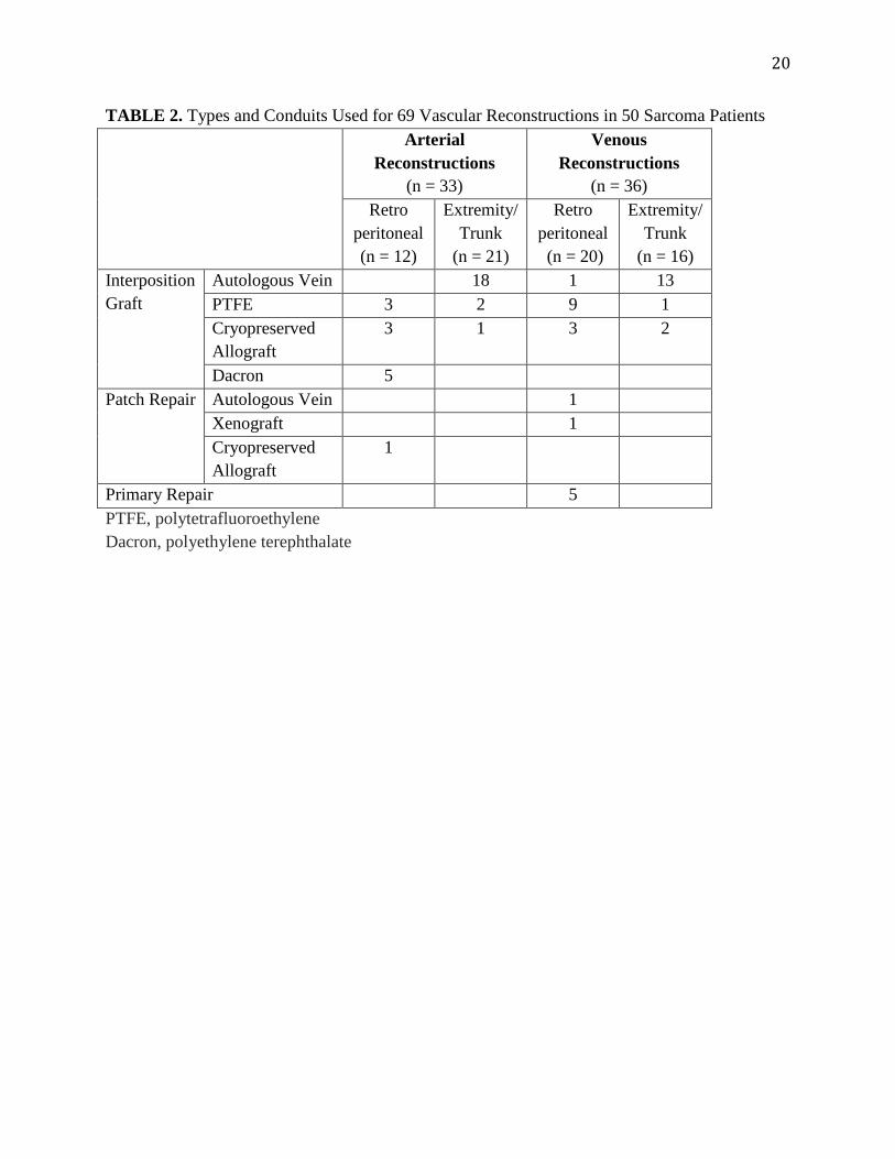
in Figure 1. Overall, 69 vessels were reconstructed in these 50 patients: 14 patients had arterial,
19 arterial and venous and 17 venous only reconstructions. The distribution of vessels
reconstructed is shown in Table 1 and the types of reconstruction (interposition graft, patch
repair, or primary repair) and choice of conduit are shown in Table 2.
The 50 VASC cases were matched in a 1:2 ratio with 100 patients (controls) who
underwent resection of sarcomas with similar clinicopathologic characteristics but did not
require vascular reconstruction (NO VASC). A comparison of basic clinicopathologic
5
characteristics between the two groups is shown in Table 3 and confirms that the two groups
were adequately matched for site, histology, grade, size, synchronous metastasis, and primary
versus repeat resection. The two groups were eventually found to be comparable by age, gender,
margin positivity, and presence of comorbidities. Neoadjuvant chemotherapy and radiotherapy
was more commonly utilized for VASC patients, whereas the rates of intraoperative radiotherapy
and adjuvant chemotherapy or radiotherapy did not differ between the two groups.
A comparison of perioperative morbidity and mortality is shown in Table 4. The VASC
group had approximately twice the estimated blood loss, operative time and intraoperative
transfusion rate of the NO VASC group. Similarly, the rates of any (74% vs. 44%, P = 0.002)
and of major (Clavien grade 3 or higher, 38% vs. 18%, P = 0.024) complications within 30 days
were significantly higher in the VASC group. As a result, median length of stay was longer by 3
days. Return to the operating room within 30 days was twice as common for the VASC group
(18% vs. 9%), however this difference did not reach statistical significance. Reasons for 30-day
reoperation in the VASC group included acute limb ischemia (n=3, two requiring graft
thrombectomy and one hip disarticulation), intestinal perforation (n=2, one leading to a
prosthetic iliofemoral graft infection), postoperative bleeding (n=2, both unrelated to the vascular
reconstruction) and extremity wound dehiscence (n = 2). In the NO VASC group, reasons for
reoperation included: intestinal perforation (n=2), extremity wound hematoma (n=2), extremity
wound infection (n=2), wound dehiscence (n=2, one abdominal and one trunk incision), and
orthopaedic hardware infection (n=1).
The 2% 30-day and 6% 90-day mortality in the VASC group were not significantly
different than the corresponding rates seen in the NO VASC group (0% and 2%). The single 30-
day mortality in the VASC group was secondary to postoperative bleeding, which was unrelated

6
to the vascular reconstruction (primary repair of the juxtarenal IVC in this case). The two
additional 90-day mortalities in the VASC group occurred after discharge from the hospital (to
skilled nursing facilities) but appeared to be related to postoperative complications. The two 90-
day mortalities in the NO VASC group included a death at home of unknown etiology and a
death from rapid progression of disease postoperatively.
Median follow-up was 24 months for the VASC and 28 months for the NO VASC
patients. Overall survival after resection was similar between the VASC and NO VASC groups
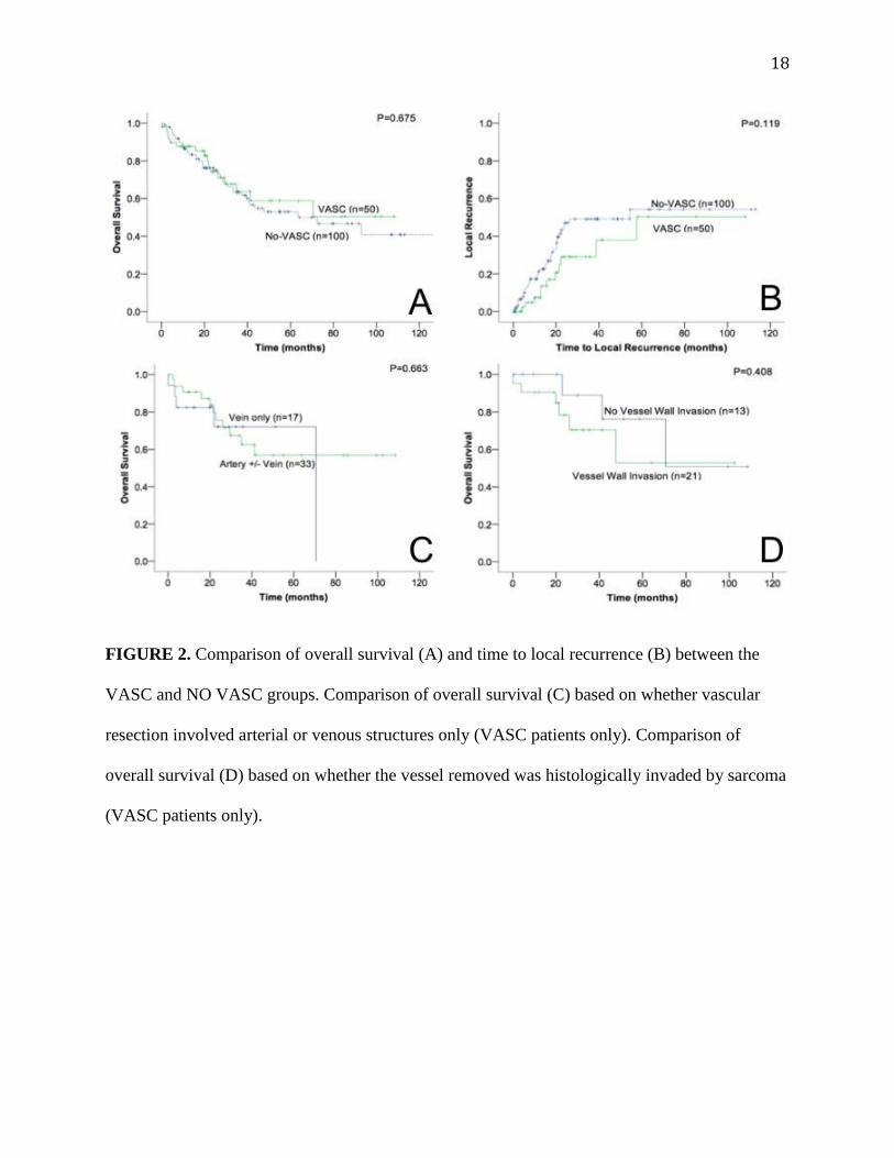
(5-year 59% vs. 53%, P = 0.67, Figure 2A). Similarly, when various subset analyses were
performed, no subgroup of patients was identified (retroperitoneum or extremity/trunk, high or
low/intermediate grade, R1 or R0, tumor size > 10 cm or < 10 cm, synchronous M1 or M0,
primary surgery or for recurrence) in whom a difference in overall survival was noted between
the VASC and NO VASC patients (data not shown). Furthermore, on multivariate analysis, high
tumor grade and presence of synchronous metastases were independent predictors of overall
survival, however there was still no association between the need for vascular reconstruction and
overall survival (Supplemental Table). Last, as local control is another significant endpoint in
the assessment of the efficacy of surgical resection for any given tumor we specifically evaluated
the time to local recurrence between the two groups: again, 5-year local recurrence rates were
similar between the two groups (51% vs. 54%, P = 0.119, Figure 2B).
We sought to examine whether specific factors within the VASC cohort, such as the type
of vessel involved or the presence of true pathologic vessel invasion, were predictive of long-
term outcome. We found no association between the type of vascular involvement (arterial vs.
venous) and overall survival (Figure 2C). Furthermore, pathology slides for 34 of the 50 VASC
cases were available for re-review to specifically assess vessel wall invasion histologically.

7
Histologic vessel wall invasion by sarcoma was noted in 21 (62%) of 34 patients and was more
common in resected veins (18/26, 69%) than resected arteries (3/16, 19%). When stratified by
sarcoma type, histologic vessel invasion was noted in all 10 leiomyosarcomas, 3 out of 7
liposarcomas, 2 out of 4 undifferentiated pleomorphic sarcomas and 1 out of 4 synovial
sarcomas. Overall survival after resection did not appear to be associated with the presence of
histology proven vessel wall invasion by the tumor (Figure 2D).
The patency rates of the vascular reconstructions are shown in Figure 3. The 5-year
primary and assisted primary patency of arterial reconstructions for retroperitoneal sarcoma was
86% and 92%, and for extremity/trunk sarcoma 56% and 56%. There were three amputations
among the 19 extremity patients (limb salvage rate of 84%). The 5-year primary patency of
venous reconstructions for retroperitoneal sarcoma was 86% and for extremity/trunk sarcoma
63%. Graft infection was noted in three patients. A popliteal artery cryopreserved allograft had to
be replaced by autologous vein 6 months postop due to the presence of an infected
pseudoaneurysm. This patient had a free myocutaneous flap at the time of the initial operation
for coverage of the resection bed, but flap ischemia required flap revision early in the
postoperative period and likely contributed to the graft infection. The second case of graft
infection was an iliac artery Dacron graft that got secondarily infected postoperatively after the
patient developed colonic perforation. This graft was removed and replaced with contralateral
superficial femoral vein. The third case of graft infection was an SMA Dacron graft that was
noted 7 years postoperatively to erode through the posterior wall of the stomach on endoscopy.
The graft had thrombosed, but the patient had developed collateral circulation to the midgut
through the inferior mesenteric artery and did not have any signs of intestinal ischemia. The graft
was removed without reconstruction. Long-term follow-up is not yet available for this case.

8
DISCUSSION
The objective of our study was to examine the impact of concomitant vascular
reconstruction on sarcoma resection outcomes. Given the fact that sarcomas involving major
blood vessels are usually more extensive than ones without vascular involvement, we utilized a
matched case-control methodology. The main finding was that the need for vascular
reconstruction almost doubled the morbidity of these resections, but was associated with a
comparable oncologic outcome (local recurrence and overall survival) to matched cases without
vascular involvement. Our study reinforces previously reported findings of a smaller case-control
study on 19 extremity sarcoma patients who underwent vascular reconstruction and were
matched with 38 patients of similar age, tumor size, anatomic location, depth, and timing of
radiotherapy, but without vascular involvement.5 Although cases and controls in this previous
study were not matched for the presence of synchronous metastases, when only M0 patients
(n=40) were examined, the 5-year disease-free survival rates were similar at 83% and 74%. Our
study corroborated this finding on a larger cohort of patients, with retroperitoneal and truncal in
addition to extremity sarcomas as well as patients undergoing resection of both primary and
recurrent disease.
The concept that long-term survival after resection may not be affected by the need for
vascular resection and reconstruction has been demonstrated for a variety of other solid tumors.
Specifically, several single-institution22-25
and multi-institutional series26
have demonstrated that
patients with pancreatic adenocarcinoma who require portal vein resection have similar survival
to resected patients not requiring portal vein resection. Similarly, our group has previously
reported our experience on outcomes after resection of pancreatic neuroendocrine tumors with
major vascular involvement,27
and others have reported on outcomes after resection of locally

9
recurrent rectal cancer involving the aortoiliac axis,28
with both studies showing long-term
survival rates comparable to historical controls with locally advanced disease but not involving
vascular structures. Taken together, these data indicate that major vascular involvement is not
necessarily a predictor of aggressive tumor biology but rather a reflection of tumor size and
location.
Histologically proven invasion of the resected vessel wall by sarcoma was noted in 62%
of 34 patients at dedicated pathologic re-review. This number is higher than previously reported
studies examining the frequency of histologic vessel infiltration found at concomitant vascular
resection for retroperitoneal (32%),10
and extremity sarcoma (43%),7 and similar to
corresponding studies on pancreatic adenocarcinoma (61%).25,29
Histologic vessel invasion was
less frequently noted in resected arteries (19%) than veins (69%) in our study, and the finding on
arterial invasion is identical to a previous study of 37 sarcoma patients who underwent arterial
resection and in whom the frequency of true pathologic invasion was 19%.8 In the absence of
clear-cut encasement or intraluminal tumor thrombus, our practice has been to initially attempt to
dissect the tumor off the surrounding vessels. However, as noted herein and in several other
studies,7,8,10,25,29
true vascular invasion is difficult to differentiate intraoperatively from peri-
tumoral inflammation and desmoplastic reaction. In addition, dissection of arteries and veins
from abutting tumors can threaten their integrity, and formal vascular resection is sometimes
necessary to prevent inadvertent venotomy or arteriotomy even in the absence of true invasion.
Nonetheless, as reported by others specifically for pancreatic adenocarcinoma,25
histologically
proven vessel wall involvement was not associated with worse survival in our study.
Perhaps more controversial is the necessity, if any, to reconstruct a major vein that has
been resected. In particular, the optimal management of the IVC after resection is debatable, with

10
some advocating ligation,14,30
others selective,12,31,32
and others routine reconstruction.11,13,16,18
The rationale for the latter is based on the need to resect several venous collaterals for complete
tumor removal, as well as the inability to predict which patients will tolerate IVC ligation
without subsequent renal insufficiency or significant lower extremity edema. Our study was not
designed to address this specific question. Due to the nature of our search (CPT codes), we were
unable to discern whether there were additional cases in which a major vein was resected and
simply ligated without being reconstructed. In general, we do not advocate venous reconstruction
in cases where the venous structure is chronically occluded, the patient does not have lower
extremity edema, and the existing collateral pathways (gonadal, adrenal vein, and abdominal
wall collaterals for retroperitoneal tumors and greater saphenous vein for extremity tumors) are
maintained during oncologic resection.
Nine (15%) of the 61 interposition graft reconstructions in our series were performed
with cryopreserved cadaveric allografts, typically in the setting of a clean contaminated field.
The safety and efficacy of cryopreserved allografts for aortoiliac reconstruction in the setting of
infection has been recently established through a multi-institutional US study of 220 patients
with a mean follow-up of 30 months, reporting low rates of aneurysm formation, recurrent
infection, aortic blowout, and limb loss.33
Our limited experience with three aortoiliac
reconstructions for patients with concomitant intestinal resection has similarly shown no
instances of allograft occlusion or infection. When used for venous reconstructions, however,
cryopreserved allografts have been shown to have decreased patency rates: in a series of 8
patients undergoing IVC replacement with cryopreserved allografts for retroperitoneal sarcoma,
graft occlusion was observed in half of the patients (three late and asymptomatic and one early
and symptomatic) likely due to the susceptibility of the pliable allograft to compression from

11
abdominal viscera.12
We have used cryopreserved allografts to reconstruct the IVC in two
instances, one of which was complicated by early and symptomatic graft thrombosis. For this
reason, we, and others,11,18
favor IVC reconstruction with externally supported (ringed)
polytetrafluoroethylene (PTFE) graft, in cases where there is no bowel contamination. We have
utilized this approach in 8 patients in this study with no instances of graft infection and only one
case of thrombosis in a patient who developed heparin-induced thrombocytopenia and
thrombosis (HITT).
Our findings should be interpreted with caution as the relatively short follow-up in our
study might have led to misclassification of certain study endpoints (death, recurrence) or under-
detection of long-term vascular graft-related complications (patency, graft infection, anastomotic
pseudoaneurysm). Furthermore, there are inherent selection biases submerged in any
retrospective analysis that are difficult to control for. Patients in whom vascular resection was
undertaken (cases) likely represent a select group with robust performance status and overall
candidacy for aggressive treatment, an attribute that might have not been consistently true for
controls, despite our efforts to match cases and controls on a variety of important
clinicopathologic factors. Last, the lack of reliable information on cause of death for a large
number of patients led us to choose overall (as opposed to disease-specific) survival as our
primary endpoint. It is very likely, however, that the vast majority of the patients in this study,
who died during follow-up, did indeed die of sarcoma, as this cohort includes patients with
advanced disease (20% recurrent, 20% with synchronous metastasis, more than half larger than
10 cm, more than half retroperitoneal, and more than half high grade). Therefore, we feel that
overall survival represents an accurate and reliable measure of treatment efficacy in this select
group of sarcoma patients.

12
In conclusion, the need for vascular resection and reconstruction should not be a deterrent
to resection for sarcoma patients, as the oncologic outcome (overall and local recurrence free
survival) appears equivalent to matched cases without vascular involvement. The need for
vascular reconstruction essentially doubles the morbidity of these operations, whose technical
complexity spans across surgical disciplines. Meticulous multidisciplinary planning and close
collaboration between surgical oncologists, orthopaedic oncologists, and vascular surgeons is
critical for a successful outcome.

13
REFERENCES
1. Brennan MF, Antonescu CR, Moraco N, et al: Lessons learned from the study of 10,000 patients with soft tissue sarcoma. Ann Surg 260:416-21; discussion 421-2, 2014 2. Bonvalot S, Raut CP, Pollock RE, et al: Technical considerations in surgery for retroperitoneal sarcomas: position paper from E-Surge, a master class in sarcoma surgery, and EORTC-STBSG. Ann Surg Oncol 19:2981-91, 2012 3. Adelani MA, Holt GE, Dittus RS, et al: Revascularization after segmental resection of lower extremity soft tissue sarcomas. J Surg Oncol 95:455-60, 2007 4. Baxter BT, Mahoney C, Johnson PJ, et al: Concomitant arterial and venous reconstruction with resection of lower extremity sarcomas. Ann Vasc Surg 21:272-9, 2007 5. Ghert MA, Davis AM, Griffin AM, et al: The surgical and functional outcome of limb-salvage surgery with vascular reconstruction for soft tissue sarcoma of the extremity. Ann Surg Oncol 12:1102-10, 2005 6. Hohenberger P, Allenberg JR, Schlag PM, et al: Results of surgery and multimodal therapy for patients with soft tissue sarcoma invading to vascular structures. Cancer 85:396-408, 1999 7. Schwarzbach MH, Hormann Y, Hinz U, et al: Results of limb-sparing surgery with vascular replacement for soft tissue sarcoma in the lower extremity. J Vasc Surg 42:88-97, 2005 8. Carpenter SG, Stone WM, Bower TC, et al: Surgical management of tumors invading the aorta and major arterial structures. Ann Vasc Surg 25:1026-35, 2011 9. Fueglistaler P, Gurke L, Stierli P, et al: Major vascular resection and prosthetic replacement for retroperitoneal tumors. World J Surg 30:1344-9, 2006 10. Schwarzbach MH, Hormann Y, Hinz U, et al: Clinical results of surgery for retroperitoneal sarcoma with major blood vessel involvement. J Vasc Surg 44:46-55, 2006 11. Bower TC, Nagorney DM, Cherry KJ, Jr., et al: Replacement of the inferior vena cava for malignancy: an update. J Vasc Surg 31:270-81, 2000 12. Fiore M, Colombo C, Locati P, et al: Surgical technique, morbidity, and outcome of primary retroperitoneal sarcoma involving inferior vena cava. Ann Surg Oncol 19:511-8, 2012 13. Hardwigsen J, Baque P, Crespy B, et al: Resection of the inferior vena cava for neoplasms with or without prosthetic replacement: a 14-patient series. Ann Surg 233:242-9, 2001 14. Hollenbeck ST, Grobmyer SR, Kent KC, et al: Surgical treatment and outcomes of patients with primary inferior vena cava leiomyosarcoma. J Am Coll Surg 197:575-9, 2003 15. Kieffer E, Alaoui M, Piette JC, et al: Leiomyosarcoma of the inferior vena cava: experience in 22 cases. Ann Surg 244:289-95, 2006 16. Kuehnl A, Schmidt M, Hornung HM, et al: Resection of malignant tumors invading the vena cava: perioperative complications and long-term follow-up. J Vasc Surg 46:533-40, 2007 17. Mann GN, Mann LV, Levine EA, et al: Primary leiomyosarcoma of the inferior vena cava: a 2-institution analysis of outcomes. Surgery 151:261-7, 2012

14
18. Quinones-Baldrich W, Alktaifi A, Eilber F, et al: Inferior vena cava resection and reconstruction for retroperitoneal tumor excision. J Vasc Surg 55:1386-93; discussion 1393, 2012 19. Song TK, Harris EJ, Jr., Raghavan S, et al: Major blood vessel reconstruction during sarcoma surgery. Arch Surg 144:817-22, 2009 20. Clavien PA, Barkun J, de Oliveira ML, et al: The Clavien-Dindo classification of surgical complications: five-year experience. Ann Surg 250:187-96, 2009 21. Niven DJ, Berthiaume LR, Fick GH, et al: Matched case-control studies: a review of reported statistical methodology. Clin Epidemiol 4:99-110, 2012 22. Harrison LE, Klimstra DS, Brennan MF: Isolated portal vein involvement in pancreatic adenocarcinoma. A contraindication for resection? Ann Surg 224:342-7; discussion 347-9, 1996 23. Hristov B, Reddy S, Lin SH, et al: Outcomes of adjuvant chemoradiation after pancreaticoduodenectomy with mesenterico-portal vein resection for adenocarcinoma of the pancreas. Int J Radiat Oncol Biol Phys 76:176-80, 2010 24. Porembka MR, Hawkins WG, Linehan DC, et al: Radiologic and intraoperative detection of need for mesenteric vein resection in patients with adenocarcinoma of the head of the pancreas. HPB (Oxford) 13:633-42, 2011 25. Tseng JF, Raut CP, Lee JE, et al: Pancreaticoduodenectomy with vascular resection: margin status and survival duration. J Gastrointest Surg 8:935-49; discussion 949-50, 2004 26. Kelly KJ, Winslow E, Kooby D, et al: Vein involvement during pancreaticoduodenectomy: is there a need for redefinition of "borderline resectable disease"? J Gastrointest Surg 17:1209-17; discussion 1217, 2013 27. Norton JA, Harris EJ, Chen Y, et al: Pancreatic endocrine tumors with major vascular abutment, involvement, or encasement and indication for resection. Arch Surg 146:724-32, 2011 28. Abdelsattar ZM, Mathis KL, Colibaseanu DT, et al: Surgery for locally advanced recurrent colorectal cancer involving the aortoiliac axis: can we achieve R0 resection and long-term survival? Dis Colon Rectum 56:711-6, 2013 29. Roder JD, Stein HJ, Siewert JR: Carcinoma of the periampullary region: who benefits from portal vein resection? Am J Surg 171:170-4; discussion 174-5, 1996 30. Daylami R, Amiri A, Goldsmith B, et al: Inferior vena cava leiomyosarcoma: is reconstruction necessary after resection? J Am Coll Surg 210:185-90, 2010 31. Ito H, Hornick JL, Bertagnolli MM, et al: Leiomyosarcoma of the inferior vena cava: survival after aggressive management. Ann Surg Oncol 14:3534-41, 2007 32. Yoshidome H, Takeuchi D, Ito H, et al: Should the inferior vena cava be reconstructed after resection for malignant tumors? Am J Surg 189:419-24, 2005 33. Harlander-Locke MP, Harmon LK, Lawrence PF, et al: The use of cryopreserved aortoiliac allograft for aortic reconstruction in the United States. J Vasc Surg 59:669-74, 2014

15
LEGENDS

16
17
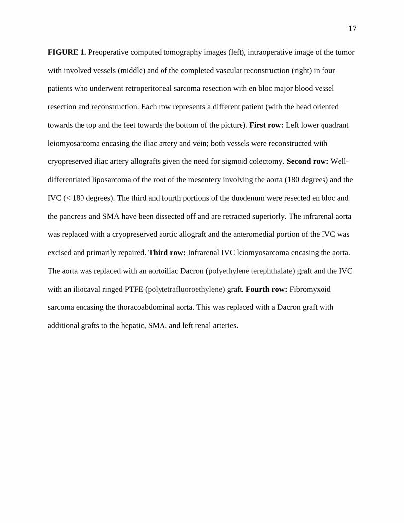
FIGURE 1. Preoperative computed tomography images (left), intraoperative image of the tumor
with involved vessels (middle) and of the completed vascular reconstruction (right) in four
patients who underwent retroperitoneal sarcoma resection with en bloc major blood vessel
resection and reconstruction. Each row represents a different patient (with the head oriented
towards the top and the feet towards the bottom of the picture). First row: Left lower quadrant
leiomyosarcoma encasing the iliac artery and vein; both vessels were reconstructed with
cryopreserved iliac artery allografts given the need for sigmoid colectomy. Second row: Well-
differentiated liposarcoma of the root of the mesentery involving the aorta (180 degrees) and the
IVC (< 180 degrees). The third and fourth portions of the duodenum were resected en bloc and
the pancreas and SMA have been dissected off and are retracted superiorly. The infrarenal aorta
was replaced with a cryopreserved aortic allograft and the anteromedial portion of the IVC was
excised and primarily repaired. Third row: Infrarenal IVC leiomyosarcoma encasing the aorta.
The aorta was replaced with an aortoiliac Dacron (polyethylene terephthalate) graft and the IVC
with an iliocaval ringed PTFE (polytetrafluoroethylene) graft. Fourth row: Fibromyxoid
sarcoma encasing the thoracoabdominal aorta. This was replaced with a Dacron graft with
additional grafts to the hepatic, SMA, and left renal arteries.
18
FIGURE 2. Comparison of overall survival (A) and time to local recurrence (B) between the
VASC and NO VASC groups. Comparison of overall survival (C) based on whether vascular
resection involved arterial or venous structures only (VASC patients only). Comparison of
overall survival (D) based on whether the vessel removed was histologically invaded by sarcoma
(VASC patients only).

19
FIGURE 3. Patency rates (continuous line: primary patency, dashed line: assisted primary
patency) of arterial (A) and venous (B) reconstructions stratified by anatomic site.
TABLE 1. Types of Vessels Reconstructed in 50 Sarcoma Patients
Artery only (n = 14) Artery and Vein (n=19) Vein only (n=17)
Aorta (n=4)* Aorta and IVC (n=2) IVC (n=13)**
Iliac (n=2) Iliac (n=4) Iliac (n=1)
Femoral (n=3) Femoral (n=9) Femoral (n=1)
Popliteal (n=1) Popliteal (n=3)
Posterior Tibialis (n=1)
Subclavian (n=2) Subclavian (n=1)
Brachial a. and Basilic v. (n=1)
SMA (n=1) SMV/PV (n=1)
*, one case with aorto-hepatic, aorto-SMA, and aorto-renal bypass
**, one case with reimplantation of the left renal vein
IVC, inferior vena cava; SMA, superior mesenteric artery; SMV, superior mesenteric vein; PV,
portal vein; SVC, superior vena cava
20
TABLE 2. Types and Conduits Used for 69 Vascular Reconstructions in 50 Sarcoma Patients
Arterial
Reconstructions
(n = 33)
Venous
Reconstructions
(n = 36)
Retro
peritoneal
(n = 12)
Extremity/
Trunk
(n = 21)
Retro
peritoneal
(n = 20)
Extremity/
Trunk
(n = 16)
Interposition
Graft
Autologous Vein 18 1 13
PTFE 3 2 9 1
Cryopreserved
Allograft
3 1 3 2
Dacron 5
Patch Repair Autologous Vein 1
Xenograft 1
Cryopreserved
Allograft
1
Primary Repair 5
PTFE, polytetrafluoroethylene
Dacron, polyethylene terephthalate

21
TABLE 3. Clinical and Pathologic Characteristics
ASA, American Society of Anesthesiologists
VASC
(n = 50)
NO VASC
(n = 100)
P
Age (years) 56 (9-90) 57 (12-88) 0.61
Female Gender 27 54 1
Site 1
Trunk 4 8
Extremity 19 38
Retroperitoneal 27 54
Primary Operation (versus for recurrence) 40 80 1
Synchronous Metastasis 11 20 0.78
Histologic Type 1
Leiomyosarcoma 14 28
Dedifferentiated Liposarcoma 7 14
Undifferentiated Pleomorphic Sarcoma 5 10
Synovial Sarcoma 5 10
Desmoid 5 10
Myxoid Liposarcoma 3 6
Well differentiated Liposarcoma 2 4
Endometrial Stromal Sarcoma 2 4
Fibromyxoid sarcoma 2 4
Extraskeletal Osteosarcoma 2 4
Chondrosarcoma 1 2
Angiosarcoma 1 2
Peripheral Nerve Sheath Tumor 1 2
Grade 0.83
Low 14 28
Intermediate 11 18
High 25 54
Tumor Size (cm) 11 (2-36) 12 (2-36) 0.77
R1 Margins 12 24 1
Any Comorbidity 29 48 0.25
ASA Score 3 or 4 28 49 0.42
Neoadjuvant Radiation (n=148) 14 (27%) 13 (13%) 0.039
Neoadjuvant Chemotherapy (n=148) 10 (20%) 8 (8%) 0.037
Intraoperative Radiation (n=149) 10 (20%) 14 (14%) 0.360
Adjuvant Chemotherapy (n=141) 15 (32%) 31 (33%) 0.90
Adjuvant Radiation (n=141) 9 (19%) 31 (33%) 0.090

22
TABLE 4. Perioperative Morbidity and Mortality
* Among patients with retroperitoneal sarcomas (27 VASC and 54 NO VASC)
IR, Interventional Radiology
VASC
(n = 50)
NO VASC
(n = 100)
P
Estimated Blood Loss (ml) 850 (50-30,000) 400 (5-14,500) 0.0036
Operating Time (minutes) 430 (88-930) 209 (28-900) <0.0001
Blood Transfusion (n=141) 33 (66%) 30 (33%) <0.001
Any Other Organ Resection* 18 (67%) 41 (76%) 0.38
Nephrectomy* 14 (52%) 27 (50%) 0.87
Bowel Resection* 8 (30%) 22 (41%) 0.33
Pancreatectomy* 2 (7%) 14 (26%) 0.06
Any Complication 37 (74%) 44 (44%) 0.002
Grade 3 or Higher Complication 19 (38%) 18 (18%) 0.024
Reoperation within 30 days 9 (18%) 9 (9%) 0.11
IR Drain for Collection 7 (14%) 4 (4%) 0.06
Sepsis 4 (8%) 5 (5%) 0.59
Reintubation 0 (0%) 3 (3%) 0.55
Renal Failure Requiring Dialysis 2 (4%) 0 (0%) 0.13
Wound Dehiscence 5 (10%) 7 (7%) 0.68
Wound Dehiscence (Extremity only n=57) 3 (16%) 7 (18%) 0.86
Discharge to Nursing Facility 10 (20%) 11 (11%) 0.17
Readmission within 90-days 19 (37.3) 22 (24.4) 0.11
30-Day Mortality 1 (2%) 0 (0%) 0.30
90-day Mortality 3 (6%) 2 (2%) 0.24
Length of Stay (days) 10 (3-55) 7 (1-63) 0.005

23
SUPPLEMENTAL TABLE. Univariate and Multivariate Analyses of Factors Associated with
Overall Survival
Univariate Analysis Multivariate Analysis
Hazard Ratio
(95% Confidence
Interval)
P Hazard Ratio
(95% Confidence
Interval)
P
Age (per year) 1.02 (1.01-1.03) 0.006 1.02 (1.00-1.04) 0.086
Female 0.62 (0.38-1.06) 0.081 -
Any Comorbidity 2.08 (1.19-3.63) 0.009 1.26 (0.65-2.45) 0.497
R1 Margin 1.95 (1.12-3.38) 0.017 1.76 (0.95-3.27) 0.073
Synchronous Metastasis 2.19 (1.21-3.94) 0.009 2.37 (1.26-4.47) 0.007
Vascular Reconstruction 0.88 (0.49-1.58) 0.675 0.94 (0.52-1.70) 0.844
Tumor Size (per cm) 1.03 (0.99-1.06) 0.069 -
Retroperitoneum vs.
Extremity/Trunk
2.01 (1.16-3.48) 0.013 1.71 (0.92-3.18) 0.092
High Grade 3.09 (1.72-5.56) <0.001 2.93 (1.60-5.35) <0.001
Surgery for Recurrence 1.12 (0.57-2.17) 0.740 -
Preoperative Chemotherapy 1.66 (0.81-3.40) 0.165 -
Preoperative Radiation 1.55 (1.83-2.90) 0.167 -