saturation of the endoplasmic reticulum retention machinery reveals anterograde bulk flow
TRANSCRIPT

The Plant Cell, Vol. 11, 2233–2247, November 1999, www.plantcell.org © 1999 American Society of Plant Physiologists
Saturation of the Endoplasmic Reticulum Retention Machinery Reveals Anterograde Bulk Flow
Andrew J. Crofts,
a,b
Nathalie Leborgne-Castel,
a,1
Stefan Hillmer,
c
David G. Robinson,
c
Belinda Phillipson,
a,2
Lena E. Carlsson,
a,3
David A. Ashford,
b
and Jürgen Denecke
a,b,4
a
Leeds Institute for Plant Biotechnology and Agriculture, School of Biology, University of Leeds, Leeds LS2 9JT, United Kingdom
b
Plant Laboratory, Department of Biology, University of York, P.O. Box 373, York YO10 5YW, United Kingdom
c
Albrecht-von-Haller-Institut für Pflanzenwissenschaften, Universität Göttingen, D-37073 Göttingen, Germany
We have studied the possible mechanisms of endoplasmic reticulum (ER) export and retention by using natural resi-dents of the plant ER. Under normal physiological conditions, calreticulin and the lumenal binding protein (BiP) are effi-ciently retained in the ER. When the ER retention signal is removed, truncated calreticulin is much more rapidlysecreted than truncated BiP. Calreticulin carries two glycans of the typical ER high-mannose form. Both glycans arecompetent for Golgi-based modifications, as determined from treatment with brefeldin A or based on the deletion ofthe ER retention motif. In contrast to BiP, calreticulin accumulation is strongly dependent on its retention signal,thereby allowing us to test whether saturation of the retention mechanism is possible. Overexpression of calreticulinled to 100-fold higher levels in dilated globular ER cisternae as well as dilated nuclear envelopes and partial secretionof both BiP and calreticulin. This result shows that both molecules are competent for ER export and supports the con-cept that proteins are secreted by default. This result also supports previous data suggesting that truncated BiP devoidof its retention motif can be retained in the ER by association with calreticulin. Moreover, even under these saturatingconditions, cellular calreticulin did not carry significant amounts of complex glycans, in contrast to secreted calreticu-lin. This result shows that calreticulin is rapidly secreted once complex glycans have been synthesized in the medial/
trans
Golgi apparatus and that the modified protein does not appear to recycle back to the ER.
INTRODUCTION
Resident chaperones and folding enzymes, termed reticulo-plasmins, constitute the majority of proteins in the lumen ofthe endoplasmic reticulum (ER) in most cell types. In con-trast, newly synthesized secretory and vacuolar proteinsusually are in the minority. The mechanism by which the cellensures efficient export of secretory proteins to the Golgiapparatus while maintaining high levels of soluble reticulo-plasmins in the ER lumen has been the subject of intense in-vestigation during the past 15 years (Pelham, 1995; Hong,1998).
The model by which soluble proteins devoid of sorting in-formation exit the cell by default (Denecke et al., 1990; Hunt
and Chrispeels, 1991; reviewed in Vitale and Denecke, 1999)implies that all molecules can diffuse freely into and out ofnascent anterograde transport vesicles formed by the ER.Therefore, it has been assumed that ER resident proteins,such as the binding protein (BiP), can escape from the ERby diffusion into anterograde transport vesicles. A con-served C-terminal motif (mainly HDEL and KDEL in plants;reviewed in Vitale et al., 1993), which is recognized in theGolgi apparatus, is responsible for the retrieval of these pro-teins by the transmembrane receptor ERD2 (for ER reten-tion defective 2; Lewis and Pelham, 1992). This recyclingmodel is required to explain how a relatively small number ofreceptors could mediate retention of a large number ofligands without affecting the mobility of the latter in the ERlumen in which they perform their function (Ceriotti andColman, 1988). Evidence for the recycling of such ligandsfrom the Golgi apparatus was obtained using a model pro-tein containing a lysosomal sorting signal competing with anER retention motif (Pelham, 1988).
Although the recycling model explains how soluble ER res-idents are sorted from secretory or vacuolar proteins to ac-cumulate in the ER, it is not clear how frequently ER resident
1
Current address: Laboratoire de Phytobiologie, Université de Bour-gogne, BP 400, 21011 Dijon Cedex, France.
2
Current address: Carlsberg Research Laboratory, Gamle CarlsbergVej 10, 2500 Valby, Denmark.
3
Current address: Department of Immunology and TransfusionMedicine, Ernst-Moritz-Arndt-University, Sauerbruchstr, D-174 87Greifswald, Germany.
4
To whom correspondence should be addressed. E-mail [email protected]; fax 44-113-2332835.
Dow
nloaded from https://academ
ic.oup.com/plcell/article/11/11/2233/6008709 by guest on 27 O
ctober 2021

2234 The Plant Cell
proteins, such as BiP or calreticulin, escape and return tothe ER. The small number of receptors would have to recy-cle much more rapidly between the ER and the Golgi appa-ratus to cope with the leakage of the large number of ligandsfrom the ER. It is unclear how the rapid export of such re-ceptors from the ER would occur simultaneously with theslow export of residents from the ER.
Recent experiments with yeast cells have shown that ER-derived anterograde transport vesicles coated with coatprotein complex II (COPII) do not contain detectableamounts of BiP (Barlowe et al., 1994). These results, as wellas reports demonstrating that secretory proteins are con-centrated during ER export (Balch et al., 1994), have led tothe notion that secretion by default may not actually occurand that sorting information is required for efficient exit fromthe ER. Default secretion (Denecke et al., 1990; Hunt andChrispeels, 1991) then would represent only an accidentaldiffusion into the lumen of the COPII vesicle. However, dueto the high secretion rates observed, this is very unlikely, atleast in some of the cases described thus far (Hunt andChrispeels, 1991). It also should be noted that unlike withplant and mammalian cells, ER retention is very easy to sat-urate in yeast (Dean and Pelham, 1990), which thus wouldnot tolerate excessive export of ER residents without suffer-ing continuous losses. It is also possible that plants or mam-mals possess another type of anterograde transport vesicleapart from COPII to accommodate bulk flow and that ERD2is transported mainly in COPII vesicles.
Recently, a diacidic signal (Asp-X-Glu, where X is anyamino acid) in the vesicular stomatitis glycoprotein has beensuggested to act as a positive ER export signal (Nishimuraand Balch, 1997). However, the vesicular stomatitis glyco-protein is a membrane-spanning protein and carries the pu-tative signal on the cytosolic face of the membrane. Signalscarried by soluble proteins in the secretory pathway wouldnot be in contact with the same machinery. Until active sort-ing signals for ER exit have been found on soluble proteins,secretion by default remains a good model to explain theexperimental data obtained so far.
ER resident proteins recently have been suggested tobe present in large complexes in the ER lumen (Tatu andHelenius, 1997). This could mean that diffusion may be lim-ited due to poor mobility and is perhaps below the limit ofdetection, explaining the conflicting results (Barlowe et al.,1994). The existence of such protein complexes in the ERlumen recently has been demonstrated in plants (Crofts andDenecke, 1998; Crofts et al., 1998), but it remains to be de-termined whether large complexes can be packed intotransport vesicles. Even if ER residents, such as BiP, proveto be less competent for packaging into transport vesicles,this by itself cannot rule out the possibility that other pro-teins are transported by bulk flow.
COPI vesicles have been found to be responsible for theretrograde traffic of type I membrane proteins with a dilysineretention motif in mammalian cells (Cosson and Letourneur,1994) and also may carry soluble ER residents back to the
ER (Spang and Schekman, 1998). However, retrogradetransport of ERD2 has been shown to be independent of thepresence of a dilysine motif (Townsley et al., 1993). Two dis-tinct Arabidopsis ERD2 homologs have been identified inthe expressed sequence tag database, and one of these hasbeen tested functionally by restoring the lethal effect of de-leting ERD2 from the yeast genome (Lee et al., 1993). Never-theless, ERD2-mediated recycling of ligands from the Golgiapparatus remains to be demonstrated in plant cells, al-though an ERD2–green fluorescent protein fusion recentlywas shown to rapidly redistribute to the ER when cells weretreated with brefeldin A (Boevink et al., 1998).
Here, we have probed the limits of ER retention capacityand addressed the question of how frequently the naturalER residents calreticulin and BiP exit the ER. The questionof which Golgi compartment actually is involved in the retro-grade transport of soluble proteins also has been addressed.In contrast to previous reports, the results show that bothER residents have ER export competence, supporting thedefault model for protein secretion. In addition, it is likelythat recycling of ER residents is limited to the
cis-
Golgi.
RESULTS
Calreticulin Is More Dependent on Its Retention Signal Than Is BiP
Transport studies using artificial passenger molecules fusedto ER retention motifs have revealed a variable but signifi-cant leakage from the ER to more distal compartments andthe cell surface (Herman et al., 1990; Denecke et al., 1993;Pueyo et al., 1995; Gomord et al., 1997). In contrast, naturalER residents, such as BiP and calreticulin, are retained ef-fectively in the ER, perhaps due to mechanisms otherthan the signal-mediated pathway (reviewed in Vitale andDenecke, 1999).
To determine whether both ER resident proteins are com-petent for ER export, as predicted by the default secretionmodel, we generated transgenic tobacco plants that synthe-sized truncated forms of BiP or calreticulin lacking the reten-tion motif HDEL (BiP
D
HDEL and calreticulin
D
HDEL) underthe control of the cauliflower mosaic virus 35S promoter.Plants containing the truncated forms of either molecule atthe same level as the wild-type endogenous forms werechosen. Protoplasts were prepared from these plants andincubated for 24 hr, and the cells and medium were ana-lyzed by protein gel blotting. Due to the lower molecularweight of the truncated forms, protein bands appeared asdoublets in the cellular extracts and can be distinguishedfrom those of the wild type (Figure 1).
Calreticulin
D
HDEL clearly accumulates in the medium.Interestingly, the molecular weight of secreted calreticu-lin
D
HDEL is significantly lower than that of cellular calreticu-lin
D
HDEL (visible as the lower portion of the doublet in
Dow
nloaded from https://academ
ic.oup.com/plcell/article/11/11/2233/6008709 by guest on 27 O
ctober 2021

Retention in the ER 2235
Figure 1), indicating that glycan processing to lower molecu-lar weight complex forms has taken place during transportfrom the ER to the cell surface. This complex form is unde-tectable in the cells, indicating rapid export from the cellonce the glycans have been modified.
In contrast to calreticulin
D
HDEL, secreted BiP
D
HDEL isonly detectable after 10-fold concentration of the medium(Figure 1). Only the lower molecular weight form corre-sponding to the truncated protein could be detected in themedium, whereas the cellular extract contained both forms.This result indicates that slow secretion of BiP
D
HDEL occurs,but at a 10-fold lower rate than that of calreticulin
D
HDEL.The very slow secretion of truncated BiP is consistent withprevious reports in other systems (Munro and Pelham, 1987).This observation has been used as an argument against thedefault model (Barlowe et al., 1994), but the more efficientsecretion of calreticulin
D
HDEL suggests that this only ap-plies to BiP and cannot be generalized.
Calreticulin Is Sensitive to Endoglycosidase H Digestion
The result from the previous experiment prompted us to an-alyze possible glycan processing on calreticulin. Resistanceto digestion by the enzyme endoglycosidase H (Endo H) isacquired when a high-mannose oligosaccharide of a glycopro-tein is modified by mannosidase II (Kornfeld and Kornfeld,1985), which is localized in the medial Golgi cisterna(Rabouille et al., 1995). Endo H digestion therefore can beused as a functional test to determine whether a glycopro-
tein has reached the medial Golgi apparatus or has passedthrough it.
Endo H digestion shows that calreticulin has two cleav-able glycans (Figure 2A). At intermediate time points duringdigestion, three distinct bands were visible; they repre-sented the undigested molecule, calreticulin lacking one ofthe two glycans, and calreticulin lacking both glycans. At in-cubation times longer than 1 hr, only the lowest molecularweight form of calreticulin was visible. These data corre-spond well with the results from Navazio et al. (1996) andshow that the vast majority of calreticulin possesses EndoH–sensitive glycans only.
Calreticulin Can Acquire Endo H Resistance
The Endo H sensitivity of the glycans suggests that calreti-culin either is very efficiently retained in the ER or is retrievedfrom a location that does not allow complex glycan forma-tion. Either conclusion is based on the assumption that cal-reticulin glycans acquire Endo H resistance when they comeinto contact with Golgi enzymes. To determine whether cal-reticulin glycans are competent for modification, we tookadvantage of the known effect of brefeldin A (BFA) in pro-moting the formation of an ER–Golgi supercompartment inmammalian cells (Scheel et al., 1997) and plants (Boevink etal., 1998). Tobacco cells were incubated in the presence orabsence of BFA, and the resulting extracts were subjectedto Endo H digestion. Figure 2B clearly shows that BFA treat-ment led to the acquisition of Endo H resistance by both cal-reticulin glycans because the highest molecular weight formof the three possible glycoforms remains visible. This glyco-form is significantly lower in molecular weight than the EndoH–sensitive glycoform of calreticulin in the absence of BFA.Together with the deletion of the HDEL motif, this result ex-plains the significantly lower molecular weight form of cal-reticulin
D
HDEL observed in the medium (Figure 1). The factthat after the first 15 min no further shift from higher to lowermolecular weight forms was observed for the BFA lanesdemonstrates that the digestion has gone to completion. Inaddition, the digestion of the control extracts shows that theenzyme has remained active during the course of the exper-iment. Therefore, we concluded that both glycans are com-petent for modification by Golgi enzymes.
The effect of BFA also was tested on constructs produc-ing the secretory protein
a
-amylase (amy). A dual expres-sion plasmid (Leborgne-Castel et al., 1999) carrying genesencoding amy as a secretory marker and
b
-glucuronidase(GUS) as a cytosolic marker was transfected into tobaccoprotoplasts, which then were incubated in the presence orabsence of BFA. Figure 2C shows that BFA causes partialinhibition of amy secretion, although the protein is not fullyrecovered in the cells. In comparison, synthesis of the cyto-solic protein GUS is not affected by the drug, indicating thatthe reduction in total amy production cannot be due to cellmortality. The addition of a retention signal to amy caused
Figure 1. Relative Importance of the Retention Motifs of BiP andCalreticulin.
Suspensions of tobacco protoplasts prepared from wild-type plants(WT) or from transgenic plants expressing either calreticulinDHDELor BiPDHDEL (D) were incubated for 24 hr. Cellular extracts (13cells)and culture medium (13med) then were analyzed by protein gelblotting. A 10-fold concentrated culture medium also was loaded(103med) to allow detection of secreted BiPDHDEL. Anti-calreticulin(Cal) and anti-BiP antibodies (BiP) were used (indicated at left). WT,wild-type calreticulin and BiP; DHDEL-hm, high-mannose glycancontaining form; DHDEL-c, complex glycan containing form; DHDEL,truncated forms of BiP and calreticulin lacking HDEL.
Dow
nloaded from https://academ
ic.oup.com/plcell/article/11/11/2233/6008709 by guest on 27 O
ctober 2021

2236 The Plant Cell
Figure 2. Sensitivity to Endo H Digestion.
(A) Endo H digestion time course of a wild-type tobacco cell extract. The numbers above the lanes refer to the seconds (lanes 1 to 3), minutes(lanes 4 to 15), and hours (lanes 16 to 21) of incubation in the presence (1) or absence (2) of Endo H. Protein gel blotting was performed usingantibodies to BiP and calreticulin (Cal), indicated at left. Fully deglycosylated calreticulin is indicated at right (open arrowhead).(B) Tobacco cells were incubated for 5 hr in the presence (1) and absence (2) of 5 mg/mL BFA before extraction and incubation in the presenceor absence of Endo H. The numbers above the lanes refer to the incubation time in minutes. The samples then were analyzed by protein gel blot-ting by using antibodies to both BiP and calreticulin (Cal), indicated at left. Fully deglycosylated calreticulin is indicated at right (open arrow-head). Note the presence of three distinct bands when Endo H digestion was performed with extracts from BFA-treated cells.(C) Assessment of the biological activity of BFA in preventing anterograde transport through the secretory pathway. a-Amylase (amy) activitywas measured in the cells (open bars) and in the medium (closed bars) and is shown at left. At right, GUS activities are shown. Enzyme activitiesare given as the percentage of the total enzyme activity of the amy suspension. Note that treatment with BFA causes a similar reduction in amysecretion as tagging with HDEL, with no significant effect on the cytosolic marker GUS.
Dow
nloaded from https://academ
ic.oup.com/plcell/article/11/11/2233/6008709 by guest on 27 O
ctober 2021

Retention in the ER 2237
an increased accumulation of the fusion protein in the cellsbut did not reduce the total yield. This shows that ER reten-tion as such is not deleterious to viability or amy production.It is possible that BFA causes ER–Golgi retention of vacu-olar proteases, which then could lead to proteolysis in theER–Golgi supercompartment.
Calreticulin
D
HDEL Is Rapidly Secreted Once It Reaches the Medial/
trans
Golgi
We concluded from the results shown in Figures 1 and 2Cthat secretion of calreticulin
D
HDEL is accompanied by trim-ming to a smaller, complex glycan-containing form that can-not be detected in the cells. However, these cells do containa high-mannose form of calreticulin
D
HDEL. This suggeststhat export of calreticulin
D
HDEL from the ER occurs slowly,but that the protein is rapidly secreted once it has acquiredEndo H resistance in the Golgi apparatus.
To confirm that secreted calreticulin
D
HDEL contains gly-cans, we treated cells with tunicamycin to prevent
N
-glyco-sylation. Figure 3 reveals that the doublet of calreticulin andcalreticulin
D
HDEL is better separated in the absence of gly-cans (in Figure 3, see cells*). A longer exposure of the samegel reveals that Endo H–resistant calreticulin present in themedium is of a higher molecular weight than the two degly-cosylated forms detected in the cells. In the presence of tuni-camycin, only the lowest molecular weight form is detectedin the medium (
D
HDEL-unglyc). This shows that secretedcalreticulin
D
HDEL does contain glycans under normal con-ditions, but they are of the complex type. This form, exhibit-ing an intermediate molecular weight (
D
HDEL-c), is notdetected in the cells. In contrast, Endo H–sensitive calreticu-lin
D
HDEL is present in the cells and is seen just below wild-type calreticulin, showing that export from the ER is rate lim-iting whereas export from the Golgi is rapid. In addition, the
result shows that glycans are not required for the secretionof calreticulin
D
HDEL. Thus, to accumulate in the ER, calreti-culin is completely dependent on its retention signal and isotherwise ER export competent, as predicted by the defaultmodel for protein secretion.
Generation of Calreticulin Overproducers
The half-life of calreticulin has been determined to be 26 hr(Crofts et al., 1998; A.J. Crofts, unpublished results). Duringits relatively long lifetime, the protein would be expected toleak out to the
cis
, medial, and
trans
Golgi cisternae as aconsequence of its ER export competence (Figure 1). Giventhe evidence for retrograde transport of soluble proteinswith retention motifs from the plasma membrane to the ER(Johannes and Goud, 1998; Lord and Roberts, 1998; Majoulet al., 1998), it was surprising not to detect any evidence forsuch events. An attempt thus was made to saturate the ma-chinery responsible for maintaining calreticulin in the ER lu-men to increase the probability of escape to post-
cis
Golgicompartments.
Agrobacterium-mediated transformation of tobacco plantswas performed with a chimeric gene coding for wild-typecalreticulin under the transcriptional control of the strongcauliflower mosaic virus 35S promoter. A total of 100 inde-pendently transformed lines of tobacco plants were ana-lyzed by protein gel blotting, and the best overproducerswere selected. Figure 4A shows that the best calreticulinoverproducers show greatly elevated calreticulin levelscompared with control plants. In contrast to BiP overpro-ducers, far greater levels of overproduction were achievedwith calreticulin (Leborgne-Castel et al., 1999; Figure 4A).
To appreciate the level of overproduction in the case ofcalreticulin, we conducted simultaneous dilution series ofcontrol and calreticulin-overproducing plants. Figure 4B
Figure 3. Secreted CalreticulinDHDEL Possesses Fully Endo H–Resistant Glycans.
Protoplasts were prepared from transgenic tobacco plants expressing calreticulinDHDEL and incubated for 24 hr in the presence (1) or absence(2) of 20 mg/mL tunicamycin. Cell and medium samples then were prepared and incubated for 16 hr in the presence or absence of Endo H. Five-fold shorter exposures of the cellular samples (cells*) are included to visualize the doublet consisting of both wild-type and truncated calreticulin(note that only the lower band is detected in the medium). WT, wild-type calreticulin; DHDEL-hm, high-mannose glycan containing form of trun-cated calreticulin lacking HDEL; DHDEL-c, complex glycan containing form of truncated calreticulin lacking HDEL; WT-unglyc and DHDEL-unglyc,unglycosylated forms of wild type and DHDEL, respectively, due to tunicamycin treatment. Note the reduction in molecular weight of secretedcalreticulinDHDEL after treatment with tunicamycin, indicating the presence of Endo H–resistant glycans.
Dow
nloaded from https://academ
ic.oup.com/plcell/article/11/11/2233/6008709 by guest on 27 O
ctober 2021

2238 The Plant Cell
indicates that the best overproducers have
z
100-fold highercalreticulin protein levels in leaves. Interestingly, we also de-termined that the level of BiP increases slightly in relation tothe level of calreticulin overproduction and vice versa. Thiscould indicate that for calreticulin in particular, overexpressionhad reached such a high level that ER stress or ER overload(Pahl and Baeuerle, 1997) could be observed. The result ob-tained is in contrast with that of overproduction of neutralHDEL- or KDEL-containing proteins that did not induce BiPlevels (Denecke et al., 1992). The reciprocal induction of BiPand calreticulin thus is not due to the production of a largenumber of HDEL ligands and may be due to the fact thatboth proteins participate in a complex (Crofts et al., 1998).
Screening of transgenic plants producing the truncatedforms of BiP and calreticulin lacking the retention signalfailed to reveal such high expression levels of the trans-genes. The best overproducers of BiP
D
HDEL or calreticu-lin
D
HDEL merely showed protein levels comparable to thatof the endogenous proteins (Figure 4C). This suggests that a
significant portion of the proteins is degraded when allowedto exit from the ER, as has been observed for artificial pas-senger molecules (Denecke et al., 1992; Wandelt et al.,1992). This means that the actual ER export rates may beeven higher than those deduced from their appearance inthe medium (Figures 1 and 3).
Calreticulin Overexprexssion Dilates the ER and the Nuclear Envelope
The very high levels of overexpression observed promptedus to characterize the plants ultrastructurally (Figures 5 and6). Compared with nontransformed plants (Figure 5A), thecalreticulin overproducers were characterized by dilation ofthe nuclear envelope (Figure 5B and 5C) and the rough ER(Figure 6). In particular, the ER was seen to swell enor-mously (Figure 6A), occasionally to a size of the nucleus(Figure 6B). Nevertheless, ribosomes at the surface of these
Figure 4. Screening of Calreticulin Overproducers by Protein Gel Blotting.
(A) Equal quantities of protein from tobacco leaf extracts of calreticulin (Cal with arrow) and BiP (BiP with arrow) overproducing plants were an-alyzed with protein gel blotting by using anti-calreticulin and anti-BiP antibodies and compared with wild-type (WT) plants. The positions of BiPand calreticulin (Cal) are indicated at left. Note that calreticulin overexpression is more successful than BiP overexpression and that plants withincreased calreticulin levels also show increased BiP levels and vice versa.(B) Protein gel blot with 10-fold dilution series of leaf extracts from wild-type (WT) and calreticulin (Cal)-overproducing tobacco plants probedwith anti-calreticulin antibodies. The numbers above the lanes refer to the amount of total protein in micrograms loaded onto the gel. Note that 3mg of protein from wild-type plants gives rise to a signal comparable to that of 0.03 mg of protein from the overproducer.(C) Protein gel blot analysis of extracts from wild-type plants (WT) and transgenic plants expressing calreticulinDHDEL (CalD) and BiPDHDEL(BiPD) by using both anti-calreticulin (Cal) and anti-BiP (BiP) antibodies. Note that the truncated proteins are present at approximately the samelevels as the endogenous proteins, and no overproduction is observed.
Dow
nloaded from https://academ
ic.oup.com/plcell/article/11/11/2233/6008709 by guest on 27 O
ctober 2021
Retention in the ER 2239
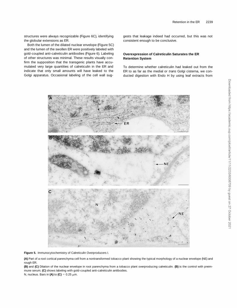
structures were always recognizable (Figure 6C), identifyingthe globular extensions as ER.
Both the lumen of the dilated nuclear envelope (Figure 5C)and the lumen of the swollen ER were positively labeled withgold-coupled anti-calreticulin antibodies (Figure 6). Labelingof other structures was minimal. These results visually con-firm the supposition that the transgenic plants have accu-mulated very large quantities of calreticulin in the ER andindicate that only small amounts will have leaked to theGolgi apparatus. Occasional labeling of the cell wall sug-
gests that leakage indeed had occurred, but this was notconsistent enough to be conclusive.
Overexpression of Calreticulin Saturates the ER Retention System
To determine whether calreticulin had leaked out from theER to as far as the medial or
trans
Golgi cisterna, we con-ducted digestion with Endo H by using leaf extracts from
Figure 5. Immunocytochemistry of Calreticulin Overproducers I.
(A) Part of a root cortical parenchyma cell from a nontransformed tobacco plant showing the typical morphology of a nuclear envelope (NE) andrough ER.(B) and (C) Dilation of the nuclear envelope in root parenchyma from a tobacco plant overproducing calreticulin. (B) is the control with preim-mune serum; (C) shows labeling with gold-coupled anti-calreticulin antibodies.N, nucleus. Bars in (A) to (C) 5 0.25 mm.
Dow
nloaded from https://academ
ic.oup.com/plcell/article/11/11/2233/6008709 by guest on 27 O
ctober 2021

2240 The Plant Cell
two individually transformed tobacco plants overproducingcalreticulin at different levels. Figure 7 clearly shows that thebest calreticulin overproducer (C
1
) had significant Endo Hresistance. The additional calreticulin band seen in the lanelabeled C
1
has the same molecular weight as singly glycosy-lated calreticulin. This band is absent when another trans-genic line producing lower levels of calreticulin was analyzed(lane C
2
) and may be due to saturation of the glycosylationmachinery. The transgenic plant used in lane C
1
shows 50-to 100-fold higher calreticulin levels, and the presence ofEndo H–resistant forms suggests that transport of calreticu-lin beyond the
cis
Golgi apparatus had occurred.
To determine whether acquisition of Endo H resistance isaccompanied by secretion of calreticulin, we prepared pro-toplasts from untransformed plants and overproducers andincubated them for different lengths of time, after whichcells and medium were separated for analysis. Cell extractsand medium samples from overproducers were diluted inproportion to the degree of overproduction to allow directcomparison with the wild-type protoplast population. Figure8A shows that only in overproducers is a small proportion ofthe total calreticulin secreted to the culture medium, demon-strating that saturation of the retention system has occurred.Figure 8B shows that calreticulin recovered from the culture
Figure 6. Immunocytochemistry of Calreticulin Overproducers II—Retention in the ER.
(A) Two dilated ER cisternae situated in the peripheral cytoplasm of a transformed root parenchyma cell.(B) At low magnification, extreme swelling of the ER is visible, giving rise to a structure similar in size to the nucleus.(C) High magnification of the ER membrane boxed in (B). Ribosomes clearly can be seen at the surface of the dilated ER (indicated by arrow-heads). Note that ribosomes are only present on the righthand side of the membrane, which corresponds to the cytoplasmic face.CW, cell wall; N, nucleus; V, vacuole. Bars in (A) and (C) 5 0.25 mm; bar in (B) 5 1 mm.
Dow
nloaded from https://academ
ic.oup.com/plcell/article/11/11/2233/6008709 by guest on 27 O
ctober 2021
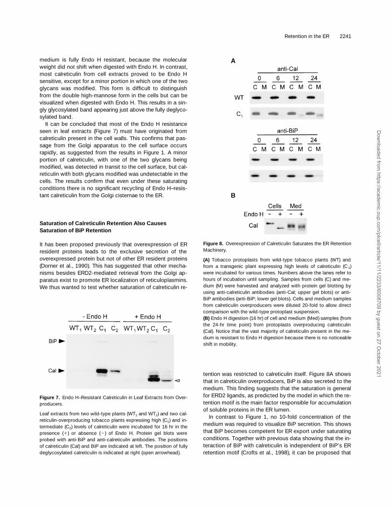
Retention in the ER 2241
medium is fully Endo H resistant, because the molecularweight did not shift when digested with Endo H. In contrast,most calreticulin from cell extracts proved to be Endo Hsensitive, except for a minor portion in which one of the twoglycans was modified. This form is difficult to distinguishfrom the double high-mannose form in the cells but can bevisualized when digested with Endo H. This results in a sin-gly glycosylated band appearing just above the fully deglyco-sylated band.
It can be concluded that most of the Endo H resistanceseen in leaf extracts (Figure 7) must have originated fromcalreticulin present in the cell walls. This confirms that pas-sage from the Golgi apparatus to the cell surface occursrapidly, as suggested from the results in Figure 1. A minorportion of calreticulin, with one of the two glycans beingmodified, was detected in transit to the cell surface, but cal-reticulin with both glycans modified was undetectable in thecells. The results confirm that even under these saturatingconditions there is no significant recycling of Endo H–resis-tant calreticulin from the Golgi cisternae to the ER.
Saturation of Calreticulin Retention Also Causes Saturation of BiP Retention
It has been proposed previously that overexpression of ERresident proteins leads to the exclusive secretion of theoverexpressed protein but not of other ER resident proteins(Dorner et al., 1990). This has suggested that other mecha-nisms besides ERD2-mediated retrieval from the Golgi ap-paratus exist to promote ER localization of reticuloplasmins.We thus wanted to test whether saturation of calreticulin re-
tention was restricted to calreticulin itself. Figure 8A showsthat in calreticulin overproducers, BiP is also secreted to themedium. This finding suggests that the saturation is generalfor ERD2 ligands, as predicted by the model in which the re-tention motif is the main factor responsible for accumulationof soluble proteins in the ER lumen.
In contrast to Figure 1, no 10-fold concentration of themedium was required to visualize BiP secretion. This showsthat BiP becomes competent for ER export under saturatingconditions. Together with previous data showing that the in-teraction of BiP with calreticulin is independent of BiP’s ERretention motif (Crofts et al., 1998), it can be proposed that
Figure 7. Endo H–Resistant Calreticulin in Leaf Extracts from Over-producers.
Leaf extracts from two wild-type plants (WT1 and WT2) and two cal-reticulin-overproducing tobacco plants expressing high (C1) and in-termediate (C2) levels of calreticulin were incubated for 16 hr in thepresence (1) or absence (2) of Endo H. Protein gel blots wereprobed with anti-BiP and anti-calreticulin antibodies. The positionsof calreticulin (Cal) and BiP are indicated at left. The position of fullydeglycosylated calreticulin is indicated at right (open arrowhead).
Figure 8. Overexpression of Calreticulin Saturates the ER RetentionMachinery.
(A) Tobacco protoplasts from wild-type tobacco plants (WT) andfrom a transgenic plant expressing high levels of calreticulin (C1)were incubated for various times. Numbers above the lanes refer tohours of incubation until sampling. Samples from cells (C) and me-dium (M) were harvested and analyzed with protein gel blotting byusing anti-calreticulin antibodies (anti-Cal; upper gel blots) or anti-BiP antibodies (anti-BiP; lower gel blots). Cells and medium samplesfrom calreticulin overproducers were diluted 20-fold to allow directcomparison with the wild-type protoplast suspension.(B) Endo H digestion (16 hr) of cell and medium (Med) samples (fromthe 24-hr time point) from protoplasts overproducing calreticulin(Cal). Notice that the vast majority of calreticulin present in the me-dium is resistant to Endo H digestion because there is no noticeableshift in mobility.
Dow
nloaded from https://academ
ic.oup.com/plcell/article/11/11/2233/6008709 by guest on 27 O
ctober 2021

2242 The Plant Cell
HDEL-less BiP probably is retained by association with cal-reticulin in the absence of saturation. However, under satu-rating conditions, this interaction no longer promotes ERretention and thus reveals ER export competence.
DISCUSSION
Why Is BiP Poorly Secreted When Lacking Its ER Retention Motif?
It already has been reported that BiP is only slowly secretedwhen devoid of its targeting signal (Munro and Pelham,1987), and our data correspond well with these previous re-ports (Figure 1). There are several possible explanations forsuch behavior. One is that there is a decrease in the stabilityof BiP once it leaves the ER, as has been observed for phos-phinothricin acetyltransferase (PAT; Denecke et al., 1992).This possibility is supported by the recent observation thatBiP overexpression leads to increased BiP degradation anda marked discrepancy between mRNA levels and proteinlevels (Leborgne-Castel et al., 1999). If BiP degradation in apost-ER compartment is more rapid, it would explain theapparent lack of secretion. This would imply that BiP is infact ER export competent, but that only a minor fractionreaches the cell surface and becomes secreted. The resultsshown in Figure 4 confirm this possibility.
A second explanation arises from the observation that themajority of cellular BiP is complexed with calreticulin andthat the retention signal of BiP is not required for this inter-action (Crofts et al., 1998). In plants, HDEL-less BiP thuscould be retained by HDEL-containing calreticulin. However,when the ER retention machinery is saturated, this would nolonger be effective and should reveal possible ER exportcompetence. Our results confirm that BiP is secreted duringsaturation (Figure 8). Thus, we propose that the lack of se-cretion of truncated BiP is due mainly to coretention withcalreticulin and that BiP is competent for ER exit. The factthat BiP was not detected in anterograde COPII vesiclesfrom the yeast Saccharomyces cerevisiae (Barlowe et al.,1994) could be due to a similar mechanism. The genomicsequence of S. cerevisiae suggests that it does not containa calreticulin-encoding gene, but it is conceivable that theclosely related molecule calnexin (Crofts and Denecke,1998) also could bind to BiP and promote retention.
Further Evidence for ER Exit as the Default Pathway for Soluble Proteins
The presence of two functional glycosylation consensussites in tobacco calreticulin has provided us with a valuablemeans of investigating how far the protein proceeds withinthe secretory pathway. We have established that both cal-reticulin glycans are competent for processing to complex
forms in the medial and trans Golgi apparatus. The absenceof any Endo H–resistant calreticulin glycans under normalphysiological conditions suggests that if calreticulin is recy-cling, then it is not from the medial or trans Golgi but mustbe from the cis Golgi or an earlier compartment devoid ofglycan-processing enzymes. These results confirm earlierobservations that glycan modification does not occur on as-sembly-defective phaseolin (Pedrazzini et al., 1997).
Calreticulin is secreted when devoid of its ER retentionsignal, and the secreted portion is fully Endo H resistant.The lack of overproduction of calreticulinDHDEL suggeststhat a major portion in fact is degraded when allowed toleave the ER. The rate of ER export thus may be very highand certainly comparable to neutral passenger molecules in-troduced into the secretory pathway (Denecke et al., 1990).The results are in good agreement with previous findings inwhich tagging of unstable passenger molecules with KDELor HDEL led to a high degree of stabilization (Denecke et al.,1992; Wandelt et al., 1992). The same appears to be true forcalreticulin and BiP, which accumulate to a 100- or 10-foldhigher level, respectively, compared with the HDEL-lessvariants. It is likely that all of the passenger molecules men-tioned are competent for rapid ER export but that in somecases, instability in a post-ER compartment prevents cleardetection of the phenomenon. However, at this stage it can-not be ruled out that deletion of HDEL has a direct effect onthe stability of the protein and accounts for the lack of over-production of the truncated forms.
In addition, our data support the model in which ER ex-port is the limiting factor in protein secretion, with every sub-sequent step being faster. Only the Endo H–sensitive ER-located transport intermediate of calreticulinDHDEL wasclearly detectable in the cells. In contrast, Endo H–resistantGolgi-located transport intermediates were difficult to ob-serve and restricted to glycoforms in which only one of thetwo glycans was modified. Taken together, the data supportthe theory that soluble proteins exit the ER by default, thatis, by passive diffusion into anterograde transport vesicles,implicating a concentration gradient along the secretorypathway. It has not been shown why, and in which compart-ment, a significant portion of calreticulin, BiP, or other pas-senger molecules appears to be degraded.
Probing the Limits of ER Retention
In both mammalian and plant cells, saturation of the ER re-tention mechanism has been difficult to achieve. Reports onpartial retention of proteins containing ER retention signals(Herman et al., 1990; Denecke et al., 1993; Pueyo et al.,1995; Gomord et al., 1997) all were based on the generationof fusion proteins of which the major portion is not normallya resident of the ER lumen. Calreticulin is a natural residentof the ER, and it does not appear to escape from the ER in adetectable fashion. Therefore, it provides a good model sys-tem to test the feasibility of saturation.
Dow
nloaded from https://academ
ic.oup.com/plcell/article/11/11/2233/6008709 by guest on 27 O
ctober 2021
Retention in the ER 2243
Due to the high stability of calreticulin in the ER lumen,overproduction of this protein leads to globular dilations ofthe ER and the nuclear envelope. These dilations demon-strate the enormous capacity of the plant ER to retain andstore soluble proteins as well as the dynamics of the ER tomaximize the enclosed volume with a minimal increase inmembrane surface. Given the fact that calreticulin is alreadyone of the most abundant ER resident proteins in plants(Denecke et al., 1995), a 100-fold overproduction and al-most complete retention would be impossible with an ER-localized receptor. No other ER protein exists that is suffi-ciently abundant to perform such a function.
Interestingly, saturation of calreticulin retention and partialsecretion of the protein appeared to be accompanied bypartial secretion of BiP. This strongly suggests that thesetwo proteins are retained by the same mechanism. In calreti-culin overproducers, saturation thus could be observed forall ligands recognized by the same receptor, regardless oftheir abundance. This result contradicts a previous report inwhich overproduction resulted in the exclusive secretion ofthe overproduced molecule (Dorner et al., 1990). This obser-vation suggested that other mechanisms aside from theERD2-mediated retrieval operate in mammalian cells andthat only this protein-specific retention system was satu-rated. Clearly, it cannot be ruled out that mammalian cellspossess other mechanisms of ER retention that are notpresent in plants.
Our current data strongly support the model in which acommon sorting receptor exists for soluble ER resident pro-teins. Given the fact that calreticulin and BiP appear to beER export competent, we postulate that anterograde trans-port of the sorting receptor must be faster and thus inde-pendent of the leakage of the ER residents.
No Evidence for Retrograde Transport from beyond the cis Golgi Apparatus
Evidence for retrograde transport first arose from work withthe fungal metabolite BFA, which caused a redistribution ofGolgi apparatus enzymes into the ER (Lippincott-Schwartzet al., 1990). The formation of an ER–Golgi supercompart-ment and the acquisition of Endo H resistance in ER resi-dents when treated with BFA (Figure 2B; Pedrazzini et al.,1997) can be explained by such a recycling mechanism aswell as by the trapping of de novo–synthesized Golgi appa-ratus enzymes in the ER. The latter explanation alone is un-likely to provide a mechanism, given the high number of ERresidents and the relatively fast enzymatic conversion ob-served with BFA treatment (Figure 2B). In the absence of re-cycling, the rapid BFA-induced redistribution of an ERD2–green fluorescent protein fusion (Boevink et al., 1998) wouldrequire trapping of rapidly de novo–synthesized moleculescombined with rapid turnover in the Golgi apparatus. This isvery unlikely, and it can be postulated that retrograde trans-port from at least the cis Golgi does exist in plants. How-
ever, the fact that BFA leads to a reduction in amyproduction (Figure 2C) could indicate that the drug doesprevent anterograde transport. In this particular instance,proteases destined for the vacuole could be trapped in theER–Golgi supercompartment and contribute to a more rapidturnover of the enzyme. Experiments in which BFA treat-ment is used to test whether a post-ER–Golgi compartmentis involved in degradation or processing of a protein there-fore should be interpreted with care.
Evidence for bacterial toxins, such as cholera toxin(Majoul et al., 1998) or Shiga toxin (Johannes and Goud,1998), reaching the ER via endocytosis and retrogradetransport through the Golgi complex due to the presence ofKDEL or RDEL motifs has led to the idea of the reversibilityof the secretory pathway (reviewed in Lord and Roberts,1998). Selective recycling from all Golgi compartments hasbeen compared with plate distillation (Miesenbock andRothman, 1995) in which each cisterna is a plate at which aprotein has a probability x to proceed further and a probabil-ity 1 2 x to return to the previous plate, thus explaining theefficiency of the sorting process.
Saturation of the ER retention system and the partialsecretion of reticuloplasmins result in leakage via the cis,medial, and trans Golgi apparatus toward the plasma mem-brane (Figure 8). The reversibility of the secretory pathway,as suggested (Lord and Roberts, 1998), appears to be in-consistent with the apparent lack of calreticulin recyclingfrom the trans Golgi cisternae under saturating conditions.Hardly any Endo H–resistant forms of calreticulin were foundin the cells, whereas the secreted portion was found to befully Endo H resistant. The low degree of Endo H resistanceproved to originate from calreticulin molecules carrying onemodified and one high-mannose form, which could be re-garded as a transport intermediate. Calreticulin moleculescarrying two modified glycans were undetectable in thecells, suggesting that the protein is rapidly exported once ithas reached the medial/trans Golgi apparatus. There was noevidence for significant retrograde transport from the medialor trans Golgi, but it cannot be ruled out completely.
If calreticulin leaks out beyond the cis Golgi, the protein israpidly secreted or degraded. Our present data are in goodagreement with findings in mammalian cells based on a verysensitive immunocytochemical approach to detect horse-radish peroxidase. When horseradish peroxidase is taggedwith the ER retention motif KDEL, it progresses through theGolgi stack no further than the cis-most element (Connollyet al., 1994). The data also correspond well with the obser-vation that tagging with KDEL prevents acquisition of EndoH resistance of phytohemagglutinin (Herman et al., 1990)and phaseolin (L. Frigerio and A. Vitale, personal communi-cation) glycans.
The results obtained here suggest an alternative explana-tion of the well-established retrograde transport of toxins.One possibility is that toxins carry additional signals to reachthe cis Golgi from the trans Golgi. Alternatively, endocytosisof toxins is followed by direct delivery to the cis Golgi from
Dow
nloaded from https://academ
ic.oup.com/plcell/article/11/11/2233/6008709 by guest on 27 O
ctober 2021

2244 The Plant Cell
the plasma membrane. Such a mechanism cannot beexcluded from the existing data and would be in agreementwith the data on ER retention of calreticulin and KDEL-tagged horseradish peroxidase. Finally, it also should betaken into account that monitoring the activity of a toxin maybe a much more sensitive way of detecting retrograde trans-port to the ER. Therefore, the biochemical and immunocy-tochemical data do not have to be in contradiction with thefindings on toxins.
Several studies with mammalian cells have shown thattransport from the ER to the Golgi is mediated by an ER–Golgi intermediate compartment (ERGIC; Hauri andSchweizer, 1992) from which escaped ER resident proteinscan be recycled back to the ER (Connolly et al., 1994; re-viewed in Hong, 1998). Despite continuous efforts, no evi-dence for the existence of an intermediate compartment hasbeen found in plants. We also failed to detect candidates forsuch a compartment in the calreticulin overproducers. Re-cent evidence has suggested that the ERGIC protein ER-GIC-53 is a mannose-selective lectin that may function as atransport receptor for secretion of a small subset of glyco-proteins (Vollenweider et al., 1998). Because calreticulin car-ries two glycans, it could be assumed that these areessential for the observed rapid export of calreticulin out ofthe ER. However, with the use of tunicamycin, we haveshown that nonglycosylated calreticulin is secreted as wellas is glycosylated calreticulin. This has been confirmed bygenetic modification to remove the two consensus sites forN-linked glycosylation (A.J. Crofts and J. Denecke, manu-script in preparation). Thus, the lack of BiPDHDEL secretioncannot be explained by the absence of glycans. The factthat saturation leads to secretion of BiP and calreticulinshows that both molecules are competent for ER export andsupports the default model of ER export.
METHODS
Plasmid Construction for Transient and Stable Expression
All DNA manipulations were performed according to established pro-cedures. The Escherichia coli MC1061 strain (Casadaban andCohen, 1980) was used for the amplification of all plasmids. The full-length calreticulin cDNA clone (Denecke et al., 1995) was cut withPstI and inserted into pDE300d, previously cut with KpnI and SmaI,treated with the Klenow fragment of DNA polymerase I, and dephos-phorylated with calf intestine alkaline phosphatase. Ligation resultedin plasmid pLC48.
The plasmid pDE314 containing the gene encoding phosphinothri-cin acetyltransferase (PAT; Denecke et al., 1992) was digested withBglII, filled in by using the Klenow fragment, and digested with SalI.The calreticulin fragment for ligation was prepared by polymerasechain reaction amplification of the calreticulin coding region by usingpLC48 as the template. To mutate the retention signal, we used theoligonucleotide 59-TGCGCTTCATCATCCTTGGA-39. This fragmentthen was digested with SalI and gel-purified together with the vector
before ligation. Ligation resulted in the replacement of the PAT cod-ing region by the calreticulin coding region, and the plasmid wastermed pDE314C.
Plasmid pDE800 (Leborgne-Castel et al., 1999) was cut with NheI,blunted with the Klenow fragment, and cut with NcoI. The resultingfragment carries the entire binding protein (BiP) coding region, ex-cept for the last eight codons. This fragment was ligated into pDE312(Denecke et al., 1992), previously cut with XbaI, blunted with the Kle-now fragment, and cut with NcoI. This resulted in the replacement ofthe PAT coding region by truncated BiP in-frame with the stop codonencoded within the XbaI site; this plasmid was termed pDE801.
For stable Agrobacterium tumefaciens–mediated plant transfor-mation, genes present in plasmids pLC48, pDE314C, pDE800, andpDE801 were inserted into pDE1001, as described previously(Denecke et al., 1992).
Plant Material and Growth Culture Conditions
Plants (Nicotiana tabacum cv Petit Havana; Maliga et al., 1973) weregrown in Murashige and Skoog medium (Murashige and Skoog,1962) and 2% sucrose in a controlled room at 258C with a 16-hr daylength at the light irradiance of 200 mE m22 sec21. Tobacco BY2 cul-tures were maintained and transformed as described previously(Nagata et al., 1992).
Plant Transformations
The plasmids derived from pDE1001 were mobilized into Agrobac-terium strain C58 (pGV2260; Deblaere et al., 1985) by using the E.coli helper strain HB101 (pRK2013). Transformed plants were ob-tained by agrobacterial infection of leaf squares with the respectivestrains. Transformants were selected on B5 salts (Sigma; dilutedaccording to the manufacturer’s instructions) supplemented with250 mg/L NH4NO3, 500 mg/L 2-(N-morpholino)ethanesulfonic acid,20 g/L glucose, 5 g/L agarose, 40 mg/L adenine, 100 mg/L kana-mycin, 500 mg/L cefotaxime, 1 mg/L 6-benzylaminopurine, and 0.1mg/L a-naphthaleneacetic acid, and brought to pH 5.7 with con-centrated KOH.
Transient Expression Experiments
Tobacco leaf protoplasts from untransformed plants were prepared,and electroporation experiments were performed as previously de-scribed (Denecke and Vitale, 1995). For each experiment, 2.5 3 106
protoplasts were used with 30 mg of DNA. After 24 hr, the protoplastswere analyzed by protein gel blotting. Harvesting of cells and culturemedium as well as the enzymatic assays was performed as de-scribed previously (Denecke and Vitale, 1995; Leborgne-Castel et al.,1999).
Transport Assays
To assess the secretion of proteins, we incubated electroporatedprotoplasts or protoplasts prepared from transgenic plants as sus-pensions for various time periods (see legends to Figures 1, 3, and 8).Protoplasts were suspended in 2 mL of TEX medium containing B5
Dow
nloaded from https://academ
ic.oup.com/plcell/article/11/11/2233/6008709 by guest on 27 O
ctober 2021
Retention in the ER 2245
salts (Sigma; diluted according to the manufacturer’s instructions)supplemented with 250 mg/L NH4NO3, 500 mg/L CaCl2·2H2O, 500mg/L 2-(N-morpholino)ethanesulfonic acid, and 136.9 g/L sucrose,brought to pH 5.7 with concentrated KOH and filter-sterilized (0.2mm). After the appropriate incubation time, the suspension was spunfor 5 min at 100g in a swing-out centrifuge (4K15; Sigma), which re-sults in the floating of the cells. Using an extra-fine Pasteur pipette,we removed 1 mL of clear supernatant from below the floating celllayer. The remainder of the suspension (1 mL) was brought to 10 mLwith 250 mM NaCl and mixed gently by inverting the tube twice. Aftera second spin of 5 min at 100g, the supernatant was removed with aperistaltic pump, and the cell pellet was placed on ice. The cells were ex-tracted in a volume of 2 mL. Equal volumes of cell extract and culturemedium were analyzed by protein gel blotting or by enzymatic anal-ysis. When indicated, the medium was concentrated by precipitationwith 60% ammonium sulfate on ice by using BSA at 0.5 mg/mL as acarrier. After incubation on ice for 2 hr and centrifugation for 15 minat 25,000g, the pellet was resuspended in a 10-fold smaller volumecompared with the amount of medium used in the precipitation.
Protein Extraction
Leaf extracts were prepared by grinding in protein extraction buffer(50 mM Tris-HCl, pH 7.5, 2 mM EDTA, 1 mL/mL b-mercaptoethanol,and 10 mM phenylmethylsulfonyl fluoride) with a mortar and pestle,using 2 mL of buffer for each gram of tissue. Protoplasts were ex-tracted with the same buffer but by using sonication for 5 sec. In allcases, extracts were cleared by a 10-min centrifugation at 25,000g at48C, and the supernatant was recovered. Protein concentrationswere determined using Bio-Rad protein assay reagent. For thescreening of transgenic plants, extracts were brought to 0.1 mg/mL.
Endoglycosidase H Digestion
In Figure 2, digestions were conducted directly on extracts madewith 50 mM Tris-HCl, pH 7.5, 2 mM EDTA, and 1 mM b-mercapto-ethanol. The extracts to be analyzed were added to an equal volumeof endoglycosidase H (Endo H; typically 30 mL) in 100 mM sodiumcitrate, pH 5.5, to give a final activity of 0.1 units in 50 mM sodiumcitrate. Control digests contained extract and enzyme buffer alone.The digests were incubated at 378C for times ranging from 30 sec to21 hr and were stopped by the addition of 2 3 SDS-PAGE loadingbuffer (see below). Samples then were analyzed by protein gel blot-ting.
Because we observed a possible degradation as well as an endog-enous deglycosylation during prolonged incubations (.2 hr), weadapted the protocol in the following way. Extracts prepared as de-scribed above were denatured by adding an equal volume of dena-turation buffer (0.1 M sodium citrate, pH 5.5, 1% SDS, and 0.2 Mb-mercaptoethanol) and subsequent boiling for 10 min. After coolingto 228C, 16 mL of the denatured sample was mixed with 1 mL of phe-nylmethylsulfonyl fluoride (10 mg/mL in methanol) and 23 mL of en-zyme (0.2 units per mL), giving a final volume of 40 mL and an activityof 0.1 units per mL.
Digestions were conducted for 16 hr at 378C. Control digests wereperformed in the same way but without Endo H. Under these condi-tions, complete digestions were ensured at all times with no degra-dation or intrinsic N-glycanase activity. This protocol was used for all
other assays except those shown in Figure 2, for which different in-cubation times were used.
Protein Gel Blotting
Samples were loaded after twofold dilution with SDS-PAGE loadingbuffer (200 mM Tris-HCl, pH 8.8, 5 mM EDTA, 1 M sucrose, and0.1% bromophenol blue). Proteins in SDS–polyacrylamide gels weretransferred onto a nitrocellulose membrane and then blocked withPBS, 0.5% Tween 20, and 5% milk powder for 1 hr. The filter thenwas incubated in blocking buffer, with primary antibody at a dilutionof 1:5000 for anti-BiP (Denecke et al., 1991) and anti-calreticulin an-tibodies (Denecke et al., 1995). After 1 hr, a 15-min wash and three5-min washes were performed with 1 3 PBS and 0.5% Tween 20.The secondary antibody used was anti–rabbit antibody conjugatedto horseradish peroxidase at a dilution of 1:5000 in 1 3 PBS, 0.5%Tween 20, and 5% milk powder. The filter was incubated with thesecondary antibody for 1 hr. A 15-min wash followed by four washesof 5 min with 1 3 PBS and 0.5% Tween 20 and a final wash with 1 3PBS ensured minimal background. Detection of antigen–antibodycomplexes was performed with enhanced chemiluminescence (Am-ersham), and the images were recorded on film.
Immunocytochemistry
Small segments of root and leaf tissue were fixed, dehydrated, andembedded according to the protocol given by Sikora et al. (1998),with the following minor alterations. The first 30 min of the primary al-dehyde fixation was performed in vacuo, and the duration of theosmium tetroxide fixation at 2208C was extended to 24 hr. Immu-nogold labeling was conducted with standard procedures (Hohl etal., 1996) by using anti-calreticulin antibodies at a primary dilution of1:200. Gold-coupled (10 nm) secondary antibodies were presentedat a dilution of 1:30. Uranyl acetate/lead citrate poststained sectionswere examined in an electron microscope (model CM 10; Philips,Eindhoven, The Netherlands) operating at 80 kV.
ACKNOWLEDGMENTS
We thank Alison Caffreys for suggesting that ER retention in plantsmay be saturated by screening more transgenic plants. We alsothank Andy Forrester, Jeff Patteson, Phil Taylor, Pat Romano, andMahita Sastry for contributing to the generation of transgenic plantsduring their Master of Research (MRes) course at the University ofYork. We thank Peter Berry for inserting the calreticulin gene intopDE1001 during his third-year project and Graham King for journalis-tic suggestions. Alessandro Vitale is thanked for communicatingunpublished results and for continuous scientific discussion. Thiswork was supported by grants from the Biotechnology and Biologi-cal Sciences Research Council (BBSRC) and the European Union(Grant No. CHRX-CT94-0590), which we gratefully acknowledge.A.J.C. is indebted to the BBSRC for a quota studentship.
Received June 16, 1999; accepted September 13, 1999.
Dow
nloaded from https://academ
ic.oup.com/plcell/article/11/11/2233/6008709 by guest on 27 O
ctober 2021

2246 The Plant Cell
REFERENCES
Balch, W.E., McCaffery, J.M., Plutner, H., and Farquhar, M.G.(1994). Vesicular stomatitis virus glycoprotein is sorted and con-centrated during export from the endoplasmic reticulum. Cell 76,841–852.
Barlowe, C., Orci, L., Yeung, T., Hosobuchi, M., Hamamoto, S.,Salama, N., Rexach, M.F., Ravazzola, M., Amherdt, M., andSchekman, R. (1994). COPII: A membrane coat formed by Secproteins that drive vesicle budding from the endoplasmic reticu-lum. Cell 77, 895–907.
Boevink, P., Oparka, K., Santa Cruz, S., Martin, B., Betteridge,A., and Hawes, C. (1998). Stacks on tracks: The plant Golgiapparatus traffics on an actin/ER network. Plant J. 15, 441–447.
Casadaban, M.J., and Cohen, S. (1980). Analysis of gene controlsignals by DNA fusion and cloning in Escherichia coli. J. Mol. Biol.138, 179–207.
Ceriotti, A., and Colman, A. (1988). Binding to membrane-proteinswithin the endoplasmic-reticulum cannot explain the retention ofthe glucose-regulated protein GRP78 in Xenopus oocytes. EMBOJ. 7, 633–638.
Connolly, C.N., Futter, C.E., Gibson, A., Hopkins, C.R., and Cutler,D.F. (1994). Transport into and out of the Golgi-complex studiedby transfecting cells with cDNAs encoding horseradish peroxi-dase. J. Cell Biol. 127, 641–652.
Cosson, P., and Letourneur, F. (1994). Coatomer interaction withdi-lysine endoplasmic reticulum retention motifs. Science 263,1629–1631.
Crofts, A.J., and Denecke, J. (1998). Calreticulin and calnexin inplants. Trends Plant Sci. 3, 396–399.
Crofts, A.J., Leborgne-Castel, N., Pesca, M., Vitale, A., andDenecke, J. (1998). BiP and calreticulin form an abundant com-plex that is independent of endoplasmic reticulum stress. PlantCell 10, 813–823.
Dean, N., and Pelham, H.R.B. (1990). Recycling of proteins from theGolgi compartment to the ER in yeast. J. Cell Biol. 111, 369–377.
Deblaere, R., Bytebier, B., De Greve, H., Debroeck, F., Schell, J.,Van Montagu, M., and Leemans, J. (1985). Efficient octopine Tiplasmid-derived vectors for Agrobacterium-mediated gene trans-fer to plants. Nucleic Acids Res. 13, 4777–4788.
Denecke, J., and Vitale, A. (1995). The use of plant protoplasts tostudy protein synthesis, quality control, protein modification andtransport through the plant endomembrane system. In Methods inCell Biology, Vol. 50, D.W. Galbraith, H.J. Bohnert, and D.P.Bourque, eds (San Diego, CA: Academic Press), pp. 335–348.
Denecke, J., Botterman, J., and Deblaere, R. (1990). Proteinsecretion in plant cells can occur via a default pathway. Plant Cell2, 51–59.
Denecke, J., Goldman, M.H.S., Demolder, J., Seurinck, J., andBotterman, J. (1991). The tobacco luminal binding protein isencoded by a multigene family. Plant Cell 3, 1025–1035.
Denecke, J., De Rycke, R., and Botterman, J. (1992). Plant andmammalian sorting signals for protein retention in the endoplas-mic reticulum contain a conserved epitope. EMBO J. 11, 2345–2355.
Denecke, J., Ek, B., Caspers, M., Sinjorgo, K.M.C., and Palva,E.T. (1993). Analysis of sorting signals responsible for the accu-mulation of soluble reticuloplasmins in the plant endoplasmicreticulum. J. Exp. Bot. 44, 213–221.
Denecke, J., Carlsson, L.E., Vidal, S., Hoglund, A.-S., Ek, B., vanZeijl, M.J., Sinjorgo, K.M.C., and Palva, E.T. (1995). Thetobacco homolog of mammalian calreticulin is present in proteincomplexes in vivo. Plant Cell 7, 391–406.
Dorner, A.J., Wasley, L.C., Raney, P., Haugejorden, S., Green,M., and Kaufman, R.J. (1990). The stress response in chinese-hamster ovary cells—Regulation of Erp72 and protein disulfideisomerase expression and secretion. J. Biol. Chem. 265, 22029–22034.
Gomord, V., Denmat, L.A., Fitchette-Laine, A.C., Satiat-Jeunemaitre, B., Hawes, C., and Faye, L. (1997). The C-terminalHDEL sequence is sufficient for retention of secretory proteins inthe endoplasmic reticulum (ER) but promotes vacuolar targetingof proteins that escape the ER. Plant J. 11, 313–325.
Hauri, H.-P., and Schweizer, A. (1992). The endoplasmic reticu-lum–Golgi intermediate compartment. Curr. Opin. Cell Biol. 4,600–608.
Herman, E.M., Tague, B.W., Hoffman, L.M., Kjemtrupp, S.E., andChrispeels, M.J. (1990). Retention of phytohemagglutinin withcarboxy terminal tetrapeptide KDEL in the nuclear envelope andthe endoplasmic reticulum. Planta 182, 305–312.
Hohl, I., Robinson, D.G., Chrispeels, M.J., and Hinz, G. (1996).Transport of storage proteins to the vacuole is mediated by vesi-cles without a clathrin coat. J. Cell Sci. 109, 2539–2550.
Hong, W. (1998). Protein transport from the endoplasmic reticulumto the Golgi apparatus. J. Cell Sci. 111, 2831–2839.
Hunt, D.C., and Chrispeels, M.J. (1991). The signal peptide of avacuolar protein is necessary and sufficient for the efficient secre-tion of a cytosolic protein. Plant Physiol. 96, 18–25.
Johannes, L., and Goud, B. (1998). Surfing on a retrograde wave:How does Shiga toxin reach the endoplasmic reticulum? TrendsCell Biol. 8, 158–162.
Kornfeld, R., and Kornfeld, S. (1985). Assembly of asparagine-linked oligosaccharides. Annu. Rev. Biochem. 54, 631–664.
Leborgne-Castel, N., Jelitto-Van Dooren, E.P.W.M., Crofts, A.J.,and Denecke, J. (1999). Overexpression of BiP in tobacco allevi-ates endoplasmic reticulum stress. Plant Cell 11, 459–470.
Lee, H.I., Gal, S., Newman, T.C., and Raikhel, N.V. (1993). TheArabidopsis endoplasmic-reticulum retention receptor functionsin yeast. Proc. Natl. Acad. Sci. USA 90, 11433–11437.
Lewis, M.J., and Pelham, H.R. (1992). Ligand-induced redistribu-tion of a human KDEL receptor from the Golgi complex to theendoplasmic reticulum. Cell 68, 353–364.
Lippincott-Schwartz, J., Donaldson, J.G., Schweizer, A., Berger,E.G., Hauri, H.P., Yuan, L.C., and Klausner, R.D. (1990). Micro-tubule-dependent retrograde transport of proteins into the ER inthe presence of brefeldin A suggests an ER recycling pathway.Cell 5, 821–836.
Lord, J.M., and Roberts, L.M. (1998). Retrograde transport: Goingagainst the flow. Curr. Biol. 8, R56–R58.
Majoul, I., Sohn, K., Wieland, F.T., Pepperkok, R., Pizza, M.,Hillemann, J., and Soling, H.D. (1998). KDEL receptor (Erd2p)–
Dow
nloaded from https://academ
ic.oup.com/plcell/article/11/11/2233/6008709 by guest on 27 O
ctober 2021

Retention in the ER 2247
mediated retrograde transport of the cholera toxin A subunit fromthe Golgi involves COPI, p23, and the terminus of Erd2p. J. CellBiol. 143, 601–612.
Maliga, P., Breznowitz, A., and Marton, L. (1973). Streptomycin-resistant plants from callus culture of haploid tobacco. Nature244, 29–30.
Miesenbock, G., and Rothman, J.E. (1995). The capacity toretrieve escaped ER proteins extends to the trans-most cisternaof the Golgi stack. J. Cell Biol. 129, 309–319.
Munro, S., and Pelham, H.R.B. (1987). A C-terminal signal preventssecretion of luminal ER proteins. Cell 48, 899–907.
Murashige, R., and Skoog, F. (1962). A revised medium for rapidgrowth and bioassays with tobacco tissue cultures. Physiol. Plant.15, 473–497.
Nagata, T., Nemoto, Y., and Hasezawa, S. (1992). Tobacco BY-2cell line as the “HeLa” cell in the cell biology of higher plants. Int.Rev. Cytol. 132, 1–30.
Navazio, L., Baldan, B., Mariani, P., Gerwig, G.J., and Vliegenthart,J.F.G. (1996). Primary structure of the N-linked carbohydrate chainsof calreticulin from spinach leaves. Glycoconj. J. 13, 977–983.
Nishimura, N., and Balch, W.E. (1997). A di-acidic signal requiredfor selective export from the endoplasmic reticulum. Science 277,556–558.
Pahl, H.L., and Baeuerle, P.A. (1997). Endoplasmic-reticulum-induced signal transduction and gene expression. Trends CellBiol. 7, 50–55.
Pedrazzini, E., Giovinazzo, G., Bielli, A., de Virgilio, M., Frigerio,L., Pesca, M., Faoro, F., Bollini, R., Ceriotti, A., and Vitale, A.(1997). Protein quality control along the route to the plant vacuole.Plant Cell 9, 1869–1880.
Pelham, H.R.B. (1988). Evidence that luminal ER proteins are sortedfrom secreted proteins in a post-ER compartment. EMBO J. 7,913–918.
Pelham, H.R.B. (1995). Sorting and retrieval between the endo-plasmic reticulum and Golgi apparatus. Curr. Opin. Cell Biol. 7,530–535.
Pueyo, J.J., Chrispeels, M.J., and Herman, E.M. (1995). Degrada-
tion of transport-competent destabilized phaseolin with a signalfor retention in the endoplasmic-reticulum occurs in the vacuole.Planta 3, 586–596.
Rabouille, C., Hui, N., Hunte, F., Kieckbusch, R., Berger, E.G.,Warren, G., and Nilsson, T. (1995). Mapping the distribution ofGolgi enzymes involved in the construction of complex oligosac-charides. J. Cell Sci. 108, 1617–1627.
Scheel, J., Pepperkok, R., Lowe, M., Griffiths, G., and Kreis, T.E.(1997). Dissociation of coatomer from membranes is required forbrefeldin A–induced transfer of Golgi enzymes to the endoplasmicreticulum. J. Cell Biol. 137, 319–333.
Sikora, A., Hillmer, S., and Robinson, D.G. (1998). Sucrose starva-tion causes a loss of immunologically detectable pyrophos-phatase and V-ATPase in the tonoplast of suspension-culturedtobacco cells. J. Plant Physiol. 152, 207–212.
Spang, A., and Schekman, R. (1998). Reconstitution of retrogradetransport from the Golgi to the ER in vitro. J. Cell Biol. 143, 589–599.
Tatu, U., and Helenius, A. (1997). Interactions between newly syn-thesized glycoproteins, calnexin and a network of resident chap-erones in the endoplasmic reticulum. J. Cell Biol. 136, 555–565.
Townsley, F.M., Wilson, D.W., and Pelham, H.R. (1993). Muta-tional analysis of the human KDEL receptor: Distinct structuralrequirements for Golgi retention, ligand binding and retrogradetransport. EMBO J. 12, 2821–2829.
Vitale, A., and Denecke, J. (1999). The endoplasmic reticulum—Gateway of the secretory pathway. Plant Cell 11, 615–628.
Vitale, A., Ceriotti, A., and Denecke, J. (1993). The role of theendoplasmic reticulum in protein synthesis, modification andintracellular transport. J. Exp. Bot. 44, 1417–1444.
Vollenweider, F., Kappeler, F., Itin, C., and Hauri, H.-P. (1998).Mistargeting of the lectin ERGIC-53 to the endoplasmic reticulumof HeLa cells impairs the secretion of a lysosomal enzyme. J. CellBiol. 142, 377–389.
Wandelt, C.I., Khan, M.R.I., Craig, S., Schroeder, H.E., Spencer,D., and Higgins, T.J.V. (1992). Vicilin with carboxy-terminal KDELis retained in the endoplasmic reticulum and accumulates to highlevels in the leaves of transgenic plants. Plant J. 2, 181–192.
Dow
nloaded from https://academ
ic.oup.com/plcell/article/11/11/2233/6008709 by guest on 27 O
ctober 2021