sc professional support and expert advice from your ... · professional support and expert advice...
TRANSCRIPT
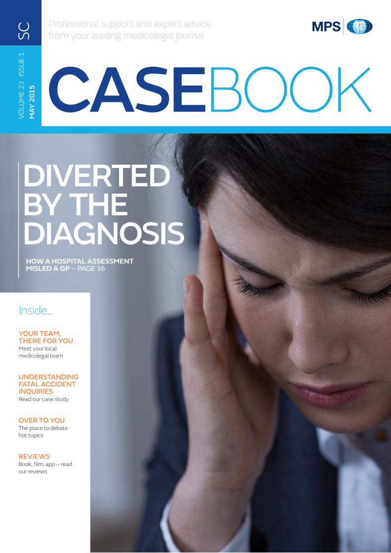
CASEBOOKDIVERTED BY THE DIAGNOSIS
Professional support and expert advice from your leading medicolegal journal
Inside...
YOUR TEAM, THERE FOR YOUMeet your local medicolegal team
UNDERSTANDING FATAL ACCIDENT INQUIRIESRead our case study
OVER TO YOUThe place to debate hot topics
REVIEWSBook, film, app – read our reviews
HOW A HOSPITAL ASSESSMENT MISLED A GP – PAGE 16
VOLU
ME
23
ISSU
E 1
MAY
201
5SC
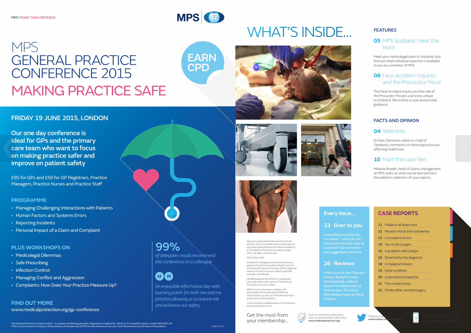
MPS GENERAL PRACTICE CONFERENCE 2015MAKING PRACTICE SAFE
MPS MORE THAN DEFENCE
The Medical Protection Society Limited – A company limited by guarantee. Registered in England No. 36142 at 33 Cavendish Square, London W1G 0PS, UK.MPS is not an insurance company. All the benefi ts of membership of MPS are discretionary as set out in the Memorandum and Articles of Association. 1680: 04/15
EARNCPD
FRIDAY 19 JUNE 2015, LONDON
Our one day conference is ideal for GPs and the primary care team who want to focus on making practice safer and improve on patient safety
£95 for GPs and £50 for GP Registrars, Practice Managers, Practice Nurses and Practice Sta�
PROGRAMME• Managing Challenging Interactions with Patients• Human Factors and Systems Errors• Reporting Incidents • Personal Impact of a Claim and Complaint
PLUS WORKSHOPS ON• Medicolegal Dilemmas • Safe Prescribing • Infection Control• Managing Confl ict and Aggression • Complaints: How Does Your Practice Measure Up?
FIND OUT MOREwww.medicalprotection.org/gp-conference
99%of delegates would recommend this conference to a colleague
An enjoyable informative day with learning points for both me and my practice allowing us to reduce risk and enhance our safety
“
“
1680 GP Conference A4 advert.indd 1 23/04/2015 16:26
CASE REPORTS
11 Pulled in all directions
12 Missed critical limb ischaemia
13 Cumulative errors
14 Too much oxygen
15 A problem with polyps
16 Diverted by the diagnosis
18 A malignant lesion
19 Fatal condition
20 A persistent headache
21 The swollen knee
22 Stroke after carotid surgery
FEATURES
05 MPS Scotland: meet the team
Meet your medicolegal team in Scotland, and find out what individual expertise is available to you as a member of MPS.
08 Fatal Accident Inquiries and the Procurator Fiscal
The Fatal Accident Inquiry and the role of the Procurator Fiscal is a process unique to Scotland. We outline a case and provide guidance.
FACTS AND OPINION
04 Welcome
Dr Nick Clements, editor-in-chief of Casebook, comments on some topical issues affecting healthcare.
10 From the case files
Melanie Rowles, head of claims management at MPS, looks at what can be learned from this edition’s collection of case reports.
Every issue...
23 Over to youA sounding board for you, the reader – what did you think about the last issue of Casebook? All comments and suggestions welcome.
26 ReviewsIn this issue Dr Sam Dawson reviews Being Mortal by Atul Gawande, while Dr Anand Naranbhai looks at Postmortem: The Doctor Who Walked Away by Maria Phalime.
WHAT’S INSIDE…
Get the most from your membership…
Visit our website for publications, news, events and other information: www.medicalprotection.org
Follow our tweets at: www.twitter.com/MPSdoctors
Top tips in the palm of your hand – download the free MPS Advice app on the App Store or Google Play
Opinions expressed herein are those of the authors. Pictures should not be relied upon as accurate representations of clinical situations. © The Medical Protection Society Limited 2015. All rights are reserved.
ISSN 2056 4449
Casebook is designed and produced twice a year by the Communications Department of the Medical Protection Society (MPS). Regional editions of each issue are mailed to all MPS members worldwide.
GLOBE (logo) (series of 6)® is a registered UK trade mark in the name of The Medical Protection Society Limited.
MPS is not an insurance company. All the benefits of membership of MPS are discretionary, as set out in the Memorandum and Articles of Association.
Cover: © KatarzynaBialasiewicz/iStockphoto/thinkstockphotos.co.uk
WELCOMEDr Nick ClementsEDITOR-IN-CHIEF
edical matters, unsurprisingly, continue to feature heavily in the headlines and the media in general. There seems to be an endless appetite
among the public for such stories, whether they are announcing the arrival of new and better treatments or procedures, or reporting shortfalls, errors or even scandals. Politicians frequently feel obliged to step in, but their attempts to remedy things don’t always have the desired result.
Inevitably this is felt by you on the wards or in your consulting rooms, with increasing patient expectations in the form of unrealistic demands or a raft of self-researched information from the internet. This can make for some challenging situations, at a time when workloads grow in intensity, perhaps due to budgetary cutbacks or other local factors.
It continues to be an important time to be part of an organisation like MPS. We work in partnership with you to protect and support your career at every stage, and this work takes many forms, beyond the litigation work that we are more traditionally associated with. This includes an extensive range of educational products such as online learning, workshops and seminars, as well as continuous consultative work with governments and policy-makers worldwide. The latter is often ‘behind the scenes’ and often not highly-publicised, but you can be reassured that our specialist teams are fighting hard to safeguard your interests.
Many of you got in touch with us following the last edition of Casebook, regarding our cover story on the case of Beth Bowen. While the emotional reaction from a number of correspondents was not surprising, I was heartened by the way the article made everyone think about their own approach to communication, openness and consent. Anger at the treatment of the Bowen family was palpable in some of your letters, and if this deeply tragic case results in reflection and changes in culture and practice, then something positive will have been achieved.
We have published a short response to this correspondence in our “Over to you” section on page 23. I hope you find this edition of Casebook an equally thought-provoking one and, as ever, I am keen to hear your feedback.
M
MPS SCOTLAND: MEET THE TEAMAs an MPS member in Scotland, you have access to a wide range of specialist support and advice. Based in our Edinburgh office, the MPS team has extensive experience of Scotland’s medicolegal landscape – find out how they are working hard on your behalf
In 2009, MPS established its base of specialist Scottish medicolegal services in our office on George Street, Edinburgh. Since then the team has grown to over 30 members of staff working together to support members practising in Scotland.
MPS Scotland also provides:
• Risk management and performance improvement: Each year we deliver up to 40 teaching sessions and lectures for hospital doctors and general practice teams across Scotland.
• Support for your development: You have access to a range of CPD-approved education and risk management resources in Scotland. Each year, MPS runs up to 20 workshops and seminars in Scotland, including half-day risk management workshops and full-day practice management seminars.
Please address all correspondence to:
Casebook editorMedical Protection SocietyVictoria House 2-3 Victoria Place Leeds LS11 5AE United Kingdom
EDITORIAL TEAM
Dr Nick Clements EDITOR-IN-CHIEF
Sara Dawson DEPUTY EDITOR
Gareth Gillespie EDITOR
CASE REPORT WRITERS
Dr John Adams Dr Mike Greenberg
Dr Dudley Bush Dr Paul Heslin
Mr Sam Dresner Dr Gerard McKeague
Mr David D’Souza Dr Janet Page
Dr Anna Fox Dr Ellen Welch
PRODUCTIONPhilip Walker, Production managerEmma Senior, Senior designerDante Marcuccio, DesignerAllison Forbes, Assistant designer
EDITORIAL BOARDAntoinette Coltsmann, Dr Marika Davies, Professor Jonathan Hardman, Dr Muiris Houston, Dr Graham Howarth, Mark Jordan, Mr Goldie Khera, Dr Sonya McCullough, Dr Gordon McDavid, Shelley McNicol, Julie Price, Dr Ewen Ross, Dr Ming-Keng Teoh
Dr Gordon McDavid EDITORIAL CONSULTANT
© C
ate Gillon
5CASEBOOK | VOLUME 23 ISSUE 1 | MAY 2015 | www.medicalprotection.org
FEATURE
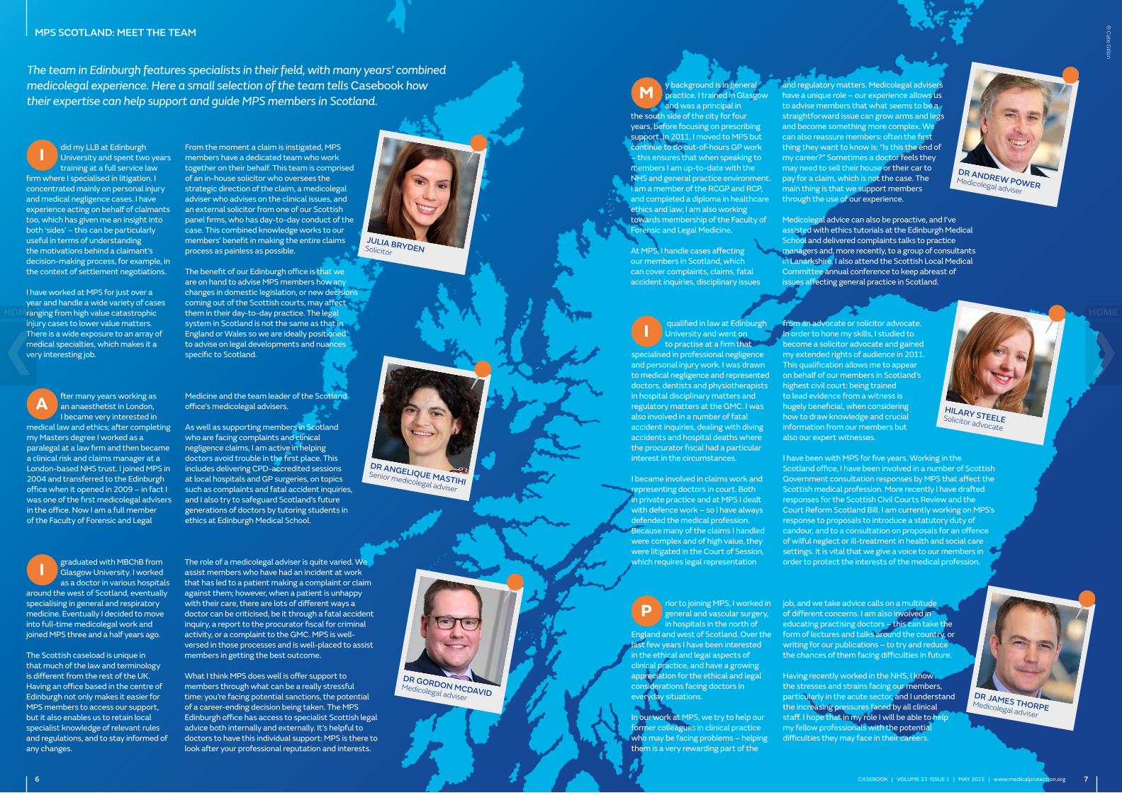
The team in Edinburgh features specialists in their field, with many years’ combined medicolegal experience. Here a small selection of the team tells Casebook how their expertise can help support and guide MPS members in Scotland.
graduated with MBChB from Glasgow University. I worked as a doctor in various hospitals
around the west of Scotland, eventually specialising in general and respiratory medicine. Eventually I decided to move into full-time medicolegal work and joined MPS three and a half years ago.
The Scottish caseload is unique in that much of the law and terminology is different from the rest of the UK. Having an office based in the centre of Edinburgh not only makes it easier for MPS members to access our support, but it also enables us to retain local specialist knowledge of relevant rules and regulations, and to stay informed of any changes.
The role of a medicolegal adviser is quite varied. We assist members who have had an incident at work that has led to a patient making a complaint or claim against them; however, when a patient is unhappy with their care, there are lots of different ways a doctor can be criticised, be it through a fatal accident inquiry, a report to the procurator fiscal for criminal activity, or a complaint to the GMC. MPS is well-versed in those processes and is well-placed to assist members in getting the best outcome.
What I think MPS does well is offer support to members through what can be a really stressful time: you’re facing potential sanctions, the potential of a career-ending decision being taken. The MPS Edinburgh office has access to specialist Scottish legal advice both internally and externally. It’s helpful to doctors to have this individual support: MPS is there to look after your professional reputation and interests.
fter many years working as an anaesthetist in London, I became very interested in
medical law and ethics; after completing my Masters degree I worked as a paralegal at a law firm and then became a clinical risk and claims manager at a London-based NHS trust. I joined MPS in 2004 and transferred to the Edinburgh office when it opened in 2009 – in fact I was one of the first medicolegal advisers in the office. Now I am a full member of the Faculty of Forensic and Legal
Medicine and the team leader of the Scotland office’s medicolegal advisers.
As well as supporting members in Scotland who are facing complaints and clinical negligence claims, I am active in helping doctors avoid trouble in the first place. This includes delivering CPD-accredited sessions at local hospitals and GP surgeries, on topics such as complaints and fatal accident inquiries, and I also try to safeguard Scotland’s future generations of doctors by tutoring students in ethics at Edinburgh Medical School.
did my LLB at Edinburgh University and spent two years training at a full service law
firm where I specialised in litigation. I concentrated mainly on personal injury and medical negligence cases. I have experience acting on behalf of claimants too, which has given me an insight into both ‘sides’ – this can be particularly useful in terms of understanding the motivations behind a claimant’s decision-making process, for example, in the context of settlement negotiations.
I have worked at MPS for just over a year and handle a wide variety of cases ranging from high value catastrophic injury cases to lower value matters. There is a wide exposure to an array of medical specialties, which makes it a very interesting job.
From the moment a claim is instigated, MPS members have a dedicated team who work together on their behalf. This team is comprised of an in-house solicitor who oversees the strategic direction of the claim, a medicolegal adviser who advises on the clinical issues, and an external solicitor from one of our Scottish panel firms, who has day-to-day conduct of the case. This combined knowledge works to our members’ benefit in making the entire claims process as painless as possible.
The benefit of our Edinburgh office is that we are on hand to advise MPS members how any changes in domestic legislation, or new decisions coming out of the Scottish courts, may affect them in their day-to-day practice. The legal system in Scotland is not the same as that in England or Wales so we are ideally positioned to advise on legal developments and nuances specific to Scotland.
y background is in general practice. I trained in Glasgow and was a principal in
the south side of the city for four years, before focusing on prescribing support. In 2011, I moved to MPS but continue to do out-of-hours GP work – this ensures that when speaking to members I am up-to-date with the NHS and general practice environment. I am a member of the RCGP and RCP, and completed a diploma in healthcare ethics and law; I am also working towards membership of the Faculty of Forensic and Legal Medicine.
At MPS, I handle cases affecting our members in Scotland, which can cover complaints, claims, fatal accident inquiries, disciplinary issues
and regulatory matters. Medicolegal advisers have a unique role – our experience allows us to advise members that what seems to be a straightforward issue can grow arms and legs and become something more complex. We can also reassure members: often the first thing they want to know is: “Is this the end of my career?” Sometimes a doctor feels they may need to sell their house or their car to pay for a claim, which is not the case. The main thing is that we support members through the use of our experience.
Medicolegal advice can also be proactive, and I’ve assisted with ethics tutorials at the Edinburgh Medical School and delivered complaints talks to practice managers and, more recently, to a group of consultants in Lanarkshire. I also attend the Scottish Local Medical Committee annual conference to keep abreast of issues affecting general practice in Scotland.
qualified in law at Edinburgh University and went on to practise at a firm that
specialised in professional negligence and personal injury work. I was drawn to medical negligence and represented doctors, dentists and physiotherapists in hospital disciplinary matters and regulatory matters at the GMC. I was also involved in a number of fatal accident inquiries, dealing with diving accidents and hospital deaths where the procurator fiscal had a particular interest in the circumstances.
I became involved in claims work and representing doctors in court. Both in private practice and at MPS I dealt with defence work – so I have always defended the medical profession. Because many of the claims I handled were complex and of high value, they were litigated in the Court of Session, which requires legal representation
from an advocate or solicitor advocate. In order to hone my skills, I studied to become a solicitor advocate and gained my extended rights of audience in 2011. This qualification allows me to appear on behalf of our members in Scotland’s highest civil court: being trained to lead evidence from a witness is hugely beneficial, when considering how to draw knowledge and crucial information from our members but also our expert witnesses.
I have been with MPS for five years. Working in the Scotland office, I have been involved in a number of Scottish Government consultation responses by MPS that affect the Scottish medical profession. More recently I have drafted responses for the Scottish Civil Courts Review and the Court Reform Scotland Bill. I am currently working on MPS’s response to proposals to introduce a statutory duty of candour, and to a consultation on proposals for an offence of wilful neglect or ill-treatment in health and social care settings. It is vital that we give a voice to our members in order to protect the interests of the medical profession.
rior to joining MPS, I worked in general and vascular surgery, in hospitals in the north of
England and west of Scotland. Over the last few years I have been interested in the ethical and legal aspects of clinical practice, and have a growing appreciation for the ethical and legal considerations facing doctors in everyday situations.
In our work at MPS, we try to help our former colleagues in clinical practice who may be facing problems – helping them is a very rewarding part of the
job, and we take advice calls on a multitude of different concerns. I am also involved in educating practising doctors – this can take the form of lectures and talks around the country, or writing for our publications – to try and reduce the chances of them facing difficulties in future.
Having recently worked in the NHS, I know the stresses and strains facing our members, particularly in the acute sector, and I understand the increasing pressures faced by all clinical staff. I hope that in my role I will be able to help my fellow professionals with the potential difficulties they may face in their careers.
I
I
I
P
M
A
JULIA BRYDEN Solicitor
HILARY STEELE Solicitor advocate
DR ANGELIQUE MASTIHI Senior medicolegal adviser
DR GORDON MCDAVID Medicolegal adviser DR JAMES THORPE Medicolegal adviser
DR ANDREW POWER Medicolegal adviser
© C
ate Gillon
6 7CASEBOOK | VOLUME 23 ISSUE 1 | MAY 2015 | www.medicalprotection.org
MPS SCOTLAND: MEET THE TEAM
The Fatal Accident Inquiry and the role of the Procurator Fiscal is a process unique to Scotland. Dr Rachel Birch, MPS medicolegal adviser, outlines a case and provides guidance
FATAL ACCIDENT INQUIRIES AND THE PROCURATOR FISCALCARE CONCERNS: A CASE STUDYMrs S was an 84-year-old who had been resident in a nursing home for the past two years. She had a history of recurrent falls, vertigo, hypertension and ischaemic heart disease. If she took her time, she was usually able to move about the nursing home with the aid of a Zimmer frame. However, she got up quickly from her chair one Saturday evening to go to the bathroom. Unfortunately she became dizzy and fell and immediately complained of pain in her hip and knee. She was given paracetamol and helped into bed.
The following day she continued to complain of pain and was unable to get herself out of bed. She needed two members of staff to transfer her to the bathroom.
The nursing home staff called Dr H on the Monday afternoon and requested a visit. Mrs S had remained in bed and was now chesty. Dr H felt that clinically Mrs S had a chest infection and a hip fracture and arranged hospital admission by ambulance.
At admission Mrs S was found to have a fractured neck of femur. A chest x-ray revealed left lower lobe pneumonia but also the presence of two healed rib fractures on the right side.
Mrs S was treated with antibiotics with the aim of surgery once her chest was improved. However, she deteriorated over the next two days and died.
WHAT HAPPENED NEXT?The family expressed concern about the care that she had received from the nursing home. They felt that the nursing staff should have contacted the family and the OOH doctor on the Saturday evening. They met with the consultant
responsible for Mrs S’s care and told him that they felt that a delay in seeking medical help for her hip fracture had contributed to her death. They were also upset to learn that Mrs S had been found to have old healed rib fractures and were concerned that recurrent falls in the home had caused this. They told the consultant that they had previously raised concerns about two of the carers with the nursing manager for the home, but that little appeared to have changed since then.
The consultant reported the death to the Procurator Fiscal (PF), since the death had occurred following a fall. He told the PF that the family had expressed concerns about the nursing home care, both recently and in the past. The family were concerned that other patients may potentially be at risk at the home. The consultant also discussed the family’s concerns with the Care Inspectorate, the body responsible for regulating nursing home care.
The PF decided to investigate the death further. As part of this investigation, Dr H was asked for a statement. As well as reviewing and admitting the patient, Dr H was the doctor responsible for conducting regular Friday morning visits to the nursing home.
The PF requested a background summary of the patient’s health over the last few months, details of previous falls and a statement regarding the final visit. The PF also asked Dr H to comment on the concerns raised about the nursing home, and whether he had previously been aware of any shortcomings in care.
Dr H called MPS for advice as he had never had to provide a written statement to the PF before. MPS assisted him with his statement and advised him that he was providing a statement as a witness
to fact. It did not appear that there would be any criticism of the care he had provided, and he had been unaware of any criticism of the nursing home in the past.
The Care Inspectorate carried out an investigation of the nursing home and made recommendations regarding deficiencies in communication and the training of new staff. A new falls policy was adopted within the home and the Care Inspectorate arranged to review the matter again later in the year.
As a result of the Care Inspectorate’s involvement, the PF felt it was no longer necessary to proceed with Fatal Accident Inquiry (FAI), as the potential public interest concern was being addressed.
WHAT DOCTORS SHOULD DO IF THE PROCURATOR FISCAL REQUESTS A STATEMENTThe PF may request a witness statement from GPs or other health professionals to investigate a death and decide whether to proceed with a FAI.
The GMC states that doctors should disclose relevant information about a patient who has died to help a PF with a Fatal Accident Inquiry.1
Not all cases proceed to an FAI. However, doctors must be prepared to give evidence at a potential FAI and should consider this when drafting their statement.
REFERENCES
1. General Medical Council, Confidentiality (2009) – www.gmc-uk.org/guidance/ethical_guidance/confidentiality.asp
2. Death and the Procurator Fiscal: Information and Guidance for Medical Practitioners (October 2008) www.sudiscotland.org.uk/wmslib/PDFs/Death_and_the_Procurator_Fiscal.pdf
3. Fatal Accidents and Sudden Deaths Inquiry (Scotland) Act (1976) – www.legislation.gov.uk/ukpga/1976/14
THE PROCURATOR FISCAL: A RECAPOne of the duties of the PF is to undertake inquiries into sudden or unusual deaths. This role is similar to that of the coroner in the remainder of the UK. There are 11 PFs each covering a specific geographical location. In total, PFs investigate around 14,000 sudden deaths each year.
There are some deaths that must be reported to the PF. These include:
• potentially violent or unnatural deaths (including accidents or suicide)
• sudden deaths of unknown cause
• deaths in prison or police custody
• deaths as a result of an industrial or infectious disease that may pose a serious risk to public health.
Doctors should also report deaths if there were contributory medical causes, including any complications or deficiencies in care. If a death is the subject of a complaint you should also discuss this with the PF.
This list is not exhaustive and if you are unsure about whether or not to report a death, you should contact the PF, who will be able to advise you further. The Crown Office and Procurator Fiscal Services have produced useful guidance for doctors in this regard.2
In addition to doctors, police officers and the Registrar of Births, Deaths and Marriages may also report a death to the PF. If a death is sudden the Procurator Fiscal will often ask the police to interview medical and nursing staff and family members to produce a report to assist them in collating the relevant information.
Deaths may sometimes be reported because no-one has been able to issue a death certificate. The PF must decide whether a postmortem is necessary to establish the cause of death.
In most cases reported to the PF, early inquiry can rule out suspicious circumstances and establish that the death was due to natural causes. However, the PF has a duty to inquire into deaths that occur due to violence, unnatural circumstances, in custody or sudden deaths with unknown cause. The PF will decide whether there is a need for a criminal prosecution, or if a Fatal Accident Inquiry should be held, under the Fatal Accident and Sudden Deaths Inquiry (Scotland) Act 1976.3
FATAL ACCIDENT INQUIRIESThe PF investigates many more cases of sudden death than actually result in FAIs. FAIs take place with nothing like the frequency of coroners’ inquests in the remainder of the UK. There are around 50 to 60 FAIs held each year.
An FAI must be held when a death was caused during employment, or while in legal custody, ie, while being held at a police station or prison.
In other circumstances, an FAI may be held where there are issues of public safety or matters of general public concern arising from a death, and there is a need to highlight or investigate dangerous circumstances or systems that have caused or contributed to it. The views of the deceased’s relatives are also taken into account when deciding whether to conduct an FAI.
The PF makes an application for an FAI to the sheriff whose sheriffdom appears to be most clearly connected with the circumstances of the death. If an FAI is held, this is independent to any other criminal or civil proceedings.
Any individuals whom the PF believes may be able to provide relevant information will be asked to give evidence. GPs will be asked for statements if they were involved in the deceased’s care prior to death. They are often also asked for a background medical statement, even if the death occurred in hospital, since they will have access to the full GP medical records and are likely to have known the patient best.
FAIs are held by the sheriff in court. They may take several days, depending on the complexity of the case. At the end of an FAI, the sheriff makes a determination. The determination will set out where and when the death occurred and the cause of death.
The sheriff may also, if he or she thinks it appropriate, comment on any precautions by which the death might have been avoided and any defects in systems that caused or contributed to the death. However, an FAI cannot make any findings of fault or blame against individuals.
Contact MPS for further advice. MPS has produced a factsheet on this topic – visit our website for more information
© sudok1/iStock/thinkstockphotos.co.uk
8 9CASEBOOK | VOLUME 23 ISSUE 1 | MAY 2015 | www.medicalprotection.org
FEATURE
FROM THE CASE FILESMelanie Rowles, head of claims management at MPS, introduces this edition’s collection of cases, and looks at how they are often viewed very differently by doctors and lawyers
am pleased for this opportunity to review the cases in this edition of Casebook from a claims
management perspective. I have been qualified as a solicitor for nearly 30 years and the majority of my career has been spent working with doctors. After a few years of working with my medical colleagues, it became clear to me that lawyers and doctors often speak a different language and look at events from a very different perspective.
So having read the cases, I thought I would highlight where I see some of those key differences – and clarify those situations where a lawyer’s advice may seem difficult to understand or even illogical.
As I was reading each case I could see where the story would end before I got there. I think this was because I was seeing them as a lawyer: seeing the whole scenario unfold and not just seeing a snapshot in time. This is exactly how a judge would see a case and I think that is worth reflecting on.
As a doctor you are often dealing with a snapshot in time, and often under significant time pressure. However, it is always worth checking that in carrying out your role, you are taking the whole picture into account. In many of the cases in this edition, this has been a failure of the doctor, which has led to cumulative errors and a chain of events leading to an adverse outcome for the patient.
Often we see claims where the patient has suffered an avoidable harm because of a whole chain of events set in motion by one person failing to act appropriately, or misdiagnosing a condition. This then leads
I
Want to join the discussion about this edition’s case reports? Visit www.medicalprotection.org and click on the “Casebook and Resources” tab.
What’s it worth?
Since precise settlement figures can be affected by issues that are not directly relevant to the learning points of the case (such as the claimant’s job or the number of children they have) this figure can sometimes be misleading. For case reports in Casebook, we simply give a broad indication of the settlement figure, based on the following scale:
HIGH £1,000,000+
SUBSTANTIAL £100,000+
MODERATE £10,000+
LOW £1,000+
NEGLIGIBLE <£1,000
to others relying on that view, even as the picture is changing or not fitting together. Think of stuffing an incorrect jigsaw piece into a space that is quite similar, yet when you stand back the picture is wrong and often there are missing pieces. If each doctor had looked at the whole picture that was emerging, then the chain of events would have been halted earlier and the outcome for the patient would probably have been better.
When a claim appears before a judge they see the whole picture with all the missing pieces and an adverse outcome. A judge will use the experts to inform him on medical issues and look at the expert opinion, but will apply legal tests and a layman’s view of common sense. With that in mind you will see how easy it is for them to reach a view that if someone had stood back and looked at all that had gone before, and assessed the issues objectively, the chain of events could have been stopped.
Interestingly, having had the opportunity to discuss this with my colleagues who deal with matters before the regulator, ‘reflection’ and ‘insight’ are words that are used repeatedly in that arena. Again, reflection can be the key to a successful outcome.
As a final thought I can see how some may wonder why compensation is still paid even though an eventual outcome for a patient is the same irrespective of the adverse event: “What has been caused?” you may ask. Legal causation is any pain and suffering that flows from an error, and which otherwise would not have been there. So any period of additional pain is compensatable, even if it is hours or days.
I will leave you with these thoughts and let you ponder again on the words we use and their different meanings, as you read the cases.
CASE REPORTS
PULLED IN ALL DIRECTIONSSPECIALTY ANAESTHETICSTHEME INTERVENTION AND MANAGEMENT
rs J was a 32-year-old female patient with a long history of neck pain following a road traffic
accident. The pain was localised to the left side of the neck and left shoulder, with only very occasional paraesthesia in her left hand. Despite regular analgesics and exercises, the pain was still troublesome and she was keen for a specialist opinion.
Mrs J was referred to Dr M, a pain consultant. Dr M noted slight restriction in neck movement on the affected side and elicited tenderness over the left C5/6 and C6/7 facet joints. Imaging revealed fusion of the C3 and C4 vertebrae and some loss of normal cervical spine curvature, but the vertebral bodies and spaces remained otherwise well-preserved.
Dr M recommended C5/6 and C6/7 facet joint treatment and told Mrs J that there was a 50% chance of getting long-term pain relief. He suggested two diagnostic injections with local anaesthetic followed by radiofrequency lesioning if benefit was felt. Dr M went through the risks of the procedure with Mrs J, including lack of benefit, relapse of pain, infection and damage to nerves.
Mrs J returned for the first of the two diagnostic blocks. The block was performed in the lateral position and Dr M injected a mixture of 0.5% levobupivacaine and triamcinolone. The block provided good pain relief and Mrs J felt it was easier to move her neck.
Mrs J later returned for the second diagnostic injection. Mrs J was placed in the prone position and local anaesthetic infiltrated into the skin. Using biplanar fluoroscopy, 22G spinal needles were inserted toward the C5/6 and C6/7 facet joints. Dr M then attempted to inject a mixture of lignocaine and triamcinolone at the lower level. Unfortunately, as soon as Dr M started the injection the patient jumped with pain and her left arm twitched. The procedure was abandoned.
Despite a normal neurological examination immediately after the procedure, the patient later the same day developed numbness in her left arm and right leg. She also
M
complained of headache when sitting up, as well as pain in her left neck and shoulder. As she felt dizzy on standing, Dr M decided to admit Mrs J for overnight monitoring and analgesia.
The next morning Mrs J was no better. She felt unsteady on her feet and complained of a burning sensation in her right leg, as well as weakness and shooting pains in her left arm. Dr M decided that a second opinion was required and referred Mrs J to a neurosurgical colleague. An MRI was arranged, which unfortunately demonstrated signal change in the cord at a level consistent with the intended facet joint injection.
Over time, the MRI changes improved but Mrs J continued to suffer from terrible neuropathic pain. It affected many aspects of her daily life and she found it difficult to return to work as she was not able to sit for any length of time. A spinal cord stimulator was inserted by another pain specialist to try and help with the pain, but this was largely unsuccessful and was later removed.
Mrs J subsequently lost her job and, following that, decided to bring a claim against Dr M.
EXPERT OPINIONThe case was reviewed for MPS by Dr F, a specialist in pain management. Dr F was of the opinion that the initial assessment and management plan were entirely appropriate. She was somewhat critical of the approach used by Dr M for the diagnostic injection as it was not consistent with the planned approach for the radiofrequency lesioning and, in her opinion, more likely to
be associated with the possibility of damage to the spinal cord. She also felt that the use of triamcinolone in the diagnostic injections could be criticised, as injection of particulate matter into the spinal cord is known to be associated with a higher risk of cord damage.
Dr W, an expert neuroradiologist, was concerned about the images he reviewed from the second diagnostic injection. He concluded that neither needle was within the respective facet joint and that the lower needle tip was within the spinal canal at the level of C5, less than 1cm from the midline. Dr W also confirmed that the MRI abnormality corresponded with the position of the lower needle tip.
Dr F concluded that insufficient images were taken to satisfactorily position the needles. She also noted that only 40 seconds had passed between the images taken for the first and second needle insertions, inferring that the procedure had been carried out with some haste.
MPS then instructed a causation expert to comment on Mrs J’s progression of symptoms. Professor I concluded that the development of neuropathic pain in the right limb was understandable, although the disabling effects were more than he would have expected. Whilst the patient did have a history of neck pain, the patient’s symptoms were consistent with a lesion affecting the spinothalamic tract on the contralateral side of the cervical spinal cord.
The case was considered indefensible and was settled for a high sum.
Learning points• Although it is commonplace for a doctor to assume multiple roles, this case highlights the risks during an individual procedure. Dr M was acting as an anaesthetist providing sedation, analgesia and reassurance, whilst at the same time carrying out the facet joint injections.
• Although Dr M warned the claimant about the possibility of nerve damage, this does not mean that a defence can necessarily be made. Both the expert pain consultant and radiologist concluded that neither needle was positioned as intended prior to the injection and that the lower needle tip was clearly within the spinal canal and thus potentially within the substance of the cord. • The experts were of the opinion that a pain medicine consultant should be confident in interpretation of live radiological imaging including needle
trajectory and accurately determine needle trajectory and position prior to performing the procedure. It is important to allow the necessary time regardless of other pressures and to follow guidelines published by professional societies/bodies, eg, International Spinal Injection Society. There is a body of opinion that advises against the use of particulate steroid injections in the cervical area. • When an elective procedure or service has been offered to a patient, the practitioner may feel an obligation to fulfil this, even when they may not be entirely confident about doing so. Where there is any doubt or concern, it is far better to abandon the procedure or seek a second opinion, particularly where a mistake may lead to a serious complication. DB JA
HIGH £1,000,000+
11CASEBOOK | VOLUME 23 ISSUE 1 | MAY 2015 | www.medicalprotection.org10
CASE REPORTS
MISSED CRITICAL LIMB ISCHAEMIASPECIALTY GENERAL PRACTICETHEME DIAGNOSIS
r S was a 60-year-old lorry driver. He was overweight and smoked, and couldn’t walk far because he
suffered with pain in his calves.
During a long drive he became aware of pain in his right calf and foot. This became so severe that he attended the out-of-hours service that evening. The GP measured both calves and found them to be the same. A history of forefoot pain but no calf tenderness was noted and a DVT was excluded. He told Mr S he likely had a problem with his circulation. Mr S was prescribed aspirin and advised to consult with his own GP for further follow-up.
Mr S struggled to sleep for the next two nights because he had a burning sensation in his right foot and lower leg, which felt cold and numb. He had to get up and walk around to relieve the pain. He made an appointment with his own GP, Dr A, the next day. Dr A noted the history of numbness and rest pain. He documented that his right foot was pale and felt cold. He requested a non-urgent Doppler assessment because he could not detect any pulses in his right foot and prescribed quinine sulphate.
Mr S’s Doppler scan was arranged for the following week but he rang his GP surgery three days later because the pain in his foot and lower leg was becoming more severe. He had to hang his foot over the edge of the bed to get relief from it. Dr A advised him to go straight to the Emergency Department (ED).
The ED doctor sent him home despite documenting limb pain at rest and a cool, pale right foot with weak pulses. The diagnosis of arterial insufficiency rather than acute ischaemia was made. Mr S was advised to stop smoking and to attend his Doppler assessment in four days’ time.
Mr S was really worried about his leg despite being reassured in the ED. He rang his GP explaining that his leg was still very painful and was becoming swollen. Dr A reassured him because he had been discharged home from the ED and advised him to come for
M
his Doppler scan the following day. When he attended the operator was unable to get a result due to swelling and pain but noted that his foot pulses were difficult to detect. Mr S was given an appointment with Dr A the next day to discuss the results.
Dr A discussed the Doppler results and documented that his right foot was cold. He made the diagnosis of “worsening peripheral vascular disease” and arranged for Mr S to attend the surgical assessment unit the following day.
Mr S was admitted urgently from the surgical assessment unit with a diagnosis of an acutely ischaemic right leg. On femoral angiography, he was found to have thrombus in the distal superficial femoral artery. He had a right femoral embolectomy, which was unsuccessful and converted to a right femoral popliteal bypass. Unfortunately his leg was still not viable following this procedure and he went on to have an above knee amputation. Mr S suffered with phantom limb pain and despite undergoing rehabilitation he remained severely limited in his daily activities.
He was devastated and made a claim of negligence against his GP. It was alleged that Dr A had not appropriately acted upon his symptoms of rest pain or made the correct diagnosis of critical limb ischaemia. It was claimed that Dr A had failed to refer him for urgent surgical review and that he had wrongly asked him to wait for a week for a Doppler scan.
HIGH £1,000,000+Learning points
• NICE has published useful guidelines on the diagnosis and management of lower limb ischaemia. • Critical limb ischaemia is characterised by any of: rest pain,
arterial ulceration or gangrene. It has a high risk of amputation. If a patient has rest pain they need same-day surgical assessment.
• You should not be completely reassured by another doctor’s assessment. In this case the GP had been reassured by the diagnosis in the ED, which was incorrect. Doctors should use their own clinical acumen.
AF
EXPERT ADVICEMPS sought the advice of an expert GP. She felt that Dr A had performed below the acceptable standard of GP care. She considered that there was sufficient evidence of critical ischaemia in the description of rest pain at night coupled with an alteration in colour and temperature of the foot. She said that this required urgent same-day surgical assessment. She felt that there was no clinical indication for quinine sulphate and the decision to request a Doppler scan, which was clearly not performed with any degree of urgency, was insufficient in the light of the history and clinical findings.
The opinion of a professor in vascular surgery was also gained. He considered that Mr S’s foot was obviously ischaemic when he presented to his GP. He thought that an amputation may well have been avoided if Mr S had been admitted earlier.
The case was settled for a high amount against both the hospital and the GP.
CASE REPORTS
CUMULATIVE ERRORSSPECIALTY OBSTETRICS & GYNAECOLOGYTHEME COMMUNICATION
rs G, 34, presented to the delivery suite at 12pm, 38 weeks into her first pregnancy.
Her antenatal care had been uneventful apart from measuring slightly “large for dates”. She was found to have a longitudinal lie with a cephalic presentation, and was experiencing three contractions every ten minutes. The midwife examined her and found her to be 2cm dilated with a fully effaced cervix and “intact membranes”.
At 3.30pm she was re-examined and found to be 3cm dilated and was given 100mg pethidine IM.
At 8.30pm she was examined by the midwife again and still found to be 3cm dilated. The cardiotocograph (CTG), which had been started one hour before, was normal, with a baseline of 140b/min and good variability and good reactivity. Mrs G was now experiencing more painful contractions and an epidural was sited.
At 10pm, she was found to be 3cm dilated and the “membranes were still intact”, despite still having regular contractions of three every ten minutes. No artificial membrane rupture was carried out; however, Mrs G was started on a syntocinon regime by the midwife. There was no documentation as to whether this was carried out after verbal advice from the doctor or not, but no written prescription could be found on the drug chart, when the notes were reviewed retrospectively.
At 12.30am the CTG had become “suspicious”, with the baseline 150b/min and typical variable decelerations and the contractions were coming five every ten minutes. Dr A, the staff grade obstetrician on-call, was notified and he advised “verbally” to stop the syntocinon infusion, change the position of Mrs G and give her oxygen. The midwife felt the CTG improved after this.
M At 3am, Mrs G was re-examined and her cervix was found to be 6cm dilated with “bulging membranes”. These were artificially ruptured and she was found to have grade II meconium. The CTG baseline had risen to 180b/min and there were deep late decelerations and the contractions were still strong, coming four every ten minutes, despite having stopped the syntocinon. Dr A was informed, but he was “busy” and had still not arrived to review the CTG by 3.35am.
He was re-contacted and came to assess Mrs G at 4am. He felt she was now “fully dilated” with the head at the level of the ischial spines. He decided to carry out a ventouse delivery, which was started at 4.15am. This was recorded as a “difficult delivery”, but no other documentation was made. The 3.9kg baby
Learning points• When things go wrong it is rarely because of a single isolated event. Errors and incidents occur within a system and usually there is a sequence of events that occur before an accident happens.
• Although the mother and the baby were “adequately” monitored throughout the whole labour, the expert witnesses felt that there was significant substandard care in the interpretation of this CTG and the communication of the findings with the doctor involved.• In this case the handover was poor throughout. A recognised handover model is a useful way of ensuring good communication and effective handover between health professionals and teams.• All verbal advice about the proposed procedures should be carefully documented in the notes, eg, position of suction cup over the flexion point on the occiput, number of pulls (ideally less than three) and time for completion (less than 15 minutes). In this case there was a 20-minute time from application to delivery.
• The patient should be reviewed by the doctor before syntocinon is prescribed. The membranes should be ruptured before this is done because there is the risk of amniotic fluid embolism. The patient should be fully assessed on an individual basis, eg, signs of fetal distress on the CTG, frequency and strength of the contractions, previous obstetric history etc.• If there is any delay in a patient being assessed by one member of a team, seek advice from a higher level to get this expedited (eg, supervisor of midwives, consultant).
• Whenever a suspected fetal compromised baby is to be delivered, the paediatric team need to be alerted, such that resuscitation can be instituted as soon as the baby is delivered. In this case the baby had to be transferred directly to NICU before appropriate resuscitation was started. AF
girl was delivered at 4.35am with an Apgar score of 3 at one minute after birth, and 6 at five minutes. The cord gases showed severe metabolic acidosis with a pH 6.9 and BE-18 (arterial). The paediatricians were called subsequently and the baby was transferred to NICU. Although the baby survived, she had significant hypoxic ischaemic encephalopathy and severe cerebral palsy as a result.
Mrs G made a claim against Dr A and his team for their failure to adequately monitor her baby and recognise signs of fetal distress. This lack of communication between the teams and lack of recognition of the severity of the condition resulted in the infant having severe cerebral palsy, requiring lifelong care.
The claim was settled for a substantial sum.
SUBSTANTIAL £100,000+
© C
hadZuber/iStocko/thinkstockphotos.co.uk
© sansara/iStock/thinkstockphotos.co.uk
12 13CASEBOOK | VOLUME 23 ISSUE 1 | MAY 2015 | www.medicalprotection.org
CASE REPORTS
TOO MUCH OXYGENSPECIALTY PAEDIATRICSTHEME INTERVENTION AND MANAGEMENT
baby was born by caesarean section at 27 weeks gestation with a birth weight of 980grams.
The baby was intubated, ventilated and endotracheal surfactant was administered.
During the first four hours of life, the baby’s oxygen saturations were recorded as ranging between 90-97%. A blood gas taken five hours after delivery showed a pH of 7.68 (normal 7.3-7.4), a PaCO2 of 1.91kPa (normal 4.5-6.0), a PaO2 of 35.84kPa (normal 5-8) and a bicarbonate level of 24.6mmol/L (normal 18-24). This demonstrated the baby was being over-ventilated.
The baby was ventilated for three days, placed on continuous positive airway pressure (CPAP), and then placed on 0.5L nasal cannula oxygen due to recurrent apnoeic spells. Overall the baby received 204 hours of oxygen with oxygen saturation levels of 96-100% throughout.
The baby was not referred at four to six weeks of age for retinopathy of prematurity (ROP) screening, and was first seen by an ophthalmologist at the age of seven months when a diagnosis of inoperable Grade 5 ROP, causing blindness, was made.
The baby’s parents made a claim against the consultant paediatrician who handled the baby’s care.
EXPERT OPINION The baby had inappropriately high transcutaneous oxygen saturation levels and PaO2 levels for a period of 204 hours. During oxygen administration to premature infants, very high blood oxygen levels can develop if saturation levels rise above 96%. Weaning of the Fraction of Inspired Oxygen (FiO2) seldom occurred
despite oxygen saturation levels of between 96% and 100%, indicating that the nursing staff had no protocol for weaning of oxygen according to oxygen saturation.
There was no record that an ophthalmological appointment for the screening of ROP was made at the recommended four to six weeks of age. The baby developed severe ROP and blindness due to excessive oxygen administration. The opportunity to limit the condition and save the infant’s vision was missed due to the fact that the child was not referred for screening for ROP. There was negligence on the part of the paediatrician and nursing, in allowing the baby to be exposed to unnecessarily high oxygen levels in his blood over a four-day period, and for not referring the child at the appropriate time for an eye examination.
The case was settled for a substantial sum.
A
Learning points• Neonatal units should have written
guidelines for oxygen saturation levels
during the administration of oxygen to very
low birth weight premature infants, and
these must be adhered to. • Attention should be paid to weaning oxygen when the saturation levels are more
than 95%. The recommended safe levels of
oxygen saturation in very premature, low
birth weight infants are between 86%-
92%. Unrestricted and prolonged oxygen
exposure in very low birth weight infants is
significantly associated with severe grades
of ROP.• ROP is a retinal disease that affects
premature infants, and can be limited by adhering to the specific guidelines for
oxygen administration and by screening of
premature infants at four to seven weeks of
age by an ophthalmologist experienced in
the identification and treatment of ROP.
MG
SUBSTANTIAL £100,000+
CASE REPORTS
A PROBLEM WITH POLYPSSPECIALTY GENERAL PRACTICETHEME DIAGNOSIS
r S was a 35-year-old taxi driver who was visiting his extended family abroad. While he was there
he decided to have a routine health check in a private clinic. He told the doctor in the health clinic that he had noticed some rectal bleeding over the previous four months. The doctor did a digital rectal examination and proctoscopy and saw two rectal polyps. He gave Mr S a letter to take to his GP at home, explaining the findings and recommending a colonoscopy to further investigate his bowel.
Mr S returned from overseas a week later and made an appointment with his GP, Dr A. He gave Dr A the letter from the overseas health clinic and explained that he had noticed occasional rectal bleeding. Dr A noted that he had seen one of his colleagues a month before who had seen external haemorrhoids that were bleeding slightly. Dr A advised Mr S to avoid constipation to help with his haemorrhoids. He filed the letter from the health clinic but did not act on it.
The following year Mr S was still bleeding occasionally. He remembered the concerns of the overseas doctor and rang his GP surgery. He was given an appointment with Dr B. He explained that he had seen maroon blood on the toilet paper and in his stool for months and was concerned about the cause. Dr B examined him externally and noticed some simple haemorrhoids. He noted that Mr S was not keen on medication so advised him to drink more fluids and increase his fibre intake. Mr S tried following this advice for six months, but the bleeding persisted so he visited Dr B again. Dr B did a purely external examination again and documented “simple external piles”. He prescribed anusol suppositories.
Over the next three months Mr S began to lose weight and feel very tired. His wife was concerned that he looked pale. He still had the bleeding and was having episodes of diarrhoea and constipation. He made an appointment with Dr C, another GP from his practice, who arranged for some blood tests, which showed significant iron deficiency anaemia. She referred Mr S to the colorectal team, who diagnosed rectal carcinoma.
M
He had a panproctocolectomy and the histological diagnosis was of two synchronous rectal carcinomas, Dukes stage C1. Multiple adenomas were found, some with high grade dysplasia, and it was considered that Mr S had Attenuated Polyposis Syndrome.
Mr S and his family were devastated. He struggled through chemotherapy and radiotherapy. He was told that it was not possible to reverse his iliostomy and that his five-year survival rate was 45-55%. He was very angry and made a claim against Dr A for not referring him earlier or taking notice of the overseas health clinic’s recommendations.
EXPERT OPINION MPS sought the advice of an expert GP. He was critical of Dr A for failing to perform any examination of his own, relying instead on a prior examination by one of his colleagues. He felt that Dr A should have taken a fuller history including possible alteration in bowel habit, weight loss and abdominal pain. He felt that choosing to ignore the recommendations of the overseas clinic without making any attempt to reach his own diagnosis to explain the rectal bleeding failed to provide a reasonable standard of care. He commented that haemorrhoids are a common cause of rectal bleeding in a 35-year-old but the decision to dismiss the clinic’s advice without adequately assessing the patient could not be defended.
The expert GP was also critical of Dr B. The notes from his two consultations gave no indication that any further history was taken. He felt that he should have conducted a digital rectal examination rather than just an external inspection and that this represented an unreasonable standard of care. He felt that a digital rectal examination would have revealed the polyps and thus a more timely referral.
The opinion of a professor in colorectal surgery was sought. He considered that if Dr A had performed a digital rectal examination at Mr S’s first presentation he would have been able to palpate the polypoid lesion in the lower rectum. This should have raised suspicions such that he would have made the referral for colonoscopy. He felt that Mr S would not have avoided a panproctocolectomy because he had multiple other polyps in his colon and was thought to have Attenuated Polyposis Syndrome. He did state that if the resection had been done closer to presentation, the tumour would have been more likely to be a Dukes A or B and he would have had a five-year survival rate of 70-95%.
The case went to court and was settled for a high amount.
HIGH £1,000,000+
Learning points
• Common, normally benign symptoms
can on occasion be more serious.
• Be prepared to reassess patients if their
symptoms are not resolving by taking
a detailed history and conducting a
thorough examination.
• A diagnosis may need to be revisited on
subsequent consultations rather than
relying solely on former colleagues’
decisions.
• Regardless of the fact someone has a
consultation overseas out of context, it is
never safe to ignore the findings of those
consultations and investigations without
properly ruling them out first.
• In the UK the National Institute for Clinical
Excellence (NICE) has produced guidelines
for referring suspected cancer cases:
www.nice.org.uk/guidance/cg27/chapter/
guidance
AF
© m
ermaidb/iStock/thinkstockphotos.co.uk
© Eraxion/iStock/thinkstockphotos.co.uk
14 15CASEBOOK | VOLUME 23 ISSUE 1 | MAY 2015 | www.medicalprotection.org
CASE REPORTS
DIVERTED BY THE DIAGNOSISSPECIALTY GENERAL PRACTICE THEME DIAGNOSIS
iss A, a 40-year-old IT consultant, was talking to a colleague at work when she developed a headache, along with blurred vision and nausea.
Her symptoms worsened so an ambulance was called. In the Emergency Department (ED), Miss A was triaged as moderate urgency and examined by Dr B who recorded that her head felt “heavy” at work and she’d felt herself breaking out in a cold sweat, with a throbbing frontal headache radiating to each temple.
The notes describe Miss A lying on a trolley covering her eyes with her hands, with temperature of 35.4, blood pressure 152/96, pulse rate 58/min, and tenderness over her temporal muscles. Her neurological examination was essentially normal. Kernig’s sign was negative and she had no sinus tenderness or neck stiffness. There was no past medical history of migraine or family history of note. She was given IM metoclopramide and diclofenac.
A record followed of a telephone discussion with another doctor, who requested that Miss A have hourly neurological observations, be given analgesia and reviewed. In the emergency observation unit, Miss A received intravenous fluid and analgesia. She had a normal full blood count, electrolytes, liver function tests, bone profile and C-reactive protein. ESR was mildly raised at 30mm/hr. Two hours later, Miss A was assessed and, although the headache was still present, she was feeling better and the blurred vision and dizziness had resolved. The raised ESR was noted with a comment that it was unlikely to represent giant cell arteritis. Following a diagnosis of migraine headache, she was discharged with analgesia and advised to return if the symptoms worsened.
Two days later, Miss A returned to work, though she still had the headache and preferred to be in a dark room. The next week she attended her GP, Dr X, who listened to her history and read the hospital letter, noting that she still had a throbbing bi-temporal headache worse on movement and relieved by being in a dark room. He recorded a blood pressure of 130/80, no carotid bruits on auscultation, and a normal neurological examination with normal cranial nerves and no papilloedema.
When Dr X asked about her social circumstances, Miss A became upset as she was worried she might lose her
job. Dr X explained that the likely cause of her headache was an acute migraine precipitated by work stress. Due to her blurred vision, Dr X decided an ophthalmology opinion and an MRI scan might be useful to rule out a vascular abnormality and this was recorded in the notes. He prescribed Maxalt wafers and asked Miss A to call him the next day to report her progress. Later, Miss A’s partner said Dr X explained the migraine might be linked to her eyesight but did not recommend an MRI or suggest that there might be anything more serious causing it.
The following day, Miss A phoned to report that her headache was much better. Dr X recorded a discussion about a possible ophthalmology opinion and follow up.
Over the next three weeks, Miss A continued to have a headache, which varied in severity. She didn’t seek further medical advice because she expected the headache to pass, after being investigated at hospital and attending her GP. Her partner said later she had no reason to doubt the advice she had been given.
One month after the headache started, Miss A left work early because of another severe headache. While brushing her teeth, she lost consciousness and collapsed. She vomited twice before an ambulance took her to the ED where, on arrival, her GCS was 6/15. Resuscitation was attempted but following a CT scan of her brain, she died. The scan confirmed a large subarachnoid haemorrhage involving the 3rd and 4th ventricle on the left side and a frontal intracerebral haemorrhage.
A claim was made, alleging delay in referring Miss A, resulting in late diagnosis of subarachnoid haemorrhage from which she died. Allegedly, Dr X had failed to notice the ED records, which showed a history of sudden onset headache. He did not act cautiously and refer Miss A for investigations for suspected SAH. After considering the possibility of a vascular anomaly, he did not act and hadn’t arranged an urgent hospital admission and investigations. He’d made an unreasonable diagnosis of migraine with respect to Miss A’s age and symptoms.
The claim also alleged that the hospital had failed to establish Miss A’s subarachnoid haemorrhage and hadn’t reviewed her appropriately in the ED.
MODERATE £10,000+
M
Learning points
• It is important to be prepared to revisit
a colleague’s diagnosis, particularly if
the patient’s condition has changed.
In this case, Dr X was misled by the
diagnosis made at the hospital, where
the necessary investigations did not take
place. On the day she presented to ED,
Miss A’s blood pressure and pulse rate
were not entirely within normal range
and this should have prompted further
investigation, ie, CT scan.
• Dr X attributed Miss A’s symptoms to
stress at work – although stress and
anxiety can cause physical symptoms,
you must ensure you have excluded any
serious physical causes first.
GMcK
EXPERT OPINION Expert opinion found that it is reasonable for GPs to rely on diagnoses made at hospital after a period of inpatient observation and investigation. In this case, however, the patient presentation to Dr X was so suggestive of a subarachnoid haemorrhage that hospital admission was essential that day to exclude a diagnosis.
Dr X had reasonably considered a vascular event as a cause of the headache. However, he’d planned to wait and arrange an MRI scan if the headache did not settle with treatment. In this case, Dr C, an expert GP instructed by MPS, said it was not reasonable to wait before arranging referral for investigations.
Dr X felt his actions were defensible. After their consultation, Miss A had his telephone number so could have phoned him at any stage. He’d instructed her to return if her condition deteriorated. He’d acted cautiously and responsibly – the patient declined medical follow-up and specialist
referral the next day. She’d been investigated at ED before attending him and the diagnosis had been migraine.
Dr X had based his own diagnosis on the reported pulsating headache lasting 4-72 hours of moderate to severe intensity, aggravated by routine exertion and associated photophobia. Miss A had work stress, which may have precipitated a migraine and reinforced the diagnosis. Migraines usually present as unilateral headaches, but bilateral headaches can also occur. Miss A’s headache was frontal to begin with and then bi-temporal when she’d attended Dr X. Although she had no history of aura, migraines without aura are more common. In Dr X’s opinion, it did not matter that Miss A had no past history of migraine – not all patients are aware they may have experienced migraines in the past.
The claim was settled against both Dr X and the hospital for a moderate sum.
© KatarzynaBialasiew
icz/iStock/thinkstockphotos.co.uk
16 17CASEBOOK | VOLUME 23 ISSUE 1 | MAY 2015 | www.medicalprotection.org
CASE REPORTS
A MALIGNANT LESION SPECIALTY GENERAL PRACTICE THEME SUCCESSFUL DEFENCE
r M, a 44-year-old architect, attended his GP, Dr C, for a skin check. Dr C diagnosed a
papilloma on his right chest wall as well as a seborrhoeic keratosis skin lesion of the upper left arm. A brief record was made in the notes, but there was no detailed description of how the lesion looked and no action was taken.
Five months later, Mr M was seen by another member of the practice, Dr B, for heartburn symptoms and Mr M also mentioned the skin lesion on his left arm. Dr B noted a “large crusty seborrhoeic wart with almost black hard surface and red flare around with warty texture”. There was no catching or bleeding. Dr B discussed removal with Mr M only “if it was a nuisance”.
The following month, a third doctor in the practice, Dr A, saw Mr M and referred him to the practice’s minor surgery clinic for removal of the lesion.
A month later, Mr M returned to the GP practice about the skin lesion – it had increased in size and was bleeding. Dr A prescribed flucloxacillin as he felt the lesion was infected. Mr M was referred urgently to a dermatologist. In the referral letter, Dr A wrote: “Pigmented lesion that he claims he has always had, although it was quite small. Over recent months it has increased in size and is now bleeding on occasions. It may be a malignant melanoma or squamous cell carcinoma. Can you see him as a matter of urgency?”
The day after the urgent referral was made, Mr M presented for minor surgery at his general practice, for the appointment that had been arranged by Dr A two months earlier. Only the crust of the lesion was removed as the doctor noted the possibility of squamous cell or “more likely a malignant melanoma”. The practice arranged for Mr M to be seen urgently by the dermatologist within two days. There were now palpable axillary nodes and melanoma seemed likely.
One month later, in March, Mr M underwent wide excision and axillary dissection, but his condition deteriorated. Unfortunately, he had developed brain metastasis by April and stage 4 malignant melanoma. He died in July of progressive metastatic disease, despite chemotherapy and radiotherapy.
Mr M’s widow made a claim against the doctors at the practice for failing to diagnose the lesion as malignant sooner.
EXPERT OPINIONClaimant expert opinion was critical of the standard of care provided and felt that Mr M should have been referred straight away, rather than three months after the initial presentation. They also felt the earlier description of the lesion was not adequate or detailed enough, quoting NICE guidelines. Lifting the crust off the top of the lesion was criticised. However, expert opinion instructed by MPS felt that the overall outcome would not have been affected by a referral after the second GP consultation, given the rate and rapid progression of the disease by the time Mr M was first seen by the dermatologist.
In summary, the practice had been in breach of duty, but this breach was not the cause of death. The case was successfully defended.
Learning points• Whenever a patient presents with a skin lesion, apply appropriate guidelines such as NICE’s seven-point checklist: http://cks.nice.org.uk/skin-cancer-suspected. Given Mr M’s age, the GP should have checked for and recorded anything sinister, especially as Mr M said he had always had the lesion. What had changed about the lesion that made Mr M attend the surgery for examination in the first place? This should have been investigated further and a full history documented.
• Meticulous record keeping is important, especially in relation to lesions and whether they are growing or changing in appearance. When referring, it is helpful to detail how the lesion looks in terms of size, colour and shape, rather than simply making a diagnosis. To find out more, MPS runs a workshop on Medical Records for GPs: www.medicalprotection.org/uk/education-and-events/medical-records-for-gps• Further reading: Watch out for the melanoma black spot, MPS Your Practice, (December 2012) www.medicalprotection.org/uk/your-practice-december-2012/watch-out-for-the-melanoma-black-spot PH
M
CASE REPORTS
FATAL CONDITION SPECIALTY GENERAL PRACTICETHEME SUCCESSFUL DEFENCE
rs J, a 62-year-old housewife, did not visit her GP often. However, she consulted Dr D with a two-
week history of coryzal symptoms. Apart from hypothyroidism, she was otherwise fit and well, but for the previous fortnight she reported lethargy, body aches and a cough productive of green sputum. Dr D recorded a temperature of 40˚C with a pulse of 102, respiratory rate of 24 and oxygen saturation levels of 95%. Despite a lack of chest signs on auscultation, he commenced treatment for a lower respiratory tract infection, prescribing co-amoxiclav and clarithromycin, which the patient had taken in the past without problems.
The following day Mrs J felt worse rather than better and her husband requested a visit at home. This time she was seen by Dr A, who found that her fever continued and she now had a sore throat and a rash. Her husband mentioned that she had been confused through the night and had been hallucinating. Dr A measured her temperature at 40.5˚C and found her throat to be red and swollen with bilateral exudates. He documented a blanching rash on her chest and back, which appeared to be erythema multiforme. He also noted bilateral conjunctivitis, for which he started chloramphenicol. Since she also complained of thrush, Dr A added canesten to his script and advised Mrs J to give the antibiotics longer to work, and to take paracetamol, ibuprofen and fluids to control her fever.
Mrs J continued to deteriorate and the following morning she called the surgery again. She spoke to Dr C, explaining that she was unable to swallow any medication due to her sore throat. The rash and fever were ongoing. Dr C converted the paracetamol and antibiotics to a dispersible form and advised she crush the clarithromycin. She advised the patient to seek medical attention if the fever persisted once she managed to swallow her medications.
Later that day, Mrs J deteriorated further and her husband called the surgery, this time speaking to Dr B. She was now unable to swallow fluids at all. Dr B advised she would need IV treatment and told them to go urgently to the Emergency Department.
The ambulance transferred them to hospital within 30 minutes. On arrival in the ED a temperature of 39 was recorded. Mrs J was noted to have macules and papules with urticarial plaques and bullous erythema multiforme over her face, scalp and neck as well as her trunk (30% of her body). Oral ulceration and conjunctivitis was present.
A diagnosis of Stevens-Johnson syndrome was made presumed secondary to penicillin or to mycoplasma pneumonia, and she was transferred to the ICU where she remained for over a month. CXR showed a left lower zone consolidation and skin swabs detected herpes simplex virus, which was treated with acyclovir. By the time of Mrs J’s discharge from ICU her skin had greatly improved, but she became colonised with pseudomonas and suffered with recurrent chest infections. She had significant muscle loss, which required intensive physiotherapy.
Another month after being discharged to the ward, Mrs J’s breathing began to deteriorate and she was transferred back to ICU with severe type 2 respiratory failure attributed to toxic epidermal necrosis (TEN), and associated bronchiolitis obliterans. She was intubated, ventilated and treated with methylprednisolone, cyclophosphamide and IV immunoglobulin. Despite this, Mrs J continued to deteriorate and died.
EXPERT OPINIONExperts reviewing the case were critical of Dr A and considered she had breached her duty of care in this case. When she visited Mrs J, there was a clear deterioration in her condition. She was febrile, hallucinating and had a widespread rash. Dr A maintained that she had been concerned about the patient but felt that hospital admission would not have changed the patient’s treatment at this point. It was unclear
whether the Stevens-Johnson syndrome was drug-induced and expert opinion agreed that it was reasonable for Dr D to have commenced antibiotics in a patient with no history of drug allergy, who had been given both of the medications in the past without problems. It proved difficult to speculate on whether or not earlier withdrawal of these medications would have affected Mrs J’s outcome.
MPS served a detailed letter of response, defending the claim on a causation basis. As a
result, the case was discontinued.
Learning points• Stevens-Johnson syndrome is a rare but potentially fatal condition, usually triggered by drugs or infection. Useful summaries and images of the condition can be accessed here for a knowledge update:
• www.patient.co.uk/doctor/stevens-johnson-syndrome• http://dermnetnz.org/reactions/sjs-ten.html
• Take care to revisit the earlier diagnosis of another doctor, especially if the condition has changed. Treatment does take time to work, but in this case, a more careful assessment was needed in light of the changes in the patient’s condition. Expert opinion agreed hospital admission should have been initiated earlier for Mrs J, but was unlikely to have made a difference to the overall outcome.• The decision as to whether to admit patients to hospital is often very difficult – documentation of observations is important so that if there is any uncertainty later regarding a hospital admission, someone reading your notes can be clear how the patient was at the time, and why you agreed on the course of action. EW
M
© A
lexRaths/iStock/thinkstockphotos.co.uk
© m
onkeybusinessimages/iStock/thinkstockphotos.co.uk
18 19CASEBOOK | VOLUME 23 ISSUE 1 | MAY 2015 | www.medicalprotection.org
CASE REPORTS
A PERSISTENT HEADACHE SPECIALTY GENERAL PRACTICE THEME SUCCESSFUL DEFENCE
Learning points• When starting new anti-hypertensives,
it is important to have a baseline measurement of renal function, and ongoing monitoring of renal function thereafter. See NICE guidelines on Clinical
Management of Primary Hypertension in
Adults for more information: www.nice.
org.uk/guidance/cg127/chapter/guidance• In a ten-minute GP consultation, blood
pressure is often checked, but may not
be the main focus of the consultation. It is
important not to overlook monitoring of
hypertension when dealing with multiple
other complaints and have systems in
place to ensure this is followed up.• In this case, MPS wrote a robust letter of
response denying liability, which led to the
claim being discontinued. EW
r H, a 45-year-old solicitor and father of three, visited his GP Dr P with a persistent headache.
He described two months of symptoms, occurring up to six times per week, mainly in the mornings and with associated nausea. Dr P took a thorough history and neurological examination, including fundoscopy. He excluded alcohol, stress or carbon monoxide poisoning as potential precipitants, and found no other ‘red flag’ symptoms. Mr H mentioned that a close friend had been diagnosed with a brain tumour a few years ago. He was not particularly worried about this, but Dr P decided it should be excluded and referred him for an early neurological opinion.
As part of his examination, Dr P checked the patient’s blood pressure and found it to be elevated at 164/89. A follow-up visit was arranged with the practice nurse a few days later and this had reduced to 132/72. No further action was taken.
Mr H was seen by neurologist Dr B some six months after his initial GP presentation, and underwent an MRI scan. The scan was normal and Dr B advised Mr H that his headaches were likely to be related to muscle tension.
Mr H didn’t see Dr P again for another two years. When he re-presented to Dr P, it was mainly to discuss some terminal dysuria. He mentioned that the headaches had been ongoing for two years and were still associated with vomiting. Dr P arranged for an MSU and bloods to be taken (CRP, LFTs, PV and PSA) and commenced sumatriptan to treat the headaches as migraine. Blood pressure was not checked. Mr H was reviewed the following week and investigations were all normal. His headache also appeared to have improved.
Three months later, Mr H returned about his headaches again. He felt sumatriptan was no longer effective and requested a trial of physiotherapy to address his muscle tension. This was arranged, along with pain clinic review, and the patient was not seen by Dr P for another six months, until he
presented with a presumed sinus infection. His blood pressure was recorded as 180/100 on this occasion, and when repeated a week later was still elevated at 166/110. Lisinopril was started at 5mg once daily. This was continued until he saw Dr P again four months later with symptoms of a UTI. Blood pressure was documented as 150/96 and lisinopril was doubled to a dose of 10mg daily.
Time went on, and apart from a blood pressure check with the practice nurse every couple of months, Mr H was not followed up until seven months later when he was called in for some routine blood tests. His renal function was notably impaired with a serum creatinine of 262 umol/l, an eGFR of 23 ml/min and a urea of 17.3 mmol/l. Investigations were initiated (renal USS was normal) and he was reviewed by consultant nephrologist Dr C. Dr C made note of recurrent UTIs during Mr H’s childhood and his hypertension, and concluded that reflux nephropathy was the most likely culprit. Dr C commented that it was likely that Mr H already had significant renal impairment when his hypertension was originally diagnosed, and although it would have been good practice to have checked renal function at this time, it was unlikely to have affected his outcome significantly. He further noted that the main tool available to delay renal deterioration is optimal control of blood pressure, using renal protective drugs like the lisinopril Mr H was given.
Mr H made a claim against Dr P for alleged breach of duty – stating that renal function could have been tested on several occasions. Mr H also claimed for causation, stating that had renal function been tested when he first presented with headaches, then he would have been diagnosed at a far earlier stage, which would have allowed him to retain his renal function by a judicious use of medication and diet.
EXPERT OPINIONExpert opinion was supportive of Dr P’s initial management. When Mr H first presented with headaches he had a single mildly elevated blood pressure reading followed by two normal results, which would not be consistent with a headache secondary to malignant hypertension or renal disease. Although outside his area of expertise to comment on a GP’s standard of care, he did comment on Dr P’s failure to follow up Mr H more intensively once his hypertension was diagnosed and for failing to assess baseline renal function in conjunction with starting lisinopril. However, since the treatment to delay renal deterioration is to use an ACE inhibitor, experts agreed that on the balance of probabilities, earlier intervention is unlikely to have significantly affected Mr H’s long-term renal prognosis.
Mr H subsequently discontinued his claim.
M
CASE REPORTS
THE SWOLLEN KNEE SPECIALTY RADIOLOGY THEME DIAGNOSIS
orty-four-year-old Ms M presented to her GP with pain and swelling of her right knee. She had experienced
similar symptoms three years earlier whilst pregnant but had not undergone any investigations at the time. The GP made a provisional clinical diagnosis of recurrent meniscal injury and referred Ms M for an MRI scan.
The radiologist, Dr A, reported the scan as normal. Plain films taken at the same time showed evidence of mild degenerative change and several small loose bodies above and below the joint, which were not considered significant. Ms M underwent a course of physiotherapy. Fourteen months later she re-presented with acute locking of the knee after an aerobics class. She was experiencing difficulty sleeping and reduced movement in the knee joint and was referred to Mr B, who noted tenderness over the medial side of the joint and a 15 degree fixed flexion deformity. He advised an arthroscopy for further evaluation. This confirmed the presence of multiple loose bodies and attached soft tissue structures. Mr B made a provisional diagnosis of a foreign body reaction and took biopsies for histology.
Interpretation of the histology proved extremely challenging and the specimens were sent to a number of eminent pathologists for review. The consensus was that this was a high grade, undifferentiated soft tissue sarcoma, although malignant pigmented villonodular synovitis (PVNS) could not be entirely excluded.
A further MRI scan was carried out, which identified a residual soft tissue mass that was also biopsied and confirmed to be consistent with the initial histology. Ms M underwent an above knee amputation followed by chemotherapy.
She subsequently made a claim against Dr A for alleged failure to properly interpret and report on the original MRI scan, thus leading to a delay in diagnosis of synovial sarcoma, which necessitated an above knee amputation.
EXPERT OPINIONIn the opinion of the MPS radiology expert, Dr J, Dr A had underreported the MRI scans in that he had failed to mention the presence of a joint effusion with non-specific tissue in the supra-patellar pouch. In his opinion, however, it would have been inappropriate on this evidence to consider a sarcoma in the differential diagnosis. In the context of a recurrent acute episode these findings were likely to represent breakdown products of blood.
Further investigation would have been dictated by the subsequent clinical course of events, albeit that this may have been influenced by the MRI findings. Mr K, the orthopaedic expert, agreed with Dr J that the MRI findings were non-specific and not indicative of malignancy. Had the MRI been reported in the terms suggested by Dr J, Mr K considered it likely that the GP would have reassured Ms B and treated her conservatively with physiotherapy, which was, in fact, what happened.
Had Ms B’s symptoms not settled down following the first MRI scan it is likely the GP would have referred Ms B to an orthopaedic surgeon who would probably have arranged an arthroscopy, and biopsied the lesion. This would have resulted in the same course of action and outcome as that which subsequently transpired. The treatment options that would have been offered would have been above knee amputation or tumour resection followed by radiotherapy. The prospects of success for the latter option would have been low, with a high risk of recurrence. In Mr K’s opinion, the only safe option was above knee amputation. He disagreed with the claimant’s expert, Mr C, that amputation would have been avoided had the diagnosis been made 14 months earlier.
MPS argued that although there was a breach of duty by Dr A in failing to report
the presence of an effusion and soft tissue within the knee joint, this would not have altered the outcome. Had Dr A reported the MRI scan correctly, management would have been dictated by the subsequent clinical course and would most likely have been conservative in the first instance. From the outset, above knee amputation would have remained the only curative treatment option, and hence the amputation could not be attributed to any failure on Dr A’s part to report the abnormalities on the original MRI scan and so causation could not be established.
Although the claimant could not be persuaded to discontinue on the causation defence alone, it enabled MPS to settle the case for a reduced amount, based on the patient’s additional pain and suffering.
F
Learning points• A poor outcome does not necessarily mean negligence.• In radiology, errors of perception or interpretation that lead to a failure to recommend further investigation may constitute a breach of duty, even if the diagnosis cannot be made from the presenting features. The same principle also applies to failure to elicit or correctly interpret clinical signs and symptoms. • Breach of duty alone is insufficient to establish negligence. The claimant must prove a causal link between the breach and the subsequent injury or harm suffered.
JP
SUBSTANTIAL £100,000+
© Ferli A
chirulli/iStock/thinkstockphotos.co.uk
© A
rtem_Furm
an/iStock/thinkstockphotos.co.uk
20 21CASEBOOK | VOLUME 23 ISSUE 1 | MAY 2015 | www.medicalprotection.org
CASE REPORTS
STROKE AFTER CAROTID SURGERYSPECIALTY GENERAL SURGERY THEME CONSENT
iss C, a 30-year-old accountant, developed an asymptomatic left-sided neck lump. A CT scan
revealed a 23 x 17 x 27mm mass at the carotid bifurcation consistent with a carotid body tumour. Miss C saw a vascular surgeon, Professor A, who noted there was no significant medical or family history and confirmed that she was normotensive with no neurological signs. He explained that this was a rare tumour with the potential for malignancy and recommended surgical excision, which he undertook the following day. Miss C signed a consent form completed by Professor A for “radical excision of left carotid body tumour”.
During surgery, the carotid bifurcation was damaged, resulting in rapid blood loss of approximately 1,100mls. Professor A recorded that the bleeding was controlled by clamping the common carotid artery three times for a total of 16 minutes. The injury was repaired “with difficulty” using a 5/0 prolene suture and at the end of the procedure there was good flow in the internal carotid artery.
Postoperatively, Miss C was transferred to the ICU where she was extubated and initially appeared drowsy, but had no obvious neurological deficit. She remained stable overnight but the following morning appeared drowsier and was noted by the nursing staff to have profound right-sided weakness. Dr B, ICU anaesthetist, reviewed Miss C and attributed her drowsiness to opiate toxicity and prescribed naloxone. Miss C’s condition did not improve and when Professor A saw her, he arranged an urgent MRI scan. This demonstrated a large left middle cerebral artery territory infarction with complete occlusion of the entire extra-cranial left common carotid, internal carotid, external carotid arteries. Despite further intervention by the ICU team and neurosurgeons, Miss C suffered permanent brain damage with severe hemiplegia and cognitive impairment requiring continuous nursing care.
The family of Miss C initiated proceedings against Professor A and Dr B, as they were critical of numerous aspects of their care.
M
EXPERT OPINIONExpert opinion agreed that arterial bleeding from excision of a carotid body tumour is a well-recognised and inherent potential risk of such surgery and Professor A handled this complication in an appropriate and timely manner. Although questioning the need for three periods of carotid clamping, it was felt that the total time of potential cerebral ischaemia was relatively short and the alternative approach of arterial shunting carried its own additional risks.
Postoperatively, Miss C initially appeared neurologically intact and experts therefore felt that the stroke had occurred several hours after surgery, as the result of thrombus formation at the site of the carotid arterial repair and/or the site of clamp application. It was also agreed that while anti-coagulation may have prevented thrombus formation, such a strategy would have carried a high risk of major haemorrhage and was contraindicated.
The experts raised concerns regarding the failure of the nursing staff to inform the medical team immediately when Miss C demonstrated neurological deterioration. Dr B was also criticised for not performing a full neurological evaluation and wrongly attributing the decreased conscious level simply to opiate toxicity. It was speculated that the resulting delay in the diagnosis and treatment of Miss C’s stroke may have led to a worse neurological outcome.
However, the main focus of criticism centred on the consent process. Experts questioned why Professor A carried out surgery the day after the initial consultation, given the slow growing nature of
carotid body tumours. Miss C’s family felt the process had been rushed and that she had not fully understood the magnitude of the risks of surgery. Indeed, there was no documented evidence that any of the major complications had ever been discussed and Professor A accepted that the process of informed consent had been inadequate.
The case was settled for a high sum, reflecting the severe neurological outcome and the need for continuous care.
Learning points• Communicating within the team is
important – the nursing staff did not inform the medical team of the patient’s
deterioration – consider a team approach for
raising concerns. • Good communication and documentation
are essential in the process of consent.
Patients must be made aware of the risks of
surgery and their implications. This should
include common complications as well as
any serious adverse outcomes, including
rare complications, which may result in
permanent disability or death. Patients need
to be able to weigh up the benefits and risks
of medical intervention so that they can
make an informed decision as to whether
they want to proceed. • Complications can and do occur and are not
necessarily a sign of negligence. • Litigation can be prevented or successfully
defended if patients are warned about
the risks in advance and this discussion is
recorded. SD
HIGH £1,000,000+
© BakiBG
/iStock/thinkstockphotos.co.uk
22
YOUOVER TO
THE STORY OF BETH BOWENCasebook 22(3), September 2014
Our cover story in the previous edition of Casebook, “The Story of Beth Bowen”, drew a powerful and emotional response from many readers – indeed your letters were so numerous that we can only print a small selection in this edition.
The two letters below capture many common themes: respect and admiration for Clare Bowen in speaking openly about her daughter’s loss and anger and disbelief at Mrs Bowen’s struggle to obtain answers and information.
Although mistakes in medicine are unavoidable, many issues in this case combined to contribute to the tragedy and its aftermath: from the surgical team’s misplaced confidence (in terms of the equipment used), to the lack of an appropriate and valid consent process. This was only exacerbated by the institutional behaviour of the hospital, which made it so difficult for the Bowen family to get the explanations and apologies that were their basic right.
MPS has long campaigned for greater openness in healthcare, particularly when things go wrong. This is a challenging and difficult process, which needs the support of culture, colleagues and organisations. The story of Beth Bowen is a stark reminder of why this is so important to everyone involved, and of the responsibilities of the medical profession, healthcare workers and managers.
Dr Nick Clements Editor-in-chief, Casebook
We welcome all contributions to Over to you. We reserve the right to edit submissions. Please address correspondence to: Casebook, MPS, Victoria House, 2 Victoria Place, Leeds LS11 5AE, UK. Email: [email protected]
EXPERT REPORTSI am writing to say how much I enjoyed reading your article “A guide to writing expert reports” in the Ireland edition of Casebook 22(3), September 2014 [for members outside Ireland, visit here to read the article: www.medicalprotection.org/ireland/casebook-september-2014/a-guide-to-writing-expert-reports].
I think that all of the important aspects of report writing were well covered in the article apart from one.
To be comprehensive and complete the article should have mentioned that, having supplied a written report, there is a small but definite chance that the expert may be called to give evidence and stand over the opinions expressed and the conclusions reached in either the Circuit or the High Court.
The expert will normally be led through his report but may then expect a sometimes rigorous cross-examination by the other side.This may include but not be limited to questioning the expert’s qualifications, impartiality, experience, opinions and conclusions.
A cross-examination, particularly one from an experienced, clever and sometimes deprecating barrister, is rarely an enjoyable experience but one that an expert should expect to undergo from time to time.
Writing a report is one thing. Standing over it in a court of law is a part of the totality of being an expert and should, I feel, have received at least a mention in an otherwise excellent article.
Dr Stephen Murphy, The Park Clinic Dublin
Response
I completely agree with the point you make regarding cross-examination in the context of formal legal proceedings. The article was intended to apply more widely to expert reports in general, many of which are written for purposes other than litigation. The role of an expert in the litigation process (depending on the jurisdiction of course) can be considerably wider and may involve attendance at conferences, provision of supplementary reports and opinions, and meeting the expert for the other side with a view to reaching an agreed, joint position.
I will ask the author of the original piece to see whether a follow-up article, dealing with some of these other issues, might be helpful.
Thank-you once again for your comments.
“
“
“
“
Responses
I am emailing to say thank you for publishing the heart-wrenching story of little Beth Bowen in the September edition of Casebook.
Her mother Clare has shown much courage and strength of character in standing up and speaking out about these harrowing events. One can but only begin to imagine the desolation of losing a daughter and subsequently a husband under such devastating circumstances.
Her words are humbling and a timely reminder for doctors regarding the privileged positions of trust and responsibility that we hold. I hope this article will provide food for thought amongst our profession and for the institutions that we work within.
Dr Rachel Jones , GP, Auckland, New Zealand
I read with much sadness the story of Beth Bowen as narrated by her mother in Casebook (2014) 22:3, pp 10-11. I wish to express my deepest sympathy to the Bowen family and concur with Mrs Bowen that the medical profession fell far short of expectations in this
case and much needs to be done.
The irony was that the child would not have died 30 years ago, before the widespread introduction of laparoscopic surgery. If she had open splenectomy, a properly qualified surgeon could have completed the operation with minimal risk. Even if a major blood vessel is torn, it could have been controlled without delay. Laparoscopic surgery denies the surgeon the important faculty of tactile sensation and stereoscopic vision. It also denies the surgeon rapid response to accidental tear of major blood vessels and organs as illustrated in this case. Worst of all, it opens a floodgate and permits the introduction of high risk instruments like the morcellator, which has killed other patients including adults. And it is not young surgeons that are dangerous; senior surgeons trained in the open classical procedures are even more dangerous if they try their hands on laparoscopic procedure without proper retraining. Is it so important to have a small scar that we should compromise safety standards?
John SM Leung, FRCSEd, Hong Kong
23CASEBOOK | VOLUME 23 ISSUE 1 | MAY 2015 | www.medicalprotection.org
YOUOVER TO
MISSED CAUDA EQUINAYou report a case of a GP missing a cauda equina syndrome in a patient with a slipped disc (page 17, Casebook September 2014). I do not believe this is within the expertise of a GP and is not even within the expertise of many specialists. I have seen several of these cases not from slipped disc but from anaesthesia either by inserting a needle into the lumbar spine or from the insertion of a plastic catheter to anaesthetise the abdomen or legs. Most anaesthetists claim the procedure is harmless and that ‘soft’ catheters can’t harm. It may be rare but it is completely false to assume it is harmless.
I recently saw a previously completely healthy middle-aged businesswoman who had weak legs and disabling and permanent urinary and faecal incontinence immediately postoperatively, after she had ‘soft’ catheter cauda equina anaesthesia. Various alternative explanations were given but the timing of her signs and symptoms were indisputable and occurred immediately after surgery.
Other neurological colleagues I have discussed this with have had similar experiences. I suggest that spinal catheters should be avoided whenever possible.
John W Norris , Emeritus Professor, Clinical Neurosciences St Georges Medical School, London
Response
Thank-you for your letter.
Our case report was, as you point out, concerned with the care provided by the GP, and was settled on the basis of expert opinions from a GP and a neurosurgeon. Our GP expert was of the view that the care provided by the GP was in this case substandard, and the neurosurgeon was of the view that an earlier admission would have (on the balance of probabilities) led to a more favourable outcome.
I quite agree that cauda equina syndrome may arise in a number of circumstances, but the key issue in this case was the delay in the GP recognising the “red flag” symptoms, and consequently failing to take appropriate timely action.
HIGH EXPECTATIONSI am rather puzzled by “High Expectations”, on pages 22 to 23 of the September 2014 issue. From the description of the case, it sounds very likely that this was indeed a case of post viral fatigue syndrome (also known as Myalgic encephalomyelitis or chronic fatigue syndrome). No explanation is given of the basis of the probable possible diagnosis of chronic fatigue or what management was given for the condition.
Post viral fatigue syndrome is a common condition probably affecting about 1% of the population. It is not difficult to diagnose as there are clear diagnostic criteria available today and it would be interesting to know whether this patient fitted the diagnostic criteria or not. They do indeed seem so bizarre to doctors that I feel a misdiagnosis would be unlikely if the criteria were properly used. In addition, in the following paragraph it is stated that the patient “… was convinced that there was a physical cause for his symptoms…” as if this rebutted the specialist opinion. However it is well-known today that chronic fatigue is indeed definitely an organically-based physical condition. This was clearly shown at the last conference of 2014 in the United States and it is no longer considered acceptable to consider a non-organic basis for the disease. It is probably a chronic encephalitis but this has not been definitely proven. There is management available for chronic fatigue syndrome.
In my opinion, it is indeed negligent to miss this diagnosis in a patient who fits the criteria for it (eg, Carruthers et al 2003 and 2011 – these are the criteria I use). In addition the patient’s prognosis can be adversely affected if proper management including management of activity scheduling is not instituted as soon as possible.
Unfortunately, at least in South Africa, this disease now occupies the same space as mental illnesses did in the dark ages and as multiple sclerosis did at the turn of the last century (“Faker’s Disease”). Patients generally do not have the energy or financial means to pursue their cases against doctors regarding this diagnosis but in my opinion it certainly should be a source of litigation because of the poor diagnostic skills of most practitioners in this regard, the ignorance about management and the stigma which doctors attach to this disease, greatly increasing the significant suffering of patients.
Dr Elizabeth Murray, Rondebosche Medical centre, Mediclinic Constantiaberg, UCT Private Academic Hospital,South Africa
Response
Thank you for your letter of 21 September, regarding the case report “High Expectations”.
By necessity, our case reports are a summarisation of the actual case, where the documents often run into many hundreds of pages. This does mean that we are only able to focus on the most salient features of the case from a medicolegal perspective.
In this particular case, even after the involvement of a number of specialists, the diagnosis was not completely certain. The claimant alleged a failure to make the diagnosis (probably a variant of chronic fatigue syndrome), as well as a failure to arrange vestibular rehabilitation. This will have been based on the advice of his solicitors and, in all probability, an expert opinion.
However, the expert opinion obtained by MPS on behalf of our member was supportive, as explained at the end of the article. It is important to bear in mind that the standard to be applied here is that of a responsible body of general practitioners, and not any higher, or different, standard. It is also the case that where there might be more than one school of thought on a particular issue, a doctor will not be negligent for choosing one over the other, as long as the option he chooses is supported by a responsible body of practitioners, skilled in that particular specialty, even if that is a minority opinion.
In this case, the claimant withdrew their claim before the matter came to court, which generally indicates that their solicitor (with the help of their expert) has advised them that their case is unlikely to succeed.
Of course, medicine is constantly changing and advancing, and what would have been acceptable practice five years ago may no longer be supportable. In the context of medical negligence litigation, the standard which applies is, of course, that which applied at the time in question.
Thank you once again for your comments.
“
“
“
“
REFERENCES
1. Hershfield M, Dahlen CP, Phimosis with balanoposthitis in previously undiagnosed diabetes mellitus, GP 1968; 38:95.
THE ELUSIVE DIAGNOSISRe: “The elusive diagnosis”, Casebook September 2014. I am very surprised from the evidence given that the claim for late diagnosis of diabetes (presumably mellitus) was successfully defended. The failure to test the plaintiff’s urine is inexcusable.
Many years ago the late Professor Peter Jackson estimated that in Cape Town there were an estimated 20,000 asymptomatic people with undiagnosed diabetes mellitus. Since then the provincial facility at which I used to practise has tested the urine of every new and returned patient for glucose et al. We were newly diagnosing two to three diabetes mellitus patients every week.
Dr Stephen A Craven, Hon Lecturer in Family Medicine, University of Cape Town, South Africa
I read “The elusive diagnosis” (Casebook 22(3), September 2014) with great interest, in particular the mention during two presentations of penile symptoms, described as “sore scratch on L-side of penis” and “a rash on the glans penis”.
Some years ago I submitted with a medical student a paper to the BMJ in the hope it would be published as “Lesson of the week”. We reported case histories of four men, aged 26, 34, 40 and 51 years, who presented to our department of genitourinary medicine in the month of July 2008 and were found on examination to have balanoposthitis, while three of them also had fissuring of the penile skin. All gave a history of or had a tight prepuce at presentation. None had a previous diagnosis of diabetes but all four were found at their first attendance to have glycosuria, with random blood sugars of 28.8 mmol/L, 14.8 mmol/L, 24.3 mmol/L and 17.5 mmol/L, in order of their ages as above. The 26-year-old gave a ten-month history of self-use of anabolic steroids for bodybuilding and was subsequently diagnosed with Type 1 diabetes requiring insulin. All four had their diabetes managed by their GPs and at least two were prescribed metformin.
These patients all presented with balanoposthitis and at some stage appeared to have associated phimosis. It has been previously suggested that the sudden appearance of these symptoms in a patient without a prior history justifies investigating such patients for possible diabetes.1
The paper was not accepted for publication as it was felt that the association with balanoposthitis and diabetes was well-known, although interestingly the 40 and 51-year-old had been advised to attend our department by their GPs.
It is difficult from the description of the penile findings in the case presented in “The elusive diagnosis” to fully assess their relevance in regard to missing the diagnosis of diabetes in this case but balanoposthitis (and vulvitis particularly when recurrent) certainly warrant at least checking the patient’s urine for glycosuria.
Dr Mike Walzman, Consultant in Genitourinary Medicine, George Eliot Hospital, Nuneaton, UK
Response (to both letters):
Thank you for your correspondence about this case.
The chronology of the symptoms relating to the skin in this case was of a sore scratch to the penis (possibly infected) in June 2006, and of a rash on the hand and penis eight months later, in February 2007.
Whether a doctor would be considered negligent in not considering diabetes in such circumstances revolves around whether their actions would be supported by a responsible body of medical opinion, skilled in the relevant specialty. In this case, the relevant specialty is general practice, and the GP expert instructed by MPS was supportive of our member’s actions.
It is important to realise that where there might be differing views as to the appropriate steps to take in an individual case, a doctor is not negligent for choosing one option over another, as long as the option he or she chooses would be supported by a responsible body of opinion.
It was on the basis of the supportive opinion that MPS decided to defend the case.
Subsequently, the claimant discontinued his case, presumably on the advice of his solicitors and any expert opinions they had obtained.
CORRECTIONThe following correction relates to a photo accompanying the case “A cannula complication” in the previous issue of Casebook. Our photographs are taken from stock image libraries and are chosen to reflect the general theme of an article or case. Here, the case related to the potential risks associated with cannulation, specifically neuropraxic damage to the radial nerve, and the image was chosen to reflect that theme. In this case a picture of venous cannulation would have been better, and we apologise for any confusion caused by this error.
“
“
24 25CASEBOOK | VOLUME 23 ISSUE 1 | MAY 2015 | www.medicalprotection.org
OVER TO YOU
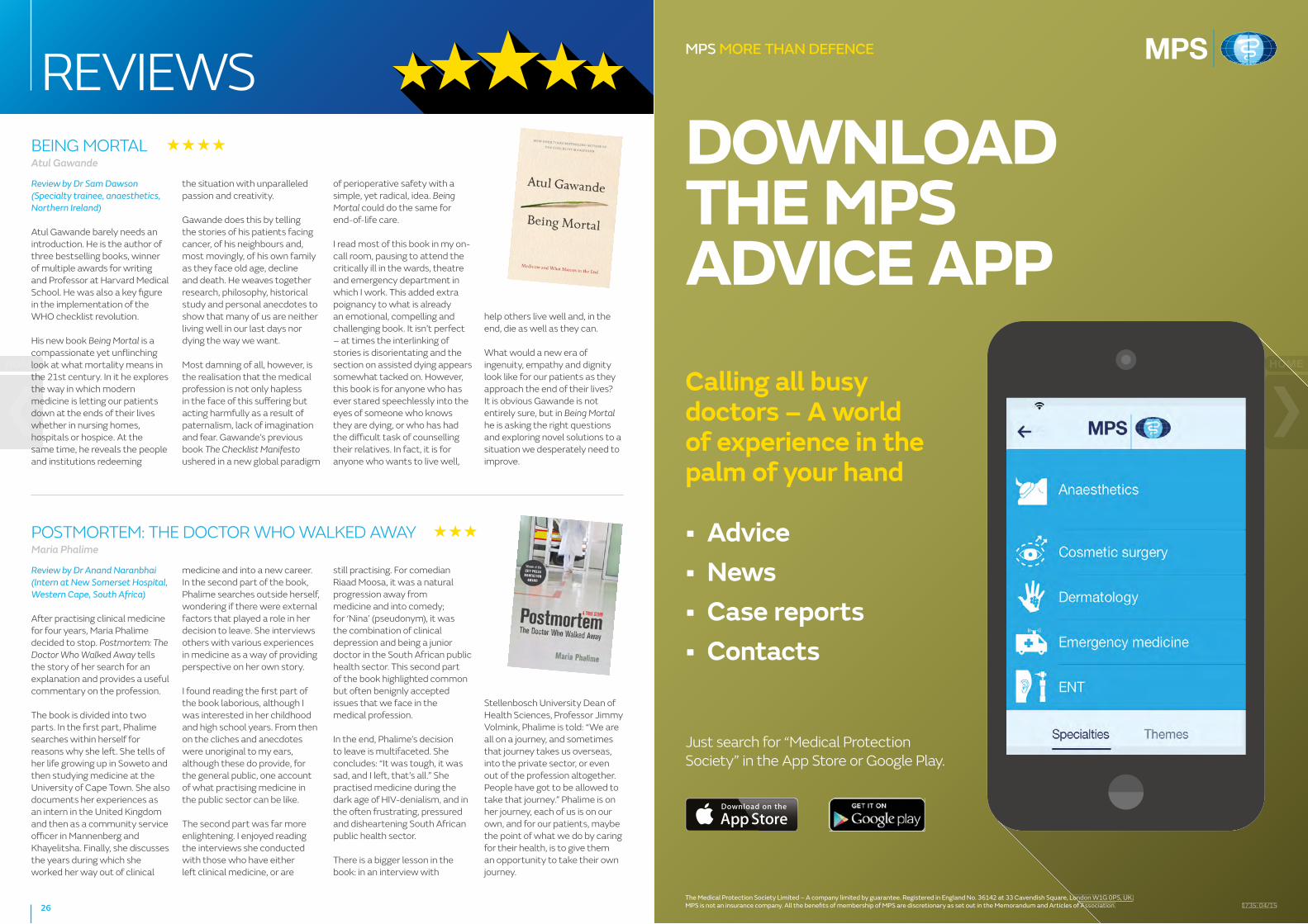
The Medical Protection Society Limited – A company limited by guarantee. Registered in England No. 36142 at 33 Cavendish Square, London W1G 0PS, UK.MPS is not an insurance company. All the benefi ts of membership of MPS are discretionary as set out in the Memorandum and Articles of Association.
MPS MORE THAN DEFENCE
1735: 04/15
Calling all busy doctors – A world of experience in the palm of your hand
• Advice• News• Case reports• Contacts
DOWNLOAD THE MPS ADVICE APP
The Medical Protection Society Limited – A company limited by guarantee. Registered in England No. 36142 at 33 Cavendish Square, London W1G 0PS, UK.MPS is not an insurance company. All the benefi ts of membership of MPS are discretionary as set out in the Memorandum and Articles of Association. 1735: 04/15
Just search for “Medical Protection Society” in the App Store or Google Play.
1735 Advicec App HPV.indd 2 10/04/2015 09:25
BEING MORTALAtul Gawande
Review by Dr Sam Dawson (Specialty trainee, anaesthetics, Northern Ireland)
Atul Gawande barely needs an introduction. He is the author of three bestselling books, winner of multiple awards for writing and Professor at Harvard Medical School. He was also a key figure in the implementation of the WHO checklist revolution.
His new book Being Mortal is a compassionate yet unflinching look at what mortality means in the 21st century. In it he explores the way in which modern medicine is letting our patients down at the ends of their lives whether in nursing homes, hospitals or hospice. At the same time, he reveals the people and institutions redeeming
the situation with unparalleled passion and creativity.
Gawande does this by telling the stories of his patients facing cancer, of his neighbours and, most movingly, of his own family as they face old age, decline and death. He weaves together research, philosophy, historical study and personal anecdotes to show that many of us are neither living well in our last days nor dying the way we want.
Most damning of all, however, is the realisation that the medical profession is not only hapless in the face of this suffering but acting harmfully as a result of paternalism, lack of imagination and fear. Gawande’s previous book The Checklist Manifesto ushered in a new global paradigm
of perioperative safety with a simple, yet radical, idea. Being Mortal could do the same for end-of-life care.
I read most of this book in my on-call room, pausing to attend the critically ill in the wards, theatre and emergency department in which I work. This added extra poignancy to what is already an emotional, compelling and challenging book. It isn’t perfect – at times the interlinking of stories is disorientating and the section on assisted dying appears somewhat tacked on. However, this book is for anyone who has ever stared speechlessly into the eyes of someone who knows they are dying, or who has had the difficult task of counselling their relatives. In fact, it is for anyone who wants to live well,
help others live well and, in the end, die as well as they can.
What would a new era of ingenuity, empathy and dignity look like for our patients as they approach the end of their lives? It is obvious Gawande is not entirely sure, but in Being Mortal he is asking the right questions and exploring novel solutions to a situation we desperately need to improve.
POSTMORTEM: THE DOCTOR WHO WALKED AWAYMaria Phalime
Review by Dr Anand Naranbhai (Intern at New Somerset Hospital, Western Cape, South Africa)
After practising clinical medicine for four years, Maria Phalime decided to stop. Postmortem: The Doctor Who Walked Away tells the story of her search for an explanation and provides a useful commentary on the profession.
The book is divided into two parts. In the first part, Phalime searches within herself for reasons why she left. She tells of her life growing up in Soweto and then studying medicine at the University of Cape Town. She also documents her experiences as an intern in the United Kingdom and then as a community service officer in Mannenberg and Khayelitsha. Finally, she discusses the years during which she worked her way out of clinical
medicine and into a new career. In the second part of the book, Phalime searches outside herself, wondering if there were external factors that played a role in her decision to leave. She interviews others with various experiences in medicine as a way of providing perspective on her own story.
I found reading the first part of the book laborious, although I was interested in her childhood and high school years. From then on the cliches and anecdotes were unoriginal to my ears, although these do provide, for the general public, one account of what practising medicine in the public sector can be like.
The second part was far more enlightening. I enjoyed reading the interviews she conducted with those who have either left clinical medicine, or are
still practising. For comedian Riaad Moosa, it was a natural progression away from medicine and into comedy; for ‘Nina’ (pseudonym), it was the combination of clinical depression and being a junior doctor in the South African public health sector. This second part of the book highlighted common but often benignly accepted issues that we face in the medical profession.
In the end, Phalime’s decision to leave is multifaceted. She concludes: “It was tough, it was sad, and I left, that’s all.” She practised medicine during the dark age of HIV-denialism, and in the often frustrating, pressured and disheartening South African public health sector.
There is a bigger lesson in the book: in an interview with
Stellenbosch University Dean of Health Sciences, Professor Jimmy Volmink, Phalime is told: “We are all on a journey, and sometimes that journey takes us overseas, into the private sector, or even out of the profession altogether. People have got to be allowed to take that journey.” Phalime is on her journey, each of us is on our own, and for our patients, maybe the point of what we do by caring for their health, is to give them an opportunity to take their own journey.
26
REVIEWS
THE MEDICAL PROTECTION SOCIETY 33 Cavendish Square London, W1G 0PS United Kingdom
www.medicalprotection.org www.dentalprotection.org
Please direct all comments, questions or suggestions about MPS service, policy and operations to:
Chief Executive Medical Protection Society 33 Cavendish Square London W1G 0PS United Kingdom
In the interests of confidentiality please do not include information in any email that would allow a patient to be identified.
MPS is the world’s leading protection organisation for doctors, dentists and healthcare professionals. We protect and support the professional interests of more than 300,000 members around the world. Our benefits include access to indemnity, expert advice and peace of mind. Highly qualified advisers are on hand to talk through a question or concern at any time.
MPS is not an insurance company. All the benefits of membership of MPS are discretionary as set out in the Memorandum and Articles of Association.
The Medical Protection Society Limited. A company limited by guarantee. Registered in England No. 36142 at 33 Cavendish Square, London, W1G 0PS
How to contact us
www.medicalprotection.org
SCOTLAND MEDICOLEGAL ADVICE Tel 0800 561 9090
Fax 0113 241 0500
SCOTLAND MEMBERSHIP ENQUIRIES Tel 0800 561 9000
Fax 0113 241 0500
MPS MORE THAN DEFENCE