schedule of accreditation united kingdom accreditation service€¦ · fine needle aspirates for...
TRANSCRIPT

Assessment Manager: HL Page 1 of 20
Schedule of Accreditation issued by
United Kingdom Accreditation Service
2 Pine Trees, Chertsey Lane, Staines-upon-Thames, TW18 3HR, UK
8415
Accredited to
ISO 15189:2012
Oxford University Hospitals NHS Foundation Trust
Issue No: 003 Issue date: 16 November 2017
Cellular Pathology
John Radcliffe Hospital
Headley Way
Headington
Oxford
OX3 9DU
Contact: Sharon Roberts-Gant
Tel: +44 (0)1865-220494
Fax: +44 (0)1865-220190
E-Mail: [email protected]
Website: www.ouh.nhs.uk
Testing performed by the Organisation at the locations specified below
Locations covered by the organisation and their relevant activities
Laboratory locations:
Location details Activity Location code
John Radcliffe Hospital Headley Way Headington Oxford OX3 9DU
Sharon Roberts-Gant (contact details above)
Cytopathology (inc. gynaecological & diagnostic) Histopathology (inc. routine & special staining, frozen sections) Immunocytochemistry In-situ hybridisation Mortuary services (inc. body receipt, storage & release)
JRH
Churchill Hospital Old Road Headington Oxford OX3 7LE
Sharon Roberts-Gant (contact details above)
Slide production, staining & mounting of fine needle aspirates for diagnostic cytology.
CHF
Site activities performed away from the locations listed above:
Location details Activity Location code
Churchill Hospital Old Road Headington Oxford OX3 7LE
Sharon Roberts-Gant (contact details above)
Body receipt, storage & release CHB
Horton General Hospital Oxford Road Banbury Oxfordshire OX16 9AL
Sharon Roberts-Gant (contact details above)
Body receipt, storage & release HH

8415
Accredited to
ISO 15189:2012
Schedule of Accreditation issued by
United Kingdom Accreditation Service 2 Pine Trees, Chertsey Lane, Staines-upon-Thames, TW18 3HR, UK
Oxford University Hospitals NHS Foundation Trust
Issue No: 003 Issue date: 16 November 2017
Testing performed by the Organisation at the locations specified
Assessment Manager: HL Page 2 of 20
DETAIL OF ACCREDITATION
Materials/Products tested
Type of test/Properties
measured/Range of measurement
Standard specifications/
Equipment/Techniques used
Location
Code
HUMAN TISSUES AND FLUIDS
Gynaecological cytopathology
Cervical/vaginal cells Preparation and screening to
identify or exclude cytological abnormalities for the purpose of diagnosis
Liquid based cytology In-house procedures based on Thin Prep method in conjunction with manufacturer’s instructions using: Hologic T5000 processor: G55, G56 Sakura Tissue Tek staining machine & Glas automated coverslipper: G02, G05
JRH
Slides prepared in house from samples listed above
Morphological assessment and interpretation/diagnosis
Microscopy (qualitative analysis) In-house procedures: CPSOP27 in conjunction with manufacturer’s instructions using microscopes: Leitz Dialux, Labourlux K, Laborlux S, Orthoplan, Orthomat II, Nikon Eclipse 50i, E200, E400 & E600, Optishot Olympus BH2, BHTU, BH-U, BHS312, BHS313, BX40, BX41, BX50, BX51
JRH
Diagnostic cytopathology Ascitic fluid Bile Billary brushings Breast cyst fluid Bronchial brushes Bronchial lavage Broncho-alvaeolar lavage Colonic brushings Hydrocele fluid Oesophageal brushings Ovarian cyst fluid Pericardial fluid Pleural fluid Sputum Urine
Examination to identify or exclude cytological abnormalities for the purpose of diagnosis
Prepatation/centrifugation using procedures: NG03 in conjunction with manufacturer’s instructions using: ThermoShandon Cytospin4, NG04 Heraeus Labofuge 400: NG08, NG40
JRH

8415
Accredited to
ISO 15189:2012
Schedule of Accreditation issued by
United Kingdom Accreditation Service 2 Pine Trees, Chertsey Lane, Staines-upon-Thames, TW18 3HR, UK
Oxford University Hospitals NHS Foundation Trust
Issue No: 003 Issue date: 16 November 2017
Testing performed by the Organisation at the locations specified
Assessment Manager: HL Page 3 of 20
Materials/Products tested
Type of test/Properties
measured/Range of measurement
Standard specifications/
Equipment/Techniques used
Location
Code
HUMAN TISSUES AND FLUIDS (Cont’d)
Diagnostic cytopathology (cont’d)
Ascitic fluid Bile Billary brushings Breast cyst fluid Bronchial brushes Bronchial lavage Broncho-alvaeolar lavage Colonic brushings Hydrocele fluid Oesophageal brushings Ovarian cyst fluid Pericardial fluid Pleural fluid Sputum Urine
Examination to identify or exclude cytological abnormalities for the purpose of diagnosis
Staining using in-house procedures for (as appropriate): Papanicoloau: NG19 May Grunwald Giemsa: NG20 Quick Diff: NG21 in conjunction with manufacturer’s instructions using: Shandon Varistain 24-4: NG02, NG04, NG19
JRH
Slides prepared in house from samples listed above
Morphological assessment and interpretation/diagnosis
Microscopy (qualitative analysis) In-house procedures: CPSOP27 in conjunction with manufacturer’s instructions using microscopes: Leitz Dialux, Labourlux K, Laborlux S, Orthoplan, Orthomat II, Nikon Eclipse 50i, E200, E400 & E600, Optishot Olympus BH2, BHTU, BH-U, BHS312, BHS313, BX40, BX41, BX50, BX51
JRH
Fine needle aspirates from: Lymph nodes Neck mass Thoracic: lung, lymph node, mediastinal and pleural Pancreatic Breast
Examination to identify or exclude cytological abnormalities for the purpose of diagnosis
Staining using in-house procedures for (as appropriate): Papanicoloau: NG19 May Grunwald Giemsa: NG20 Quick Diff: NG21 in conjunction with manufacturer’s instructions using: Shandon Varistain 24-4: NG02, NG04, NG19
JRH CHF
Slides prepared in house from samples listed above
Morphological assessment and interpretation/diagnosis
Microscopy (qualitative analysis) Using procedures: CPSOP27 in conjunction with manufacturer’s instructions using microscopes listed above
JRH CHF
8415
Accredited to
ISO 15189:2012
Schedule of Accreditation issued by
United Kingdom Accreditation Service 2 Pine Trees, Chertsey Lane, Staines-upon-Thames, TW18 3HR, UK
Oxford University Hospitals NHS Foundation Trust
Issue No: 003 Issue date: 16 November 2017
Testing performed by the Organisation at the locations specified
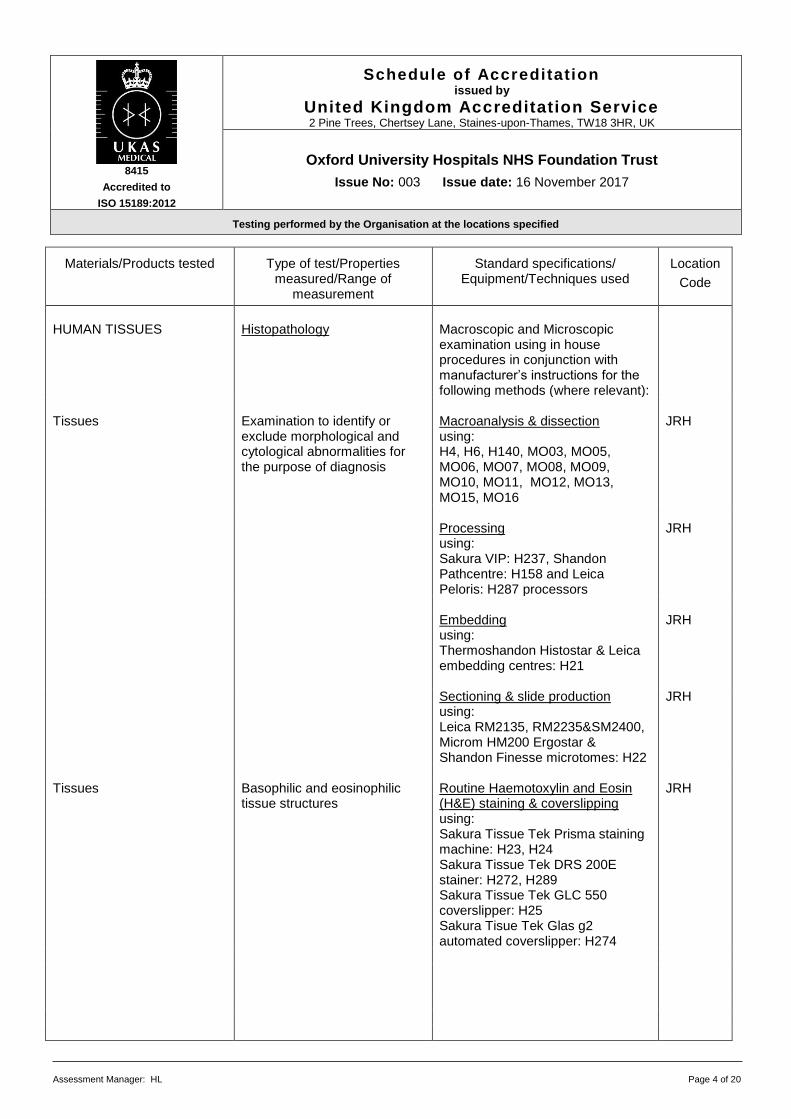
Assessment Manager: HL Page 4 of 20
Materials/Products tested
Type of test/Properties
measured/Range of measurement
Standard specifications/
Equipment/Techniques used
Location
Code
HUMAN TISSUES Histopathology
Macroscopic and Microscopic examination using in house procedures in conjunction with manufacturer’s instructions for the following methods (where relevant):
Tissues Examination to identify or
exclude morphological and cytological abnormalities for the purpose of diagnosis
Macroanalysis & dissection using: H4, H6, H140, MO03, MO05, MO06, MO07, MO08, MO09, MO10, MO11, MO12, MO13, MO15, MO16
JRH
Processing
using: Sakura VIP: H237, Shandon Pathcentre: H158 and Leica Peloris: H287 processors
JRH
Embedding
using: Thermoshandon Histostar & Leica embedding centres: H21
JRH
Sectioning & slide production
using: Leica RM2135, RM2235&SM2400, Microm HM200 Ergostar & Shandon Finesse microtomes: H22
JRH
Tissues Basophilic and eosinophilic
tissue structures
Routine Haemotoxylin and Eosin (H&E) staining & coverslipping using: Sakura Tissue Tek Prisma staining machine: H23, H24 Sakura Tissue Tek DRS 200E stainer: H272, H289 Sakura Tissue Tek GLC 550 coverslipper: H25 Sakura Tisue Tek Glas g2 automated coverslipper: H274
JRH
8415
Accredited to
ISO 15189:2012
Schedule of Accreditation issued by
United Kingdom Accreditation Service 2 Pine Trees, Chertsey Lane, Staines-upon-Thames, TW18 3HR, UK
Oxford University Hospitals NHS Foundation Trust
Issue No: 003 Issue date: 16 November 2017
Testing performed by the Organisation at the locations specified
Assessment Manager: HL Page 5 of 20
Materials/Products tested
Type of test/Properties
measured/Range of measurement
Standard specifications/
Equipment/Techniques used
Location
Code
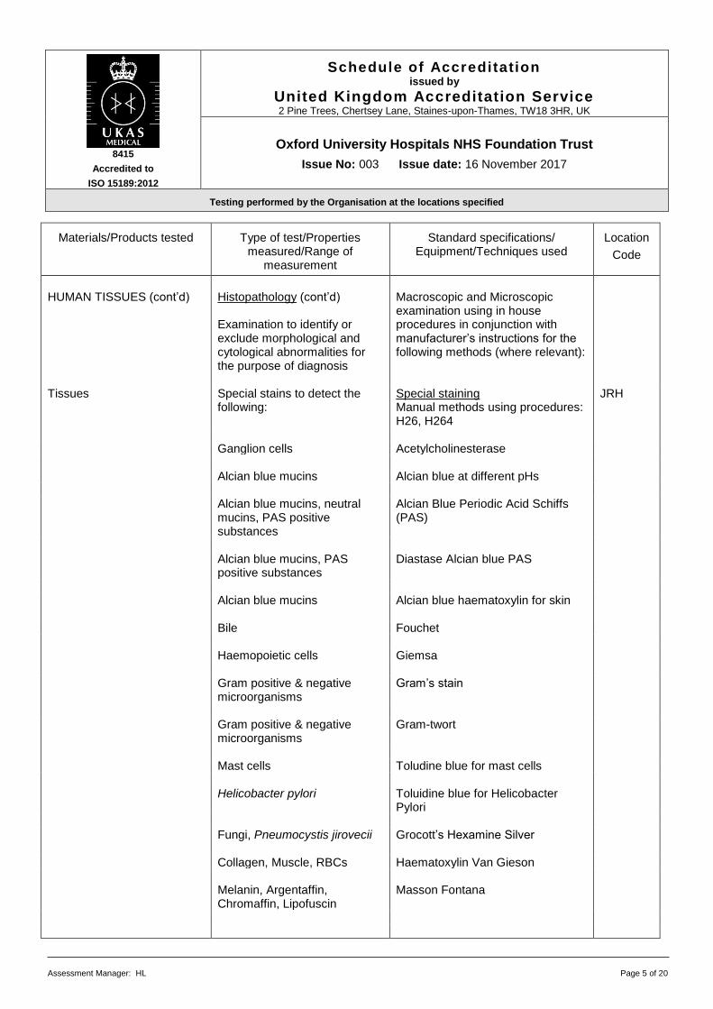
HUMAN TISSUES (cont’d) Histopathology (cont’d)
Examination to identify or exclude morphological and cytological abnormalities for the purpose of diagnosis
Macroscopic and Microscopic examination using in house procedures in conjunction with manufacturer’s instructions for the following methods (where relevant):
Tissues Special stains to detect the
following: Special staining Manual methods using procedures: H26, H264
JRH
Ganglion cells Acetylcholinesterase Alcian blue mucins Alcian blue at different pHs Alcian blue mucins, neutral
mucins, PAS positive substances
Alcian Blue Periodic Acid Schiffs (PAS)
Alcian blue mucins, PAS
positive substances Diastase Alcian blue PAS
Alcian blue mucins Alcian blue haematoxylin for skin Bile Fouchet Haemopoietic cells Giemsa Gram positive & negative
microorganisms Gram’s stain
Gram positive & negative
microorganisms Gram-twort
Mast cells Toludine blue for mast cells Helicobacter pylori Toluidine blue for Helicobacter
Pylori
Fungi, Pneumocystis jirovecii Grocott’s Hexamine Silver Collagen, Muscle, RBCs Haematoxylin Van Gieson Melanin, Argentaffin,
Chromaffin, Lipofuscin Masson Fontana

8415
Accredited to
ISO 15189:2012
Schedule of Accreditation issued by
United Kingdom Accreditation Service 2 Pine Trees, Chertsey Lane, Staines-upon-Thames, TW18 3HR, UK
Oxford University Hospitals NHS Foundation Trust
Issue No: 003 Issue date: 16 November 2017
Testing performed by the Organisation at the locations specified
Assessment Manager: HL Page 6 of 20
Materials/Products tested
Type of test/Properties
measured/Range of measurement
Standard specifications/
Equipment/Techniques used
Location
Code
HUMAN TISSUES (cont’d) Histopathology (cont’d)
Examination to identify or exclude morphological and cytological abnormalities for the purpose of diagnosis
Macroscopic and Microscopic examination using in house procedures in conjunction with manufacturer’s instructions for the following methods (where relevant):
Tissues Special stains to detect the
following: Special staining Manual methods (Cont’d)
JRH
Fibrin, Muscle, Collagen,
RBCs MSB trichrome
Neutral lipds Oil red o HBsAg (Hep B), Elastin Orcein for HBSag (Shikata) Glycogen PAS Glycogen PAS Diastase Cryptococcus Southgates Mucicarmine Acid/alcohol fast bacilli Ziehl Neelsen Leprosy bacilli, RBCs Wade-Fite Mineralised bone, Canaliculi &
Lacunae in the centre of the traberculae, Bone marrow, Osteoid seams
Von kossa
Collagen Fast green / Sirius red Amyloid Sirius red Copper, Copper-associated
protein Rhodanine
Amyloid Thioflavin T
8415
Accredited to
ISO 15189:2012
Schedule of Accreditation issued by
United Kingdom Accreditation Service 2 Pine Trees, Chertsey Lane, Staines-upon-Thames, TW18 3HR, UK
Oxford University Hospitals NHS Foundation Trust
Issue No: 003 Issue date: 16 November 2017
Testing performed by the Organisation at the locations specified
Assessment Manager: HL Page 7 of 20
Materials/Products tested
Type of test/Properties
measured/Range of measurement
Standard specifications/
Equipment/Techniques used
Location
Code
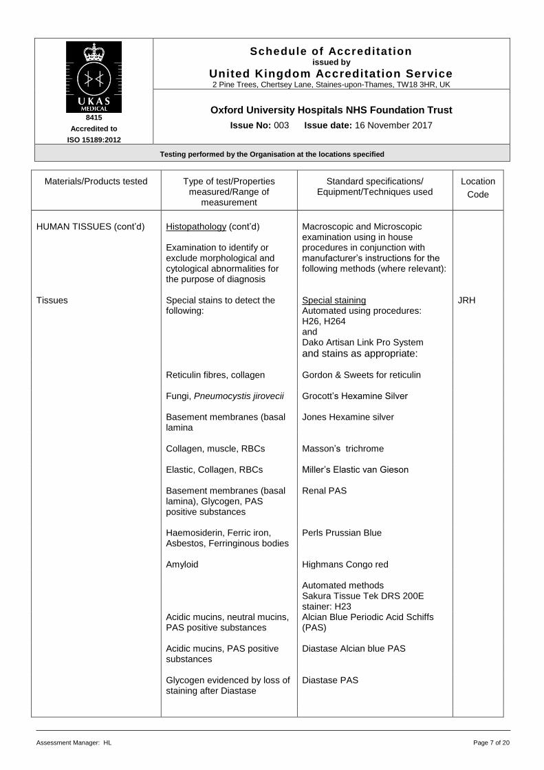
HUMAN TISSUES (cont’d) Histopathology (cont’d)
Examination to identify or exclude morphological and cytological abnormalities for the purpose of diagnosis
Macroscopic and Microscopic examination using in house procedures in conjunction with manufacturer’s instructions for the following methods (where relevant):
Tissues Special stains to detect the
following: Special staining Automated using procedures: H26, H264 and Dako Artisan Link Pro System
and stains as appropriate:
JRH
Reticulin fibres, collagen Gordon & Sweets for reticulin Fungi, Pneumocystis jirovecii Grocott’s Hexamine Silver Basement membranes (basal
lamina Jones Hexamine silver
Collagen, muscle, RBCs Masson’s trichrome Elastic, Collagen, RBCs Miller’s Elastic van Gieson Basement membranes (basal
lamina), Glycogen, PAS positive substances
Renal PAS
Haemosiderin, Ferric iron,
Asbestos, Ferringinous bodies Perls Prussian Blue
Amyloid Highmans Congo red Automated methods
Sakura Tissue Tek DRS 200E stainer: H23
Acidic mucins, neutral mucins, PAS positive substances
Alcian Blue Periodic Acid Schiffs (PAS)
Acidic mucins, PAS positive substances
Diastase Alcian blue PAS
Glycogen evidenced by loss of staining after Diastase
Diastase PAS

8415
Accredited to
ISO 15189:2012
Schedule of Accreditation issued by
United Kingdom Accreditation Service 2 Pine Trees, Chertsey Lane, Staines-upon-Thames, TW18 3HR, UK
Oxford University Hospitals NHS Foundation Trust
Issue No: 003 Issue date: 16 November 2017
Testing performed by the Organisation at the locations specified
Assessment Manager: HL Page 8 of 20
Materials/Products tested
Type of test/Properties
measured/Range of measurement
Standard specifications/
Equipment/Techniques used
Location
Code
HUMAN TISSUES (cont’d) Histopathology (cont’d)
Examination to identify or exclude morphological and cytological abnormalities for the purpose of diagnosis
Macroscopic and Microscopic examination using in house procedures in conjunction with manufacturer’s instructions for the following methods (where relevant):
Special staing (cont’d)
Automated methods Sakura Tissue Tek DRS 200E stainer: H23
Mucins, Glycogen, PAS positive substances – fungi
PAS
Tissues Immunocytochemistry to detect the following:
Immunocytochemistry Automated using procedures: CPP31, H151, H236, H237, H279 and Bond III and antibodies as appropriate:
JRH
Smooth muscle Actin (1A4) Adenovirus Adenovirus Epithelium AE1/3 (multi CK) Tissues Immunocytochemistry to
detect the following: Immunocytochemistry (cont’d)
JRH
Alpha fetal protein AFP Anaplastic lymphoma kinase ALK1 Alpha 1 anti-chymotrypsin Alpha 1 C Trypsin Alpha 1 anti-trypsin Alpha 1AT Racemase (Prostate ca) AMACR (racemase) Amyloid A Amyloid A (MC1) Lymphoma panel Annexin Androgen receptor Androgen receptor
8415
Accredited to
ISO 15189:2012
Schedule of Accreditation issued by
United Kingdom Accreditation Service 2 Pine Trees, Chertsey Lane, Staines-upon-Thames, TW18 3HR, UK
Oxford University Hospitals NHS Foundation Trust
Issue No: 003 Issue date: 16 November 2017
Testing performed by the Organisation at the locations specified
Assessment Manager: HL Page 9 of 20
Materials/Products tested
Type of test/Properties
measured/Range of measurement
Standard specifications/
Equipment/Techniques used
Location
Code
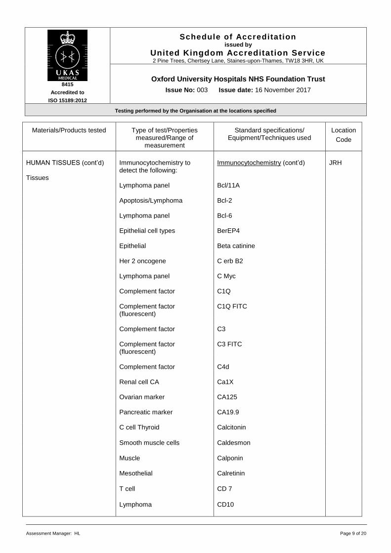
HUMAN TISSUES (cont’d) Tissues
Immunocytochemistry to detect the following:
Immunocytochemistry (cont’d)
JRH
Lymphoma panel Bcl/11A Apoptosis/Lymphoma Bcl-2 Lymphoma panel Bcl-6 Epithelial cell types BerEP4 Epithelial Beta catinine Her 2 oncogene C erb B2 Lymphoma panel C Myc Complement factor C1Q Complement factor
(fluorescent) C1Q FITC
Complement factor C3 Complement factor
(fluorescent) C3 FITC
Complement factor C4d Renal cell CA Ca1X Ovarian marker CA125 Pancreatic marker CA19.9 C cell Thyroid Calcitonin
Smooth muscle cells Caldesmon
Muscle Calponin Mesothelial Calretinin
T cell CD 7
Lymphoma CD10

8415
Accredited to
ISO 15189:2012
Schedule of Accreditation issued by
United Kingdom Accreditation Service 2 Pine Trees, Chertsey Lane, Staines-upon-Thames, TW18 3HR, UK
Oxford University Hospitals NHS Foundation Trust
Issue No: 003 Issue date: 16 November 2017
Testing performed by the Organisation at the locations specified
Assessment Manager: HL Page 10 of 20
Materials/Products tested
Type of test/Properties
measured/Range of measurement
Standard specifications/
Equipment/Techniques used
Location
Code
HUMAN TISSUES (cont’d) Histopathology (cont’d) Examination to identify or exclude morphological and cytological abnormalities for the purpose of diagnosis
Macroscopic and Microscopic examination using in house procedures in conjunction with manufacturer’s instructions for the following methods (where relevant):
Tissues Immunocytochemistry to detect the following:
Immunocytochemistry (cont’d) JRH
Hogkins lymphoma CD15 Leu M1
Monocytes CD163 B Cell CD19 Langerhans cells CD1a T cell CD2 B cell CD20 (L26) Follicular dendritic cells CD21 (I2G9)
Follicular dendritic cells CD23 (MHM6)
T cells CD3 Hogkins lymphoma CD30 Vascular CD31 (Jc70) Myeloid CD33 Vascular/stromal CD34 (Qbend) T cell CD4 368 T cell CD43 (DFT1) Leucocyte common antigen CD45 (LCA) T cell CD5 Neuroendocrine CD56
8415
Accredited to
ISO 15189:2012
Schedule of Accreditation issued by
United Kingdom Accreditation Service 2 Pine Trees, Chertsey Lane, Staines-upon-Thames, TW18 3HR, UK
Oxford University Hospitals NHS Foundation Trust
Issue No: 003 Issue date: 16 November 2017
Testing performed by the Organisation at the locations specified
Assessment Manager: HL Page 11 of 20
Materials/Products tested
Type of test/Properties
measured/Range of measurement
Standard specifications/
Equipment/Techniques used
Location
Code
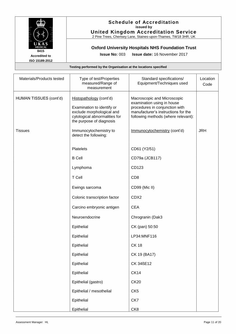
HUMAN TISSUES (cont’d) Histopathology (cont’d)
Examination to identify or exclude morphological and cytological abnormalities for the purpose of diagnosis
Macroscopic and Microscopic examination using in house procedures in conjunction with manufacturer’s instructions for the following methods (where relevant):
Tissues Immunocytochemistry to detect the following:
Immunocytochemistry (cont’d) JRH
Platelets CD61 (Y2/51) B Cell CD79a (JCB117)
Lymphoma CD123
T Cell CD8
Ewings sarcoma CD99 (Mic II)
Colonic transcription factor CDX2
Carcino embryonic antigen CEA
Neuroendocrine Chrogranin (Dak3
Epithelial CK (pan) 50:50
Epithelial LP34:MNF116 Epithelial CK 18 Epithelial CK 19 (BA17) Epithelial CK 34ßE12 Epithelial CK14 Epithelial (gastro) CK20 Epithelial / mesothelial CK5 Epithelial CK7 Epithelial CK8
8415
Accredited to
ISO 15189:2012
Schedule of Accreditation issued by
United Kingdom Accreditation Service 2 Pine Trees, Chertsey Lane, Staines-upon-Thames, TW18 3HR, UK
Oxford University Hospitals NHS Foundation Trust
Issue No: 003 Issue date: 16 November 2017
Testing performed by the Organisation at the locations specified
Assessment Manager: HL Page 12 of 20
Materials/Products tested
Type of test/Properties
measured/Range of measurement
Standard specifications/
Equipment/Techniques used
Location
Code
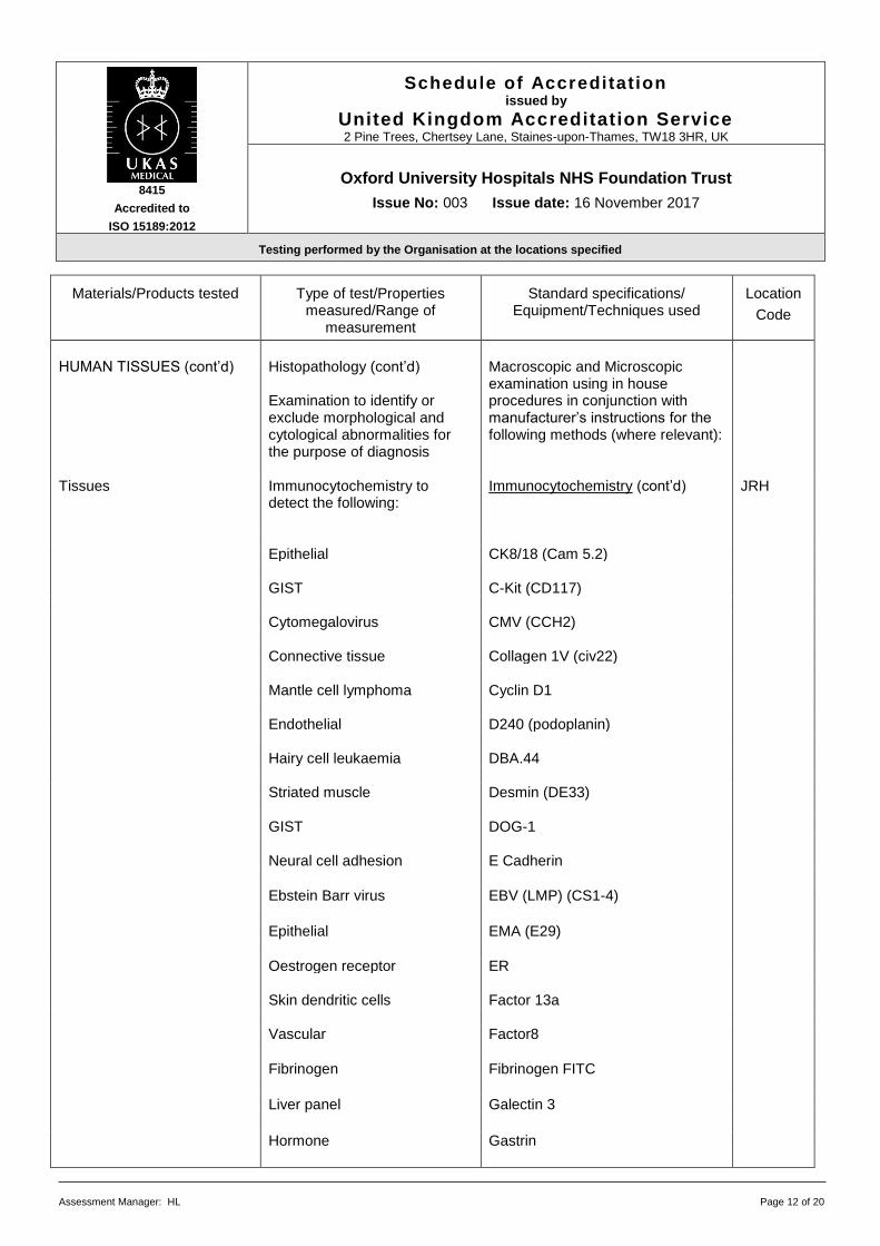
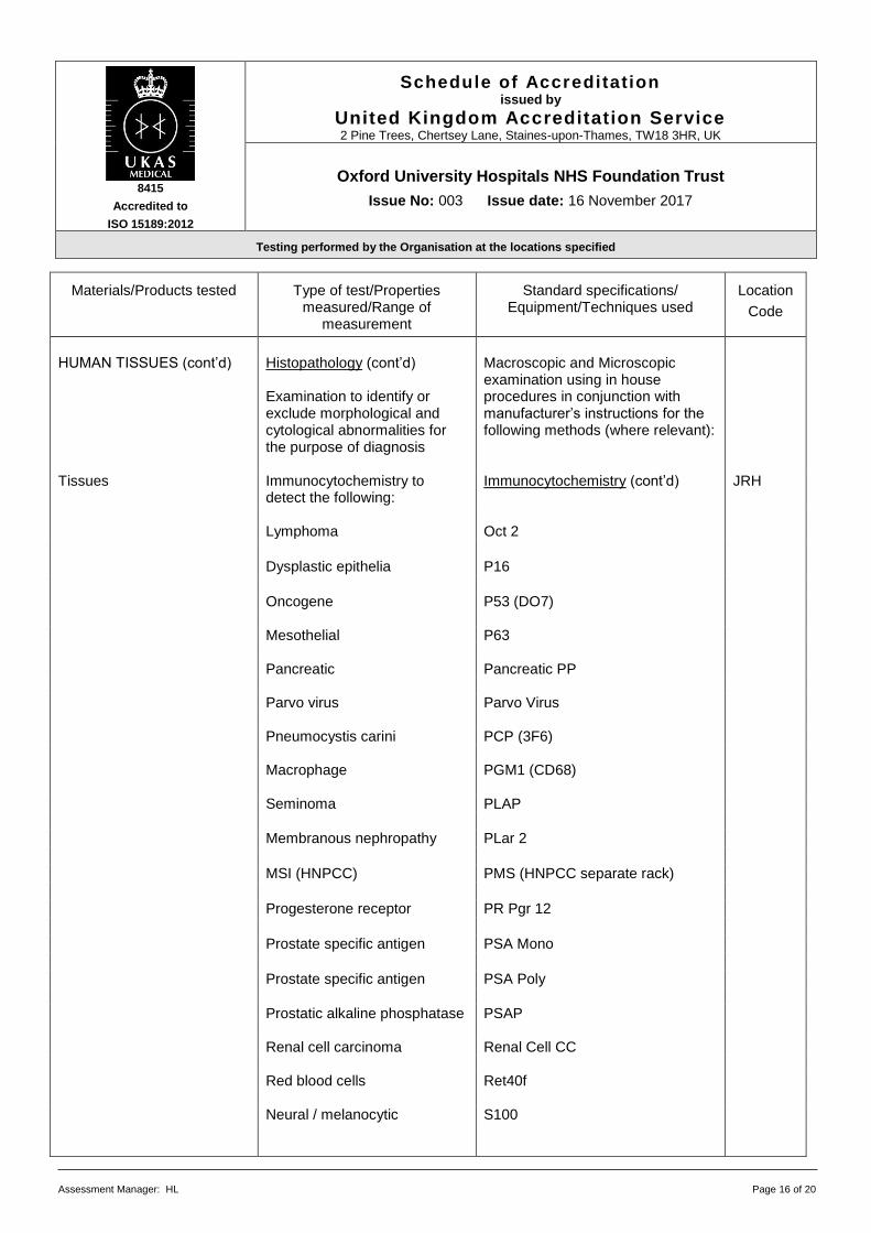
HUMAN TISSUES (cont’d) Histopathology (cont’d)
Examination to identify or exclude morphological and cytological abnormalities for the purpose of diagnosis
Macroscopic and Microscopic examination using in house procedures in conjunction with manufacturer’s instructions for the following methods (where relevant):
Tissues Immunocytochemistry to
detect the following:
Immunocytochemistry (cont’d) JRH
Epithelial CK8/18 (Cam 5.2) GIST C-Kit (CD117) Cytomegalovirus CMV (CCH2) Connective tissue Collagen 1V (civ22) Mantle cell lymphoma Cyclin D1 Endothelial D240 (podoplanin) Hairy cell leukaemia DBA.44 Striated muscle Desmin (DE33) GIST DOG-1 Neural cell adhesion E Cadherin
Ebstein Barr virus EBV (LMP) (CS1-4)
Epithelial EMA (E29)
Oestrogen receptor ER Skin dendritic cells Factor 13a Vascular Factor8
Fibrinogen Fibrinogen FITC
Liver panel Galectin 3
Hormone Gastrin

8415
Accredited to
ISO 15189:2012
Schedule of Accreditation issued by
United Kingdom Accreditation Service 2 Pine Trees, Chertsey Lane, Staines-upon-Thames, TW18 3HR, UK
Oxford University Hospitals NHS Foundation Trust
Issue No: 003 Issue date: 16 November 2017
Testing performed by the Organisation at the locations specified
Assessment Manager: HL Page 13 of 20
Materials/Products tested
Type of test/Properties
measured/Range of measurement
Standard specifications/
Equipment/Techniques used
Location
Code
HUMAN TISSUES (cont’d) Histopathology (cont’d)
Examination to identify or exclude morphological and cytological abnormalities for the purpose of diagnosis
Macroscopic and Microscopic examination using in house procedures in conjunction with manufacturer’s instructions for the following methods (where relevant):
Tissues Immunocytochemistry to
detect the following: Immunocytochemistry (cont’d) JRH
Bladder carcinoma Gata 3 Hormone Glucagon Liver panel Glutamine Synthetase Liver panel Glypican 3 Helicobacter pylori H Pylori
Human Chorionic gonadotrophin
HCG
Hep B virus Hep b core
Hep B virus He b surface
Hepatocyte Hepar 1
Smooth muscle HHF35
Mesothelial HMBE 1
Smooth muscle HHV-8 Human papilloma virus HPV Herpes simplex virus HSV Type II Immunoglobulin IgA Immunoglobulin fluorescent IgA FITC Immunoglobulin B cell IgD Immunoglobulin IgG

8415
Accredited to
ISO 15189:2012
Schedule of Accreditation issued by
United Kingdom Accreditation Service 2 Pine Trees, Chertsey Lane, Staines-upon-Thames, TW18 3HR, UK
Oxford University Hospitals NHS Foundation Trust
Issue No: 003 Issue date: 16 November 2017
Testing performed by the Organisation at the locations specified
Assessment Manager: HL Page 14 of 20
Materials/Products tested
Type of test/Properties
measured/Range of measurement
Standard specifications/
Equipment/Techniques used
Location
Code
HUMAN TISSUES (cont’d) Histopathology (cont’d)
Examination to identify or exclude morphological and cytological abnormalities for the purpose of diagnosis
Macroscopic and Microscopic examination using in house procedures in conjunction with manufacturer’s instructions for the following methods (where relevant):
Tissues Immunocytochemistry to
detect the following: Immunocytochemistry (cont’d) JRH
Immunoglobulin fluorescent IgG FITC Immunoglobulin IgG1 Immunoglobulin IgG2 Immunoglobulin IgG3 Immunoglobulin IgG4 Immunoglobulin B Cell IgM Immunoglobulin Fluorescent IgM FITC Germ cell Inhibin Pancreatic Insulin Light chain Kappa Light chain Kappa FITC Macrophage KP1 (CD68)
Light chain Lambda
Light chain fluorescent Lambda FITC Connective tissue Laminin Epithelial LP34 Macrophage Lysozyme Mast cell Mast Cell Tryptase
8415
Accredited to
ISO 15189:2012
Schedule of Accreditation issued by
United Kingdom Accreditation Service 2 Pine Trees, Chertsey Lane, Staines-upon-Thames, TW18 3HR, UK
Oxford University Hospitals NHS Foundation Trust
Issue No: 003 Issue date: 16 November 2017
Testing performed by the Organisation at the locations specified
Assessment Manager: HL Page 15 of 20
Materials/Products tested
Type of test/Properties
measured/Range of measurement
Standard specifications/
Equipment/Techniques used
Location
Code
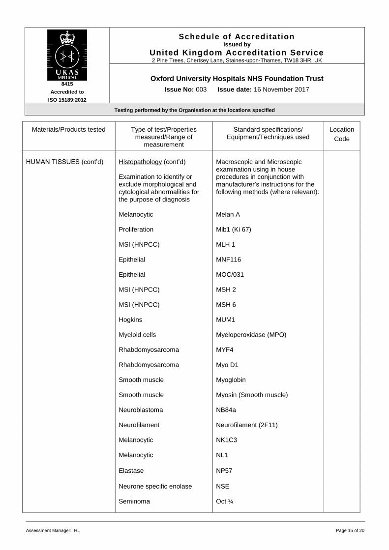
HUMAN TISSUES (cont’d) Histopathology (cont’d)
Examination to identify or exclude morphological and cytological abnormalities for the purpose of diagnosis
Macroscopic and Microscopic examination using in house procedures in conjunction with manufacturer’s instructions for the following methods (where relevant):
Melanocytic Melan A Proliferation Mib1 (Ki 67) MSI (HNPCC) MLH 1 Epithelial MNF116 Epithelial MOC/031 MSI (HNPCC) MSH 2 MSI (HNPCC) MSH 6 Hogkins MUM1 Myeloid cells Myeloperoxidase (MPO) Rhabdomyosarcoma MYF4 Rhabdomyosarcoma Myo D1 Smooth muscle Myoglobin Smooth muscle Myosin (Smooth muscle) Neuroblastoma NB84a Neurofilament Neurofilament (2F11) Melanocytic NK1C3 Melanocytic NL1
Elastase NP57
Neurone specific enolase NSE Seminoma Oct ¾
8415
Accredited to
ISO 15189:2012
Schedule of Accreditation issued by
United Kingdom Accreditation Service 2 Pine Trees, Chertsey Lane, Staines-upon-Thames, TW18 3HR, UK
Oxford University Hospitals NHS Foundation Trust
Issue No: 003 Issue date: 16 November 2017
Testing performed by the Organisation at the locations specified
Assessment Manager: HL Page 16 of 20
Materials/Products tested
Type of test/Properties
measured/Range of measurement
Standard specifications/
Equipment/Techniques used
Location
Code
HUMAN TISSUES (cont’d) Histopathology (cont’d)
Examination to identify or exclude morphological and cytological abnormalities for the purpose of diagnosis
Macroscopic and Microscopic examination using in house procedures in conjunction with manufacturer’s instructions for the following methods (where relevant):
Tissues Immunocytochemistry to
detect the following: Immunocytochemistry (cont’d) JRH
Lymphoma Oct 2
Dysplastic epithelia P16
Oncogene P53 (DO7) Mesothelial P63 Pancreatic Pancreatic PP Parvo virus Parvo Virus Pneumocystis carini PCP (3F6) Macrophage PGM1 (CD68) Seminoma PLAP
Membranous nephropathy PLar 2
MSI (HNPCC) PMS (HNPCC separate rack)
Progesterone receptor PR Pgr 12
Prostate specific antigen PSA Mono
Prostate specific antigen PSA Poly
Prostatic alkaline phosphatase PSAP Renal cell carcinoma Renal Cell CC Red blood cells Ret40f Neural / melanocytic S100
8415
Accredited to
ISO 15189:2012
Schedule of Accreditation issued by
United Kingdom Accreditation Service 2 Pine Trees, Chertsey Lane, Staines-upon-Thames, TW18 3HR, UK
Oxford University Hospitals NHS Foundation Trust
Issue No: 003 Issue date: 16 November 2017
Testing performed by the Organisation at the locations specified
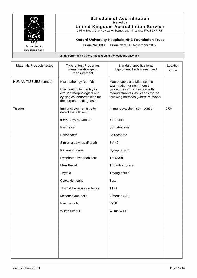
Assessment Manager: HL Page 17 of 20
Materials/Products tested
Type of test/Properties
measured/Range of measurement
Standard specifications/
Equipment/Techniques used
Location
Code
HUMAN TISSUES (cont’d) Histopathology (cont’d)
Examination to identify or exclude morphological and cytological abnormalities for the purpose of diagnosis
Macroscopic and Microscopic examination using in house procedures in conjunction with manufacturer’s instructions for the following methods (where relevant):
Tissues Immunocytochemistry to
detect the following: Immunocytochemistry (cont’d) JRH
5 Hydroxytryptamine Serotonin Pancreatic Somatostatin Spirochaete Spirochaete Simian aids virus (Renal) SV 40 Neuroendocrine Synaptohysin Lymphoma lymphoblastic Tdt (339) Mesothelial Thrombomodulin Thyroid Thyroglobulin Cytotoxic t cells Tia1 Thyroid transcription factor TTF1 Mesenchyme cells Vimentin (V9) Plasma cells Vs38 Wilms tumour Wilms WT1

8415
Accredited to
ISO 15189:2012
Schedule of Accreditation issued by
United Kingdom Accreditation Service 2 Pine Trees, Chertsey Lane, Staines-upon-Thames, TW18 3HR, UK
Oxford University Hospitals NHS Foundation Trust
Issue No: 003 Issue date: 16 November 2017
Testing performed by the Organisation at the locations specified
Assessment Manager: HL Page 18 of 20
Materials/Products tested
Type of test/Properties
measured/Range of measurement
Standard specifications/
Equipment/Techniques used
Location
Code
HUMAN TISSUES (cont’d) Histopathology (cont’d) Tissues Immunocytochemistry to
detect the following: Immunocytochemistry Automated using Ventana Benchmark ULTRA Testing Platform SOP H295 Use of the Ventana BenchMark ULTRA staining machine and antibodies as appropriate:
JRH
Her 2 oncogene HER-2 Helicobacter pylori H Pylori HHV-8 HHV-8 Dysplastic epithelia P16 Histopathology (cont’d)
Examination to identify or exclude morphological and cytological abnormalities for the purpose of diagnosis
Tissues In-situ hybridisation to detect
the following: In-situ hybridisation Automated using procedures: H236 and Bond Max and probes as appropriate:
JRH
Epstein Barr virus EBER Lambda light chain Lambda Kappa light chain Kappa

8415
Accredited to
ISO 15189:2012
Schedule of Accreditation issued by
United Kingdom Accreditation Service 2 Pine Trees, Chertsey Lane, Staines-upon-Thames, TW18 3HR, UK
Oxford University Hospitals NHS Foundation Trust
Issue No: 003 Issue date: 16 November 2017
Testing performed by the Organisation at the locations specified
Assessment Manager: HL Page 19 of 20
Materials/Products tested
Type of test/Properties
measured/Range of measurement
Standard specifications/
Equipment/Techniques used
Location
Code
HUMAN TISSUES (cont’d) Histopathology (cont’d) Slides prepared in house from samples listed above
Morphological assessment and interpretation/diagnosis
Microscopy (qualitative analysis) Using procedures: CPSOP27 and microscopes listed above
JRH
Tissues Frozen section examination to
identify or exclude morphological and cytological abnormalities for the purpose of diagnosis
Cryotomy Using procedures: H10, H201 and Leica CM1850 UV & CM1950 cryostats
JRH
Basophilic and eosinophilic
tissue structures
Routine H&E staining Manual metjods using procedures: H23, H289, H171, H179
JRH
Special stains to detect the
following: Special staining Manual methods using procedures: H26, H264 and stains as appropriate:
Ganglion cells Acetylcholinesterase Neutral lipds Oil red o Tissues Immunocytochemistry to
detect the following: Immunocytochemistry Automated using procedures: CPP31, H151, H236, H237, H279 and Bond Max and antibodies as appropriate:
Complement factor
(fluorescent) C1Q FITC
Complement factor
(fluorescent) C3 FITC
Fibrinogen Fibrinogen FITC Immunoglobulin fluorescent IgA FITC Immunoglobulin fluorescent IgG FITC

8415
Accredited to
ISO 15189:2012
Schedule of Accreditation issued by
United Kingdom Accreditation Service 2 Pine Trees, Chertsey Lane, Staines-upon-Thames, TW18 3HR, UK
Oxford University Hospitals NHS Foundation Trust
Issue No: 003 Issue date: 16 November 2017
Testing performed by the Organisation at the locations specified
Assessment Manager: HL Page 20 of 20
Materials/Products tested
Type of test/Properties
measured/Range of measurement
Standard specifications/
Equipment/Techniques used
Location
Code
HUMAN TISSUES (cont’d) Histopathology (cont’d)
Frozen section examination to
identify or exclude morphological and cytological abnormalities for the purpose of diagnosis
Macroscopic and Microscopic examination using in house procedures in conjunction with manufacturer’s instructions for the following methods (where relevant):
Immunocytochemistry to
detect the following: Immunocytochemistry (cont’d)
JRH
Immunoglobulin Fluorescent IgM FITC Light chain Kappa FITC Light chain fluorescent Lambda FITC Slides prepared in house as above
Morphological assessment and interpretation/diagnosis
Microscopy (qualitative analysis) Using procedures: CPSOP27 and microscopes listed above
JRH
END