science china life sciences - home - springer · 746 dong j, et al. sci china life sci august...
TRANSCRIPT

SCIENCE CHINA Life Sciences
© The Author(s) 2013. This article is published with open access at Springerlink.com life.scichina.com www.springer.com/scp
*Corresponding author (email: [email protected]; [email protected])
• RESEARCH PAPER • August 2013 Vol.56 No.8: 745–750
doi: 10.1007/s11427-013-4503-3
Quantitative assessment of acute kidney injury by noninvasive arterial spin labeling perfusion MRI: a pilot study
DONG Jian1, YANG Li2, SU Tao2, YANG XueDong1, CHEN Bin3, ZHANG Jue3,4*, WANG XiaoYing1* & JIANG XueXiang1
1Department of Radiology, Peking University First Hospital, Beijing 100034, China; 2Department of Nephrology, Peking University First Hospital, Beijing 100034, China;
3Academy for Advanced Interdisciplinary Studies, Peking University, Beijing 100871, China; 4College of Engineering, Peking University, Beijing 100871, China
Received January 22, 2013; accepted April 24, 2013; published online June 3, 2013
The kidneys are essential for maintaining homeostasis, are responsible for the reabsorption of water, glucose and amino acids, and filter the blood by removing waste. Acute kidney injury (AKI) is a syndrome characterized by the rapid loss of renal ex-cretory function and the accumulation of end metabolic products of urea and creatinine. AKI is associated with the later de-velopment of chronic kidney disease and end-stage kidney disease, and may eventually be fatal. Early diagnosis of AKI and assessments of the effects of treatment, however, are challenging. The pathophysiological mechanism of AKI is thought to be the imbalance between oxygen supply and demand in the kidneys. We have assessed the ability of arterial spin labeling (ASL) perfusion magnetic resonance imaging (MRI), without the administration of contrast media, to quantify renal blood flow (RBF) non-invasively. We found that RBF was significantly lower in AKI patients than in healthy volunteers. These results suggest that ASL perfusion MRI, a noninvasive measurement of RBF, may be useful in the early diagnosis of AKI.
arterial spin labeling (ASL), acute kidney injury (AKI), renal blood flow (RBF), magnetic resonance imaging (MRI)
Citation: Dong J, Yang L, Su T, et al. Quantitative assessment of acute kidney injury by noninvasive arterial spin labeling perfusion MRI: a pilot study. Sci China Life Sci, 2013, 56: 745–750, doi: 10.1007/s11427-013-4503-3
Acute kidney injury (AKI) is a frequently observed condi-tion linked with the later development of chronic kidney disease, end-stage kidney disease, and mortality. AKI has been reported to affect more than 5000 per million people not on dialysis therapy and 295 per million on dialysis therapy per year [1–3]. The advent of the RIFLE (risk, in-jury, failure, loss, end-stage kidney disease) and Acute Kidney Injury Network (AKIN) criteria has improved the diagnosis of AKI [4], and decreased renal blood flow (RBF), which causes renal ischemia, has been proposed as central to the pathogenesis of septic acute renal failure [5]. These criteria, however, have been based on serum creatinine
concentration and urine output, both of which have rela-tively poor specificity and sensitivity during the early period of AKI [6–9]. At present, therefore, there are no effective biomarkers for AKI, making early diagnosis of AKI and the assessment of the effects of therapy difficult [10].
Noninvasive functional magnetic resonance imaging (MRI) provides a variety of effective methods to evaluate renal function in vivo without administration of contrast media. The arterial spin labeling (ASL) perfusion technique, in which inflowing blood is labeled magnetically, can be used in the absolute quantification of renal blood flow (RBF), especially in patients with impaired renal function [11–15]. ASL was found to be better able to quantify corti-cal RBF than dynamic contrast enhanced MRI [16]. The

746 Dong J, et al. Sci China Life Sci August (2013) Vol.56 No.8
reproducibility of the flow-sensitive alternating inversion recovery (FAIR) ASL perfusion technique over a broad range of renal function has been demonstrated in the corti-ces of native and transplanted kidneys [17]. Moreover, by measuring kidney perfusion, ASL demonstrated a potential capability for assessing renal disease in both native and transplanted kidneys [12,17,18]. To our knowledge, howev-er, quantitative ASL analysis has not been used in the diag-nosis of AKI. We therefore performed a pilot study to in-vestigate the feasibility of noninvasive ASL perfusion MRI in the diagnosis of AKI.
1 Materials and methods
1.1 Subjects
From October 1, 2011 to December 1, 2012, 13 AKI pa-tients and 18 healthy volunteers were recruited. Patients were included if they (a) were adults diagnosed with AKI by referring nephrologists, (b) were scheduled to undergo percutaneous renal biopsy three days after MR examination and prior to therapy, (c) had no signs of other kidney dis-eases, such as renal artery stenosis and urolithiasis, as shown by ultrasonography in our hospital while being in patients, and (d) had normal cardiac function. Healthy vol-unteers were included if they (a) were adults with no history of renal or cardiac diseases, and (b) had normal serum cre-atinine concentrations one week before MR scanning.
Patients underwent MR examinations before initial of therapy. All subjects refrained from food and fluids for 6 h and were trained in breath-holding techniques training be-fore MR examination. Serum creatinine concentrations were measured in all subjects before MR examination.
This prospective study was compliant with the Health Insurance Portability and Accountability Act (HIPAA) and was approved by our institutional review board. All subjects provided written informed consent and were compatible with MR scanning.
1.2 MR protocols
All examinations were performed on a 3.0T MR scanner (Signa Excite; GE Healthcare, Milwaukee, WI) using a commercial Torso PA coil (GE Medical Systems). The scout images were scanned with a gradient echo sequence in three oblique planes through the center of each kidney. Oblique-coronal T2-weighted imaging through the center of the kidney was performed for anatomical evaluation using a single shot fast spin echo (SSFSE) sequence with the param-eters: TR=1100 ms, TE=60 ms, slice thickness=5 mm, 1 mm interval, matrix 192×256, flip angle 130°, bandwidth= 62 kHz, FOV 35–40 cm.
ASL images were acquired for quantitative measure-ments of RBF by applying the fluid alternating inversion recovery (FAIR) sequence with the combination of a sin-
gle-shot fast spin-echo (SSFSE) imaging (FAIR-SSFSE). One single oblique coronal slice of ASL was scanned in the same position through the center of the kidney with the pa-rameters: field of view (FOV)=40 cm, matrix=128×128, slice thickness=5 mm, TR=3500 ms, TE=60 ms, flip angle = 90°, bandwidth=62.5 kHz. The slab thickness was set at 20 mm for section selective inversion in the FAIR preparation, to avoid artifacts from incompletely inverted spins at the margins of the readout slice, with no feeding vessels in-cluded.
At the beginning of the scanning process, inversion ra-diofrequency pulses were used to label the protons in the arterial blood feeding both kidneys, with the magnetically tagged blood acting as an endogenous tracer. All six ASL images were scanned at the same slice, including three tag images with section selective inversion and three control images with global inversion. The inversion time (TI) be-tween the tag and control images was fixed at 1400 ms to ensure the tagged protons reached the capillary bed and exchanged with protons in the surrounding tissue; the tag image was then subtracted from the control image to re-move signals of static tissue, yielding residual signals for quantification of RBF. M0 images were acquired using the SSFSE sequence, with the same parameters as the prior FAIR-SSFSE sequence without FAIR preparation, and the repetition time (TR) was set at 6 s to allow for the complete recovery of blood signals between measurements.
All participants were instructed to hold their breath for 20–30 s during each acquisition, with ASL imaging per-formed twice. The total acquisition time for ASL imaging was limited to 1 min.
1.3 Data processing
A quantitative model for calculating RBF from FAIR tech-niques, using extended Block equations [19], was used to simulate ΔM(TI), the difference in longitudinal magnetiza-tion between FAIR images with section-selective prepara-tion and those with global preparation:
1/sel nonsel 0( ) ( ) ( ) 2 e ,TI Tf
M TI M TI M TI M TI
(1)
where M is the difference in magnetization between sec-tion-selective (Msel) and nonselective (Mnonsel) measurements; M0 represents the tissue equilibrium magnetization per unit mass of tissue; T1 is the longitudinal relaxation time of tis-sue; f is the perfusion rate (usually expressed in milliliters per 100 g per minute); and is the blood-tissue water parti-tion coefficient, which is thought to be nearly constant at 0.80. Perfusion maps can be calculated pixel-by-pixel by analyzing ΔM at a given TI, M0 and the T1 using the equa-tion:
1/
0
( )= e .
2TI TM TI f
fTI M
(2)
All ASL images were post-processed in Matlab software

Dong J, et al. Sci China Life Sci August (2013) Vol.56 No.8 747
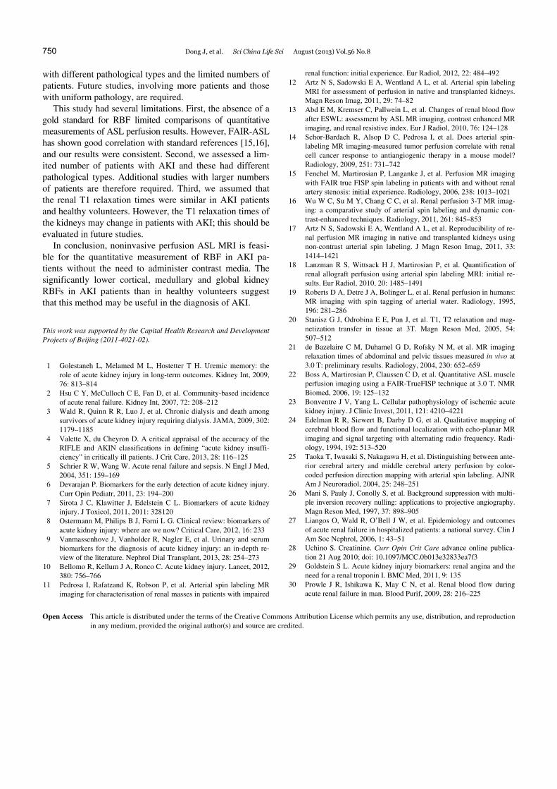
(MathWorks Inc. Natick, MA). After correction for motion artifacts, the final M images were generated by averaging and subtracting section-selective and global inversion im-ages acquired in two sets. The T1 values for the renal cortex and medulla were set at 1142 and 1545 ms, respectively [20, 21]. Pixels with perfusion values higher than 600 mL min1 100 g1 in the cortex and higher than 250 mL min1 100 g1 in the medulla were excluded from the RBF evaluation. Multiple regions of interest (ROIs) with areas of 50–100 mm2 in the perfusion map, were drawn in the cortex and medulla of both kidneys (Figure 1) [16], and RBFs in the cortex and medulla were separately calculated by averaging all ROI data in these areas. The measurements were per-formed on both ASL images for each subject, and the aver-aged value was used as the final RBF.
1.4 Statistical analysis
All statistical analyses were performed using SPSS 16.0 (SPSS Inc., Chicago, IL). Quantitative data are presented as mean±standard deviation (SD). The RBFs of the cortical and medullary kidney were compared using independent t tests. The correlations between serum creatinine concentra-tions and the RBFs of cortical and medullary kidney were determined. A P-value less than 0.05 was defined as statis-tically significant.
2 Results
MR examinations were performed in 13 patients (eight males and five females, ranging in age from 31 to 67 years) and 18 healthy volunteers (11 males, seven females, ranging in age from 32 to 66 years). Although all received respira-tory coaching, two patients were unable to hold their breath and were excluded for breathholding failure, which resulted in unsolved motion artifacts even using the nonlinear image registration method. Finally, 11 patients (seven males and four females, ranging in age from 31 to 67 years) and the 18 healthy volunteers were included. There were no statistical-ly significant between-group differences in age and sex dis-tribution. Subsequent percutaneous renal biopsy in these 11 patients showed that six had acute tubular interstitial nephritis, two had membranous nephropathy, two had IgA nephropathy, and one had granulomatous interstitial nephritis.
RBFs were measured in the cortex and medulla of each kidney in both groups, with each group showing similar RBFs in the cortex and medulla of the left and right kidneys (Table 1).
Perfusion values in the control group ranged from 357 to 426 mL min1 100 g1 in the cortex and from 107 to 126 mL min1 100 g1 in the medulla, similar to previous findings [15,17,22]. RBFs in patients with AKI ranged from 223 to
Figure 1 Regions of interests (ROIs) in the kidneys. Multiple ROIs, 50–100 mm2 in area, were drawn in the renal cortex (A) and medulla (B), with aver-aged values calculated as the cortical and medullary RBFs, respectively.
Table 1 RBFs (mL min1 100 g1) of the cortex and medulla in the left and right kidneys of the control and AKI groups
Left Right P-value Control Cortex 396.7±17.6 400.9±21.3 0.78 Medulla 121.3±18.7 115.9±20.2 0.61 Study Cortex 285.8±44.1 294.9±51.2 0.43 Medulla 110.7±18.2 101.6±15.5 0.41
Table 2 RBFs (mL min1 100 g1) of the cortex and medulla in the con-trol and AKI groups
Control Study P-value
Cortex 398.8±19.7 291.8±47.1 <0.01
Medulla 118.0±19.8 105.7±17.0 <0.01
392 mL min1 100 g1 in the cortex and from 89 to 116 mL min1 100 g1 in the medulla, with both being significantly lower than in the healthy volunteers (Table 2). Functional perfusion maps of ASL showed differences between the study and control groups (Figure 2).
Serum creatinine concentrations ranged from 52 to 111 mol L1 in the control group and from 65.8 to 752 mol L1 in the AKI group. In the control group, RBF in the cor-tex showed a significant positive correlation with serum creatinine concentration (r=0.71, P=0.001), with no sig-nificant correlation in the medulla (r=0.39, P=0.24). In the AKI group, however, RBF in the cortex (r=0.31, P=0.28) and medulla (r=0.27, P=0.48) was not correlated with se-rum creatinine concentration (Figure 3).

748 Dong J, et al. Sci China Life Sci August (2013) Vol.56 No.8
Figure 2 Oblique coronal image of ASL and perfusion map of subjects in the (A and B) and AKI (C and D) groups. A and B, Results in a 32-year-old male volunteer, showing a clear corticomedullary edge of the kidney (A), and normal renal blood flow (RBF) (cortex, 401.6 mL min1 100 g1; medulla, 123.0 mL min1 100 g1) (B). C and D, Results in a 48-year-old man diagnosed with tubular interstitial nephrology, showing an obscure boundary between the cortex and medulla of the kidney (C), with lower RBFs in the cortex (272.2 mL min1 100 g1) and medulla (97.6 mL min1 100 g1) (D).
3 Discussion
To our knowledge, this is the first study to assess RBF using ASL MRI in patients with AKI. The results suggest that this noninvasive method, using FAIR-SSFSE sequences, yields reliable quantitative measurements of RBF in both AKI patients and healthy volunteers without administration of contrast media. Reduction in RBF in both the cortex and medulla can help diagnose AKI, especially for patients with impaired renal function.
ASL imaging, a noninvasive functional MRI technique, has been widely used to assess perfusion changes in patients with cerebral artery diseases, and has yielded promising results [23–25]. Application of ASL to the kidneys, however, is limited by respiration and magnetic susceptibility artifacts in abdominal organs. Using the nonlinear image registration method, ASL images can be acquired with acceptable qual-ity for calculating perfusion maps of RBF. In addition, the background suppression technique was used to zero static tissue to enhance the low signal-to-noise ratio in perfusion maps, thus increasing perfusion signal [26]. ASL imaging with FAIR true FISP has been found suitable for quantita-tive assessment of RBF in patients with renal artery stenosis
[15]. Moreover, ASL imaging has been used in a mouse renal cell carcinoma model to characterize early changes in response to antiangiogenic therapy, with these changes pre-dicting later therapeutic responses [14]. We found that, in healthy volunteers, mean RBF was (398.8±19.7) mL min1 100 g1 in the cortex and (118±19.8) mL min1 100 g1 in the medulla, with no significant differences between the left and right kidneys. These results are consistent with studies showing the reliability and reproducibility of the ASL tech-nique.
AKI refers to a clinical syndrome characterized by rapid decreases in renal function within a few hours to a few days, with accumulation of products of nitrogen metabolism such as creatinine and urea [10]. Causes of AKI vary from re-duced kidney perfusion and prerenal azotemia to direct re-nal toxicity and postrenal obstruction, with AKI accounting for 1.9% of hospital inpatients [27]. Although increased serum creatinine and urea concentrations are the standard diagnostic laboratory criteria for AKI, they are relatively insensitive, becoming abnormal only when glomerular fil-tration rate decreases by more than 50%. Furthermore, they do not show dynamic changes in glomerular filtration rates [28]. Although several novel biomarkers were recently re-

Dong J, et al. Sci China Life Sci August (2013) Vol.56 No.8 749
Figure 3 Correlation between serum creatinine concentration and cortical and medullary renal blood flow (RBF) in the control (A and B) and AKI (C and D) groups. A, Cortical RBF was positively correlated with serum creatinine concentration in the control group (r=0.71, P=0.001). B–D, There were no correlations between serum creatinine concentration and medullary RBF in the control group (B) and between serum creatinine and cortical (C) and medul-lary (D) RBFs in the AKI group.
ported to diagnose AKI earlier and to reflect dynamic alter-ations to therapy, their clinical usefulness requires addition-al confirmation [6,7,29]. The dominant mechanism in the pathogenesis of AKI is a significant reduction in RBF [30].
Although multiple modalities have been used to measure RBF, including nuclear medicine and contrast enhanced CT, impaired renal function restricts their application.
The most important advantages of ASL in quantifying RBF are that it is completely non-invasive method and does not require administration of contrast media. This prevents any potential risk of contrast medium induced nephrology, which is of extreme importance for patients with impaired renal function. ASL MRI has been used in patients with impaired renal allograft function, and its reproducibility and reliability have been confirmed [11,17]. To assess the sta-bility of ASL, each sequence was scanned twice in each participant, with RBF determined by the average of the two perfusion values. We found that the renal perfusion exami-nation based on our FAIR SSFSE sequence was robust, with the main perfusion signal recorded by this sequence coming
from low velocity spins in capillary blood in the steady state, which stayed in the imaging slice for a relatively long period of time. RBF was significantly lower in AKI patients than in healthy volunteers, in accordance with the patho-physiological changes that occur during AKI [5,30]. There-fore, this noninvasive imaging modality is potentially capa-ble of diagnosing AKI in patients with impaired renal func-tion.
To date, AKI has been diagnosed from increases in se-rum creatinine concentration [10,28]. This relatively insen-sitive marker of glomerular filtration rate was found to be affected by many factors, including muscle mass, age, sex, muscle injury, use of steroids and aggressive fluid resuscita-tion [10]. To avoid these factors, all subjects were evaluated after a 6-h fast; moreover, their demographic and clinical characteristics were similar. The only positive correlation we observed between serum creatinine and RBF was in the cortex of the control group. The absence of a correlation between medullary RBF and serum creatinine in patients with AKI may have resulted from our inclusion of patients
750 Dong J, et al. Sci China Life Sci August (2013) Vol.56 No.8
with different pathological types and the limited numbers of patients. Future studies, involving more patients and those with uniform pathology, are required.
This study had several limitations. First, the absence of a gold standard for RBF limited comparisons of quantitative measurements of ASL perfusion results. However, FAIR-ASL has shown good correlation with standard references [15,16], and our results were consistent. Second, we assessed a lim-ited number of patients with AKI and these had different pathological types. Additional studies with larger numbers of patients are therefore required. Third, we assumed that the renal T1 relaxation times were similar in AKI patients and healthy volunteers. However, the T1 relaxation times of the kidneys may change in patients with AKI; this should be evaluated in future studies.
In conclusion, noninvasive perfusion ASL MRI is feasi-ble for the quantitative measurement of RBF in AKI pa-tients without the need to administer contrast media. The significantly lower cortical, medullary and global kidney RBFs in AKI patients than in healthy volunteers suggest that this method may be useful in the diagnosis of AKI.
This work was supported by the Capital Health Research and Development Projects of Beijing (2011-4021-02).
1 Golestaneh L, Melamed M L, Hostetter T H. Uremic memory: the role of acute kidney injury in long-term outcomes. Kidney Int, 2009, 76: 813–814
2 Hsu C Y, McCulloch C E, Fan D, et al. Community-based incidence of acute renal failure. Kidney Int, 2007, 72: 208–212
3 Wald R, Quinn R R, Luo J, et al. Chronic dialysis and death among survivors of acute kidney injury requiring dialysis. JAMA, 2009, 302: 1179–1185
4 Valette X, du Cheyron D. A critical appraisal of the accuracy of the RIFLE and AKIN classifications in defining “acute kidney insuffi-ciency” in critically ill patients. J Crit Care, 2013, 28: 116–125
5 Schrier R W, Wang W. Acute renal failure and sepsis. N Engl J Med, 2004, 351: 159–169
6 Devarajan P. Biomarkers for the early detection of acute kidney injury. Curr Opin Pediatr, 2011, 23: 194–200
7 Sirota J C, Klawitter J, Edelstein C L. Biomarkers of acute kidney injury. J Toxicol, 2011, 2011: 328120
8 Ostermann M, Philips B J, Forni L G. Clinical review: biomarkers of acute kidney injury: where are we now? Critical Care, 2012, 16: 233
9 Vanmassenhove J, Vanholder R, Nagler E, et al. Urinary and serum biomarkers for the diagnosis of acute kidney injury: an in-depth re-view of the literature. Nephrol Dial Transplant, 2013, 28: 254–273
10 Bellomo R, Kellum J A, Ronco C. Acute kidney injury. Lancet, 2012, 380: 756–766
11 Pedrosa I, Rafatzand K, Robson P, et al. Arterial spin labeling MR imaging for characterisation of renal masses in patients with impaired
renal function: initial experience. Eur Radiol, 2012, 22: 484–492 12 Artz N S, Sadowski E A, Wentland A L, et al. Arterial spin labeling
MRI for assessment of perfusion in native and transplanted kidneys. Magn Reson Imag, 2011, 29: 74–82
13 Abd E M, Kremser C, Pallwein L, et al. Changes of renal blood flow after ESWL: assessment by ASL MR imaging, contrast enhanced MR imaging, and renal resistive index. Eur J Radiol, 2010, 76: 124–128
14 Schor-Bardach R, Alsop D C, Pedrosa I, et al. Does arterial spin- labeling MR imaging-measured tumor perfusion correlate with renal cell cancer response to antiangiogenic therapy in a mouse model? Radiology, 2009, 251: 731–742
15 Fenchel M, Martirosian P, Langanke J, et al. Perfusion MR imaging with FAIR true FISP spin labeling in patients with and without renal artery stenosis: initial experience. Radiology, 2006, 238: 1013–1021
16 Wu W C, Su M Y, Chang C C, et al. Renal perfusion 3-T MR imag-ing: a comparative study of arterial spin labeling and dynamic con-trast-enhanced techniques. Radiology, 2011, 261: 845–853
17 Artz N S, Sadowski E A, Wentland A L, et al. Reproducibility of re-nal perfusion MR imaging in native and transplanted kidneys using non-contrast arterial spin labeling. J Magn Reson Imag, 2011, 33: 1414–1421
18 Lanzman R S, Wittsack H J, Martirosian P, et al. Quantification of renal allograft perfusion using arterial spin labeling MRI: initial re-sults. Eur Radiol, 2010, 20: 1485–1491
19 Roberts D A, Detre J A, Bolinger L, et al. Renal perfusion in humans: MR imaging with spin tagging of arterial water. Radiology, 1995, 196: 281–286
20 Stanisz G J, Odrobina E E, Pun J, et al. T1, T2 relaxation and mag-netization transfer in tissue at 3T. Magn Reson Med, 2005, 54: 507–512
21 de Bazelaire C M, Duhamel G D, Rofsky N M, et al. MR imaging relaxation times of abdominal and pelvic tissues measured in vivo at 3.0 T: preliminary results. Radiology, 2004, 230: 652–659
22 Boss A, Martirosian P, Claussen C D, et al. Quantitative ASL muscle perfusion imaging using a FAIR-TrueFISP technique at 3.0 T. NMR Biomed, 2006, 19: 125–132
23 Bonventre J V, Yang L. Cellular pathophysiology of ischemic acute kidney injury. J Clinic Invest, 2011, 121: 4210–4221
24 Edelman R R, Siewert B, Darby D G, et al. Qualitative mapping of cerebral blood flow and functional localization with echo-planar MR imaging and signal targeting with alternating radio frequency. Radi-ology, 1994, 192: 513–520
25 Taoka T, Iwasaki S, Nakagawa H, et al. Distinguishing between ante-rior cerebral artery and middle cerebral artery perfusion by color- coded perfusion direction mapping with arterial spin labeling. AJNR Am J Neuroradiol, 2004, 25: 248–251
26 Mani S, Pauly J, Conolly S, et al. Background suppression with multi-ple inversion recovery nulling: applications to projective angiography. Magn Reson Med, 1997, 37: 898–905
27 Liangos O, Wald R, O’Bell J W, et al. Epidemiology and outcomes of acute renal failure in hospitalized patients: a national survey. Clin J Am Soc Nephrol, 2006, 1: 43–51
28 Uchino S. Creatinine. Curr Opin Crit Care advance online publica-tion 21 Aug 2010; doi: 10.1097/MCC.0b013e32833ea7f3
29 Goldstein S L. Acute kidney injury biomarkers: renal angina and the need for a renal troponin I. BMC Med, 2011, 9: 135
30 Prowle J R, Ishikawa K, May C N, et al. Renal blood flow during acute renal failure in man. Blood Purif, 2009, 28: 216–225
Open Access This article is distributed under the terms of the Creative Commons Attribution License which permits any use, distribution, and reproduction
in any medium, provided the original author(s) and source are credited.