science china life sciences - springer · science china life sciences ... 1state key laboratory of...
TRANSCRIPT
SCIENCE CHINA Life Sciences
© The Author(s) 2016. This article is published with open access at link.springer.com life.scichina.com link.springer.com
*Corresponding author (email: [email protected]) **Corresponding author (email: [email protected])
• RESEARCH PAPER • September 2016 Vol.59 No.9: 940–949
doi: 10.1007/s11427-015-0277-8
Baicalein protects against the development of angiotensin II-induced abdominal aortic aneurysms by blocking JNK and p38
MAPK signaling
Fang Wang1, Houzao Chen1, Yunfei Yan1, Yue Liu1, Shuyang Zhang2*& Depei Liu1**
1State Key Laboratory of Medical Molecular Biology, Institute of Basic Medical Sciences, Chinese Academy of Medical Sciences & Peking Union Medical College, Beijing 100005, China;
2Department of Cardiology, Peking Union Medical College Hospital, Chinese Academy of Medical Sciences & Peking Union Medical College, Beijing 100005, China
Received April 7, 2016; accepted May 8, 2016; published online June 21, 2016
An abdominal aortic aneurysm (AAA) is a permanent, localized dilatation of the abdominal aorta. In western countries, the morbidity of AAA is approximately 8%. Currently, pharmacotherapies for AAA are limited. Here, we demonstrate that bai-calein (BAI), the main component of the Chinese traditional drug “Huang Qin”, attenuates the incidence and severity of AAA in Apoe/ mice infused with angiotensin II (AngII). Mechanically, BAI treatment decreases AngII-induced reactive oxygen species (ROS) production in the aortic wall. Moreover, BAI inhibits inflammatory cell accumulation in the aortas of mice in-fused with AngII. It also inhibits AngII-induced activation of matrix metalloproteinase 2 (MMP-2) and MMP-9 to maintain elastin content in vivo. In addition, it blocks AngII cascade by downregulating angiotensin type 1 receptor (AT1R) and inhibit-ing mitogen-activated protein kinases (MAPKs). Taken together, our findings show that BAI is an effective agent for AAA prevention.
baicalein, abdominal aortic aneurysm, oxidative stress, vascular inflammation, extracellular matrix degradation, AT1R, MAPKs
Citation: Wang, F., Chen, H., Yan, Y., Liu, Y., Zhang, S., and Liu, D. (2016). Baicalein protects against the development of angiotensin II-induced abdominal aortic aneurysms by blocking JNK and p38 MAPK signaling. Sci China Life Sci 59, 940–949. doi: 10.1007/s11427-015-0277-8
INTRODUCTION
An abdominal aortic aneurysm (AAA) is defined as a per-manent, localized dilatation of the abdominal aorta to 1.5-fold more than the normal diameter. In western coun-tries, the morbidity of AAA is approximately 8%. Owing to the lack of typical syndrome, the mortality rate associated with aortic rupture exceeds 80% (Nordon et al., 2011; Sakalihasan et al., 2005). Currently, AAA therapies mainly include pharmacotherapy and surgical approaches (open repair and endovascular aneurysm repair) (Brewster et al.,
2003). The chief function of pharmacotherapy, which con-trols the risk factors, is to indirectly attenuate aneurysm development and aorta rupture by regulating blood pressure, serum lipid level, and blood coagulation (Bergoeing et al., 2006; Miyake and Morishita, 2009). Therefore, developing new drugs that target the mechanism of AAA development is of great significance to AAA prevention and treatment.
The involved pathophysiological processes mainly in-clude oxidative stress, vascular inflammation, and extracel-lular matrix degradation (Lopez-Candales et al., 1997; Thompson et al., 1997, 2002). Apoe/ mice infused with angiotensin II (AngII) is the most commonly employed animal model to study the pathogenesis of AAA since it

Wang, F., et al. Sci China Life Sci September (2016) Vol.59 No.9 941
mimics almost all the clinical characteristics of human AAA (Daugherty and Cassis, 2004; Weiss et al., 2001). Notably, AngII cascade is upregulated in human AAA samples (Annambhotla et al., 2008; Golledge and Powell, 2007). Baicalein (BAI) , which is involved in oxidative stress, in-flammation, tumorigenesis, and angiogenesisis, is the main component of the Chinese traditional drug “Huang Qin”, (Gao et al., 1999). It has been reported that BAI remarkably retards is chemia/reperfusion-induced myocardium injury (Liu et al., 2013; Wang et al., 2013). However, its exact mechanism remains elusive and its role in AAA develop-ment has not been elucidated.
In the present study, we examined the role of BAI in AAA induced by AngII treatment in Apoe/ mice. From the view of oxidative stress, vascular inflammation, and extra-cellular matrix degradation, the relevant mechanisms were studied. The AngII cascade activation was also evaluated under BAI treatment. We believe that our findings may provide a new drug for AAA prevention and treatment.
RESULTS
BAI decreases AAA incidence in vivo
BAI did not alter the gross morphology of the aortas in Apoe/ mice (Figure 1A). Consistent with previous findings (Satoh et al., 2009; Wang et al., 2012), the incidence of
AAA in Apoe/ mice treated with vehicle after AngII infu-sion for 4 weeks was 88% in our study. In contrast, only 47% of BAI-treated Apoe/ mice developed AAA after AngII treatment (Figure 1B). No AAAs were detected in saline-infused mice.
BAI attenuated AAA severity (the ratio of aorta weight to body weight and maximal abdominal aortic diameter) in vivo
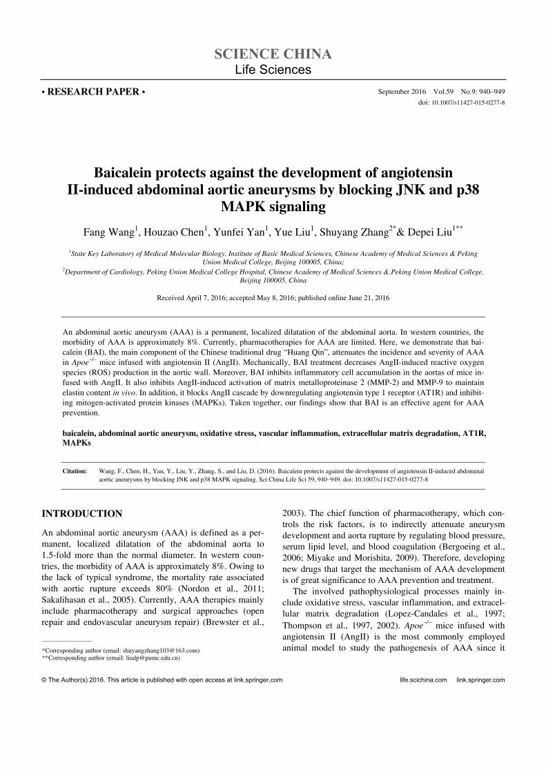
The maximal abdominal aortic diameter and the total aortic weight significantly increased after AngII infusion for 4 weeks in all groups compared to that in saline-infused mice. Compared to AngII-infused mice treated with vehicle, the BAI-treated mice had decreased maximal abdominal aortic diameter and total aortic weight (Figure 2A–C). As shown in Tables 1 and 2, SBP, HR, and serum lipid levels did not differ between Apoe/ mice treated with BAI and those receiving vehicle after AngII infusion for 4 weeks.
Taken together, BAI represses AngII-induced AAA in vivo, but it did not affect hemodynamic and lipid metabolic indices.
BAI treatment attenuated AngII-induced ROS production in vivo
Since oxidative stress is critical in AAA formation (Satoh et al., 2009; Thomas et al., 2006), we first determined the oxi-
Figure 1 (color online) Baicalein (BAI) treatment represses abdominal aortic aneurysms (AAA) formation in Apoe/ mice infused with angiotensin II (AngII). Mice were pre-treated with vehicle (Veh) or BAI (30 mg kg1 day1) via intraperitoneal injection for 1 week before infusion with saline or AngII for 4 weeks. Treatment with vehicle or BAI (30 mg kg1 day1) via intraperitoneal injection was performed at the same time-point every day during the saline or AngII infusion. A, Representative photographs showing macroscopic features of aneurysms induced by AngII following treatment with vehicle or BAI. The arrows show typical AAAs (scale bars, 5 mm). B, The incidence of AngII-induced AAA in vehicle-treated Apoe/ mice (n=17) relative to BAI-treated Ap-oe/ mice (n=17). AAAs were not detected in saline-infused mice (n=6 per group). *, P<0.05.
Table 1 Blood pressure and heart rate in micea)
Groups SBP
(Baseline, mmHg) SBP
(AngII, mmHg) HR
(Baseline, beats min1) HR
(AngII, beats min1) Veh Apoe/ 98.6±5.8 129.3±3.8* 553.8±13.6 524.6±16.0
BAI Apoe/ 97.4±6.4 126.6±5.7* 538.3±18.6 535.7±22.4
a) n=5 in baseline group; n=10 in AngII group; *, P<0.05 compared to baseline; data are mean±SE.
942 Wang, F., et al. Sci China Life Sci September (2016) Vol.59 No.9
Table 2 Serum lipid levels in AngII-infused micea)
Groups (AngII-infused) Cholesterol (mmol L1) Triglyceride (mmol L1) HDL-C (mmol L1) LDL-C (mmol L1)
Veh Apoe/ 16.36±1.40 2.17±0.15 0.85±0.09 2.61±0.26
BAI Apoe/ 15.73±0.98 2.43±0.41 0.91±0.04 2.71±0.27
a) Results are expressed as mean±SE, n=10 in each group.
Figure 2 Baicalein (BAI) treatment attenuates abdominal aorta dilatation in Apoe/ mice infused with angiotensin II (AngII). A and C, The ratio of the aorta weight to body weight (A) and maximal abdominal aortic diameter (C) in saline- and AngII-infused mice following treatment with vehicle or BAI (n=5 in each group of Apoe/ mice infused with saline; n=14 for AngII-infused Apoe/ mice following treatment with vehicle; n=15 for AngII-infused Apoe/ mice following treatment with BAI). B, Representative H&E staining of suprarenal aortas from saline- and AngII-infused mice treated with vehicle or BAI. Scale bars, 100 μm. Error bars in A and C represent the mean±SE. *, P<0.05; **, P<0.01.
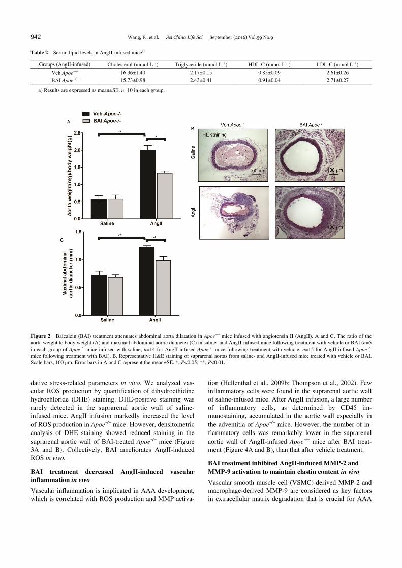
dative stress-related parameters in vivo. We analyzed vas-cular ROS production by quantification of dihydroethidine hydrochloride (DHE) staining. DHE-positive staining was rarely detected in the suprarenal aortic wall of saline- infused mice. AngII infusion markedly increased the level of ROS production in Apoe/ mice. However, densitometric analysis of DHE staining showed reduced staining in the suprarenal aortic wall of BAI-treated Apoe/ mice (Figure 3A and B). Collectively, BAI ameliorates AngII-induced ROS in vivo.
BAI treatment decreased AngII-induced vascular inflammation in vivo
Vascular inflammation is implicated in AAA development, which is correlated with ROS production and MMP activa-
tion (Hellenthal et al., 2009b; Thompson et al., 2002). Few inflammatory cells were found in the suprarenal aortic wall of saline-infused mice. After AngII infusion, a large number of inflammatory cells, as determined by CD45 im-munostaining, accumulated in the aortic wall especially in the adventitia of Apoe/ mice. However, the number of in-flammatory cells was remarkably lower in the suprarenal aortic wall of AngII-infused Apoe/ mice after BAI treat-ment (Figure 4A and B), than that after vehicle treatment.
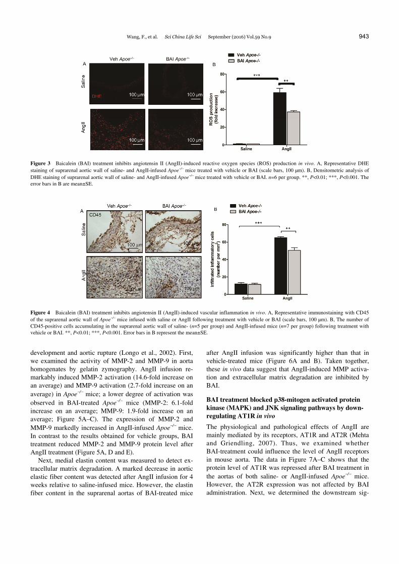
BAI treatment inhibited AngII-induced MMP-2 and MMP-9 activation to maintain elastin content in vivo
Vascular smooth muscle cell (VSMC)-derived MMP-2 and macrophage-derived MMP-9 are considered as key factors in extracellular matrix degradation that is crucial for AAA
Wang, F., et al. Sci China Life Sci September (2016) Vol.59 No.9 943
Figure 3 Baicalein (BAI) treatment inhibits angiotensin II (AngII)-induced reactive oxygen species (ROS) production in vivo. A, Representative DHE staining of suprarenal aortic wall of saline- and AngII-infused Apoe/ mice treated with vehicle or BAI (scale bars, 100 µm). B, Densitometric analysis of DHE staining of suprarenal aortic wall of saline- and AngII-infused Apoe/ mice treated with vehicle or BAI. n=6 per group. **, P<0.01; ***, P<0.001. The error bars in B are mean±SE.
Figure 4 Baicalein (BAI) treatment inhibits angiotensin II (AngII)-induced vascular inflammation in vivo. A, Representative immunostaining with CD45 of the suprarenal aortic wall of Apoe/ mice infused with saline or AngII following treatment with vehicle or BAI (scale bars, 100 µm). B, The number of CD45-positive cells accumulating in the suprarenal aortic wall of saline- (n=5 per group) and AngII-infused mice (n=7 per group) following treatment with vehicle or BAI. **, P<0.01; ***, P<0.001. Error bars in B represent the mean±SE.
development and aortic rupture (Longo et al., 2002). First, we examined the activity of MMP-2 and MMP-9 in aorta homogenates by gelatin zymography. AngII infusion re-markably induced MMP-2 activation (14.6-fold increase on an average) and MMP-9 activation (2.7-fold increase on an average) in Apoe/ mice; a lower degree of activation was observed in BAI-treated Apoe/ mice (MMP-2: 6.1-fold increase on an average; MMP-9: 1.9-fold increase on an average; Figure 5A–C). The expression of MMP-2 and MMP-9 markedly increased in AngII-infused Apoe/ mice. In contrast to the results obtained for vehicle groups, BAI treatment reduced MMP-2 and MMP-9 protein level after AngII treatment (Figure 5A, D and E).
Next, medial elastin content was measured to detect ex-tracellular matrix degradation. A marked decrease in aortic elastic fiber content was detected after AngII infusion for 4 weeks relative to saline-infused mice. However, the elastin fiber content in the suprarenal aortas of BAI-treated mice
after AngII infusion was significantly higher than that in vehicle-treated mice (Figure 6A and B). Taken together, these in vivo data suggest that AngII-induced MMP activa-tion and extracellular matrix degradation are inhibited by BAI.
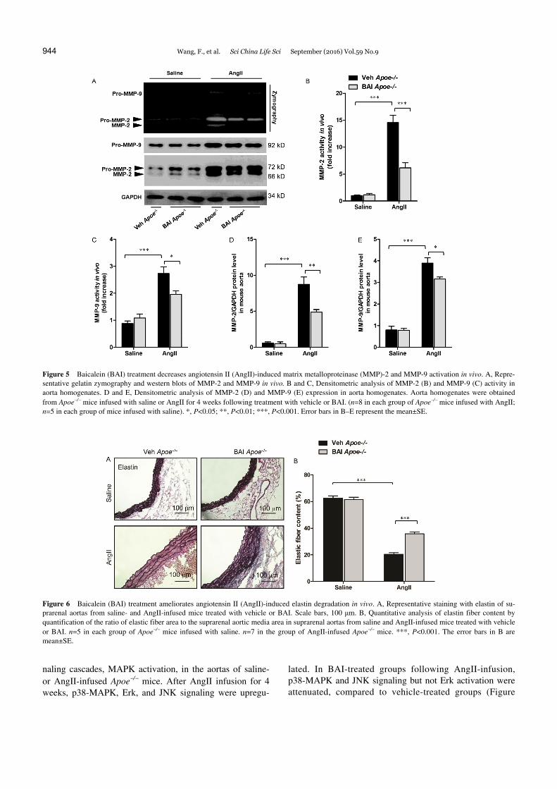
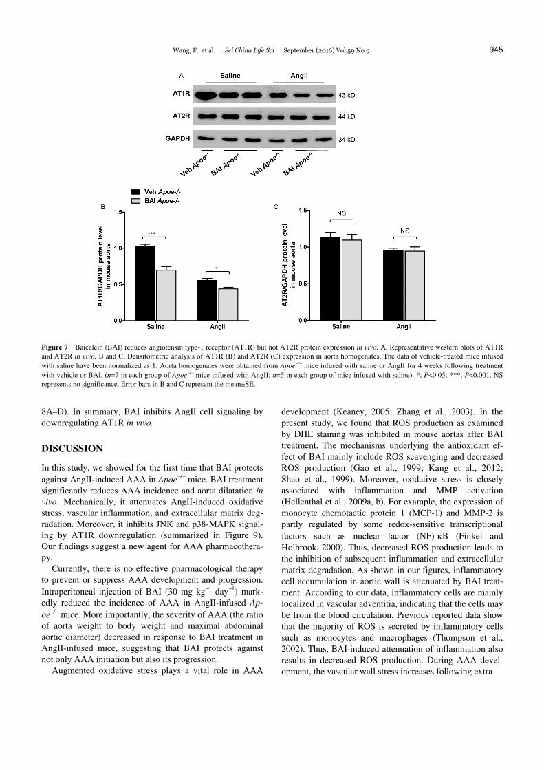
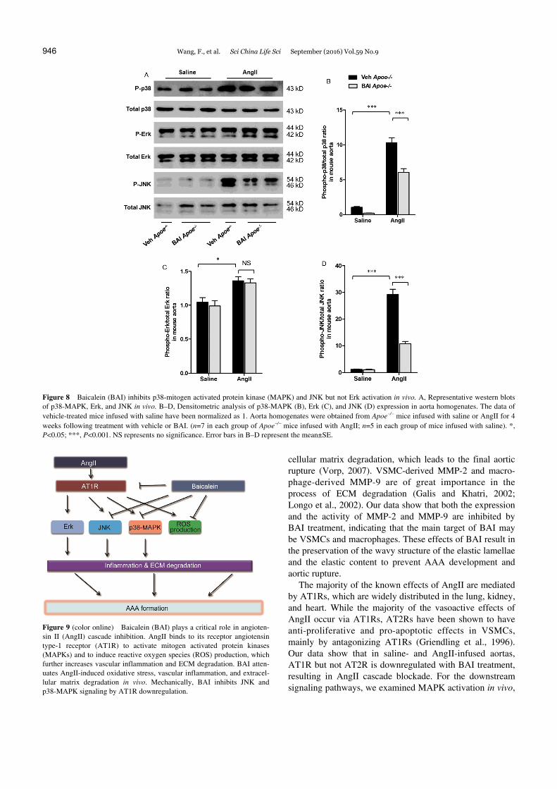
BAI treatment blocked p38-mitogen activated protein kinase (MAPK) and JNK signaling pathways by down-regulating AT1R in vivo
The physiological and pathological effects of AngII are mainly mediated by its receptors, AT1R and AT2R (Mehta and Griendling, 2007). Thus, we examined whether BAI-treatment could influence the level of AngII receptors in mouse aorta. The data in Figure 7A–C shows that the protein level of AT1R was repressed after BAI treatment in the aortas of both saline- or AngII-infused Apoe/ mice. However, the AT2R expression was not affected by BAI administration. Next, we determined the downstream sig-
944 Wang, F., et al. Sci China Life Sci September (2016) Vol.59 No.9
Figure 5 Baicalein (BAI) treatment decreases angiotensin II (AngII)-induced matrix metalloproteinase (MMP)-2 and MMP-9 activation in vivo. A, Repre-sentative gelatin zymography and western blots of MMP-2 and MMP-9 in vivo. B and C, Densitometric analysis of MMP-2 (B) and MMP-9 (C) activity in aorta homogenates. D and E, Densitometric analysis of MMP-2 (D) and MMP-9 (E) expression in aorta homogenates. Aorta homogenates were obtained from Apoe/ mice infused with saline or AngII for 4 weeks following treatment with vehicle or BAI. (n=8 in each group of Apoe/ mice infused with AngII; n=5 in each group of mice infused with saline). *, P<0.05; **, P<0.01; ***, P<0.001. Error bars in B–E represent the mean±SE.
Figure 6 Baicalein (BAI) treatment ameliorates angiotensin II (AngII)-induced elastin degradation in vivo. A, Representative staining with elastin of su-prarenal aortas from saline- and AngII-infused mice treated with vehicle or BAI. Scale bars, 100 μm. B, Quantitative analysis of elastin fiber content by quantification of the ratio of elastic fiber area to the suprarenal aortic media area in suprarenal aortas from saline and AngII-infused mice treated with vehicle or BAI. n=5 in each group of Apoe/ mice infused with saline. n=7 in the group of AngII-infused Apoe/ mice. ***, P<0.001. The error bars in B are mean±SE.
naling cascades, MAPK activation, in the aortas of saline- or AngII-infused Apoe/ mice. After AngII infusion for 4 weeks, p38-MAPK, Erk, and JNK signaling were upregu-
lated. In BAI-treated groups following AngII-infusion, p38-MAPK and JNK signaling but not Erk activation were attenuated, compared to vehicle-treated groups (Figure
Wang, F., et al. Sci China Life Sci September (2016) Vol.59 No.9 945
Figure 7 Baicalein (BAI) reduces angiotensin type-1 receptor (AT1R) but not AT2R protein expression in vivo. A, Representative western blots of AT1R and AT2R in vivo. B and C, Densitometric analysis of AT1R (B) and AT2R (C) expression in aorta homogenates. The data of vehicle-treated mice infused with saline have been normalized as 1. Aorta homogenates were obtained from Apoe/ mice infused with saline or AngII for 4 weeks following treatment with vehicle or BAI. (n=7 in each group of Apoe/ mice infused with AngII; n=5 in each group of mice infused with saline). *, P<0.05; ***, P<0.001. NS represents no significance. Error bars in B and C represent the mean±SE.
8A–D). In summary, BAI inhibits AngII cell signaling by downregulating AT1R in vivo.
DISCUSSION
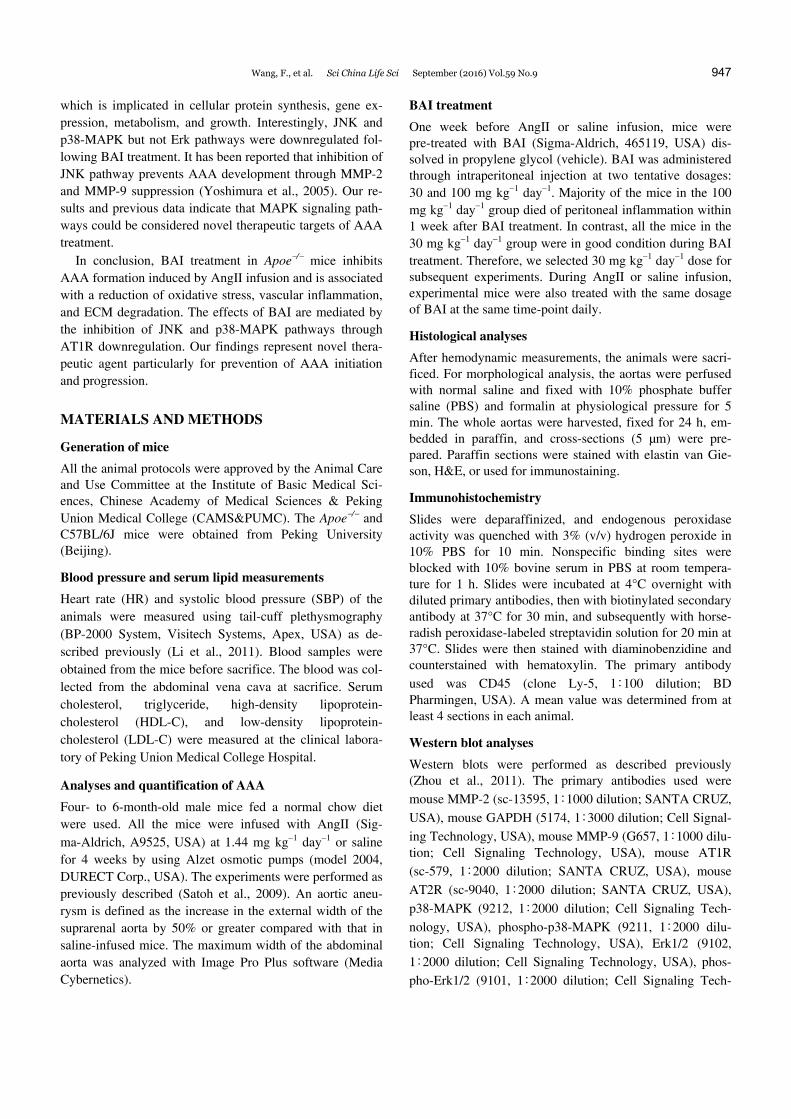
In this study, we showed for the first time that BAI protects against AngII-induced AAA in Apoe/ mice. BAI treatment significantly reduces AAA incidence and aorta dilatation in vivo. Mechanically, it attenuates AngII-induced oxidative stress, vascular inflammation, and extracellular matrix deg-radation. Moreover, it inhibits JNK and p38-MAPK signal-ing by AT1R downregulation (summarized in Figure 9). Our findings suggest a new agent for AAA pharmacothera-py.
Currently, there is no effective pharmacological therapy to prevent or suppress AAA development and progression. Intraperitoneal injection of BAI (30 mg kg1 day1) mark-edly reduced the incidence of AAA in AngII-infused Ap-oe/ mice. More importantly, the severity of AAA (the ratio of aorta weight to body weight and maximal abdominal aortic diameter) decreased in response to BAI treatment in AngII-infused mice, suggesting that BAI protects against not only AAA initiation but also its progression.
Augmented oxidative stress plays a vital role in AAA
development (Keaney, 2005; Zhang et al., 2003). In the present study, we found that ROS production as examined by DHE staining was inhibited in mouse aortas after BAI treatment. The mechanisms underlying the antioxidant ef-fect of BAI mainly include ROS scavenging and decreased ROS production (Gao et al., 1999; Kang et al., 2012; Shao et al., 1999). Moreover, oxidative stress is closely associated with inflammation and MMP activation (Hellenthal et al., 2009a, b). For example, the expression of monocyte chemotactic protein 1 (MCP-1) and MMP-2 is partly regulated by some redox-sensitive transcriptional factors such as nuclear factor (NF)-B (Finkel and Holbrook, 2000). Thus, decreased ROS production leads to the inhibition of subsequent inflammation and extracellular matrix degradation. As shown in our figures, inflammatory cell accumulation in aortic wall is attenuated by BAI treat-ment. According to our data, inflammatory cells are mainly localized in vascular adventitia, indicating that the cells may be from the blood circulation. Previous reported data show that the majority of ROS is secreted by inflammatory cells such as monocytes and macrophages (Thompson et al., 2002). Thus, BAI-induced attenuation of inflammation also results in decreased ROS production. During AAA devel-opment, the vascular wall stress increases following extra
946 Wang, F., et al. Sci China Life Sci September (2016) Vol.59 No.9
Figure 8 Baicalein (BAI) inhibits p38-mitogen activated protein kinase (MAPK) and JNK but not Erk activation in vivo. A, Representative western blots of p38-MAPK, Erk, and JNK in vivo. B–D, Densitometric analysis of p38-MAPK (B), Erk (C), and JNK (D) expression in aorta homogenates. The data of vehicle-treated mice infused with saline have been normalized as 1. Aorta homogenates were obtained from Apoe/ mice infused with saline or AngII for 4 weeks following treatment with vehicle or BAI. (n=7 in each group of Apoe/ mice infused with AngII; n=5 in each group of mice infused with saline). *, P<0.05; ***, P<0.001. NS represents no significance. Error bars in B–D represent the mean±SE.
Figure 9 (color online) Baicalein (BAI) plays a critical role in angioten-sin II (AngII) cascade inhibition. AngII binds to its receptor angiotensin type-1 receptor (AT1R) to activate mitogen activated protein kinases (MAPKs) and to induce reactive oxygen species (ROS) production, which further increases vascular inflammation and ECM degradation. BAI atten-uates AngII-induced oxidative stress, vascular inflammation, and extracel-lular matrix degradation in vivo. Mechanically, BAI inhibits JNK and p38-MAPK signaling by AT1R downregulation.
cellular matrix degradation, which leads to the final aortic rupture (Vorp, 2007). VSMC-derived MMP-2 and macro-phage-derived MMP-9 are of great importance in the process of ECM degradation (Galis and Khatri, 2002; Longo et al., 2002). Our data show that both the expression and the activity of MMP-2 and MMP-9 are inhibited by BAI treatment, indicating that the main target of BAI may be VSMCs and macrophages. These effects of BAI result in the preservation of the wavy structure of the elastic lamellae and the elastic content to prevent AAA development and aortic rupture.
The majority of the known effects of AngII are mediated by AT1Rs, which are widely distributed in the lung, kidney, and heart. While the majority of the vasoactive effects of AngII occur via AT1Rs, AT2Rs have been shown to have anti-proliferative and pro-apoptotic effects in VSMCs, mainly by antagonizing AT1Rs (Griendling et al., 1996). Our data show that in saline- and AngII-infused aortas, AT1R but not AT2R is downregulated with BAI treatment, resulting in AngII cascade blockade. For the downstream signaling pathways, we examined MAPK activation in vivo,
Wang, F., et al. Sci China Life Sci September (2016) Vol.59 No.9 947
which is implicated in cellular protein synthesis, gene ex-pression, metabolism, and growth. Interestingly, JNK and p38-MAPK but not Erk pathways were downregulated fol-lowing BAI treatment. It has been reported that inhibition of JNK pathway prevents AAA development through MMP-2 and MMP-9 suppression (Yoshimura et al., 2005). Our re-sults and previous data indicate that MAPK signaling path-ways could be considered novel therapeutic targets of AAA treatment.
In conclusion, BAI treatment in Apoe/ mice inhibits AAA formation induced by AngII infusion and is associated with a reduction of oxidative stress, vascular inflammation, and ECM degradation. The effects of BAI are mediated by the inhibition of JNK and p38-MAPK pathways through AT1R downregulation. Our findings represent novel thera-peutic agent particularly for prevention of AAA initiation and progression.
MATERIALS AND METHODS
Generation of mice
All the animal protocols were approved by the Animal Care and Use Committee at the Institute of Basic Medical Sci-ences, Chinese Academy of Medical Sciences & Peking Union Medical College (CAMS&PUMC). The Apoe/ and C57BL/6J mice were obtained from Peking University (Beijing).
Blood pressure and serum lipid measurements
Heart rate (HR) and systolic blood pressure (SBP) of the animals were measured using tail-cuff plethysmography (BP-2000 System, Visitech Systems, Apex, USA) as de-scribed previously (Li et al., 2011). Blood samples were obtained from the mice before sacrifice. The blood was col-lected from the abdominal vena cava at sacrifice. Serum cholesterol, triglyceride, high-density lipoprotein- cholesterol (HDL-C), and low-density lipoprotein- cholesterol (LDL-C) were measured at the clinical labora-tory of Peking Union Medical College Hospital.
Analyses and quantification of AAA
Four- to 6-month-old male mice fed a normal chow diet were used. All the mice were infused with AngII (Sig-ma-Aldrich, A9525, USA) at 1.44 mg kg1 day1 or saline for 4 weeks by using Alzet osmotic pumps (model 2004, DURECT Corp., USA). The experiments were performed as previously described (Satoh et al., 2009). An aortic aneu-rysm is defined as the increase in the external width of the suprarenal aorta by 50% or greater compared with that in saline-infused mice. The maximum width of the abdominal aorta was analyzed with Image Pro Plus software (Media Cybernetics).
BAI treatment
One week before AngII or saline infusion, mice were pre-treated with BAI (Sigma-Aldrich, 465119, USA) dis-solved in propylene glycol (vehicle). BAI was administered through intraperitoneal injection at two tentative dosages: 30 and 100 mg kg1 day1. Majority of the mice in the 100 mg kg1 day1 group died of peritoneal inflammation within 1 week after BAI treatment. In contrast, all the mice in the 30 mg kg1 day1 group were in good condition during BAI treatment. Therefore, we selected 30 mg kg1 day1 dose for subsequent experiments. During AngII or saline infusion, experimental mice were also treated with the same dosage of BAI at the same time-point daily.
Histological analyses
After hemodynamic measurements, the animals were sacri-ficed. For morphological analysis, the aortas were perfused with normal saline and fixed with 10% phosphate buffer saline (PBS) and formalin at physiological pressure for 5 min. The whole aortas were harvested, fixed for 24 h, em-bedded in paraffin, and cross-sections (5 µm) were pre-pared. Paraffin sections were stained with elastin van Gie-son, H&E, or used for immunostaining.
Immunohistochemistry
Slides were deparaffinized, and endogenous peroxidase activity was quenched with 3% (v/v) hydrogen peroxide in 10% PBS for 10 min. Nonspecific binding sites were blocked with 10% bovine serum in PBS at room tempera-ture for 1 h. Slides were incubated at 4°C overnight with diluted primary antibodies, then with biotinylated secondary antibody at 37°C for 30 min, and subsequently with horse-radish peroxidase-labeled streptavidin solution for 20 min at 37°C. Slides were then stained with diaminobenzidine and counterstained with hematoxylin. The primary antibody used was CD45 (clone Ly-5, 1:100 dilution; BD Pharmingen, USA). A mean value was determined from at least 4 sections in each animal.
Western blot analyses
Western blots were performed as described previously (Zhou et al., 2011). The primary antibodies used were mouse MMP-2 (sc-13595, 1:1000 dilution; SANTA CRUZ,
USA), mouse GAPDH (5174, 1:3000 dilution; Cell Signal-ing Technology, USA), mouse MMP-9 (G657, 1:1000 dilu-tion; Cell Signaling Technology, USA), mouse AT1R (sc-579, 1:2000 dilution; SANTA CRUZ, USA), mouse AT2R (sc-9040, 1:2000 dilution; SANTA CRUZ, USA),
p38-MAPK (9212, 1:2000 dilution; Cell Signaling Tech-nology, USA), phospho-p38-MAPK (9211, 1:2000 dilu-tion; Cell Signaling Technology, USA), Erk1/2 (9102, 1:2000 dilution; Cell Signaling Technology, USA), phos-pho-Erk1/2 (9101, 1:2000 dilution; Cell Signaling Tech-
948 Wang, F., et al. Sci China Life Sci September (2016) Vol.59 No.9
nology, USA), JNK (9252, 1:2000 dilution; Cell Signaling
Technology, USA), and phospho-JNK (9251, 1:2000 dilu-tion; Cell Signaling Technology, USA). Western blots were quantified densitometrically with Quantity One software (Bio-Rad), and the intensity values were normalized to GAPDH.
Matrix metalloproteinase (MMP) activity
The detection of MMP-2 and MMP-9 activity was per-formed as previously reported (Hawkes et al., 2010). Five micrograms of protein in the concentrated aortic homoge-nates was electrophoresed on SDS-PAGE gels containing gelatin. Gels were washed in 2.5% Triton X-100 for 30 min and incubated for 12–40 h with zymography development buffer at 37°C. Subsequently, the gel was stained with Coomassie brilliant blue.
ROS analysis
The aortic tissue was harvested, and the suprarenal ab-dominal aorta were embedded in OCT (C1400, APPLYGEN, Beijing) and snap-frozen. DHE (5 µmol L1, D-23107, Molecular Probes, USA) was topically applied to the freshly cut, frozen aortic sections (10 µm) for 30 min at 37°C to detect the presence of ROS as red fluorescence (585 nm) by confocal microscopy (Olympus).
Statistical analyses
Quantitative results are expressed as the mean±SE. Fisher’s exact test was applied to the comparisons of AAA incidence and mortality. Comparisons of parameters between two groups were performed using the unpaired Student t-test or a nonparametric test. Comparisons of parameters between more than two groups were performed by one-way ANOVA, and comparisons of different parameters between each group were made by a post hoc analysis using the Bonferroni test. Statistical significance was evaluated with GraphPad Prism 5. A P value less than 0.05 was considered statistically significant.
Compliance and ethics The author(s) declare that they have no conflict of interest.
Acknowledgements This work was supported by the National Natural Science Foundation of China (31571193, 81422002, 91339201).
Annambhotla, S., Bourgeois, S., Wang, X., Lin, P., Yao, Q., and Chen, C. (2008). Recent advances in molecular mechanisms of abdominal aortic aneurysm formation. World J Surg 32, 976–986.
Bergoeing, M.P., Thompson, R.W., and Curci, J.A. (2006). Pharmacological targets in the treatment of abdominal aortic aneurysms. Expert Opin Ther Targets 10, 547–559.
Brewster, D.C., Cronenwett, J.L., Hallett, J.W., Jr., Johnston, K.W., Krupski, W.C., and Matsumura, J.S. (2003). Guidelines for the treatment of abdominal aortic aneurysms. Report of a subcommittee of the Joint Council of the American Association for Vascular Surgery and Society for Vascular Surgery. J Vasc Surg 37, 1106–1117.
Daugherty, A., and Cassis, L.A. (2004). Mouse models of abdominal aortic aneurysms. Arterioscler Thromb Vasc Biol 24, 429–434.
Finkel, T., and Holbrook, N.J. (2000). Oxidants, oxidative stress and the biology of ageing. Nature 408, 239–247.
Galis, Z.S., and Khatri, J.J. (2002). Matrix metalloproteinases in vascular remodeling and atherogenesis: the good, the bad, and the ugly. Circ Res 90, 251–262.
Gao, Z., Huang, K., Yang, X., and Xu, H. (1999). Free radical scavenging and antioxidant activities of flavonoids extracted from the radix of Scutellaria baicalensis Georgi. Biochim Biophys Acta 1472, 643–650.
Golledge, J., and Powell, J.T. (2007). Medical management of abdominal aortic aneurysm. Eur J Vasc Endovasc Surg 34, 267–273.
Griendling, K.K., Lassegue, B., and Alexander, R.W. (1996). Angiotensin receptors and their therapeutic implications. Annu Rev Pharmacol Toxicol 36, 281–306.
Hawkes, S.P., Li, H., and Taniguchi, G.T. (2010). Zymography and reverse zymography for detecting MMPs and TIMPs. Methods Mol Biol 622, 257–269.
Hellenthal, F.A., Buurman, W.A., Wodzig, W.K., and Schurink, G.W. (2009a). Biomarkers of AAA progression. Part 1: extracellular matrix degeneration. Nat Rev Cardiol 6, 464–474.
Hellenthal, F.A., Buurman, W.A., Wodzig, W.K., and Schurink, G.W. (2009b). Biomarkers of abdominal aortic aneurysm progression. Part 2: inflammation. Nat Rev Cardiol 6, 543–552.
Kang, K.A., Zhang, R., Piao, M.J., Chae, S., Kim, H.S., Park, J.H., Jung, K.S., and Hyun, J.W. (2012). Baicalein inhibits oxidative stress-induced cellular damage via antioxidant effects. Toxicol Ind Health 28, 412–421.
Keaney, J.F., Jr. (2005). Oxidative stress and the vascular wall: NADPH oxidases take center stage. Circulation 112, 2585–2588.
Li, L., Zhang, H.N., Chen, H.Z., Gao, P., Zhu, L.H., Li, H.L., Lv, X., Zhang, Q.J., Zhang, R., Wang, Z., She, Z.G., Wei, Y.S., Du, G.H., Liu, D.P., and Liang, C.C. (2011). SIRT1 acts as a modulator of neointima formation following vascular injury in mice. Circ Res 108, 1180–1189.
Liu, X., Gu, J., Fan, Y., Shi, H., and Jiang, M. (2013). Baicalin attenuates acute myocardial infarction of rats via mediating the mitogen-activated protein kinase pathway. Biol Pharm Bull 36, 988–994.
Longo, G.M., Xiong, W., Greiner, T.C., Zhao, Y., Fiotti, N., and Baxter, B.T. (2002). Matrix metalloproteinases 2 and 9 work in concert to produce aortic aneurysms. J Clin Invest 110, 625–632.
Lopez-Candales, A., Holmes, D.R., Liao, S., Scott, M.J., Wickline, S.A., and Thompson, R.W. (1997). Decreased vascular smooth muscle cell density in medial degeneration of human abdominal aortic aneurysms. Am J Pathol 150, 993–1007.
Mehta, P.K., and Griendling, K.K. (2007). Angiotensin II cell signaling: physiological and pathological effects in the cardiovascular system. Am J Physiol Cell Physiol 292, C82–C97.
Miyake, T., and Morishita, R. (2009). Pharmacological treatment of abdominal aortic aneurysm. Cardiovasc Res 83, 436–443.
Nordon, I.M., Hinchliffe, R.J., Loftus, I.M., and Thompson, M.M. (2011). Pathophysiology and epidemiology of abdominal aortic aneurysms. Nat Rev Cardiol 8, 92–102.
Sakalihasan, N., Limet, R., and Defawe, O.D. (2005). Abdominal aortic aneurysm. Lancet 365, 1577–1589.
Satoh, K., Nigro, P., Matoba, T., O’Dell, M.R., Cui, Z., Shi, X., Mohan, A., Yan, C., Abe, J., Illig, K.A., and Berk, B.C. (2009). Cyclophilin A enhances vascular oxidative stress and the development of angiotensin II-induced aortic aneurysms. Nat Med 15, 649–656.
Shao, Z.H., Li, C.Q., Vanden Hoek, T.L., Becker, L.B., Schumacker, P.T., Wu, J.A., Attele, A.S., and Yuan, C.S. (1999). Extract from Scutellaria baicalensis Georgi attenuates oxidant stress in cardiomyocytes. J Mol Cell Cardiol 31, 1885–1895.
Thomas, M., Gavrila, D., McCormick, M.L., Miller, F.J., Jr., Daugherty, A., Cassis, L.A., Dellsperger, K.C., and Weintraub, N.L. (2006). Deletion of p47phox attenuates angiotensin II-induced abdominal aortic aneurysm formation in apolipoprotein E-deficient mice. Circulation 114, 404–413.
Thompson, R.W., Geraghty, P.J., and Lee, J.K. (2002). Abdominal aortic
Wang, F., et al. Sci China Life Sci September (2016) Vol.59 No.9 949
aneurysms: basic mechanisms and clinical implications. Curr Probl Surg 39, 110–230.
Thompson, R.W., Liao, S., and Curci, J.A. (1997). Vascular smooth muscle cell apoptosis in abdominal aortic aneurysms. Coron Artery Dis 8, 623–631.
Vorp, D.A. (2007). Biomechanics of abdominal aortic aneurysm. J Biomech 40, 1887–1902.
Wang, S., Zhang, C., Zhang, M., Liang, B., Zhu, H., Lee, J., Viollet, B., Xia, L., Zhang, Y., and Zou, M.H. (2012). Activation of AMP-activated protein kinase alpha2 by nicotine instigates formation of abdominal aortic aneurysms in mice in vivo. Nat Med 18, 902–910.
Wang, X., He, F., Liao, Y., Song, X., Zhang, M., Qu, L., Luo, T., Zhou, S., Ling, Y., Guo, J., and Chen, A. (2013). Baicalin pretreatment protects against myocardial ischemia/reperfusion injury by inhibiting mitochondrial damage-mediated apoptosis. Int J Cardiol 168, 4343–4345.
Weiss, D., Kools, J.J., and Taylor, W.R. (2001). Angiotensin II-induced hypertension accelerates the development of atherosclerosis in apoE-deficient mice. Circulation 103, 448–454.
Yoshimura, K., Aoki, H., Ikeda, Y., Fujii, K., Akiyama, N., Furutani, A., Hoshii, Y., Tanaka, N., Ricci, R., Ishihara, T., Esato, K., Hamano, K., and Matsuzaki, M. (2005). Regression of abdominal aortic aneurysm by inhibition of c-Jun N-terminal kinase. Nat Med 11, 1330–1338.
Zhang, J., Schmidt, J., Ryschich, E., Mueller-Schilling, M., Schumacher, H., and Allenberg, J.R. (2003). Inducible nitric oxide synthase is present in human abdominal aortic aneurysm and promotes oxidative vascular injury. J Vasc Surg 38, 360–367.
Zhou, S., Chen, H.Z., Wan, Y.Z., Zhang, Q.J., Wei, Y.S., Huang, S., Liu, J.J., Lu, Y.B., Zhang, Z.Q., Yang, R.F., Zhang, R., Cai, H., Liu, D.P., and Liang, C.C. (2011). Repression of P66Shc expression by SIRT1 contributes to the prevention of hyperglycemia-induced endothelial dysfunction. Circ Res 109, 639–648.
Open Access This article is distributed under the terms of the Creative Commons Attribution License which permits any use, distribution, and reproduction
in any medium, provided the original author(s) and source are credited.