science of the total environment - unina.it · 2018-06-28 · (pal et al., 2013). indeed cytotoxic,...
TRANSCRIPT

Science of the Total Environment 640–641 (2018) 862–873
Contents lists available at ScienceDirect
Science of the Total Environment
j ourna l homepage: www.e lsev ie r .com/ locate /sc i totenv
Effects of environmental cocaine concentrations on the skeletalmuscle ofthe European eel (Anguilla anguilla)
Anna Capaldo a,⁎, Flaminia Gay b, Marilena Lepretti b, Gaetana Paolella b, StefaniaMartucciello b, Lillà Lionetti b,c,Ivana Caputo b,c, Vincenza Laforgia a
a Department of Biology, University of Naples Federico II, Via Cinthia, Edificio 7, 80126 Naples, Italyb Department of Chemistry and Biology “A. Zambelli”, University of Salerno, Via Giovanni Paolo II, 132 - 84084 Fisciano - Salerno, Italyc ELFID (European Laboratory for Food-Induced Diseases), University of Naples Federico II, via Pansini 5, 80131 Naples, Italy
H I G H L I G H T S G R A P H I C A L A B S T R A C T
• The effects of cocaine on the eel skeletalmuscle were studied.
• Silver eels were exposed to environ-mental concentrations of cocaine.
• Morphology, caspase, COX, muscle pro-tein profile and serum enzymes werestudied.
• Cocaine altered the eel skeletal musclemorphology and physiology.
• Cocaine could hinder the reproductivemigration of this species.
⁎ Corresponding author at: Department of Biology, UniE-mail addresses: [email protected], (A. Capaldo)
(S. Martucciello), [email protected], (L. Lionetti), icaputo@
https://doi.org/10.1016/j.scitotenv.2018.05.3570048-9697/© 2018 Elsevier B.V. All rights reserved.
a b s t r a c t
a r t i c l e i n f oArticle history:Received 3 March 2018Received in revised form 26 May 2018Accepted 28 May 2018Available online xxxx
Editor: Yolanda Picó
The presence of illicit drugs in the aquatic environment represents a newpotential risk for aquatic organisms, dueto their constant exposure to substanceswith strong pharmacological activity. Currently, little is known about theecological effects of illicit drugs. The aim of this study was to evaluate the influence of environmental concentra-tions of cocaine, an illicit drug widespread in surface waters, on the skeletal muscle of the European eel (Anguillaanguilla). The skeletalmuscle of silver eels exposed to 20 ng L−1 of cocaine for 50 dayswere compared to control,vehicle control and two post-exposure recovery groups (3 and 10 days after interruption of cocaine). The eelsgeneral health, the morphology of the skeletal muscle and several parameters indicative of the skeletal musclephysiology were evaluated, namely the muscle whole protein profile, marker of the expression levels of themainmuscle proteins; cytochrome oxidase activity, markers of oxidative metabolism; caspase-3, marker of apo-ptosis activation; serum levels of creatine kinase, lactate dehydrogenase and aspartate aminotransferase,markers of skeletalmuscle damages. Cocaine-exposed eels appeared hyperactive but they showed the same gen-eral health status as the other groups. In contrast, their skeletalmuscle showed evidence of serious injury, includ-ing muscle breakdown and swelling, similar to that typical of rhabdomyolysis. These changes were still present10 days after the interruption of cocaine exposure. In fact, with the exception of the expression levels of themain
Keywords:Illicit drugsCocaine water pollutionEuropean eelSkeletal muscle injuryApoptosisOxidative metabolism
versity Federico II, Via Cinthia, Edificio 7, 80126 Naples, Italy., [email protected], (F. Gay), [email protected], (M. Lepretti), [email protected], (G. Paolella), [email protected],unisa.it, (I. Caputo), [email protected] (V. Laforgia).

863A. Capaldo et al. / Science of the Total Environment 640–641 (2018) 862–873
muscle proteins, which remained unchanged, all the other parameters examined showed alterations thatpersisted for at least 10 days after the interruption of cocaine exposure. This study shows that even low environ-mental concentrations of cocaine cause severe damage to themorphology and physiology of the skeletal muscleof the silver eel, confirming the harmful impact of cocaine in the environment that potentially affects the survivalof this species.
© 2018 Elsevier B.V. All rights reserved.
1. Introduction
Many illicit drugs and their breakdown products are detected in sur-face waters (Li et al., 2016; Pal et al., 2013; Rosi-Marshall et al., 2015)and in seawater (Aligizakis et al., 2016; Seabra-Pereira et al., 2016)throughout the world, for two main reasons: the enormous worldwideuse of these substances and the variable efficiency with which they areremoved from sewage effluent in sewage treatment plants, which ishighly dependent upon the technology used (Pal et al., 2013). The envi-ronmental fate and ecological effects of illicit drugs are not well under-stood. However, despite the very low concentrations of thesesubstances in surface waters (0.4 to 44 ng L−1 for cocaine), the firststudies showed toxic effects to the aquatic organisms, as expected fora constant exposure to substances with strong pharmacological activity(Pal et al., 2013). Indeed cytotoxic, genotoxic (Binelli et al., 2012) andsub-lethal (Parolini et al., 2013) effects were induced in the freshwatermussel Dreissena polymorpha by environmental concentrations of co-caine, and the cocaine metabolite benzoylecgonine, respectively. Fur-ther studies showed that in zebrafish embryos environmentalconcentrations of cocaine induced cytotoxic and genotoxic effects(Parolini et al., 2017) and alteration of the protein profile ofmanydiffer-ent proteins, including cytoskeletal proteins (Parolini et al., 2018) andthe impairment of skeletal muscle development in zebrafish larvae(Monaco et al., 2016). Since data on the effects of environmental con-centrations of illicit drugs on fish were lacking, we started a studyaimed to evaluate and compare the effects on fish of the most frequentillicit drug found in surface waters. Our first studies, concerning the ef-fects of environmental concentrations of cocaine on the European eel(Anguilla anguilla), showed that chronic exposure induced the accumu-lation of cocaine in its tissues (Capaldo et al., 2012) and alterations in itsendocrine system (Gay et al., 2013). Since these results suggested thepresence of histological changes, we also evaluated the condition ofthe peripheral tissues. Indeed the histological features described inthese studies may be considered as suitable biomarkers for the evalua-tion of the health of fish exposed to contaminants (Yancheva et al.,2016). Our first results, showing changes in the skin and the intestine,were reported in a previous manuscript (Gay et al., 2016). In thisstudy, we describe the effects of cocaine exposure on skeletal muscleof the silver eel.
In eels, as in most teleosts, the skeletal muscle has red and whitemuscle fibres, organized to form red and white muscles involved intwo kinds of swimming activity. The red muscle, having aerobic, slow-contracting, fibres, is related to sustained activity, while the whitemus-cle, having anaerobic, fast-contracting and fast-fatiguing fibres, is re-lated to short, strong bursts of motion (Mumford et al., 2007; Tesch,2003). The red muscle is confined to a zone beneath the lateral linewhereas the white muscle makes up the bulk of the fish (Altringhamand Ellerby, 1999). The skeletal muscle was chosen because it accumu-lates cocaine in large amounts after chronic exposure (Capaldo et al.,2012). Moreover, due to the peculiar life cycle of the European eel, thestudy of the health condition of this tissue is particularly interesting. In-deed, at the silver stage, the eel migrates across 6000 km of open seawithout feeding to the spawning grounds of the Sargasso sea (Rightonet al., 2016). This means that, in addition to sufficient energy reserves,the eel needs a healthy skeletal muscle and an efficient aerobic metab-olism, in order to complete successfully its migration. Finally, the
European eel is an edible species, and food resource (Arai, 2014).Since the skeletal muscle is the edible part of the eel, the study of thechanges induced by the aquatic contaminants is informative from ahuman health point of view. The effects of chronic exposure to cocainewere observed by evaluating the general health of the eels, the generalmorphology of the skeletal muscle and a number of different parame-ters indicative of skeletal muscle physiology: the muscle whole proteinprofile, as a marker of the expression levels of themainmuscle proteins(Fedorova et al., 2009); cytochrome oxidase (COX) activity, as a markerof oxidativemetabolism (Lee andHüttemann, 2014); caspase-3 activityas a marker of apoptosis activation, since caspase-3 is the major playerin the apoptotic pathway (Brentnall et al., 2013); serum levels of crea-tine kinase (CK), lactate dehydrogenase (LDH) and aspartate amino-transferase (AST), all well-established biomarkers of skeletal muscledamage (Brancaccio et al., 2010).
2. Materials and methods
2.1. Chemicals
Cocaine free-base was purchased from Sigma-Aldrich Inc. (St. Louis,MO, USA). Ethyl 3-aminobenzoate, methanesulfonic acid salt 98% (MS-222) was purchased from Aldrich Chemical Corporation Inc. (Milwau-kee, WI, USA).
2.2. Animals
150 adult male specimens of the European eel (Anguilla anguilla),caught as glass eels and raised in farms (38.85 ± 0.39 cm; 85.38 ±1.60 g; mean ± s.d.) (silver eel stage), were obtained from a local fishdealer. They were acclimatized to the laboratory for 1 month, in 300-Lglass aquaria under a natural photoperiod, in dechlorinated, well-aer-ated tap water, with the following physicochemical conditions: salinity0, ammonia b0.1 mg L−1, temperature 15 °C ± 1 °C, pH 7.3 ± 0.2, dis-solved oxygen 8.1 ± 0.5 mg L−1; mean ± s.d., as previously described(Gay et al., 2016). The water, which was not recycled, was renewedevery 24 h. Since the eels during the silver stage undergo a natural star-vation period, they were not fed. Fish exposure experiment was per-formed in accordance to EU Directive 2010/63/EU for animalexperiments and authorized by the General Direction of Animal Healthand Veterinary Drugs of the Italian Ministry of Health. Efforts weremade to avoid animal suffering and minimize the number of animalsused. The eels were maintained in accordance with the institutionalguidelines for care and use of laboratory animals.
2.3. Experimental design
After acclimatization, the eels from the aquaria were randomly di-vided into five groups (untreated control, vehicle control, cocaine ex-posed and two post-exposure recovery groups), each containing tenspecimens. Each group was kept in a 300-L glass aquarium, under thepreviously described conditions. In each aquarium the water wasrenewed every 24 h. The nominal concentration of cocaine selected(20 ng L−1) corresponds to the mean cocaine concentration detectedin surface waters (Li et al., 2016; Pal et al., 2013). A stock solution of0.006 mgmL−1 cocaine free-base in ethanol was prepared (Gay et al.,
864 A. Capaldo et al. / Science of the Total Environment 640–641 (2018) 862–873
2016); the concentration of cocaine in the stock solutionwas verified bymass spectrometry. Three cocaine exposed groups received daily anominal concentration of 20 ng L−1 cocaine (1mL of the stock solution,administered directly into the aquaria, every 24 h, after renewing thewater; the percentage ethanol concentration in the water was0.000333%). The untreated control group was exposed daily only totap water and the vehicle control group was exposed daily to thesame percentage ethanol concentration (0.000333%) as the treatedgroup, in the same conditions. The treatment lasted for 50 days. At theend of the exposure period, two of the three cocaine exposed groups(named post-exposure recovery groups) were deprived of cocaine andonly exposed to tap water, for 3 and 10 days, respectively, in order toverify the ability of the eels to recover from the changes induced by co-caine exposure. The experimentwas carried out in triplicate. All eels (30specimens per treatment, 150 specimens in all) were anesthetised withMS-222 at a concentration of 100 mg L−1 (Gay et al., 2013, 2016),weighed andmeasured. Bloodwas collected from the posterior cardinalvein with a 5 mL syringe, allowed to clot in Eppendorf tubes for 2–4 h,centrifuged for 15 min at 2000g, and serum was collected and storedat−22 °C until assayed. The anesthetized animals were killed by decap-itation immediately after collection of blood samples; all of them weremales, as revealed by visual inspection of their gonads. From each ani-mal, blocks of superficial skeletal muscle tissue were removed fromthe lateral line region and processed 1) for lightmicroscopy, to evaluatethe morphology of the skeletal muscle and the sarcomeric actin fila-ments or 2) weighed, immediately frozen in liquid nitrogen and storedat−80 °C until the evaluation of proteins profile and the measurementof the activities of caspase-3 and COX.
2.4. Histology
To assess the general morphology, the samples of skeletal musclewere fixed in Bouin's solution, dehydrated in graded alcohols, clearedin Histolemon, embedded in Paraplast and cut into 6 μm serial sections.The sections were processed for routine histological analysis andstained with Mallory trichromic stain. To assess the diameter of thered and white fibres, samples of skeletal muscle of the specimensfrom each experimental group (ten samples for each experimentalgroup) were cut transversely into 5 μm serial sections. The sectionswere stained by silver impregnation to highlight the reticularargyrophilic fibres (Bradbury and Gordon, 1980), with the aid of a com-mercial kit (Bio-Optica s.p.a., Milan, Italy). Sectionswere de-waxed, pre-treated with trivalent iron and later treated with ammoniacal solutionas a silver source, based on the recommendations of the manufacturer.Five sections for each sample tissue (250 sections in all), taken fromthe middle part of the sample, where the cross-section was largest,were examined. Observations and measurements were performedusing a Zeiss Axioskop microscope (Carl Zeiss MicroImaging s.p.a.,Milan, Italy). Images were captured with a camera attached to an IBMcomputer running theKontron Elektronik KS 300 image analysis system(Carl Zeiss MicroImaging s.p.a., Milan, Italy) and Adobe Photoshop. Thelesser diameter of the muscle fibre was used in calculations (Dubowitzet al., 1985). To assess themorphology of the sarcomeric actin filaments,the sections were de-waxed and stained with 0.1 mgml−1 fluoresceinisothiocyanate (FITC)-labelled phalloidin (Sigma-Aldrich, Milan, Italy)for 60 min at room temperature. After washing with PBS, stained sec-tions were observed with an Axioskop 40 fluorescent microscope. Im-ages were acquired and processed using the Axiovision software (CarlZeiss MicroImaging s.p.a., Milan, Italy).
2.5. Sodium dodecyl sulfate (SDS)–polyacrylamide gel electrophoresis(PAGE) of muscle proteins
To highlight differences in muscle protein expression between un-treated and cocaine exposed eels, total protein electrophoretic profileswere analysed by SDS-PAGE. Samples of skeletal muscle were
homogenized in cold lysis buffer (50 mM HEPES, pH 7.4, 5 mM CHAPS,5 mM dithiothreitol) by using the T10 basic ULTRA-TURRAX S10N-5G(two cycles of 2 min each). Homogenates were then centrifuged at16,000g for 15 min at 4 °C to remove tissue debris. The supernatantwas transferred into a new tube and protein content was determinedby the Bradford Protein Assay (Bio-Rad Laboratories, Milan, Italy). Pro-teins were then separated on a 10% SDS–page and both amini-chamber(gel dimension 8.3 cm × 7.3 cm) and a large-chamber (gel dimension16 cm × 16 cm) from Bio-Rad Laboratories (Milan, Italy) were used. Inorder to resolve well high molecular weight (HMW myosins) and lowmolecular weight (actins and tropomyosin) proteins, 60 μg of proteinsin themini-chamber and 45 μg of proteins in the large-chamber, respec-tively, were loaded onto a 10% gel. Gels were stained with a solution ofCoomassie brilliant blue for 1 h. Excess colorant was removed by wash-ing the gels with a destaining solution for 48 h. Gel images were ac-quired by using the densitometer GS800 from Bio-Rad Laboratories(Milan, Italy).
2.6. Cytochrome oxidase (COX) activity
To investigate the effect of cocaine exposure on mitochondrial oxi-dative metabolism, the activity of cytochrome oxidase (COX), a well-known marker of this metabolism, was evaluated. Cytochrome oxidase(COX) activity was measured polarographically with a Clark-type elec-trode at 25 °C in medium containing 30 μM cytochrome c, 4 μM rote-none, 0.5 mM dinitrophenol, 10 mM Na-malonate, 75 mM HEPES, pH7.4 (Lionetti et al., 2004). To detect COX activity, 100mgof skeletalmus-clewere diluted in Chappel and Perrymedium(1mMATP, 100mMKCl,5 mMMgCl2, 1 mM EDTA, 5 mM EGTA, 50 mMHEPES pH 7.4) contain-ing Lubrol PX (225 μg/mg protein) and incubated for 30 min in ice tounmask enzyme activity. At the end of the incubation, COX activity inthewhole homogenate wasmeasured as oxygen consumed in the pres-ence of 4 mM ascorbate + 0.3 mM tetramethyl-p-phenylenediammine(TMPD) (Barrè et al., 1997).
2.7. Caspase-3 activity assay
To determinewhether cocaine exposure caused apoptosis in musclecells, a caspase-3 activity assay was performed on muscle protein ex-tracts from different untreated and cocaine-exposed specimens. Sam-ples of skeletal muscle were homogenized and protein content wasdetermined as described above for SDS-PAGE of muscle proteins. Cas-pase-3 activity was detected by the Colorimetric Caspase 3 Assay Kit(Sigma-Aldrich, Milan, Italy), according to the manufacturers protocol.The assay is based on the hydrolysis of the synthetic tetrapeptide, ace-tyl-Asp-Glu-Val-Asp, labelled with p-nitroanilide, pNA. Proteins wereincubated for 2 h at 37 °C in a reaction mixture containing the labelledsubstrate (200 μM). Hydrolysis of the labelled substrate results in therelease of free p-NA which has an absorbance at a wavelength of 405nm. Caspase-3 activity was expressed as nmol of free pNA normalizedfor μg of proteins and time (min). When requested the caspase-3 inhib-itor Ac-DEVD-CHO was used at 20 μM.
2.8. Serum enzymes
To assess the presence of skeletal muscle damage, the serum levelsof some enzymes that are well established biomarkers of muscle dam-age were evaluated. Creatine kinase (EC 2.7.3.2; adenosine triphos-phate: creatine N-phosphotransferase; CK); lactate dehydrogenase (EC1.1.1.27; L-lactate: NAD+ oxidoreductase: LDH) and aspartate amino-transferase (EC 2.6.1.1; L-Aspartate: 2-Oxoglutarate Aminotransferase,AST or AspAT; Glutamate Oxaloacetate Transaminase, GOT)were deter-mined with the aid of commercial kits (CK-NAC FL; LDH FL (DGKG);GOT/AST FL; Chema-Diagnostica, Monsano, AN, Italy). The principle ofthe CK assay is as follows: CK catalyzes the conversion of creatine phos-phate and ADP to creatine and ATP. ATP and glucose are converted to

865A. Capaldo et al. / Science of the Total Environment 640–641 (2018) 862–873
ADP and glucose-6-phosphate by hexokinase. Glucose-6 dehydroge-nase oxidizes glucose-6-phosphate to 6-phosphogluconate, reducingNADP to NADPH. The rate of conversion of NADP/NADPH, monitoredat 340 nm, is proportional to CK activity. N-acetyl cysteine (NAC) wasadded as an activator of CK. The principle of the LDH assay is as follows:LDH catalyzes the conversion of pyruvate to L-lactate in the presence ofNADH, which is converted to NAD+. The rate of conversion of NADH/NAD+,monitored at 340 nm, is proportional to LDH activity. The princi-ple of the AST assay is as follows: AST catalyzes the transaminase reac-tion between L-Aspartate and α-ketoglutarate. The 2-Oxalacetateformed is reduced to malate in the presence of malate dehydrogenase(MDH). As the reactions proceed, NADH is oxidized toNAD+. The disap-pearance of NADH per unit time is followed by measuring the decreasein absorbance at 340 nm. The changes in absorbance at 340 nm (ΔA/min) were measured by using the Smart Spec Plus Spectrophotometer(Bio-Rad Laboratories). The CK, LDH and AST activities were expressedas UI L−1, multiplying the ΔA/min by the factor indicated from the
Fig. 1. Light micrographs of the skeletal muscle of Anguilla anguilla: control specimens (n = 30wedge along the lateral line, whereas the white (w) muscle formed the greatest volume ointensely stained, and the white (w) fibres, larger and less intensely stained. The connectivediameters; both types of fibres had a well-organized contractile apparatus, showing, in longconnective reticular tissue (c) was weakly argyrophilic in the red muscle (E) and deeply argy(s) of both types of fibres. Scale bar: a: 200 μm; b, c, d, e, f: 25 μm. (For interpretation of the refere
producer. The sensitivity/limit of detection were 1 UI L−1, 31 UI L−1
and 0.463 UI L−1 for CK, LDH and AST, respectively.
2.9. Statistical analysis
The quantitative data on the diameter of the muscle fibres, COX andcaspase-3 activities, and serum enzymeswere subject to statistical anal-ysis; the values were expressed as means ± standard error of mean(SEM). All the data were first tested for normality and homogeneity ofvariance to meet statistical assumptions; the homogeneity of variancewas assessed by the Bartlett test. The data were compared by one-wayanalysis of variance (ANOVA), followed by the Tukey-Kramer multiplecomparison test. All statistical analyses were performed using commer-cial software (SigmaStat Version 4.0; SPSS); differences were consid-ered significant when P b 0.05.
). (A, B, C, D: Mallory staining; E, F. silver impregnation). (A) The red (r) muscle lied as af the body tissue. (B, C) In transverse section, the red (r) fibres appeared rounded andreticular tissue (c) was more abundant in the red muscle. The white fibres had differentitudinal section (D), myofibrils (m) regularly aligned and parallel each other. (E, F) Therophilic, black stained, in the white muscle (F). No stain was observed in the sarcoplasmnces to colour in this figure legend, the reader is referred to theweb version of this article.)
866 A. Capaldo et al. / Science of the Total Environment 640–641 (2018) 862–873
3. Results
3.1. General health condition
Throughout the experiment, all the specimens were monitored forclinical signs of disease; neither mortality nor signs of disease were ob-served. Every day of the treatment, after renewing the water and ad-ministering cocaine or vehicle, all the experimental groups wereobserved for 60 min, by five different observers. Eels exposed to cocaineappeared to be hyperactive, compared to the control animals, showingaccelerated swimming. Since no differences were observed betweenthe two control groups for any of the parameters evaluated, further de-scription of vehicle-treated controls will be omitted.
3.2. General morphology
In the control eels, the red muscle formed a wedge along the lateralline, just beneath the skin, whereas the white muscle formed thegreatest volume of body tissue (Fig. 1A). The red fibres appeared to berounded and intensely stained (Fig. 1B); the white fibres larger and
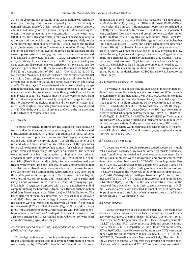
Fig. 2. Light micrographs of the skeletal muscle of Anguilla anguilla: exposed specimens (n =(w) (C, D) fibres showed signs of injury as laceration (arrow) and transversal fragmentation (red (E) and white (F) fibres were silver-stained and argyrophilic. Scale bar: 25 μm. (For interpversion of this article.)
less intensely stained than red ones (Fig. 1C). Both red and white fibreswere surrounded by a reticular connective tissue that was more con-spicuous in the redmuscle (Fig. 1B, C). Thewhite fibres were character-ized by different diameters, giving the tissues a typical mosaicappearance; however, mainly large fibres were visible, as expected inthe silver stage (Tesch, 2003) (Fig. 1C). In the sarcoplasm (the cyto-plasm of the muscle fibre), the contractile apparatus of both types of fi-bres appeared compact, withmyofibrils regularly aligned and parallel toeach other (Fig. 1B–D). After silver impregnation, the connective reticu-lar tissue appeared weakly argyrophilic in the red muscle (Fig. 1E) anddeeply argyrophilic, black stained, in the white muscle (Fig. 1F). Nostain was observed in the sarcoplasm of either type of fibre.
After the exposure to cocaine, both red (Fig. 2A, B) and white (Fig.2C, D) fibres showed signs of injury, such as laceration and transversalfragmentation. Silver impregnation showed that the sarcoplasm ofboth types of muscle fibre appeared silver stained (Fig. 2E,F); the red fi-bres (Fig. 2E) appeared larger than control ones.
Three days (Fig. 3A, B) and 10 days (Fig. 3C, D, E) after the interrup-tion of cocaine exposure, both red (Fig. 3A, C) and white (Fig. 3B, D, E)fibres showed signs of injury and muscle breakdown. In particular, the
30). (A, B, C, D: Mallory staining; E, F. silver impregnation). Both red (r) (A, B) and whitearrowhead). (E, F) In addition to the connective (c) tissue, the sarcoplasm (s) of both theretation of the references to colour in this figure legend, the reader is referred to the web

Fig. 3. Light micrographs of the skeletal muscle of Anguilla anguilla: post-exposure recovery three (n = 30) and 10 days (n = 30) specimens. (A, B, C, D, E: Mallory staining; F. silverimpregnation). Three days (A, B) and 10 days (C, D, E) after the interruption of cocaine exposure, both red (r) (A, C) and white (w) (B, D, E) fibres showed signs of injury andbreakdown (arrow). (C). The red (r) fibres showed signs of swelling (asterisk), rarefaction of the myofibrils and disorganization of the contractile apparatus. (F) Ten days after theinterruption of cocaine exposure, the sarcoplasm of both types of fibre appeared silver stained. Scale bar: a, b, c, d, e; 25 μm; f: 200 μm. (For interpretation of the references to colour inthis figure legend, the reader is referred to the web version of this article.)
Table 1Diameter (μm) of the red and white muscle fibres in control (n= 30), exposed (n= 30)and post-exposure recovery three (n= 30) and ten (n= 30) days specimens. Values aremeans ± SE of the mean. *: P b 0.05 vs. control.
Red fibres (μm) White fibres (μm)
Control (n = 30) 19.76 ± 5.90 24.41 ± 10.38Exposed *27.91 ± 15.19 26.72 ± 13.65Post-exposure recovery 3 days *29. 27 ± 10.05 30.35 ± 14.03Post-exposure recovery 10 days *33.55 ± 6.96 *33.37 ± 16.52
867A. Capaldo et al. / Science of the Total Environment 640–641 (2018) 862–873
red fibres (Fig. 3A,C) appeared to be more severely affected than whitefibres, showing signs of swelling, rarefaction of themyofibrils and disor-ganization of the contractile apparatus. The sarcoplasm of both types ofmuscle fibre was still silver stained 10 days after the interruption of co-caine exposure (Fig. 3F).
Assessment of the diameter of the red and white fibres revealed anincrease in the mean diameter of both types of muscle fibre. This in-crease was significant (P ˂ 0.05) in the red fibres of the exposed andpost-exposure recovery specimens, whereas in the white fibres differ-ences in fibre diameter became significant only 10 days after the inter-ruption of cocaine exposure (Table 1).
Assessment of the morphology of the sarcomeric actin filamentswith FITC-phalloidin (Fig. 4) confirmed previous observations. In con-trol eels, actin filaments showed a regular organization (Fig. 4A)whereas, after the exposure to cocaine, the filaments showed signs ofinjury, such as laceration and transversal fragmentation (Fig. 4B).Three days (Fig. 4C) and 10 days (Fig. 4D) after the interruption of co-caine exposure, the alterations were still present.
3.3. Muscle protein profile
An electrophoretic analysis of muscle protein composition in sam-ples from different experimental groups was performed. SDS-page pro-files did not show evident qualitative differences in protein bandsbetween groups, either in the range of high molecular weight proteins(HMW myosins) (about 210 kDa) (Fig. 5A) or low molecular weight

868 A. Capaldo et al. / Science of the Total Environment 640–641 (2018) 862–873
proteins (actins and tropomyosins) (about 32–45 kDa) (Fig. 5B). Spo-radic small differences of band intensities, which were not related to aspecific experimental group, might be due to individual variability inprotein abundance.
3.4. Cytochrome oxidase (COX) activity
Compared to control eels, COX activity was slightly increased in co-caine-exposed eels. Significant (P b 0.05) increases in COX activitieswere observed 3 days and 10 days after the interruption of cocaine ex-posure, respectively (Fig. 6).
3.5. Caspase-3 activity
Compared to control eels, caspase-3 activity significantly (P b 0.05)increased in cocaine-exposed specimens, and further increased (P b
0.02) three days and ten days after the interruption of cocaine exposure(Fig. 7A). To verify that the absorbances registered were due to specificcaspase-3 activity, the caspase-3 inhibitor was used in representativesamples from each experimental group. A significant (P b 0.05) reduc-tion of the absorbances in all the samples was observed, as expected(Fig. 7B).
3.6. Serum enzymes
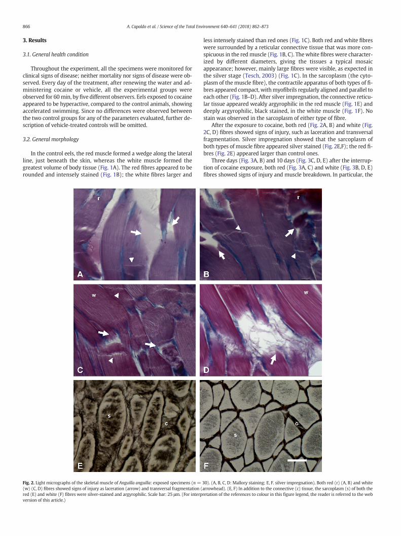
After the exposure to cocaine, changes were observed in the serumlevels of CK (Fig. 8A), LDH (Fig. 8B) and AST (Fig. 9). The serum levelsof CK significantly (P b 0.05) increased, remained steady at 3 days andfurther increased 10 days after the interruption of cocaine exposure(Fig. 8A). The serum levels of LDH showed a gradual increase that be-came significant (P b 0.05) only 10 days after the interruption of cocaineexposure (Fig. 8B).
The serum levels of AST significantly (P b 0.05) increased after theexposure to cocaine and reached a maximum 3 days after the
Fig. 4. Fluorescent micrographs of the skeletal muscle of Anguilla anguilla: FITC-phalloidin stai(B) Exposed specimens (n = 30). Actin filaments showed signs of laceration (arrow) aninterruption of cocaine exposure and (D) 10 days (n = 30) after the interruption of cocaine ex
interruption of cocaine exposure. The AST levels then decreased 10days after the interruption of cocaine exposure, remaining howeverhigher than in controls (Fig. 9).
4. Discussion
The results of the present study show, for the first time, that low en-vironmental cocaine concentrations damage the skeletal muscle of theEuropean eel, inducing alterations that persist after the interruption ofcocaine exposure.
4.1. General health condition
On the basis of personal observations, the cocaine exposed eels,compared to the control/vehicle ones, showed signs of an increasedmotor activity, namely accelerated swimming. This finding is consistentwith the properties of cocaine, a psychomotor stimulant well-known toproduce motor stimulation and increase vigilance and alertness (Grillyand Salamone, 2012). Hyperactivity was also observed in the zebrafishDanio rerio, but only during cocaine withdrawal and not following thesingle administration of a range of cocaine doses (Lòpez-Patiño et al.,2008). It is likely that such a difference could be related to the differencebetween the species examined, and/or the treatments performed(doses administered, acute vs. chronic administration etc.). In anycase, these results indicate that even at very low concentrations, chroniccocaine exposure has effects on eels that are similar to those observed inhumans (Grilly and Salamone, 2012).
4.2. General morphology and protein profile of the skeletal muscle
With regard to the general morphology of the skeletal muscle, rou-tine histological analysis showed that chronic exposure to environmen-tal cocaine concentrations heavily damaged both red and white musclefibres, both of which showed signs of injury, such as fibre breakdown
ning. (A) Control specimens (n = 30). Actin (a) filaments showed a regular organizationd transversal fragmentation (arrowhead), still evident (C) 3 days (n = 30) after theposure. Scale bar: a: 50 μm; b, c, d, e: 100 μm.

1801309572554334
CTR EXP REC-3 REC-10
CTR EXP REC-3 REC-10
43
34
Range of myosin heavy chain (210 kDa)
Range of actin (42 kDa) and tropomyosin (35 kDa)
Range of actin (42 kDa) and tropomyosin(35 kDa)
Range of myosin light chains (16-21 kDa)
A
B
Fig. 5. SDS-page representative profiles of proteins from samples of skeletalmuscles from control (CTR,n=30), exposed (EXP, n=30), post-exposure recovery three days (REC-3,n=30)and ten days (REC-10, n=30) specimens. No differences in the range of highmolecularweight (HMWmyosins) (A) or lowmolecularweight (actins and tropomyosins) (B) proteinswereobserved.
869A. Capaldo et al. / Science of the Total Environment 640–641 (2018) 862–873
and swelling. This damage was still present 10 days after the interrup-tion of cocaine exposure, and was confirmed by FITC-phalloidin stain-ing, which revealed damage to the sarcomeric actin filaments, bothduring exposure and after the interruption of the cocaine exposure.The alterations observed were similar to those typical of rhabdomyoly-sis, a syndrome that is characterized by breakdown of the muscle fibresand dispersion into the circulatory system of the intracellular
0
50
100
150
200
250
Control Exposed
CO
X ac
tivity
(nm
ol O
2/min
x m
g pr
otei
n)
Fig. 6. Cytochrome oxydase (COX) activity in control (n=30), exposed (n=30), post-exposurO2/minxmgprotein. COX activity significantly increased three and 10 days after the interruptionfrom the control values.
components, including the enzymes CK, LDH and AST, myoglobin andelectrolytes (Keltz et al., 2013; Khan, 2009). Our finding of greatly in-creased serum levels of CK, LDH and AST in eels both during the cocaineexposure (CK, AST) and after its interruption (CK, LDH, AST) is reminis-cent of rhabdomyolysis. The development of rhabdomyolysis has beenassociated with a wide variety of diseases, injuries, medications, toxins,alcohol and drugs of abuse such as opiates, amphetamines and other
Post-exposure recovery three days
Post-exposure recovery ten days
*
*
e recovery three (n=30), and ten (n=30) days specimens. Values are expressed as nmolof cocaine exposure. Values aremean±SE of themean. *: significantly (P b 0.05) different

B
0
5
10
15
20
25
30
Control Exposed Post-exposure recovery three days
Post-exposure recovery ten days
(nm
ol p
-NA*
dil)/
(min
* mg
prot
eins
)
w/o inhibitorwith inhibitor
* ** *
A
0
5
10
15
20
25
Control Exposed Post-exposure recovery three days
Post-exposure recovery ten days
nmol
p-NA
*dil)
/(min
*m
g pr
otei
ns)
*
* *
Fig. 7. Caspase-3 activity in the skeletal muscle of control (n= 30), exposed (n= 30), post-exposure recovery three (n= 30) and ten (n=30) days specimens. Values are expressed asnmol of free pNA normalized for μg of proteins and time (min). (A) Caspase-3 activity significantly increased in cocaine-exposed and post-exposure recovery specimens. (B) Inrepresentative samples from each experimental group, the use of caspase-3 inhibitor reduced the registered absorbances of all samples. Values are the mean ± SE of the mean. *:significantly (P b 0.05) different from the control values.
870 A. Capaldo et al. / Science of the Total Environment 640–641 (2018) 862–873
stimulants including cocaine (Brazeau et al., 1995; Keltz et al., 2013;Khan, 2009). It has been postulated that cocaine-induced rhabdomyol-ysis may occur through both a direct drug effect on the muscle and/orrepeated ischemic events mediated by the vasoconstrictor propertiesof cocaine and its metabolites. Further damage to the muscle tissue re-sults from the subsequent reperfusion and the generation of free radi-cals that escape the radical scavenging systems of the muscle andassociated vasculature. This in turn damages the sarcolemma, themem-brane enclosing the skeletalmuscle fibres, leading to increased cytosolicenzyme release (Brazeau et al., 1995). It is possible that such mecha-nisms are activated in eels during and after the interruption of cocaineexposure. Another effect of the disruption of sarcolemma is leakageinto the circulation of large quantities of myoglobin, a cytoplasmic he-moprotein expressed in cardiac myocytes and in skeletal muscle fibres(Ordway andGarry, 2004). If myoglobin levels exceed the protein-bind-ing capacity of the plasma, it can precipitate in the glomerular filtrate,causing renal tubular obstruction, direct nephrotoxicity, and acuterenal failure (Khan, 2009). Our preliminary results have highlightedrenal damages in A. anguilla chronically exposed to cocaine, strengthen-ing our hypothesis of a cocaine-induced rhabdomyolysis syndrome.
An indirect confirmation of the structural damage of the skeletalmuscle fibres come from the silver impregnation technique, that nor-mally stains the argyrophilic reticular fibres of the connective tissue,but not the muscle tissue. During the cocaine exposure and after its in-terruption, both the red and white fibres appeared silver stained. Thebreakdown of the skeletal muscle fibres probably allowed silver toenter the sarcoplasm and bind to the cell structures. Since proteinsbind silver ionswith either a silver nitrate or a silver-ammonia complexsolution (Chevallet et al., 2006), it is conceivable that the cell structuresinvolved in the silver binding were the muscle proteins, although theseare difficult to identify with certainty without specific stainingtechniques.
The increase in the mean diameter of the muscle fibres may be con-sidered indicative ofmuscle damage. Indeed, it has been shown that theswelling of the muscle fibres is one of the clinical features of
rhabdomyolysis (Alessandrino and Balconi, 2013; Nance andMammen, 2015). The red muscle appeared to be the most sensitive tococaine, since the mean diameter of the red fibres increased in boththe exposed eels and the post-exposure recovery eels. Instead, themean diameter of the white fibres significantly increased only 10 daysafter the interruption of cocaine exposure. In silver eels, which performa very long reproductivemigration to the Sargasso sea, without feeding,the swimming activity is sustained by an increase in the activity of theslow red aerobic muscle. Indeed, in the silver stage, the volume of redmuscle and the activity of aerobic enzymes, like COX, are increasedcompared to the yellow, non-migrating, stage (Mortelette et al.,2010). It is possible that these features of the red muscle make it moresensitive to the vasoconstrictor properties of cocaine (Brazeau et al.,1995), compared to the fast white anaerobic muscle. Moreover, it is in-teresting to observe that the effect of cocaine on the skeletal muscle tis-sue was completely different to that observed in smooth muscle tissueof the eel intestine (Gay et al., 2016). Indeed, cocaine, probably throughthe increase in plasma prolactin levels, restored the smooth intestinalmusculature that in control eels showed signs of degeneration (Gay etal., 2016), whereas it seriously damaged the skeletal muscle, with an ef-fect similar to that observed in humans (Brazeau et al., 1995; Keltz et al.,2013; Khan, 2009).
Our electrophoretic analysis of the muscle protein profile did nothighlight qualitative differences between the different experimentalgroups. These findings indicate that chronic cocaine exposure does notinfluence expression of highmolecular weight (myosins) or lowmolec-ularweight (actins and tropomyosins)muscle proteins. However, in therat, cocaine is known to influence the cell cytoskeleton. In the rat nu-cleus accumbens cocaine increases actin cycling. Indeed, acute andwithdrawal from repeated cocaine administration produces reversibleand enduring elevations in F-actin, through changes in the content orphosphorylation state of actin binding proteins, or reduced depolymer-ization and actin cycling respectively (Toda et al., 2006). In contrast, co-caine increased the total protein content and the expression of beta-myosin heavy-chain protein in rat cardiac ventricular myocytes
A
B
0
20000
40000
60000
80000
100000
120000
140000
160000
180000
200000
Control Exposed Post-exposure recovery three days
Post-exposure recoveryten days
Seru
m C
K(UI
L-1
) * *
*
0
20000
40000
60000
80000
100000
Control Exposed Post-exposure recovery three days
Post-exposure recoveryten days
Seru
m L
DH(U
I L-1
)
*
Fig. 8. (A) Serum creatine kinase (CK) and (B) serum lactate dehydrogenase (LDH) levels in control (n=30), exposed (n=30), post-exposure recovery three (n=30) and ten (n=30)days specimens. Values are expressed as UI L−1. (A) The serum CK levels significantly increased in the exposed specimens to be steady 3 days and further increase 10 days after theinterruption of cocaine exposure. (B) The serum LDH levels significantly increased only 10 days after the interruption of cocaine exposure. Values are the mean ± SE of the mean. *:significantly (P b 0.05) different from the control values.
871A. Capaldo et al. / Science of the Total Environment 640–641 (2018) 862–873
(Henning et al., 2000) and it alters the protein profile of many differentproteins, including cytoskeletal proteins in zebrafish embryos (Paroliniet al., 2018). In any case, a better understanding of the effects of cocaineon the muscle protein profile of eels will require further studies.
4.3. Cytochrome oxidase (COX) and caspase-3 activities
Our results showed an increase in COX activity that became signifi-cant after the interruption of cocaine exposure. Cytochrome oxidase(COX), a well-known biomarker of oxidative metabolism (Lee andHüttemann, 2014), catalyzes thefinal step in themitochondrial electrontransfer chain (ETC) and the reduction of oxygen to water. ETC plays akey role in cell metabolism producing most of the cellular energy. Theincrease in COX activity observed in skeletal muscle homogenatesmight indicate an increase in specific mitochondrial enzyme activityand/or in mitochondrial protein mass. This increase might play an im-portant role in the physiological adaptation to cocaine exposure by
supporting the increased energy requirements for the detoxificationprocess. However, cocaine is known to induce cardiovascularmitochon-drial dysfunction (Graziani et al., 2017) and to reduce COXactivity in therat prefrontal cortex (Vélez-Hernández et al., 2014). The contrast be-tween these data and our finding could be due to the different animal/cellular models and/or to different doses and times of exposure to co-caine. Noteworthy are previous studies that suggest that chronic co-caine exposure leads to repeated ischemic events mediated by thevasoconstrictor properties of cocaine and its metabolites. The subse-quent reperfusion and the generation of free radicals could induce rhab-domyolysis and/or muscle damage (Brazeau et al., 1995). Furtherstudies showed, during the progression of reperfusion injury, critical al-terations in COXactivity, inwhich three stageswere proposed: an ische-mic starvation phase, a reperfusion-induced hyperactivation phase, anda mitochondrial dysfunction phase. It was hypothesized that ischemia-induced stress alters the phosphorylation state of COX, that becomeshyperactive and leads to mitochondrial ROS generation during the

0
200
400
600
800
1000
Control Exposed Post-exposure recovery three days
Post-exposure recoveryten days
Seru
m A
ST (U
I L-1
)
* *
*
Fig. 9. Serum aspartate aminotransferase (AST) levels in control (n = 30), exposed (n = 30), post-exposure recovery three (n = 30) and ten (n = 30) days specimens. Values areexpressed as UI L−1. The serum AST levels significantly increased in the exposed specimens to further hugely increase 3 days and to decrease 10 days after the interruption of cocaineexposure, when AST levels were however significantly higher than the control levels. Values are the mean ± SE of the mean. *: significantly (P b 0.05) different from the control values.
872 A. Capaldo et al. / Science of the Total Environment 640–641 (2018) 862–873
beginning of reperfusion. In turn, ROS inducemitochondrial dysfunctionduring late reperfusion, and activate the intrinsic apoptotic pathway,committing the cell to death (Hüttemann et al., 2012). The increase inCOX activity observed in eels after the interruption of cocaine exposureis consistent with the hyperactivity of this enzyme typical of the ische-mia/reperfusion injury, and could coincide with reperfusion-inducedhyperactivation phase. This hypothesis should be confirmed by furtherexperiments. In accordance with this hypothesis, the evaluation of theactivity of caspase-3, the major executioner caspase in the apoptoticpathway (Brentnall et al., 2013) showed a significant increase in boththe exposed and the post-exposure recovery eels. Cocaine is known toinduce caspase activation (Cunha-Oliveira et al., 2006) and apoptosis(Li et al., 2005; Su et al., 2003) with a mechanism involving the releaseof cytochrome c from the mitochondria into the cytosol, and the subse-quent activation of caspase-9 and caspase-3 (He et al., 2000). The in-crease in caspase-3 activity observed in the eels during the exposureand after the interruption of cocaine exposure agrees with this mecha-nismand further supports the hypothesis that the increase in COX activ-ity might be associated with the ischemic/reperfusion injury.
4.4. Serum enzymes
Our results showed that chronic exposure to cocaine increased theserum levels of creatine kinase (CK), lactate dehydrogenase (LDH) andaspartate aminotransferase (AST), albeit with a different pattern. In-deed, the increases in CK andAST levelswere significant both during co-caine exposure and after its interruption, whereas the increase in LDHlevels became significant only 10 days after the interruption of cocaineexposure. CK, LDH and AST are well-established serum markers of thefunctional state of themuscle, and their increase may be an index of tis-sue injury (Brancaccio et al., 2010). CK catalyzes the reversible reactionof creatine and ATP forming phosphocreatine and ADP; it plays there-fore a key role in energy homeostasis of cells and it is typical of tissueswith high energy demand, such as skeletal muscle, heart and brain.LDH is involved in the interconversion of pyruvate and lactate, withconcomitant interconversion of NADH and NAD during the final reac-tions of glycolysis. It is present in the cytoplasm of all the cells and,
since it is released during tissue damage, it is a marker of common inju-ries and diseases such as heart failure and muscle injury. AST catalyzesthe interconversion of aspartate and α-ketoglutarate to oxaloacetateand glutamate, providing energy to the cells; the enzyme is localizedprimarily in the skeletal and myocardial muscle, liver and erythrocytes(Brancaccio et al., 2010; Yousaf and Powell, 2012). The increase in theserum levels of these enzymes observed both during the cocaine expo-sure (CK, AST) and after its interruption (CK, LDH, AST) confirms themorphological observations of structural damage to the skeletal muscleand could be indicative of a rhabdomyolysis syndrome (Keltz et al.,2013; Khan, 2009). However, considering that every enzyme has differ-ent tissue-specific isoforms (Brancaccio et al., 2010) that were not eval-uated here, since only the total amount of every enzyme wasdetermined, the increases observed could also be indicative of heartand/or liver damage. A correlation between muscle injury and the in-crease in serum muscle enzyme levels was also found in the Atlanticsalmon, in which increases in serum CK and LDH levels, and CK levels,were associated with heart and skeletal muscle inflammation (Yousafand Powell, 2012), and with acute skeletal myopathy, respectively(Rodger et al., 1991). Our results, showing an increase in the serum en-zymes considered main markers of muscle tissue injury, agree withthese data and confirm the relationship between muscle damage andchange in the serum levels of these enzymes.
4.5. Conclusion
In conclusion, the results of this study showed that low cocaine con-centrations damaged themorphology and physiology of the eel skeletalmuscle. At the silver stage, the eel undertakes a swimming migration6000 km long (Righton et al., 2016) needing sufficient energy reservesand a healthy skeletal muscle tissue, to be successfully completed. Ourresults suggest that cocaine, at environmental concentrations, couldcompromise both the sustained swimming, ensured by the slow redmuscle, and the burst swimming, ensured by the fast, white muscle.The European eel is under threat and in serious decline, due to manycauses, including overfishing, habitat loss, presence of parasites, climatechange and water pollution (Dekker, 2003). Our results suggest that
873A. Capaldo et al. / Science of the Total Environment 640–641 (2018) 862–873
environmental cocaine concentrations contribute to the threat of waterpollution to this susceptible species. Finally, the European eel is an edi-ble species used as food resource (Arai, 2014). Since the skeletal muscleis the edible part of the eel, and bioaccumulates cocaine to a large extent(Capaldo et al., 2012), these results suggest the possibility that cocainecould be taken by humans with food, although further studies areneeded to verify this hypothesis.
Competing interests
The Authors declare no competing or financial interests.
Funding
This research received no specific grant from any funding agency inthe public, commercial or not-for-profit sectors.
Acknowledgements
The authors would like to thank Prof. Elisabeth Anne Illingworth(University of Fisciano, Italy) for providing language help.
References
Alessandrino, F., Balconi, G., 2013. Complications of muscle injuries. J. Ultrasound 16,215–222.
Aligizakis, N.A., Gago-Ferrero, P., Borova, V.L., Pavlidou, A., Hatzianestis, I., Thomaidis, N.S.,2016. Occurrence and spatial distribution of 158 pharmaceuticals, drugs of abuse andrelated metabolites in offshore seawater. Sci. Total Environ. 541, 1097–1105.
Altringham, J.D., Ellerby, D.J., 1999. Fish swimming: patterns in muscle function. J. Exp.Biol. 202, 3397–3403.
Arai, T., 2014. Do we protect freshwater eels or do we drive them to extinction?Springerplus 3, 534–544.
Barrè, H., Bailly, L., Rouanet, J.L., 1997. Increased oxidative capacity in skeletal muscle fromcold-acclimated ducklings: a comparison with rats. Comp. Biochem. Physiol. 88,B5119–B5522.
Binelli, A., Pedriali, A., Riva, C., Parolini, M., 2012. Illicit drugs as new environmental pol-lutants: cyto-genotoxic effects of cocaine on the biological model Dreissenapolymorpha. Chemosphere 86, 906–911.
Bradbury, P., Gordon, K.C., 1980. Connective tissues and stain. In: Bancroft, J.D., Stevens, A.(Eds.), Theory and Practice of Histological Techniques. Churchill Livingstone, NewYork, pp. 119–142.
Brancaccio, P., Lippi, G., Maffulli, N., 2010. Biochemical markers of muscular damage. Clin.Chem. Lab. Med. 48, 757–767.
Brazeau, G.A., McArdle, A., Jackson, M.J., 1995. Effects of cocaine on leakage of creatine ki-nase from skeletal muscle: in vitro and in vivo studies in mice. Life Sci. 57,1569–1578.
Brentnall, M., Rodriguez-Menocal, L., Ladron De Guevara, R., Cepero, E., Bpise, L.H., 2013.Caspase-9, caspase-3 and caspase-7 have distinct roles during intrinsic apoptosis.BMC Cell Biol. 14:32. http://www.biomedcentral.com/1471-2121/14/32.
Capaldo, A., Gay, F., Maddaloni, M., Valiante, S., De Falco, M., Lenzi, M., Laforgia, V., 2012.Presence of cocaine in the tissues of the European eel, Anguilla anguilla, exposed toenvironmental cocaine concentrations. Water Air Soil Pollut. 223, 2137–2143.
Chevallet, M., Luche, S., Rabilloud, T., 2006. Silver staining of proteins in polyacrylamidegels. Nat. Protoc. 1, 1852–1858.
Cunha-Oliveira, T., Rego, A.C., Cardoso, S.M., Borges, F., Swerdlow, R.H., Macedo, T., deOliveira, C.R., 2006. Mitochondrial dysfunction and caspase activation in rat corticalneurons treated with cocaine or amphetamine. Brain Res. 1089, 44–54.
Dekker, W., 2003. Did lack of spawners cause the collapse of the European eel, Anguillaanguilla? Fish. Manag. Ecol. 10, 365–376.
Dubowitz, V., Dubowitz, V., Sewry, C., Dubowitz, V., Oldfors, A., Sewry, C., 1985. Muscle Bi-opsy. A Practical Approach. Bailliere Tindall, London.
Fedorova, M., Kuleva, N., Hoffmann, R., 2009. Reversible and irreversible modifications ofskeletal muscle proteins in a rat model of acute oxidative stress. Biochim. Biophys.Acta 1792, 1185–1193.
Gay, F., Ferrandino, I., Monaco, A., Cerulo, M., Capasso, G., Capaldo, A., 2016. Histologicaland hormonal changes in the European eel (Anguilla anguilla) after exposure to envi-ronmental cocaine concentration. J. Fish Dis. 39, 295–308.
Gay, F., Maddaloni, M., Valiante, S., Laforgia, V., Capaldo, A., 2013. Endocrine disruption inthe European eel, Anguilla anguilla, exposed to an environmental cocaine concentra-tion. Water Air Soil Pollut. 224:1579. https://doi.org/10.1007/s11270-013-1579-0.
Graziani, M., Sarti, P., Arese, M., Magnifico, M.C., Badiani, A., Saso, L., 2017. Oxid. Med. Cell.Longev. 2017. Article ID 3034245. https://doi.org/10.1155/2017/3034245.
Grilly, M.G., Salamone, J.D., 2012. Drugs, Brain and Behavior. Pearson, Boston.
He, J., Xiao, Y., Casiano, C.A., Zhang, L., 2000. Role of mitochondrial cytochrome c in co-caine-induced apoptosis in coronary artery endothelial cells. J. Pharmacol. Exp.Ther. 295, 896–903.
Henning, R.J., Silva, J., Reddy, V., Kamat, S., Morgan, M.B., Li, Y.X., Chiou, S., 2000. Cocaineincreases beta-myosin heavy-chain protein expression in cardiac myocytes.J. Cardiovasc. Pharmacol. Ther. 5, 313–322.
Hüttemann, M., Helling, S., Sanderson, T.H., Sinkler, C., Samavati, L., Mahapatra, G.,Varughese, A., Lu, G., Liu, J., Ramzan, R., Vogt, S., Grossman, L.I., Doan, J.W., Marcus,K., Lee, I., 2012. Regulation of mitochondrial respiration and apoptosis through cellsignaling: cytochrome c oxidase and cytochrome c in ischemia/reperfusion injuryand inflammation. Biochim. Biophys. Acta 1817, 598–609.
Keltz, E., Khan, F.Y., Mann, G., 2013. Rhabdomyolysis. The role of diagnostic and prognos-tic factors. Muscles Ligaments Tendons J. 3, 303–312.
Khan, F.Y., 2009. Rhabdomyolysis: a review of the literature. Neth. J. Med. 67, 272–283.Lee, I., Hüttemann, M., 2014. Energy crisis: the role of oxidative phosphorylation in acute
inflammation and sepsis. Biochim. Byophys. Acta 1842, 1579–1586.Li, G., Xiao, Y., Zhang, L., 2005. Cocaine induces apoptosis in fetal rat myocardial cells
through the p38 mitogen-activated protein kinase and mitochondrial/cytochrome cpathways. J. Pharmacol. Exp. Ther. 312, 112–119.
Li, K., Du, P., Xu, Z., Gao, T., Li, X., 2016. Occurrence of illicit drugs in surface waters inChina. Environ. Pollut. 213, 295–402.
Lionetti, L., Crescenzo, R., Mollica, M.P., Tasso, R., Barletta, A., Liverini, G., Iossa, S., 2004.Modulation of hepatic mitochondrial energy efficiency with age. Cell. Mol. Life Sci.61, 1366–1371.
Lòpez-Patiño, M.A., Yu, L., Cabral, H., Zhdanova, I.V., 2008. Anxiogenic effects of cocainewithdrawal in zebrafish. Physiol. Behav. 93, 160–171.
Monaco, A., Capaldo, A., Laforgia, V., Grimaldi, M.C., Ferrandino, I., 2016. Environmentalcocaine concentration effects on Danio rerio development. Eur. H. Histochem. 60, 9.
Mortelette, H., Amérand, A., Sébert, P., Belhomme, M., Calvès, P., Moisan, C., 2010. Effect ofexercise training on respiration and reactive oxygen species metabolism in eel redmuscle. Respir. Physiol. Neurobiol. 172, 201–205.
Mumford, S., Heidel, J., Smith, C., Morrison, J., Mac Connell, B., Blazer, V., 2007. Fish Histol-ogy and Histopathology. U.S. Fish & Wildlife Service-National Conservation TrainingCenter https://nctc.fws.gov/resources/course-resources/fish-histology/.
Nance, R.J., Mammen, A.L., 2015. Diagnostic evaluation of rhabdomyolysis. Muscle Nerve51:793–810. https://doi.org/10.1002/mus.24606.
Ordway, G.A., Garry, D.J., 2004. Myoglobin: an essential hemoprotein in striated muscle.J. Exp. Biol. 207, 3441–3446.
Pal, R., Megharaj, M., Kirkbride, K.P., Naidu, R., 2013. Illicit drugs and the environment-areview. Sci. Total Environ. 463–464, 1079–1092.
Parolini, M., Bini, L., Magni, S., Rizzo, A., Ghilardi, A., Landi, C., Armini, A., Del Giacco, L.,Binelli, A., 2018. Environ. Pollut. 232, 603–614.
Parolini, M., Ghilardi, A., Della Torre, C., Magni, S., Prosperi, L., Calvagno, M., Del Giacco, L.,Binelli, A., 2017. Environmental concentrations of cocaine and its main metabolitesmodulated antioxidant response and caused cyto-genotoxic effects in zebrafish em-bryo cells. Environ. Pollut. 226, 504–514.
Parolini, M., Pedriali, A., Riva, C., Binelli, A., 2013. Sub-lethal effects caused by the cocainemetabolite benzoylecgonine to the freshwater mussel Dreissena polymorpha. Sci.Total Environ. 444, 43–50.
Righton, D., Westerberg, H., Feunteun, E., Økland, F., Gargan, P., Amilhat, E., Metcalfe, J.,Lobon-Cervia, J., Sjöberg, Simon, J., Acou, A., Vedor, M., Walker, A., Trancart, T.,Brämick, U., Aerestrup, K., 2016. Empirical observations of the spawning migrationof European eels: the long and dangerous road to the Sargasso Sea. Sci. Adv. 2,e1501694.
Rodger, H.D., Murphy, T.M., Drinan, E.M., Rice, D.A., 1991. Acute skeletal myopathy infarmed Atlantic salmon Salmo salar. Dis. Aquat. Org. 12, 17–23.
Rosi-Marshall, E.J., Snow, D., Bartelt-Hunt, S.L., Paspalof, A., Tank, J.L., 2015. A review ofecological effects and environmental fate of illicit drugs in aquatic ecosystems.J. Hazard. Mater. 282, 18–25.
Seabra-Pereira, C.D., Maranho, L.A., Cortez, F.S., Pusceddu, F.H., Santos, A.R., Ribeiro, D.A.,Cesar, A., Guimarães, L.L., 2016. Occurrence of pharmaceuticals and cocaine in a Bra-zilian coastal zone. Sci. Total Environ. 548–549, 148–154.
Su, J., Li, J., Li, W., Altura, B.T., Altura, B.M., 2003. Cocaine induces apoptosis in cerebral vas-cular muscle cells: potential roles in strokes and brain damage. Eur. J. Pharmacol. 482,61–66.
Tesch, S.W., 2003. The Eel. Blackwell Science, Ltd., Oxford.Toda, S., Shen, H.W., Peters, J., Cagle, S., Kalivas, P.W., 2006. Cocaine increases actin cy-
cling: effects in the reinstatement model of drug seeking. J. Neurosci. 26, 1579–1587.Vélez-Hernández, M.E., Padilla, E., Gonzalez-Lima, F., Jiménez-Rivera, C.A., 2014. Cocaine
reduces cytochrome oxidase activity in the prefrontal cortex and modifies its func-tional connectivity with brainstem nuclei. Brain Res. 1542, 56–69.
Yancheva, V., Velcheva, I., Stoyanova, S., Georgieva, E., 2016. Histological biomarkers infish as a tool in ecological risk assessment and monitoring programs: a review.Appl. Ecol. Environ. Res. 14, 47–75.
Yousaf, M.N., Powell, M.D., 2012. The effects of heart and skeletal muscle inflammationand cardiomyopathy syndrome on creatine kinase and lactate dehydrogenase levelsin atlantic salmon (Salmo salar L.). Sci. World J. 2012:741302. https://doi.org/10.1100/2012/741302.