scientific update on functional tissue engineering of ... · scale up challenges digitized...
TRANSCRIPT
Scientific update on functional tissue
engineering of cartilage
Department of Biomedical Engineering
Columbia University
New York, NY 10027
Clark T. Hung, Ph.D.
Professor
2016 ICRS Sorrento Italy

Cartilage Repair: Basic Science Towards Clinical Translation
Approach 1
Cartilage
Allografts
Native Cartilage
Tissue
Engineered
Cartilage
Engineered
Cartilage
Approach 2
NIAMS 2002, Lawrence+ 1989
Focal Defect
Defect Repair

Cartilage Repair: Basic Science Towards Clinical Translation
Approach 1
Cartilage
Allografts
NIAMS 2002, Lawrence+ 1989
Focal Defect
Defect Repair
Native Cartilage
Cartilage allograft
Challenges:
• Declining cell viability
• Loss of matrix components
• Decreasing cell synthesis
Standard cartilage storage protocol • Fetal bovine serum (FBS)
• Hypothermal refrigeration (4˚C)
To develop a tissue culture protocol (37˚C) for
preserving cartilage allografts • Serum-free chondrogenic media (CM) (Lima+, OAC 2007)
• Dexamethasone
Brighton+ 1979; Dumont+ 1999; Bugbee+ 2002; Williams+ 2004; Pearsall+ 2004; Williams+2005
Cook+, Vet Surg, 2008
Compromised clinical outcome

FBS
0
0.5
1
1.5
2
2.5
day 14 day 28 day 42
Norm
aliz
ed V
olu
me
† † †
• Serum constructs swell
• Cells are alive in both media
Explants: Serum (FBS) vs. serum-free medium (CM)
FBS CM CM
Bian+ J Biomech 2008; Bian+ Am J Sports Med 2010
37˚C CO2 incubator
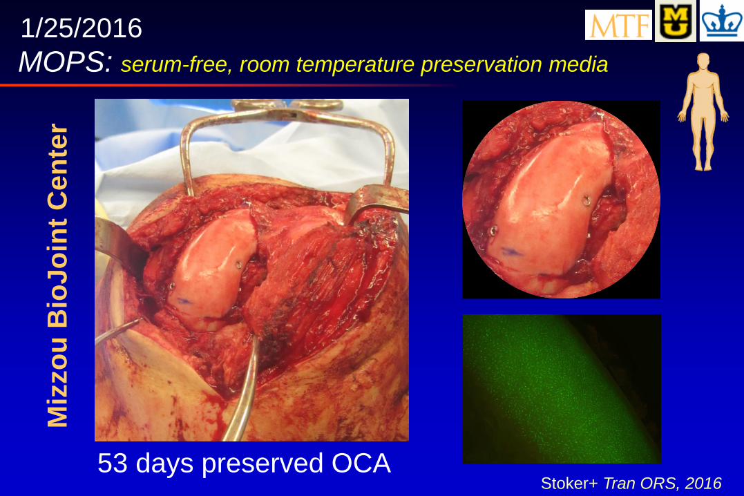
53 days preserved OCA
MOPS: serum-free, room temperature preservation media
Stoker+ Tran ORS, 2016
1/25/2016 M
izzo
u B
ioJ
oin
t C
en
ter

Cartilage Repair: Basic Science Towards Clinical Translation
Tissue
Engineered
Cartilage
Engineered
Cartilage
Approach 2
NIAMS 2002, Lawrence+ 1989
Focal Defect
Defect Repair
• Dynamic Loading
• Scale Up & Channels
• Chemical Protection
• Tissue Integration
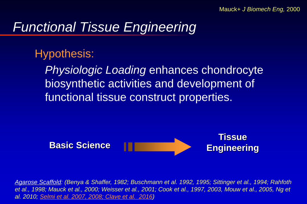
Functional Tissue Engineering
Hypothesis:
Physiologic Loading enhances chondrocyte
biosynthetic activities and development of
functional tissue construct properties.
Basic Science Tissue
Engineering
Agarose Scaffold: (Benya & Shaffer, 1982; Buschmann et al. 1992, 1995; Sittinger et al., 1994; Rahfoth
et al., 1998; Mauck et al., 2000; Weisser et al., 2001; Cook et al., 1997, 2003, Mouw et al., 2005, Ng et
al. 2010; Selmi et al. 2007, 2008; Clave et al. 2016)
Mauck+ J Biomech Eng, 2000

Dynamic Loading
Axial Loading: unconfined compression
0
100
200
300
400
500
600
700
Day 0 Day 14 Day 28 Day 42 Day 56
EY(k
Pa
)
Free Swelling
• Expansion Media (DMEM, 10% FBS, TGF-1, FGF-2, PDGF-)
• Allogeneic, Passage 2 cells, Loading starting day 28 (10% deformation, 1 Hz, 3h/day)
• Continuous growth factor supplementation (TGF-3)
native knee cartilage
Bian+ Tissue Eng 2010
Dynamic Loading
*
* *
Ng+ Tissue Eng 2010
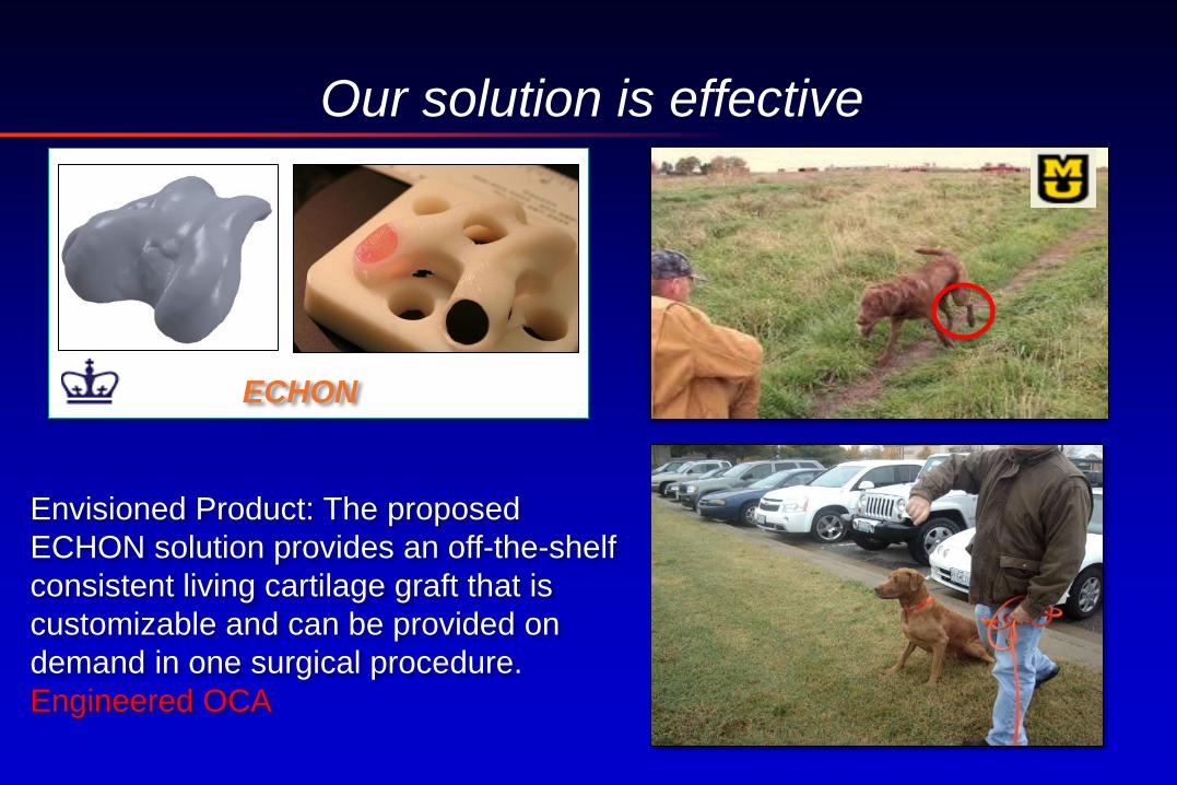
Our solution is effective
ECHON
Envisioned Product: The proposed
ECHON solution provides an off-the-shelf
consistent living cartilage graft that is
customizable and can be provided on
demand in one surgical procedure.
Engineered OCA

Scale Up Challenges
Digitized Topography
Data
CAD Rendering of
Mold
G-Code Automated MRI
Segmentation
CNC Milling
Machine
Hung+ J Biomech Eng 2002
Roach+ Methods 2014
Nutrient limitations Human Patella

Articular Surface-Sized Constructs
FEBio Modeling Glucose Consumption
Rate
3-piece mold to
create channels Rack and Shaking
Cadaver
hum
an k
nee
P2 cells, EY: 450 kPa; [GAG]: 8.1%
ww; [Collagen]: 2.6% ww
Bian+ OAC, 2009; Nims+ Tissue Eng C
Methods, 2015; Cigan+ Trans ORS 2016
Day 56
Initial diameter: 40 mm; 2.33 mm thick
Cells and Construct
Fabrication
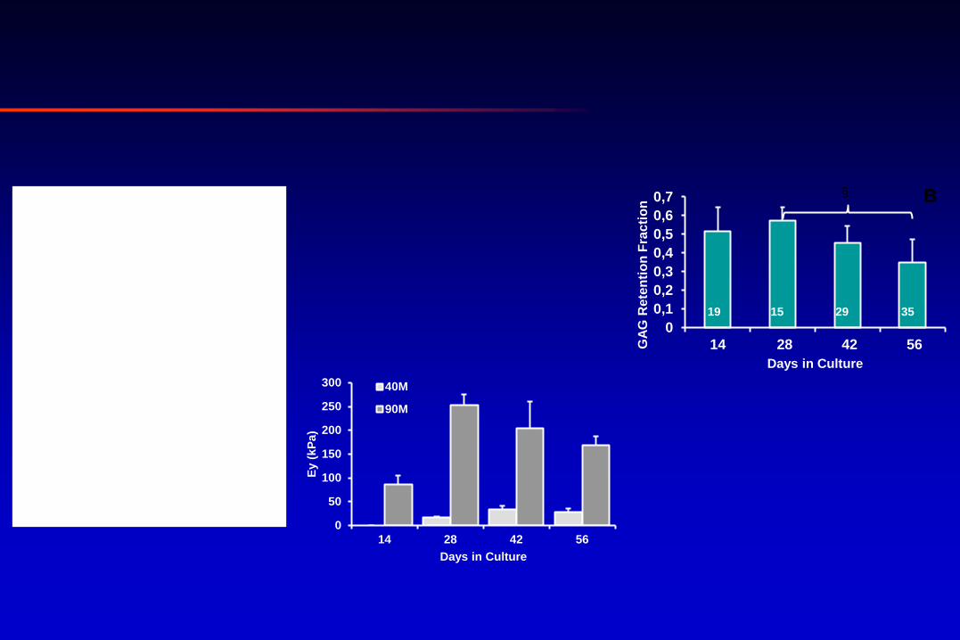
0
0,1
0,2
0,3
0,4
0,5
0,6
0,7
14 28 42 56GA
G R
ete
nti
on
Fra
cti
on
Days in Culture
B §
19 15 29 35
0
50
100
150
200
250
300
14 28 42 56
Ey (
kP
a)
Days in Culture
40M
90M
†
*§
† †
*§
* C
4 4 5 3 3 3 2 0
2
4
6
8
10
12
14
0
0,5
1
1,5
2
2,5
28 42 56 14 28 42 56
%/D
0 w
w
GA
G (
µg
)
Days in Culture
Media
Construct
40M
90M D
4 4 5 3 2 3 3
R² = 0,7578
0
50
100
150
200
250
300
0 2 4 6 8
Ey (
kP
a)
[GAG] (%/ww)
Donor 1
Donor 2
Donor 3
Donor 4
Donor 5
A N = 92
Constructs: 3 × 2.3 mm
Translation to Human Constructs Cigan+ Tissue Eng 2016 in press
Recently expired
grafts
*Best to Date: Ey: 367.8±121 kPa, 8.91±0.46 %GAG/ww, 1.67±0.14 %COL/ww, 30M cells/ml (n=6)- AR Tan
Chemical Protection of Engineered Grafts
• Incorporation of dexamethasone delivery for
chondroprotection and pain/inflammation therapy.
Intra-articular steroid injections for
pain/inflammation, 3X per year maximum
due to concerns of local and systemic
side effects. Huebner+ J Orthop Res 2014
Heard+ J Orthop Res 2015
Roach+ Tissue Eng 2016
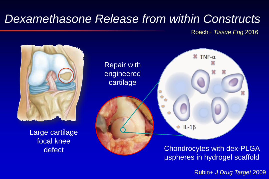
Dexamethasone Release from within Constructs
Repair with
engineered
cartilage
Large cartilage
focal knee
defect Chondrocytes with dex-PLGA
µspheres in hydrogel scaffold
Roach+ Tissue Eng 2016
Rubin+ J Drug Target 2009
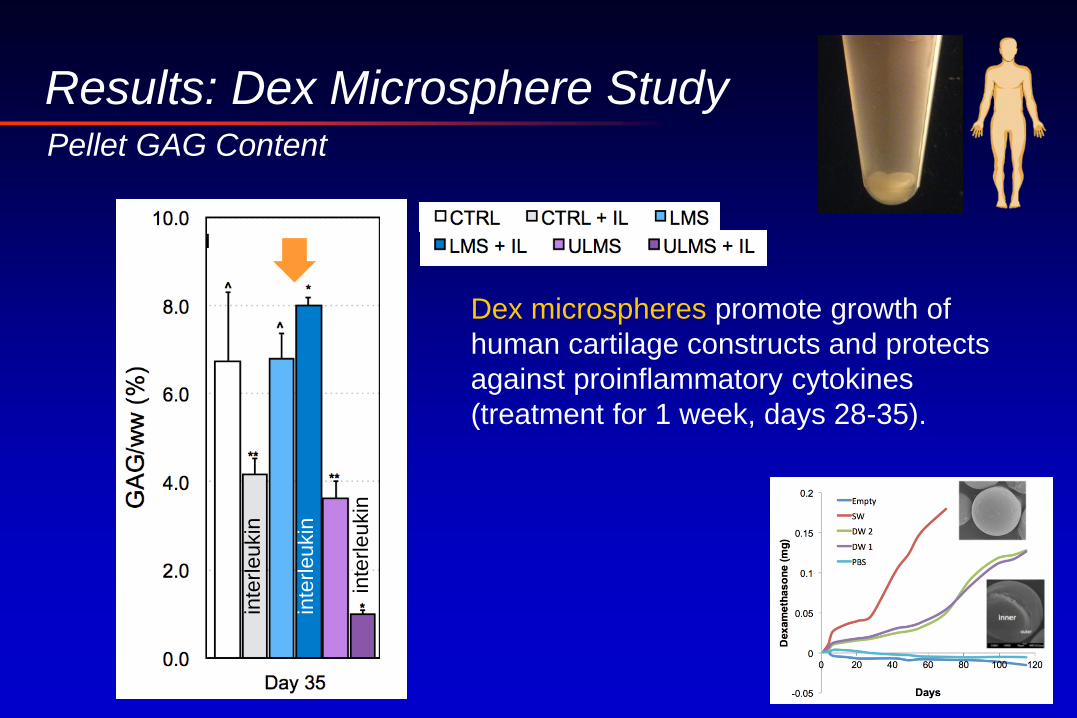
Results: Dex Microsphere Study
Dex microspheres promote growth of
human cartilage constructs and protects
against proinflammatory cytokines
(treatment for 1 week, days 28-35).
Pellet GAG Content
inte
rleu
kin
inte
rleu
kin
inte
rleu
kin

Integrative Repair: Electric field induced migration
Synovial Cells- DiI membrane dye: yellow
Chondrocytes- DAPI nuclear dye: blue
CONTROL E: 25 V/cm
Synovial Explant Placed Adjacent to Cartilage Explant with Central Defect
Top
Side
Stefani+ 2017 ORS submitted
defect defect

Integrative Repair: High Intensity Focused Ultrasound
Nover+ Med Eng Phys 2016; Tan+ 2017 ORS submitted
Albumin solder
Push-out Test
Acknowledgements
NIH R01 AR068133
NIH R01 AR060361
NIH 5P41EB002520
NIH T32AR059038 (AMS, BLR)
NSF GRF (ART, BLR)
Gerard A. Ateshian
Elisa E. Konofagou
James L. Cook
Kacey G. Marra