seattle/king county emt-b class topics 1234 trauma: chapter 21 soft tissue injuries: chapter 24...
TRANSCRIPT

Seattle/King County EMT-B Class

Topics
1
2
3
4
Trauma: Chapter 21
Soft Tissue Injuries: Chapter 24
Shock: Chapter 23
Bleeding: Chapter 22
5 Intro to AED
1 Trauma

Kinematics of Trauma
• Injuries are the leading cause of death among children and young adults.
1

Traumatic Injuries Blunt trauma
• Caused by a force to the body• Injuries do not penetrate soft tissue
or organsPenetrating trauma
• Caused by objects such as knives and bullets
• Injuries pierce the surface of the body
1

Mechanism of Injury (MOI)
• MOI is the way in which traumatic injuries occur.
• Different MOIs produce many types of injuries.– Isolated to one body system– Injuries to many body systems
1

Vehicular Crashes and MOI
• By assessing the crash, the MOI may be determined.
• By determining the MOI, you may be able to predict the types of injuries that may have happened at the time of impact.
1

Vehicular Collisions
Three types of crashes• Collision of car against another
car or object• Collision of passenger(s) against
interior of car• Collision of passenger’s internal
organs against the solid structures of the body
1

Significant MOI
• Severe deformities to the frontal part of the vehicle
• Moderate intrusion from a T-bone accident
• Severe damage from the rear• Collisions in which rotation is
involved
1

Types of Motor Vehicle Collisions• Frontal• Lateral• Rear-
end• Rollover
s• Spins
1

Frontal Collisions• Evaluate seat belts
and airbags.• Remember that
supplemental restraint systems cannot prevent all injuries.
• You should still suspect that serious injuries have occurred.
1

Frontal Collisions, continued
• Check for contact points.
• Steering wheels can also cause chest injuries, especially if no airbag is present.
1

Rear-End Collisions
• Commonly cause whiplash-type injuries
• Unrestrained passengers will be thrust forward into the dashboard.
1

Rear-End Collisions, continued• Back seat
passengers wearing only lap belts might have a higher incidence of lumbar and thoracic spine injury.
1
Quiz Question Alert...!

Lateral Collisions• Responsible for
the highest incidence of deaths.
• Lateral whiplash injury is the result.
• There may be intrusion into the passenger compartment.
1

Rollover Crashes• Injury patterns differ
if patients are unrestrained.
• The most unpredictable injuries are to unrestrained passengers.
• Ejection is the most common life-threatening injury.
1

Spins
• Vehicle is put into rotational motion.
• Vehicle often strikes a fixed object, combining forces of rotation with lateral impact.
1

Car-Versus-Pedestrian Collisions• Often cause serious injuries to body
systems• Evaluate MOI to determine:
1. Whether patient was thrown and how far, OR
2. Whether patient was struck and pulled under car.
• Presume injury to the spinal cord and maintain immobilization.
1

Falls• Injury potential is related
to the height of the fall.
• A fall either 10' or 2 times the person’s height is considered significant.
• Suspect internal injuries from a significant fall.
1

Considerations for Falls• The height of the fall
• The surface struck
• The part of the body that hit first, followed by the path of energy displacement
• Always consider syncope or other medical conditions as an underlying cause.
1
"Can you tell me what happened before you fell?"
"Can you tell me what happened before you fell?"

Penetrating Trauma
• 2nd largest cause of death in the United States after blunt trauma.
• Penetration can be low-energy, or medium- or high-velocity.
• The greater the speed of penetration, the greater the injuries.
1

Penetrating TraumaLow-Energy• Caused accidentally
by an object or intentionally with a weapon
• Injury caused by the sharp edges of the object moving through the body
1
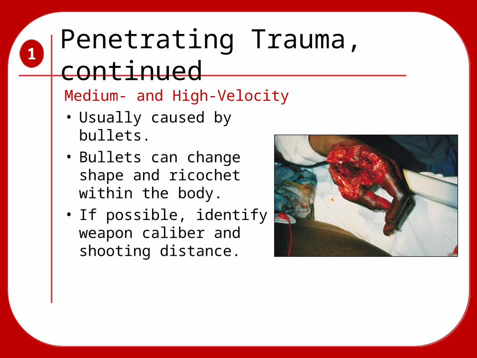
Penetrating Trauma, continued
1
Medium- and High-Velocity • Usually caused by
bullets.• Bullets can change
shape and ricochet within the body.
• If possible, identify weapon caliber and shooting distance.

Injuries to the Head
• Bruising or tearing of the brain• Bleeding or swelling inside the skull is
often life threatening.• Some patients may not have signs
and symptoms.
1

Injuries to the Neck
• Tearing or swelling of trachea can cause life-threatening airway problems.
• Injury to large blood vessels in the neck may produce swelling that prevents blood flow to the brain.
• Open wounds to neck vein bleed heavily or allow air to enter the circulatory system.
1

Injuries to the Chest
• Broken ribs may interfere with chest’s ability to expand normally.
• Large vessels may tear, causing massive bleeding.
1

Pneumothorax
• Air collecting between lung tissue and chest wall
• Compression of lung tissue interferes with oxygen exchange.
• May also interfere with the functioning of the heart (tension pneumothorax)
1

Abdominal Injuries
• Solid organs can tear, lacerate, or fracture, causing serious bleeding and death.
• Hollow organs can leak digestive fluids. • Trauma patients who complain of
abdominal pain may have abdominal bleeding.
1

Mutisystem Trauma Patient
• A patient whose injuries involve more than one body system
1

2 Bleeding

Cardiovascular System2
The cardiovascular system is responsible for supplying and
maintaining adequate blood flow.
The cardiovascular system is responsible for supplying and
maintaining adequate blood flow.
Consists of 3 parts:
• Heart (the pump)
• Blood vessels (the pipes)
• Blood and body fluids (fluids)

Significance of Bleeding
• The body will not tolerate an acute blood loss of greater than 20% of blood volume.
• In the typical adult, 20% is 1 liter or 2 pints.
• A 1-year-old infant typically has 800 mL. A loss of 200 mL is significant.
2

Characteristics of Bleeding2
ArterialArterialBlood is bright red and spurts:

Characteristics of Bleeding2
ArterialArterialBlood is dark red and does not spurt:
VenousVenous

Characteristics of Bleeding2
ArterialArterialBlood oozes out and is controlled easily:
VenousVenous
CapillaryCapillary

Blood Clotting2
• Bleeding normally stops within 10 minutes.
• Some medications interfere with clotting.
• Some injuries will be unable to clot.
• Patients with hemophilia lack clotting factors.

Perfusion2
Circulation in adequate amounts to meet the cells’ needs for oxygen,
nutrients, and waste removal.
Circulation in adequate amounts to meet the cells’ needs for oxygen,
nutrients, and waste removal.
• The heart demands a constant supply of blood.
• The brain and spinal cord can survive for 4 to 6 minutes.
• The kidneys may survive 45 minutes.• The skeletal muscles may last 2 hours.

External Bleeding2
1. Follow BSI precautions.2. Ensure patient has an open airway and
adequate breathing.3. Provide oxygen if necessary.
There are several methods to control bleeding:
• Direct pressure• Elevation• Pressure points

External Bleeding2
Direct pressureDirect pressure• Most common and
effective.• Apply pressure with
gloved finger or hand.

External Bleeding2
• Elevating a bleeding extremity often stops venous bleeding.
• Use both direct pressure and elevation whenever possible.
Direct pressureDirect pressure
ElevationElevation

External Bleeding2
• If bleeding continues, apply pressure on pressure point.
• Pressure points are located where a blood vessel lies near a bone.
Direct pressureDirect pressure
ElevationElevation
Pressure pointsPressure points

Splints2
• Splints can help control bleeding associated with a fracture.
• Air splints can be used to control bleeding of soft-tissue injuries.

Bleeding from the Nose, Ears, and Mouth
2
Causes:• Skull fractures• Facial injuries• Sinusitis• High blood pressure• Coagulation disorders• Digital trauma

Controlling a Nosebleed2
• Help the patient sit and lean forward.
• Apply direct pressure by pinching the patient’s nostrils.
• Or place a piece of gauze bandage under the patient’s upper lip and gum.
• Apply ice over the nose.

Bleeding from Skull Fractures2
• Do not attempt to stop the blood flow.• Do not attempt to push contents back in.• Loosely cover bleeding site with sterile gauze.• Note presence of cerebrospinal fluid coming
from the ears or nose.

Internal Bleeding2
Internal bleeding may not be readily apparent.
Assess patient’s:
• Mechanism of injury• Nature of illness

Signs/Symptoms of Internal Bleeding
2
• Ecchymosis: bruising• Hematoma: bleeding beneath the skin• Hematemesis: blood in vomit• Melena: black, tarry stool• Hemoptysis: coughing up blood• Pain, tenderness, bruising, guarding,
or swelling• Broken ribs, bruises over the lower
chest, or rigid, distended abdomen

Signs of Hypoperfusion (Shock)
2
• Change in mental status• Tachycardia• Weakness• Thirst• Nausea or vomiting• Cold, moist skin• Shallow, rapid breathing

Signs of Hypoperfusion (Shock)
2
• Dull eyes• Dilated pupils• Weak, rapid pulse• Decreased blood pressure• Altered level of consciousness

Emergency Medical Care2
1. Take BSI precautions.2. Decide SICK/NOT SICK.3. Maintain airway and administer oxygen.4. Control external bleeding.5. Quickly assess pulse rate and quality.6. Determine skin condition, color, and
temperature.7. Elevate legs and keep patient warm.8. Transport immediately.

3 Shock

What is Shock?
• State of collapse and failure of the cardiovascular system.
• Leads to inadequate circulation.• Without adequate blood flow, cells
cannot get rid of metabolic wastes. • The result of shock causes the organ,
then organ systems, to fail.
3

Heart (Pump Function)
Damage to the heart by disease or injury.
It cannot move blood adequately to support
perfusion.
Blood Vessels (Pipe Function)
If all the vessels dilate at once, the normal amount of blood volume is not enough
to fill the system and provide adequate perfusion
to the body.
Blood (Content Function)
If blood or plasma is lost, the volume in the container is not enough to support the perfusion needs of the body.
Perfusion Triangle3

Characteristics of Shock (cardiac)
3
Cardiogenic Shock (pump failure)
• Inadequate function of the heart
• Causes a backup of blood into the lungs
• Results in pulmonary edema
• Pulmonary edema leads to impaired ventilation

Characteristics of Shock (cardiac)
3
Neurogenic Shock (pipe failure)
• Damage to the cervical spine may affect control of the size and muscular tone of blood vessels.
• The vascular system increases.
• Blood in the body cannot fill the enlarged system.

Characteristics of Shock (cardiac)
3
Hypovolemic Shock (content failure)
• Results from fluid or blood loss.
• Blood is lost through external or internal bleeding.
• Severe thermal burns cause plasma loss.
• Dehydration aggravates shock.

Characteristics of Shock (cardiac)
3
Combined Pipe and Content Failure
• Some patients with severe bacterial infections, toxins, or infected tissues contract septic shock.
• Toxins damage vessel walls, causing leaking and impairing ability to contract.
• Leads to dilation of vessels and loss of plasma, causing shock.

Non-cardiac Causes of Shock3
Respiratory Insufficiency
• Patient with a severe chest injury or airway obstruction may be unable to breathe adequate amounts of oxygen.
• Insufficient oxygen in the blood will produce shock.

Non-cardiac Causes of Shock3
Anaphylactic Shock
• Occurs when a person reacts violently to a substance.
• Four categories of common causes:
– Injections
– Stings
– Ingestion
– Inhalation

Non-cardiac Causes of Shock3
Psychogenic shock
• Caused by sudden reaction of the nervous system that produces a temporary, generalized vascular dilation.
• Commonly referred to as fainting or syncope.
• Can be brought on by serious causes: irregular heartbeat, brain aneurysm.
• Can be brought on by fear, bad news, unpleasant sights.

Progression of Shock3
Compensated Shock
Compensated Shock
When the body compensates for blood loss.
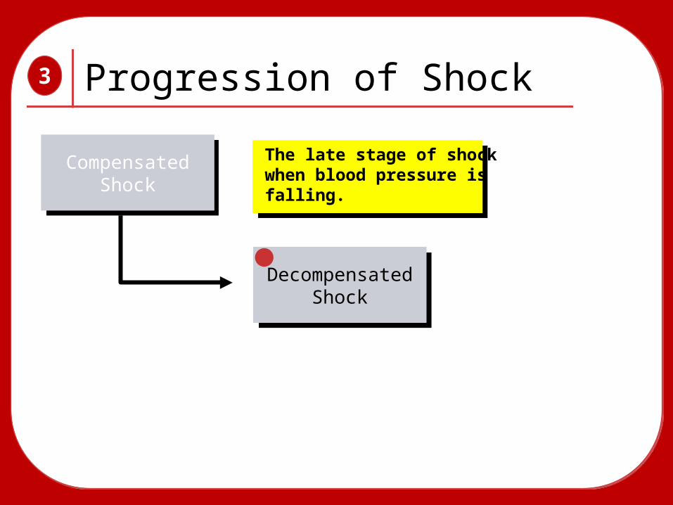
Progression of Shock3
Compensated Shock
Compensated Shock
The late stage of shock when blood pressure is falling.
Decompensated Shock
Decompensated Shock
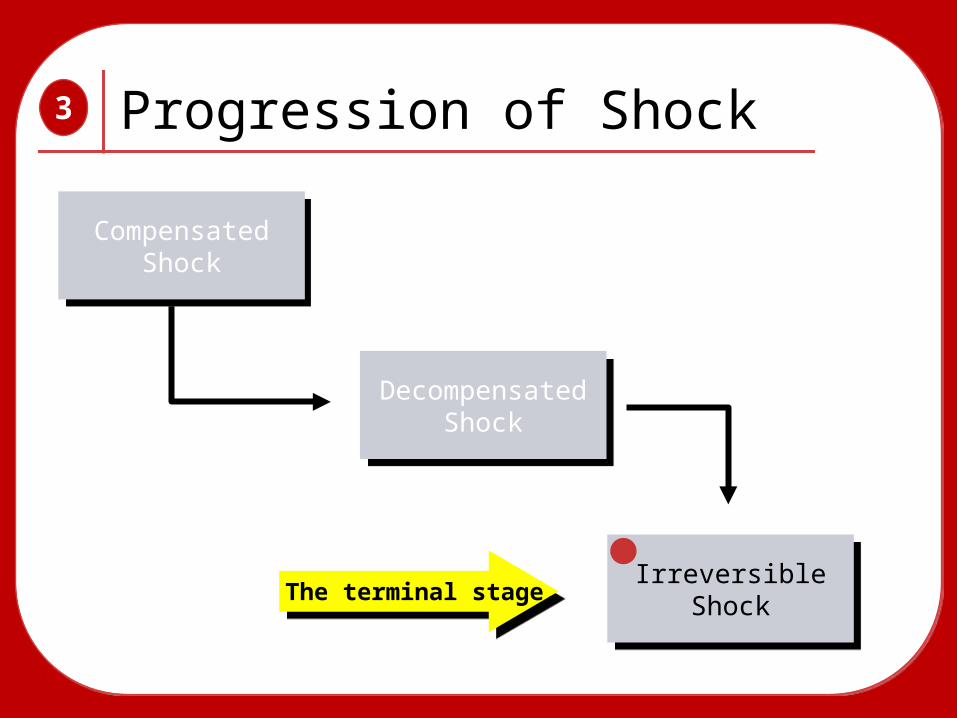
Progression of Shock3
Compensated Shock
Compensated Shock
The terminal stage.
Decompensated Shock
Decompensated Shock
IrreversibleShock
IrreversibleShock

Compensated Shock• Sustained
tachycardia (heart rate of 100-120 or higher)
• Anxiety• Restlessness• Feeling of
impending doom• Weak pulse
3
• Altered mental status• Clammy skin• Pallor• Shallow, rapid
breathing• Shortness of breath• Nausea or vomiting• Delayed capillary
refill• Marked thirst

Decompensated Shock
• Falling blood pressure (<90 mm Hg in an adult)
• Labored, irregular breathing• Ashen, mottled, cyanotic skin• Thready or absent pulse• Dull eyes, dilated pupils• Poor urinary output
3

When to Expect Shock
• Multiple severe fractures• Abdominal or chest injuries• Spine injuries• Severe infection• Major heart attack• Anaphylaxis
3

Emergency Medical Care
• Decide SICK/NOT SICK.
• Ensure patent airway.
• Keep patient supine.
• Control external bleeding.
3

Emergency Medical Care, cont'd• Splint any
broken bones or joint injuries.
• Always provide oxygen.
• Place blankets under and over patient.
3

Emergency Medical Care, cont'd• If not contra-
indicated, elevate feet 6" to 12".
• Do not give the patient anything by mouth.
3

Pneumatic Antishock Garment• Some localities allow EMTs to apply a
pneumatic antishock garment (PASG) for some patients in decompensated shock.
• Know your local protocol regarding their usage.
3

Treating Shock3
Cardiogenic Shock (pump failure)
• Patient may breathe better in a sitting or semi-sitting position.
• Administer high-flow oxygen.
• Assist ventilations as necessary.
• Have suction nearby in case the patient vomits.
• Transport promptly.

Treating Shock3
Neurogenic Shock (pipe failure)
• Maintain airway.
• Assist breathing as needed.
• Keep patient warm.
• Transport promptly.

Treating Shock3
Hypovolemic Shock (content failure)
• Control obvious bleeding.
• Splint any bone or joint injuries. If no contra-indication, raise legs 6" to 12".
• Secure and maintain airway.
• Give oxygen as soon as you suspect shock.
• Transport rapidly.

Treating Shock3
Septic Shock
• Transport as promptly as possible while giving all general support available.
• Give high-flow oxygen during transport.
• Use blankets to conserve body heat.

Treating Shock3
Anaphylactic Shock
• Administer epinephrine.
• Provide prompt transport.
• Provide all possible support:
– Oxygen
– Ventilatory assistance

Treating Shock3
Psychogenic Shock
• It is usually self-resolving.
• Assess patient for injuries from fall.
• If patient has difficulties after regaining consciousness, suspect another problem.

4Soft Tissue Injuries

Anatomy of the Skin4

Function of the Skin
• Protection• Sensation• Temperature control
4

Soft-Tissue InjuriesClosed injuries
• Soft-tissue damage beneath the skin
Open injuries• Break in the surface of the skin
Burns• Soft tissue receives more energy
than it can absorb
4

Contusion• Results from blunt force striking the body
4

Hematoma
• Pool of blood that has collected in the body
4

Crushing Injury
• Occurs when a great amount of force is applied to the body
4

• Observe for hazards. • Take BSI precautions.• You may be able to
identify bleeding before even reaching patient.
• Look for MOI indicators.
4
1. Scene Size-up
Scene Size-up

• Decide SICK/NOT SICK.• Does patient have any
apparent life threats?• Look for hidden injuries.• Ensure patent airway.• Protect patient from
further spinal injury.• Quickly assess breathing.• Palpate chest wall for
DCAP-BTLS.
4
1. Scene Size-up
Initial Assessment
2. Initial Assessment

• If soft-tissue injury is discovered on chest or abdomen:– Check for clear and
symmetrical breath sounds.
• Quickly assess pulse rate and quality.– Pulse will indicate how
aggressively you need to treat for shock.
– Closed soft-tissue injuries do not have visible signs of bleeding.
4
1. Scene Size-up
Initial Assessment, continued
2. Initial Assessment

Focused physical exam• Focus assessment on the
isolated closed injury, complaint, and affected body region.
Rapid physical exam• Perform if significant
trauma has likely affected multiple systems.
• Make sure cervical collar is applied.
4
1. Scene Size-up
Focused History/Physical Exam
2. Initial Assessment
3. Focused History/ Physical Exam

• Any time there is a significant MOI, perform detailed physical exam if time permits.
4
1. Scene Size-up
Detailed Physical Exam
2. Initial Assessment
3. Focused History/ Physical Exam4. Detailed Physical Exam

• Repeat the initial assessment.
• Reassess vital signs frequently.
• Communication and documentation• Provide accurate
account of how you treated injuries.
4
1. Scene Size-up
Ongoing Assessment
2. Initial Assessment
3. Focused History/ Physical Exam4. Detailed Physical Exam
5. Ongoing Assessment

Baseline Vital Signs
• Closed-injury patients may rapidly become unstable.
• Look for tachycardia; tachypnea; low blood pressure; weak pulse; and cool, moist skin.
• Soft-tissue injuries, even without a significant MOI, can cause shock.
4

SAMPLE History
• Obtain from responsive patient or bystanders/family.
• Look for medical ID jewelry or cards.
4

Interventions
• Provide complete spinal immobilization early if spinal injuries are suspected.
• Provide high-flow oxygen.• Treat aggressively for shock.• Request ALS if necessary.• Do not delay transport.
4

RICES• Rest—keep patient quiet and
comfortable as possible.• Ice slows bleeding.• Compression over an injury slows
bleeding.• Elevation above the level of the heart
reduces swelling.• Splinting decreases bleeding and
reduces pain.
4

Common Injuries
Abrasions
4
• Caused by friction

Common Injuries, continuedAbrasion
4
• Jagged cut
Laceration

Common Injuries, continuedAbrasion
4
• Separation of various layers of the skinLaceration
Avulsion

Common Injuries, continuedAbrasion
4
• Results from a sharp pointed objectLaceration
AvulsionPenetrating wound

Common Injuries, continuedAbrasion
4
• Gunshot wounds have unique characteristicsLaceration
AvulsionPenetrating wound
Gunshot wound

Common Injuries, continuedAbrasion
4
• May involve damaged internal organs or broken bones
Laceration
AvulsionPenetrating wound
Gunshot wound
Crushing open wound

• Wear BSI.• Do not touch equipment
with bloody gloves; wear several pairs.
• Beware of contaminating one patient with another patient’s blood.
• Wear eye protection.• Consider MOI.
4
1. Scene Size-up
Scene Size-up

• Decide SICK/NOT SICK.• There may be internal
underlying injuries.• Injuries can affect
airway and breathing.• Provide spinal
immobilization.• If the patient has an
open chest wound, evaluate for bubbling or sucking sounds.
4
1. Scene Size-up
Initial Assessment
2. Initial Assessment

• Quickly place an occlusive dressing over wound.
• Provide high-flow oxygen.
• Assess pulse and skin for shock.
• Control significant bleeding.
4
1. Scene Size-up
Initial Assessment, continued
2. Initial Assessment

Focused physical exam• Focus on isolated injury,
complaint, and affected body region.
Rapid physical exam• Perform if there is
significant trauma affecting multiple systems.
• Look for DCAP-BTLS.• Be sure that spine is
stabilized.
4
1. Scene Size-up
Focused History/Physical Exam
2. Initial Assessment
3. Focused History/ Physical Exam

• Perform if patient is stable and time allows.
4
1. Scene Size-up
Detailed Physical Exam
2. Initial Assessment
3. Focused History/ Physical Exam4. Detailed Physical Exam

• Reassess all bandaging.• Reassess ABCs.• Communication and
documentation• Include description of
MOI and patient’s position.
• Describe location, size, depth of injury.• Provide accurate account
of how you treated injuries.
4
1. Scene Size-up
Ongoing Assessment
2. Initial Assessment
3. Focused History/ Physical Exam4. Detailed Physical Exam
5. Ongoing Assessment

Baseline Vital Signs/SAMPLE HistoryBaseline vital signs
• Will help determine if patient is going into shock
SAMPLE history• Anemia and hemophilia• Medications that thin the blood
(aspirin, prescribed blood thinners)
4

Interventions
• Control bleeding.• If bleeding is not significant, control
later in assessment.• Stabilize spine and assist breathing.• Splint painful, swollen, deformed
extremities.
4

Emergency Medical Care
• Use proper BSI precautions.
• Decide SICK/NOT SICK.
• Administer oxygen.
• Treatment priority is ABCs—including controlling bleeding.
4

Emergency Medical Care, cont'd• Apply dry,
sterile dressing over entire wound.
• Maintain pressure and secure dressing with a roller bandage.
4

Emergency Medical Care, cont'd• Leave original
dressing in place if bleeding continues.
• Apply a second dressing on top of first and secure.
• Splint the extremity.
4

Abdominal Wounds• Open wound in
abdomen may expose organs.
• Organ protruding through abdomen is called an evisceration.
4

• Do not touch exposed organs.
• Cover organs with a moist sterile dressing.
• Transport immediately.
4 Abdominal Wound Management

Impaled Objects• Do not attempt to move
or remove object.
• Control bleeding and stabilize object.
4
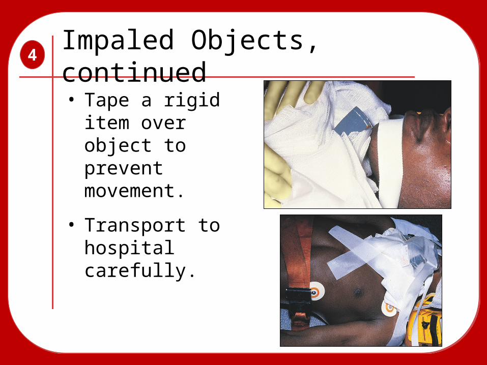
Impaled Objects, continued
• Tape a rigid item over object to prevent movement.
• Transport to hospital carefully.
4

Amputations• Immobilize partial
amputation with bulky dressings and splint.
• Wrap amputated part in dry sterile dressing and place in plastic bag (then place that in another plastic bag).
• Put bag(s) in container filled with ice. Do not let object freeze!
• Transport severed part with patient.
4

Neck Injuries• An open neck injury can
be life threatening.
• Air can get into the veins and cause an air embolism.
4

Neck Injuries, continued• Cover the wound
with an occlusive dressing.
• Apply manual pressure.
• Secure a pressure dressing loosely over the neck and firmly through the opposite axilla.
4

Burns
• Burns account for over 10,000 deaths/year.
• Burns are the most serious and painful injuries.
• Remember to perform a complete assessment on burn patients for other injuries.
4

Determining Burn Severity• What is the depth of the burn?
• What is the extent of the burn?
• Are any critical areas involved?
• Are there any preexisting medical conditions or other injuries?
• Is the patient younger than 5 years or older than 55 years of age?
4

Depth of Burns
Superficial: 1st degree
4
• Involve only top skin layer.

Depth of Burns, continued
Superficial: 1st degree
4
• Involve epidermis and some portion of dermis.
Partial-thickness: 2nd degree

Depth of Burns, continued
Superficial: 1st degree
4
• Extend through all layers of skin.
Partial-thickness: 2nd degree
Full-thickness: 3rd degree
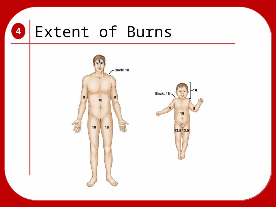
Extent of Burns4

Critical Burns
• Full-thickness burns involving hands, feet, face, upper airway, genitalia, or circumferential burns of other areas
• Full-thickness burns covering more than 10% of total body surface area
• Partial-thickness burns covering more than 30% of total body surface area
• Burns associated with respiratory injury
4

Critical Burns, continued
• Burns complicated by fractures• Burns on patients younger than 5 years
old or older than 55 years old that would be classified as moderate on young adults
4

Moderate Burns
• Full-thickness burns involving 2% to 10% of total body surface area excluding hands, feet, face, upper airway, or genitalia
• Partial-thickness burns covering 15% to 30% of total body surface area
• Superficial burns covering more than 50% of total body surface area
4

Minor Burns
• Full-thickness burns involving less than 2% of the total body surface area
• Partial-thickness burns covering less than 15% of the total body surface area
• Superficial burns covering less than 50% of the total body surface area
4

Pediatric Needs
• Burns to children are considered more serious than burns to adults.
• Children have more surface area relative to body mass than adults.
• Many burns result from abuse.• Report all suspect cases of abuse to the
authorities.
4

Critical Burns in Infants and Children• Full-thickness burns covering more
than 20% of total body surface area• Burns involving hands, feet, face,
upper airway, genitalia
4

Moderate Burns in Infants and Children• Partial-thickness burns covering 10%
to 20% of total body surface area
4

Minor Burns in Infants and Children• Partial-thickness burns covering less
than 10% of total body surface area
4

Emergency Care for Burns• Follow proper BSI
precautions.
• Move patient away from burning area.
• Immerse affected area in cool sterile water or saline solution and cover with cool, wet dressing.
4

Emergency Care for Burns, cont'd• Provide high-flow oxygen.• Prevent body heat loss.• Rapidly estimate the burn’s
severity.• Check for traumatic injuries.
4

Emergency Care for Burns• Treat the patient for shock.
• Provide prompt transport.
4

Chemical Burns• Occur whenever a
toxic substance contacts the body
• Eyes are particularly vulnerable.
• Fumes can cause burns.
• To prevent exposure, wear appropriate gloves and eye protection.
4

Care for Chemical Burns
• Remove the chemical from the patient.
• If it is a powder chemical, brush off first.
• Remove all contaminated clothing.
4

Care for Chemical Burns, cont'd• Flush burned area
with large amounts of water for about 15 to 20 minutes.
• Transport quickly.
4

Chemical Burn to the Eye
• Hold open eyelid while flooding eye with a gentle stream of water.
• Continue flushing en route to hospital.
4

Electrical Burns• Make sure power
is off before touching patient.
• There will be two wounds (an entrance and an exit wound) to bandage.
• Transport patient and be prepared to administer CPR.
4

Small Animal Bites
• All small animal bites should be considered potentially infected.
• Occasionally bites require surgical repair.
• Apply a dry, sterile dressing and transport.
4

Rabies• Potentially fatal viral infection• May be transmitted through biting or
licking an infected wound• Some commons carriers are bats,
squirrels, skunks, foxes, raccoons, and stray dogs.
• Refer to local resources for identification and capture.
• All patients with bites need medical attention.
4

Human Bites
• Very serious injury• Promptly
immobilize with a splint or bandage.
• Apply a dry, sterile dressing.
• Provide transport.
4

Dressing and Bandaging
• Control bleeding.
• Protect wound.
• Prevent contamination.
4

Dressings and Bandages
• Sterile dressings
T̶Used to cover wounds
• Bandaging
T̶Used to keep dressing in place
4

5 AED

AEDAutomated External Defibrillator • Various models.• A specialized computer
recognizes heart rhythms that require defibrillation.
• Some operator interaction required.
5

Cardiac Arrest5
The complete cessation of cardiac activity, either
electrical, mechanical, or both.
The complete cessation of cardiac activity, either
electrical, mechanical, or both.
Ventricular fibrillation

Potential AED Problems• Battery is dead.
• Patient is moving.
• Patient is responsive and has a rapid pulse.
5
AED Advantages
• ALS providers do not need to be on scene.
• Remote, adhesive defibrillator pads are used.
• Efficient transmission of electricity.
5
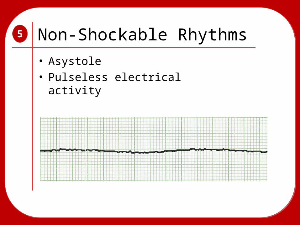
Non-Shockable Rhythms
• Asystole• Pulseless electrical activity
5

• Early defibrillation is the third link in the chain of survival.
• A patient in ventricular fibrillation needs to be defibrillated within 2 minutes.
5
early accessearly
accessearly CPR
early CPR
early defibearly defib
early BLS/ALS
early BLS/ALS
Rationale for Defibrillation

Preparation• Make sure the electricity injures no one.• Do not defibrillate a patient lying in
pooled water.• Dry a soaking wet patient’s chest first.• Do not defibrillate a patient who is
touching metal.• Remove nitroglycerin patches.• Shave a hairy patient’s chest if needed.
5

Using an AED• Assess responsiveness.
• Stop CPR if in progress.
• Check breathing and pulse.
• If patient is unresponsive and not breathing adequately, give two ventilations no more than 1 second in duration.
5

Transport Considerations
Transport:• Keep AED attached.• Check pulse frequently. • Stop ambulance to use an AED.
5

Cardiac Arrest During Transport
5
Check unconscious patient’s pulse every 30 seconds. If pulse is not present:
• Stop the vehicle.• Perform CPR until AED is available.• Analyze rhythm.• Deliver shock.• Continue resuscitation according to
local protocol.

Defibrillator Protocol

AED / CPR Standing Orders• Research continues to indicate a need for more
uninterrupted CPR.
• Longer periods of CPR help keep VF coarse!
• Longer periods of CPR provide greater coronary / cerebral perfusion.
• Pump Cavitations
• Cases of VF on decline.
• Conversion with 1st shock 95%.
5

Standing Orders
• ALL periods of CPR now are 2 minutes in duration.
• Pulse checks eliminated except when a “no shock” is indicated.
• Patient should be removed from PAD device ASAP.
5

Keys To Improved ResuscitationEarly
Access
Early Access
Early CPREarly CPR
Early Defib
Early Defib
Early ALSEarly ALS
5

What Do We Say On a Call?CRITICAL:
• Name of firefighter
• Agency and unit
• Approximate age/gender of patient
• Estimated down time
• By-stander CPR (yes/no/unknown)
"Steve Perry…King County Medic One with a 54 y/o male…approximate downtime of
6-12 minutes…no bystander CPR."
"Steve Perry…King County Medic One with a 54 y/o male…approximate downtime of
6-12 minutes…no bystander CPR."
5

What Do We Say On a Call, cont'dIMPORTANT:
• Applying patches
• Clear before analyze / shocking
• Witnessed / un-witnessed ?
• Pulse checks
• Arrival of medics (most commonly omitted information)
5

When Do We Send a Case?
ANYTIME we do CPR.This includes cases when the defibrillator was not attached.
ANYTIME we do CPR.This includes cases when the defibrillator was not attached.
5

• What questions do you have?
Questions
To review this presentation, go to:http://www.emsonline.net/emtb
To review this presentation, go to:http://www.emsonline.net/emtb
5