secondary complications following cleft palate …
TRANSCRIPT

DISSERTATION ON
SECONDARY COMPLICATIONS FOLLOWING
CLEFT PALATE REPAIR AND MANAGEMENT
M.Ch. PLASTIC SURGERY
(BRANCH III)
MADRAS MEDICAL COLLEGE, CHENNAI
TAMILNADU Dr. M.G.R. MEDICAL UNIVERSITY CHENNAI
FEBRUARY 2010
For August 2012
brought to you by COREView metadata, citation and similar papers at core.ac.uk
provided by ePrints@TNMGRM (Tamil Nadu Dr. M.G.R. Medical University)

BONAFIDE CERTIFICATE
This is to certify that this dissertation is a bonafide record of
the work done by Dr. M. Sundararaj, post graduate student in M. Ch.
Plastic surgery during the period between June 1995 to March 1997 in
the Department of Plastic Surgery, Madras Medical College and
Refresher Course during March 2009 to August 2009 in the Department
of Plastic Surgery, Coimbatore Medical College.
Prof. S. Palanivelu., M.S., M.Ch., Head of the Department, Plastic Surgery,
Coimbatore Medical College, Coimbatore.
DEAN MADRAS MEDICAL COLLEGE Chennai – 600 003
Dr. V. Kumaran M.S., M.Ch., Dean Incharge
Coimbatore Medical College, Coimbatore.
Dr. Anand Subramanian.
R Professor & HOD Department
of Plastic &reconstructive surgery
Madras Medical College

DECLARATION
I, Dr. M. Sundararaj, solemnly declare that the dissertation
titled “SECONDARY COMPLICATIONS FOLLOWING
CLEFT PALATE REPAIR AND MANAGEMENT” is a
bonafide work done by me while doing Post Graduate Course in
Plastic Surgery at Madras Medical College, Chennai during the
academic year of June 1995 to March 1997 under the
supervision and guidance of Prof. D. Kamalakar Rao, M.S.,
M.Ch., Head of the department, Plastic surgery, Madras Medical
College, Chennai and during the refresher course done at
Coimbatore Medical College from March 2009 to August 2009
under the supervision and guidance of Prof. S. Palanivelu.,
M.S., M.Ch., Head of the department, Plastic Surgery,
Coimbatore Medical College.
Place: Coimbatore Dr. M. Sundararaj
Date : 27.09.2009

ACKNOWLEDGEMENT
This study has been done with the permission and guidance
of Professor D. Kamalakar Rao, Formerly Head of the Department of
Plastic surgery, Madras Medical college, Chennai and Professor
S.Palanivelu, Head of the department of Plastic surgery, Coimbatore
Medical College, Coimbatore.
I sincerely thank them for their guidance given to me in doing
this dissertation work on “ Secondary complications following cleft
palate repair and management”.
I am deeply indebted to all my Assistant professors, Department
of Plastic surgery at Madras Medical college and Coimbatore
Medical college for taking active interest in formulating my
dissertation.

CONTENTS
S.NO. PARTICULARS PAGE NO.
1 INTRODUCTION 1
2 EMBRYOLOGY AND ANATOMY OF CLEFT PALATE
2
3 AIM OF THE STUDY 26
4 MATERIALS AND METHODS 27
5 OBSERVATION AND STUDY RESULTS 28
6 DISCUSSION 34
7 CONCLUSION 51
8 REFERENCES 52
9 PROFORMA 55
10 MASTER CHART 56

INTRODUCTION
The clinical evaluation, classification and management of cleft
lip and cleft palate requires the surgeon to rely heavily on a thorough
knowledge of embryology, head & neck anatomy and physical findings.
The cleft lip and palatal defects not only gives functional and
aesthetic problems to the patient but also gives psychic trauma to the
parents as well as the patient.
Even though various techniques are available for repair of cleft lip
and palate, secondary complications do occur in some cases.
In this study the various secondary complications after cleft
palate repair and their management were studied and discussed.
1

EMBRYOLOGY AND ANATOMY OF PALATE AND IN
CLEFT PALATE
EMBRYOLOY
Development of face:
The Stomodeum is bounded by the mandibular arch which
produces the floor of the mouth, lower jaw, and lower lip and two
maxillary processes. Its cranial boundary is the forebrain capsule from
which the frontonasal process grows. The frontonasal process is
intended by two nasal pits which divide it into median and two
lateral nasal processes.
2

The median process is characterized by a pair of converse
globular processes. The lateral nasal processes encircle the eye and
meet together along the line of the nasolacrimal duct.
The maxillary processes unite in the midline below the nostril
to produce the whole of the upper lip and the maxilla. The
frontonasal process produces the premaxilla which emerges on the
facial skeleton is covered by medial extensions of maxilla. From each
maxillary process a flange known as the palatal process grows medially
across the dorsum of the tongue.
3

The two palatal processes and the nasal septum meet and unite
from before backwards thus separating the nasal cavities from each
other and from the mouth forming the nasal capsule.
Chondrification of the nasal capsule occurs and by the sixth
week the nasal walls and hard palate are out lined by a thin layer of
hyaline cartilage.Association of the cartilaginous nasal capsule begins
almost at once spreading from several centers.
In the upper part of the nose the hyaline cartilage is replaced by
bone, but in the lower part of the septum there is deposition of
membranous bone on each surface of the hyaline cartilage. The
cartilage thus sandwitched between two layers of membranous bone is
not absorbed until some time after birth.
The nerve supply of all these structures, inside and outside is
derived from the fifth cranial nerve. The frontonasal process and its
derivatives are supplied by the maxillary division of the fifth nerve and
the lower jaw by the mandibular division of the fifth nerve.
4

Defects of development
The commonest abnormality is cleft lip and palate which may
or may not coexist. Cleft lip is usually lateral and the cleft runs down
from the nostril. The median part of the lip is derived in these cases
from the opposite maxillary process or perhaps from the frontonasal
process, which later normally does not produce any part of the upper
lip. Cleft lip may be bilateral in which case the central part of the lip
between the two clefts is obviously derived abnormally from the
frontonasal process.
Cleft Palate
5

Cleft Palate may be partial or complete. The two palatal processes
unite with each other and with the nasal septum from before backwards.
Arrest of union thus results in a posterior defect that varies from the
mildest form of bifid uvula to gum margin.
In the latter case the cleft almost always runs between premaxilla
and maxilla and involves the jaw between the lateral incisor and canine
teeth. Irregular formation of incisor and canine teeth however often
accompany these defects of palatal development. Very rarely a midline
cleft may separate the two halves of the premaxilla.
According to stark: The primitive palate consists of prolabium,
premaxilla, and cartilaginous septum developing as one unit. If the
mesodermal penetration is not adequate, then the epithelial wall ruptures
forming clefts in which side it is deficient. Three mesodermal masses
within the epithelial wall constitutes the primary palate.
The palate proper which developed behind the nasopalatine
foramen is termed as secondary palate, occurs between 7th - 12th week of
intra uterine life.
6

ANATOMY
Hard palate forms a partition between the nasal and oral cavities.
Its anterior two thirds are formed by the palatine processes of the
maxillae and its posterior one third by the horizontal plates of palatine
bones. The posterior margin gives attachment to the soft palate which
separates the nasopharynx from oropharynx.
MUSCLES OF PALATE
TENSOR VELI PALATINI
The tensor veli palatini is a flat muscle arising from the scaphoid
fossa at the base of the medial pterygoid plate from the spina angularis of
the sphenoid and from the anterolateral aspect of the cartilage of the
eustachian tube.
It runs inferiorly and narrows down towards hamulus, where some
of its bundles are attached. However most of the bundles pass in to a
tendon that turns at right angle around the hamulus and widens like a fan
towards the centre of the palate. It terminates either on the oral side of the
aponeurosis which occupies the whole anterior third of the velum or
directly in to it.
7
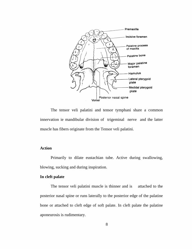
The tensor veli palatini and tensor tymphani share a common
innervation ie mandibular division of trigeminal nerve and the latter
muscle has fibers originate from the Tensor veli palatini.
Action
Primarily to dilate eustachian tube. Active during swallowing,
blowing, sucking and during inspiration.
In cleft palate
The tensor veli palatini muscle is thinner and is attached to the
posterior nasal spine or runs laterally to the posterior edge of the palatine
bone or attached to cleft edge of soft palate. In cleft palate the palatine
aponeurosis is rudimentary.
8

LEVATOR VELI PALATINI
This is a cylindrical muscle, the posterior bundles of which arise
from the undersurface of the apex of the petrous part of temporal bone
and anteromedially from the edge of the canal for the passage of the
internal carotid artery. The anterior bundles arise from the base of
cartilaginous portion of the eustachian tube.
9

Action
Elevates and shifts the soft palate backwards and dilates the
eustachian tube.
In cleft palate
The levator muscles are hypoplastic. In most cases the posterior
bundles pass between the palatopharyngeus to the base of the uvula and
join them. The medial muscle bundles radiate like a fan in to the margin
of the cleft. The anterior bundles are attached by triangular tendinous
portion to the posterior nasal spine and laterally to the posterior edge of
the hard palate while the lateral distinct part of these tendinous bundles
merge with the tensor tendon
10.

PALATOPHARYNGEUS
This muscle is divided into palatine part, pterygopharyngeal part
and the salphingopharyngeal part.
Palatine part arises from the thyroid cartilage and the adjacent part
of the pharyngeal wall through the palatopharyngeal arch to its fan
shaped insertion in the raphe.
Pterygopharyngeal part arises from the posterior and lateral aspects
of the pharynx and inserts on the hamulus and the palatine aponeurosis
intermingling with the pterygopharyngeal part of the superior constrictor.
The salphingopharyngeal part is the weakest portion and become
attached to the inferior edge of the cartilage of the eustachian tube.
Action
Narrows the pharyngonasal isthmus by bringing the
palatopharyngeal arches together. The soft palate is drawn
posteroinferiorly as the palatopharyngeal arches stretch and adduct.
At the same time the thyroid portion lifts the larynx and pharynx mainly
during deglutition. The tubal portion facilitates dilatation of the
eustachian tube by stabilizing its cartilage.
11

In Cleft Palate
The fibrous transformation of palatopharyngeus is less significant
in comparison with tensor and levator. Its palatine insertion differed from
the normal. Even though the smaller part of its fibres ended in the cleft
margin, most of its bundles pass forward along this margin and gets
inserted on the posterior edge of the hard palate and on the posterior nasal
spine.
PALATOGLOSSUS
It is a slender muscle arising from the transverse bundles of the
tongue. It passes up into the palatoglossus arch and gets inserted in a fan
shaped manner into the muscles of the soft palate.
12

Action
Together with its opposite muscle it forms the anterior pretonsillar
sphincter which narrows the Pharyngo oral isthmus. It is antagonistic to
the levator. The palatoglossus lifts the tongue and propels food.
In cleft palate
The palatoglossus passes in a posteroanterior direction in the cleft
margin to the posterior edge of the hard palate, where it gets broadened
and flattened as it reaches its insertion. Its palatal attachment extends in
many cases beyond the posterior edge of the hard palate and become
inserted more anteriorly.
UVULA
The uvular muscles are a cylindrical pair arising from the palatine
aponeurosis and from the posterior nasal spine. They pass nasalwards
from the other palatine muscles on either side of the medial sagittal plane
to the top of the uvula where they end. They lift and bend uvula
backwards and shortens it and the entire soft palate longitudinally.
13

In Cleft Palate
The muscles pass in the cleft margin and their bundles intermingle
with those of the palatopharyngeus and the levator. The isolation of its
fibres is very difficult.
SUPERIOR PHARYNGEAL CONSTRICTOR MUSCLE
This is quadrangular muscle surrounding the upper third of the
pharyngeal wall from behind and laterally. It is the deepest of the
pharyngeal constrictors. The superior Pharyngeal Constrictor muscle
originates from the pterrygoid hamulus, the lateral pterygoid plate , the
pterygomaxillary ligament , the mandible and the floor of the mouth,
and inserts in the posterior pharyngeal raphe. Passavant's ridge is formed
by the upper fibres of the superior constrictor muscle.
14

BLOOD SUPPLY OF PALATE
The descending palatine artery a branch of the maxillary artery
divides in to greater and lesser palantine arteries. The greater palatine
artery is the predominant blood supply to the hard palate. It enters the oral
side of the palate through the grater palatine foramen. The lesser palatine
artery turns posteriorly to supply the side of the anterior half of the
velum.
The posterior septal branches of the nasopalatine artery which is a
branch of maxillary artery supplies the nasal side of the palate. The
posterior alveolar artery arises from maxillary artery supplies the
15

maxillary dentition.. There is vast network between these arteries.
The ascending palatine artery which is a branch of facial artery supplies
the soft palate and its musculature.
NERVE SUPPLY
The tensor veli palatini is innervated by mandibular branch of
trigeminal nerve. Nerve fibres from the pharyngeal plexus innervate the
muscles involved in the movement of velum.. The muscular uvula
receives innervation from the glassopharyngeal nerve through lessor
palatine nerve. Sensory innervation to the alveolus and hard and soft
16

palates is derived from infraorbital branch of the maxillary division of
trigeminal nerve.
Branches from the infraorbital nerve contribute to the formation of
pterygopalatine ganglion from which the greater and lesser palatine
nerves arise to receive sensation from the posterior portion of hard palate
and oral surface of soft palate respectively. The nasopalatine branch of
the infraorbital nerve passes through incisive foramen to reach anterior
hard palate.
OVERALL MUSCLE ARRANGEMENT IN CLEFT PALATE
In cleft palate the muscles extending towards the central line of the
soft palate cannot attach themselves to a fixed point in the raphe of the
soft palate. So they insert at various substitute points, which prevent the
muscles from becoming fully functional, so that their development is
retarded.
The palatoglossus and palatopharyngeus muscles through their
palatine portions find attachment at a very acute angle. Thus most of their
muscle bundles easily bypass the margin of the cleft palate and find a
reliable substitution for insertion in line with their long axis on the
17

posterior edge of the hard palate. The fact is that in all forms of
clefts the cleft muscle becomes thicker in the postnatal life because of the
increasing demands made upon the substitute muscular insertion.
Palatoglossus in cleft palate is hypoplastic. This with
palatopharyngeus are alike in that each of these muscles form a muscular
sling with thinner compact central part with fan shaped edges, which
radiate into mobile organs.
The Levator as it advances to its insertion point in the midline
of the palate almost at a right angle in a less favorable situation. It
approaches the margin of the cleft, but fails to secure a sufficiently
firm print of insertion with the tensor anteriorly and uvulas and
platopharyngeons posteriorly.
This mutual conjugation of all three main muscles of the soft
palate is a typical cleft palate arrangement. Because of the absence of
usual muscular insertions the levator cannot function adequately and
undergo atrophy, as also the tensor muscle.
18

Because of a typical arrangement of the muscles; certain bony
changes usually seen in clefts like large hamulus.
The pterygopharyngeal part of the palatopharyngeus and the
superior constrictors compensate for the loss of function of the soft
palate on the pharyngeal side. Elevation of the soft palate plays the
main part in velopharyngeal closure.
The degree of hypolasia of the levator is directly proportional
to the severity of the cleft palate and by pulling the soft palate supra
laterally causing a further enduring of the cleft. Detaching the cleft
insertions and joining the muscles of both halves of the soft palate in
the midline was therefore be considered the basic principle of cleft
palate surgery.
19

ETIOLOGY OF CLEFT PALATE
1. Genetic factor.
2. Old parental age.
3. Racial Influences- more in oriental and Caucasians.
4. Patients with multiple anomalies like treacher collins syndrome or
stickler syndrome.
5. Environmental factors in utero -Hypoxia, rubella and diabetes.
6. Anaemia, riboflavin, folic, pantothenic and nicotinic acid
deficiencies.
7. Ionizing radiation, hormones, cortisone and alkylating agents - in
utero.
20

CLASSIFICATIONS BY DAVIS AND RITCHIE
Group I - Prealveolar clefts, unilateral, median or bilateral.
Group II - Postalveolar clefts involving the soft palate
only, the soft and hard palates or a submucous
cleft.
Group III - Alveolar clefts, unilateral, bilateral or median.
VEAU'S CLASSIFICATION
Group I - Cleft of the soft palate only
Group II - Cleft of the hard and soft palate tending no
further than the incisive foramen thus involving
the secondary palate alone.
Group III - Complete unilateral cleft, extending from
the Uvula to the incisive foramen in the midline,
then deviating to one side and usually extending
through the alveolus at the position of the future
lateral incisor tooth.
21

Group IV - Complete bilateral cleft, resembling group III
with two clefts extending forward from the
incisive foramen through the alveolus, the small
anterior element of the palate commonly referred
to as the premaxilla, remains suspended from the
nasal septum.
NAGPUR BALAKRISHNAN'S CLASSIFICATION
Group I - cleft lip alone
Group I a - cleft lip with cleft alveolus
Group II - cleft palate
Group III - cleft lip and palate
Submucous cleft
HARKINS CLASSIFICATION BASED ON THE EMBYONIC
PRINCIPLES USED BY KERNAHAN AND STARK
1. Cleft of Primary Palate
A. Cleft lip
1. Unilateral : right, left
a) extent : onethird, twothirds, complete
2. Bilateral : right, Left
a) extent : One third, two thirds, complete.
22

3. Median
a) extent : One third, two thirds, complete
4. Prolabium : Small, Medium, large
5. Congenital scar : right, left median
a) extent : One third, two thirds, complete
B. Cleft of Alveolus
1. Unilateral : right , left
a) extent : One third, two thirds, complete
2. Bilateral : right, Left
a) extent : One third, two thirds, complete
3. Median
a) extent : one third, two thirds, complete
4. Submucous : right, left, median
5. Absent incisor tooth
2. Cleft of Palate
A. Soft Palate
1. Postero anterior : one third, two thirds, complete. 2. Width. maximum (mm)
3. Palatal shortness none, slight, moderate, marked.
4. Submucons cleft a) extent : one third, two thirds, complete.
23

B. Hard Palate
1. Postero anterior : one third, two thirds, complete.
2. Width. maximum (mm)
3. vomer attachment : right, left, absent.
4. Submucous cleft a) extent : one third, two thirds, complete.
KERNAHAN AND STARK CLASSIFICATION
Clefts of primary palate (lip and premaxilla) only
Unilateral (right or left) : Total, Subtotal.
Median : Total (premaxilla absent).
Subtotal (premaxilla rudimentary).
Bilateral : Total, Subtotal.
Clefts of the secondary palate only : Total, Subtotal, Submucous.
Clefts of the primary and secondary palates
Unilateral (right or left) : Total, Subtotal
Median : Total, Subtotal
Bilateral : Total, Subtotal
24

THE STRIPED Y CLASSIFICATION OF KERNAHAN
Circle represents incisive foramen
1 and 4 represents the lip
2 and 5 represents the alveolus
3 and 6 represents the primary palate
7, 8, 9 represents the hard and soft portions of the secondary palate.
25

AIM OF THE STUDY
The main objectives of this study are
1. To evaluate the secondary complications following cleft palate
repair and their management.
2. To discuss about the various types of secondary complications
and methods of treatment.
3. To study about the post operative recurrences.
4. To study about the results.
26

MATERIALS AND METHODS
Materials
Eighteen cases with Secondary complications following cleft
palate repair were seen during the period between June 1995 to March
1997 & March 2009 to August 2009 in the Department of Plastic surgery,
Madras & Coimbatore Medical College respectively.
Methods
The methods include obtaining history regarding the nature of
primary illness, nature of primary surgery and its secondary
complications. Those patients with secondary complications were
evaluated according to their Age, sex, time of primary surgery, immediate
& late post operative history, interval between the primary surgery and its
complications.
Patients with complications were operated according to the nature
of their secondary complications. Regular immediate & late post
operative follow up were made and the results were enumerated.
27

OBSERVATION AND STUDY RESULTS
In our study 18 cases of secondary complications following cleft
palate repair were seen.
Incidence of
Velopharyngeal
incompetence
Incidence of palatal
fistula
Other complications
(malocclusion &
recurrent middle ear
infection)
11 5 1+1
11 cases presented with Velopharyngeal incompetence.
5 cases with palatal fistula
1 case with malocclusion and
1 case with recurrent middle ear infection.
Incidence of Secondary Complications
28
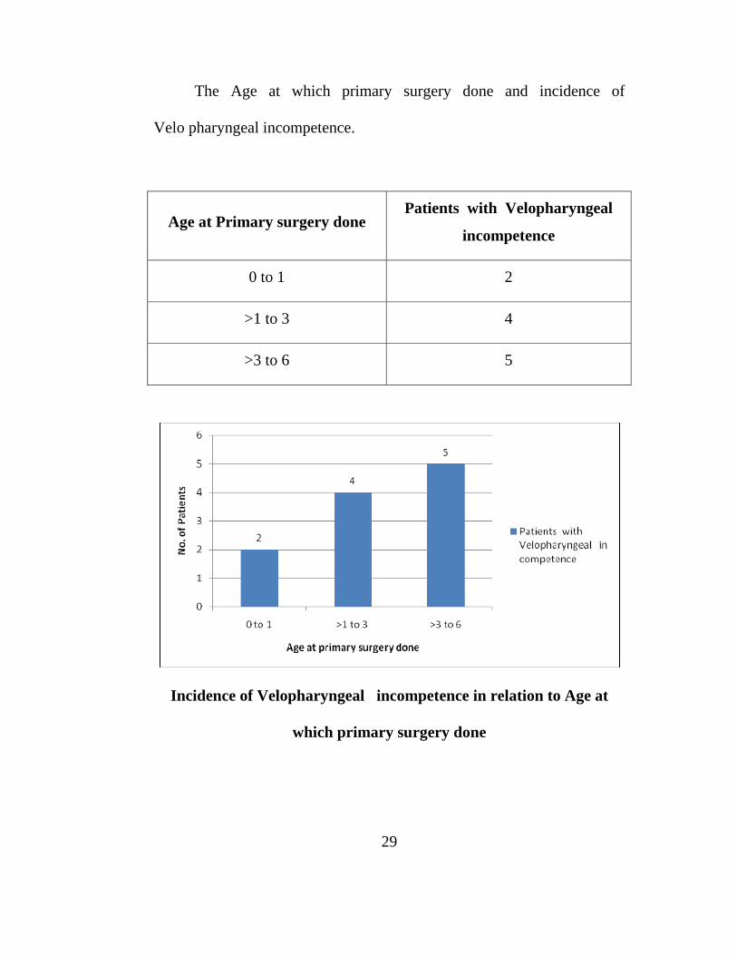
The Age at which primary surgery done and incidence of
Velo pharyngeal incompetence.
Age at Primary surgery done Patients with Velopharyngeal
incompetence
0 to 1 2
>1 to 3 4
>3 to 6 5
Incidence of Velopharyngeal incompetence in relation to Age at
which primary surgery done
29

The Age at which primary surgery done and incidence of Palatal fistula
Age at Primary surgery done Patients with Palatal fistula
0 to 1 0
>1 to 3 5
>3to 6 0
Size of Fistula
Size of Fistula No. of Patients
1.25 cm diameter 1
0.75 to 1 cm 4
Type of cleft prior to primary surgery and incidence of Velopharyngeal in
competence and Palatal fistula
Type of cleft - prior
to primary surgery
Velopharyngeal
incompetence Palatal fistula
Unilateral 11 4
Bilateral 0 1
30

Location of Fistula No. of Patients
At the Junction of hard and soft
palate
1
In the hard palate 4
31

The incidence of male Vs females with secondary complications
following cleft palate repair.
Nature of secondary
complications Male Female
Palatal fistula 3 2
Velopharyngeal
incompetence 6 5
Recurrent middle ear
infection 1 0
Malocclusion 0 1
All patients with Velopharyngeal incompetence were operated with
superiorly based pharyngeal flaps.
32

Out of 5 patients with palatal fistula 3 cases were operated with
hinge flap for nasal layer and local transposition of palatal mucosa for
oral layer and 2 cases with Tongue flap. 1 case had fistula in the junction
of hard and soft palate and 4 cases had fistula in the hard palate.
One patient had fistula measuring 1.25cm in diameter in the
anterior hard palate and the rest measured 0.75 to 1cm in diameter.
Patient with recurrent middle ear infection was treated
conservatively and one patient with malocclusion was treated with palatal
expander.
33

DISCUSSION
The goal of cleft palate surgery is to close the palate with a
technique and timing that produce optimal speech and minimize facial
growth disturbances.
The Operative procedures for cleft palate are
1. Veau -Wardill - Kilner - push back palatoplasty
This has the advantage of providing increased length for the palate
and keeps the levetor muscle in a more favorable position.
Wardill – Kilner
34

2. Furlow's - Double opposing Z plasty
By alternating reversing Z plasties of the nasal and the oral flaps
and keeping the levator palatini within the most posterior flap. Oral and
nasal surfaces of the soft palate in opposite direction. For both of the
Z plasties the central limb is the cleft margin and the posteriorly based
flap is designed to include the levator muscle.
Furlow's - Double opposing Z plasty
35

3. Two flap palatoplasty :
Two flap palatoplasty is freeing mucoperiosteal flaps from the cleft
margins only. The more extensive two flap palatoplasty is a modification
of Langenbeck technique, extending the relaxing jncisions along the
alveolar margins to the edge of the cleft.
36

4. Von Langenbeck operation
Von Langenbeck operation is simple approximation of the cleft
margins with a relaxing incision that begins posterior to the maxillary
tuberosity and follows the posterior portion of the alveolar ridge.
Von Langenbeck Method
5. Vomer flap
Vomer flap is taken by reflecting the mucosa from the septum near
the cleft margin dissecting only enough to close the nasal mucosa of the
opposite side. In bilateral cleft this requires a midline incision along the
septum and two flaps are reflected in each direction.
37

Vomer flap
6. Primary palatoplasty with addition of pharyngeal flap
7. Primary palatoplasty with intravelar veloplasty
Intravelar veloplasty is by re-approximating the levator palatini in
the midline.
COMPLICATIONS AND MANAGEMENT
Immediate
1. Impaired airway-Post operative laryngotracheal oedema should be
treated by steam inhalation, systemic steroids, traction suture in
the tip of the tongue. Rarely tracheostomy may be needed.
2. Haemorrhage and blood loss- bleeding is rarely brisk and is easily
controlled by electrocoagulation. Blood replacement is rarely
necessary.
3. Wound disruption or dehiscence-Suture of the mucoperiosteal
flaps and velum under tension causes wound disruption. Sedation
will help control of crying. Fracture of hamulus reduces tension.
4. Infection should be controlled with appropriate antibiotics.
38

Early
1. Fistula
Tension and wound disruption will cause fistula. An added cause
of fistula is residual epithelium resulting from failure to excise
completely or to incise the cleft margins prior to palatoplasty. Fistula may
close spontaneously if small. If the fistula is less than 5 mm in diameter it
rarely account for either nasal escape of food or nasal speech. Usually
only posterior fistulae are detrimental to speech.
39

2. Nasal speech
Rhinolalia aperta or speech typical of cleft palate may result, if
palatoplasty is done late or if the palate even though closed surgically has
a short anteroposterior dimension, due to mesodermal deficiency of the
velum or from improper surgical technique.
3. Middle ear infection
It should be treated and speech therapy should be given after
evaluating the hearing loss.
Late
1.Velopharyngeal incompetence
40

2. Fistula in the palate
Palatal Fistula
41

3. Midfacial growth abnormality
Decreased maxillary width and the resulting cross bite are managed
by orthodontic maxillary expansion with a fixed appliance. Once
expansion is completed the optimum time for bone grafting is chosen
according to the stage of canine development. Maxillary retrusion or
midface hypoplasia is managed in childhood with a palatofacial device to
aid horizontal growth. Leforte-I advancement osteotomy is delayed until
adolescence to avoid malocclusion related to mandibular growth.
4. Recurrent middle ear infections
Recurrent middle ear infections may need myringotomy with
placement of ventilating tubes.
5. Dentitional problems like malocclusion
Orthodontic treatment is needed to correct dental arch
irregularities, its maintenance and correction of occlusion.
42

SECONDARY SURGICAL PROCEDURES
1. With a failed palatoplasty with Rhinolalia aperta persisting for
atleast .6 months the patient usually needs a v-y retroposition or a
pharyngeal flap operation.
2. For fistula : A soft palate fistula can frequently be excised and the
defect closed in two layers by hinge flap and tongue flap or by
retromolar buccal flap.
Tongue Flap
43

For fistula in the alveolus and adjoining hard palate a simple two layer
closure normally leaves a dead space between the nasal and oral closure.
A buccal flap combined with secondary bone grafting can be used with
autogenous bone taken from iliac crest, caranium or rib. For fistula in the
hard palate Hinge flap and transposition of local palatal mucosa and for
large defects more than 1 cm diameter tongue flap can be used for oral
layer.
Tongue Flap after Division
44

3. Velopharyngeal incompetence should be corrected by
(i) Posterior pharyngeal flaps
The technique of pharyngeal flap surgery involves longitudinal
incisions through the mucosa and muscle down to the
prevertebral fascia on each side of the posterior pharyngeal
wall. Dissection is continued along the prevertebral fascia.
Posterior pharyngeal flap
45

Posterior pharyngeal flap
A superiorly based flap is transversely incised inferiorly and raised
to the level of the palatal plane which usually corresponds to 1-2cm
above the tubercle of the atlas.The flap is usually inset on the nasal side
of uvula with or without opening the midline palate repair.
46

(ii) Reconstruction of velopharyngeal sphincter by
a. Jackson's modification of the orticochea procedure
In this the sphincter is contructed from the posterior tonsillar
pillars and the palatopharyngeus muscle is included in the flaps. The
flaps are sutured together with a small superiorly based posterior flap.
b. Hynes sphincter pharyngoplasty
(iii) Augmentation of posterior pharyngeal wall can be done by
autogenous materials like cartilage or by alloplastic materials like
silicone, Teflon and proplast.
4. Midface Hypoplasia
Leforte-I advancement osteotomy is delayed until adolescence to
avoid mal occlusion related to mandibular growth for midface hypoplasia
causing maxillary retrusion.
Orthodontic treatment is needed to correct dental arch irregularities
it's maintenance and correction of occlusion.
47

5. Recurrent middle ear infections
Recurrent middle ear infections may need myring otomy with
placement of ventilating tubes.
In this study, the incidence of Velopharyngeal incompetence
was found to be 61% and that of palatal fistula was 28%. This proves
that among the secondary complications Velopharyngeal incompetence
was more common than palatal fistula.
Out of the 11 patients operated for Velo pharyngeal incompetence
5 patients were operated for cleft palate in the age between 3-6 years and
4 patients were operated for cleft palate in the age between 1-3 years and
1 patient got operated at the age of 1 year. This proves the incidence of
Velopharyngeal incompetence is increased as the age of the primary
repair is increased - Journal of craniofacial surgery 2009, USA,
September – cutting – Sullivan SR, Marinom EM.
According to cleft palate craniofacial journal USA 1998 March,
Velopharyngeal incompetence is decreased with early cleft palate repair.
48

Journal of craniofacial surgery USA 2009 proved increased
incidents of Velopharyngeal incompetence in patients who underwent
primary surgery for cleft palate in late age.
All the patients with Velopharyngeal incompetence were operated
with superiorly based posterior pharyngeal flap. Among the operated
cases, 10 had improvement in speech and 1 patient had persistent
hypernasality.
For palatal fistula, hinge flap with local palatal mucosa flap was
done for 3 cases and tongue flap was done for 2 cases who had larger and
anteriorly placed fistula.
Out of the 18 patients 17 patients were operated for their cleft
palate primarily with Veau Wardill Kilner push back palatoplasty and 1
patient with Furlow's repair. All 5 patients who developed fistula were
primarily repaired for their cleft palate with Veau Wardill Kilner push
back palatoplasty.
According to journal American society of plastic surgery, the type
of cleft palate and type of repair influence the out come of secondary
49

complications. In complications like fistula 43% occurred after
Wardill type of palatal repair, 10% in Furlow’s repair and 23% in
Van Langenburgh procedure.
Wide clefts after primary repair are more prone to develop fistula
than persons who got operated primarily for narrow unilateral cleft.
In this study 1 patient got primarily operated for bilateral cleft
palate and the secondary complication for this patient was palatal fistula.
It was successfully treated with Hinge flap and tongue flap.
1 patient with malocclusion was treated with palatal expander and
obtained satisfactory occlusion.
50
CONCLUSION
The secondary complications after cleft palate repair like fistula
and Velopharyngeal incompetence can be corrected by secondary repair.
Orthodontic treatment is necessary to correct dental arch irregularities
and correction of occlusion.
Complications like recurrent middle ear infections may require
myringotomy and ventilating tube placements and midface hypoplasia
may require surgical correction with procedures like Leforte I
advancement osteotomy at late teens.
Patients operated for cleft palate at late ages develop speech
difficulties and middle ear infections. Early surgical intervention of cleft
palate avoid the complications and gives better results. Approximating
flaps without tension during primary surgery for cleft palate and control
of infection reduces the risk of secondary fistula.
The addition of pharyngeal flap to primary palatoplasty or with
intravelar veloplasty reduces the secondary complication like
velopharyngeal incompetence
51.

REFERENCES
1. Cleft palate craniofacial journal USA 1998 March. Marinam EM,
Mulliken JB
2. Cleft palate craniofacial journal 2008 March. Study done by Phua,
dechalin, university of Auckland.
3. Annales os plastic surgery 2008 October 1 edition
4. Journal of craniofacial surgery USA 2009 Sulivan SR, Marinam
EM
5. BJ Plastic surgery volume 37 issue 3 July 1984 auther RW Pigott
& FW Reger
6. Journal of oral and maxillofacial surgery volume 45 issue 4
November 1987 auther Posnick & Stanley B. Gets
7. Journal of oral and maxillofacial surgery volume 46 issue 10
October 1988.
8. Kernahan DA on cleft lip and palate classifications plastic & re
consuctive surgery 1973
9. Skoog, T. The pharyngeal flap operation in cleft palate Br. J.
plastic surgery 18 : 265, 1965.
52

10. Bresica, N.J Anatomy of the lip and palate. In W.C. Grab, S.W.
Rosenstein, and K.R. Bzoch (eds) Cleft lip and palate. Boston :
little , Brown, 1971.
11. Fogh. Anderson, P. incidences and Ediology. In M.Edwards and
A.C.H. Watson ( eds.) advances in the management of cleft palate .
Edinburgh Churchill Livingstone , 1980.
12. Millard DR. Jr Cleft craft The evalution of its surgery Volume 3
alveolar and palatel deformities., Boston , Little, Brown, 1980.
13. Grabb and Smith’s Plastic Surgery 6 th Edition
14. Mathes Plastic surgery Volume 4 pediatric plastic surgery.
15. Jhonson , M.C. Embryology of the head and neck. in J.G Mc
Carthy ( ed ) Plastic surgery Philadelphia Saunders 1990 P 2451 –
2495
16. langman J. Head and neck Embryology in Langman ( ed ) medical
Embryology ( 4 th ed ) Baltimore Williams & Wilkins 1981
17. Nomina Embryologica ( 3rd ed ) in Nomina Anatomica ( 6th ed )
Edinburgh Churchill livingstone
18. Millard, D.R. Cleft craft The evaluation of its surgery Bilateraland
rare deformities. Volume II Boston Little, Brown 1977.
53

19. Cohen S.R. Kalinowski J., La Rossa D., et al cleft palate fistulas a
multivariate statistical analysis of prevalence etiology and surgical
management plast reconstr surg 87 : 1041, 1991
20. dorf, D., Curtin J.W. Early cleft palate repair and speech outcome
plast reconstr Surg 70: 75 1982
21. Lewin, M.J., et al VPI due to hypoplasia of the muscular uvulae
and occult submucos cleft palate plaste reconst surg 65 : 585 1980
22. Lewis , M. Secondary soft tissue procedures for cleft lip and palate
mastry of plastic and reconstructive surgery Boston Little Brown &
co vol I chap 44, 1994.
23. Marsh, J.L cleft lip and palate Residual deformity decision making
in oplastic surgery mMarsh St Louis Mosby – Yearbook 86 : 1993
24. Miilard, D.R. Jr, Latham, R.A. improved primary surgical and
dental treatment of clefts plast reconstr surg 86 : 856 – 871, 1990
25. Witt, T.D. D’ Antonio L.L Velopharyngeal insufficiency and
secondary palatal management . A new look at an ols problem clin
plast surg 4 : 707, 1993.
54

PROFORMA
Sl No. :
Name :
Age / Sex :
IP No :
Presentation - secondary complications:
Pre – Operative diagnosis :
Type of cleft :
Previous Surgery done for cleft palate :
Age at which previous surgery done :
Procedure for secondary complications:
Operative Details :
Post Operative Details :
Follow Up :
Exclusion Criteria
Patients with identifiable syndromes, Central Nervous System disorders,
Communicatively significant hearing loss, and non syntromic Robin’s
sequence were excluded.
55

MASTER CHART
S.No Age Sex Pre – Operative diagnosis
Type of cleft Palate prior to
primary surgery
Previous Primary surgery
Age at previous
surgery done
Procedure for secondary complication
Follow Up
1. 7 M Palatal fistula Unilateral Veau Wardill kilner pushback palatoplasty
2 Hinge flap & local transposition flap
No recurrence
2. 9 F Palatal fistula Unilateral Push back palatoplasty 2 Hinge flap & local transposition flap
-do-
3. 12 M Palatal fistula Unilateral Push back palatoplasty 3 Hinge flap & local transposition flap
-do-
4. 14 M Palatal fistula Unilateral Push back palatoplasty 3 Hinge flap with tongue flap -do-
5. 10 F Palatal fistula Bilateral Vomerine flap & push back palatoplasty
2 Hinge flap with tongue flap -do-
6. 9 M Velopharngeal incompetence
Unilateral Veau Wardill kilner pushback palatoplasty
1 Superiorly based posterior pharyngeal flap
Speech improvement +
7. 15 F Velopharngeal incompetence
Unilateral Push back palatoplasty 3 Superiorly based posterior pharyngeal flap
-do-
8. 17 M Velopharngeal incompetence
Unilateral Push back palatoplasty 4 Superiorly based posterior pharyngeal flap
-do-
9. 20 M Velopharngeal incompetence
Unilateral Push back palatoplasty 2 Superiorly based posterior pharyngeal flap
-do-
56

10. 14 F Velopharngeal incompetence
Unilateral Push back palatoplasty 4 Superiorly based posterior pharyngeal flap
-do-
11. 8 M Velopharngeal incompetence
Unilateral Push back palatoplasty 3 Superiorly based posterior pharyngeal flap
Speech improvement +
12. 11 F Velopharngeal incompetence
Unilateral Push back palatoplasty 4 Superiorly based posterior pharyngeal flap
-do-
13. 18 M Velopharngeal incompetence
Unilateral Push back palatoplasty 2 Superiorly based posterior pharyngeal flap
-do-
14. 7 M Velopharngeal incompetence
Unilateral Push back palatoplasty 1 Superiorly based posterior pharyngeal flap
-do-
15. 9 F Velopharngeal incompetence
Unilateral Push back palatoplasty 4 Superiorly based posterior pharyngeal flap
-do-
16. 13 F Velopharngeal incompetence
Unilateral Furlow’s palatoplasty 5 Superiorly based posterior pharyngeal flap
Hypernasality persist
17. 8 M Recurrent middle ear infection
Unilateral Push back palatoplasty 3 Conservative controlled
18. 12 F Malocclusion Unilateral Push back palatoplasty
2 Palatal expander satisfactory
57