section 3, the four components of asthma · pdf filesection 3, the four components of asthma...
TRANSCRIPT

Section 3, The Four Components of Asthma Management
35
August 28, 2007
SECTION 3, THE FOUR COMPONENTS OF ASTHMA MANAGEMENT
Introduction
The Expert Panel Reports presenting clinical practice guidelines for the diagnosis and management of asthma have organized recommendations for asthma care around four components considered essential to effective asthma management:
Measures of assessment and monitoring, obtained by objective tests, physical examination, patient history and patient report, to diagnose and assess the characteristics and severity of asthma and to monitor whether asthma control is achieved and maintained
Education for a partnership in asthma care
Control of environmental factors and comorbid conditions that affect asthma
Pharmacologic therapy
This section updates information on each of these four components, based on the Expert Panel’s review of the scientific literature. The sections that follow present specific clinical recommendations for managing asthma long term and for managing exacerbations that incorporate the four components
Section 3, Component 1: Measures of Asthma Assessment and Monitoring
36
August 28, 2007
SECTION 3, COMPONENT 1: MEASURES OF ASTHMA ASSESSMENT AND MONITORING
Introduction
See section 1, “Overall Methods Used To Develop This Report,” for literature search strategy and tally of results for the EPR—3: Full Report 2007 on this component, Measures of Asthma Assessment and Monitoring. Two Evidence Tables were prepared: 1, Predictors of Exacerbation; and 2, Usefulness of Peak Flow Measurement.
Recommendations for “Component 1: Measures of Asthma Assessment and Monitoring” are presented in five sections: “Overview of Assessing and Monitoring Severity, Control, and Responsiveness in Managing Asthma;” “Diagnosis of Asthma;” “Initial Assessment: Characterization of Asthma and Classification of Asthma Severity;” “Periodic Assessment and Monitoring of Asthma Control Essential for Asthma Management;” and “Referral to an Asthma Specialist for Consultation or Comanagement.” The recommendations are based on the opinion of the Expert Panel and review of the scientific literature.
Overview of Assessing and Monitoring Asthma Severity, Control, and Responsiveness in Managing Asthma
K E Y P O I N T S : O V E R V I E W O F M E A S U R E S O F A S T H M A A S S E S S M E N T A N D M O N I T O R I N G
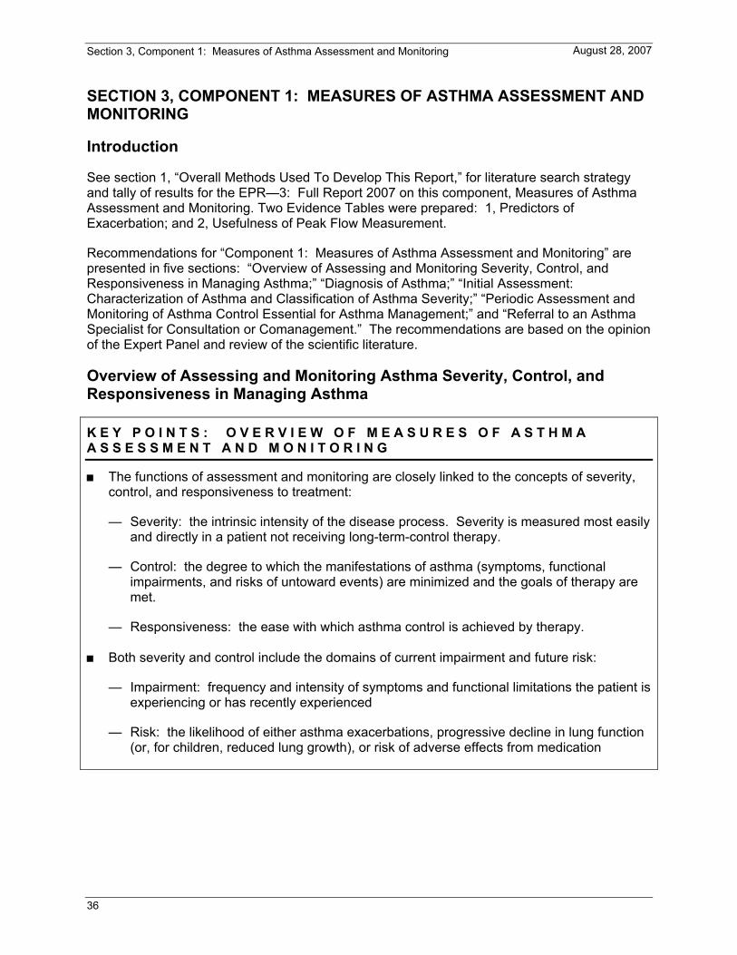
The functions of assessment and monitoring are closely linked to the concepts of severity, control, and responsiveness to treatment:
— Severity: the intrinsic intensity of the disease process. Severity is measured most easily and directly in a patient not receiving long-term-control therapy.
— Control: the degree to which the manifestations of asthma (symptoms, functional impairments, and risks of untoward events) are minimized and the goals of therapy are met.
— Responsiveness: the ease with which asthma control is achieved by therapy.
Both severity and control include the domains of current impairment and future risk:
— Impairment: frequency and intensity of symptoms and functional limitations the patient is experiencing or has recently experienced
— Risk: the likelihood of either asthma exacerbations, progressive decline in lung function (or, for children, reduced lung growth), or risk of adverse effects from medication

Section 3, Component 1: Measures of Asthma Assessment and Monitoring
37
August 28, 2007
The concepts of severity and control are used as follows for managing asthma:
— During a patient’s initial presentation, if the patient is not currently taking long-term control medication, asthma severity is assessed to guide clinical decisions on the appropriate medication and other therapeutic interventions.
— Once therapy is initiated, the emphasis thereafter for clinical management is changed to the assessment of asthma control. The level of asthma control will guide decisions either to maintain or adjust therapy.
— For population-based evaluations, clinical research, or subsequent characterization of the patient’s overall severity, asthma severity can be inferred after optimal therapy is established by correlating levels of severity with the lowest level of treatment required to maintain control. For clinical management, however, the emphasis is on assessing asthma severity for initiating therapy and assessing control for monitoring and adjusting therapy.
K E Y D I F F E R E N C E S F R O M 1 9 9 7 A N D 2 0 0 2 E X P E R T P A N E L R E P O R T S
The key elements of assessment and monitoring are refined to include the separate, but related, concepts of severity, control, and responsiveness to treatment. Classifying severity is emphasized for initiating therapy; assessing control is emphasized for monitoring and adjusting therapy. Asthma severity and control are defined in terms of two domains: impairment and risk.
The distinction between the domains of impairment and risk for assessing asthma severity and control emphasizes the need to consider separately asthma’s effects on quality of life and functional capacity on an ongoing basis (i.e., in the present) and the risks it presents for adverse events in the future, such as exacerbations and progressive loss of pulmonary function. These domains of asthma may respond differentially to treatment.
Diagnosing a patient as having asthma is only the first step in reducing the symptoms, functional limitations, impairment in quality of life, and risk of adverse events that are associated with the disease. The ultimate goal of treatment is to enable a patient to live with none of these manifestations of asthma, and an initial assessment of the severity of the disease allows an estimate of the type and intensity of treatment needed. Responsiveness to asthma treatment is variable; therefore, to achieve the goals of therapy, followup assessment must be made and treatment should be adjusted accordingly. Even patients who have asthma that is well controlled at the time of a clinical assessment must be monitored over time, for the processes underlying asthma can vary in intensity over time, and treatment should be adjusted accordingly.
Section 3, Component 1: Measures of Asthma Assessment and Monitoring
38
August 28, 2007
The functions of assessment and monitoring are closely linked to the concepts of severity, control, and responsiveness to treatment:
Severity: the intrinsic intensity of the disease process. Severity is most easily and directly measured in a patient who is not currently receiving long-term control treatment.
Control: the degree to which the manifestations of asthma (symptoms, functional impairments, and risks of untoward events) are minimized and the goals of therapy are met.
Responsiveness: the ease with which control is achieved by therapy.
An important point linking asthma severity, control, and responsiveness is that the goals are identical for all levels of baseline asthma severity. A patient who has severe persistent asthma compared to a patient who has mild persistent asthma, or a patient who is less responsive to therapy may require more intensive intervention to achieve well-controlled asthma; however, the goals are the same: in well-controlled asthma, the manifestations of asthma are minimized by therapeutic intervention.
Although the severity of disease is most accurately assessed in patients before initiating long-term control medication, many patients are already receiving treatment when first seen by a new health care provider. In such cases, severity can be inferred from the least amount of treatment required to maintain control. This approach presumes that the severity of asthma is closely related to its responsiveness to treatment. Although this assumption may not be true for all forms of asthma and all treatments, it does focus attention on what is important in managing patients who have asthma: achieving a satisfactory level of control.
Both asthma severity and asthma control can be broken down into two domains: impairment and risk. Impairment is an assessment of the frequency and intensity of symptoms and functional limitations that a patient is experiencing or has recently experienced. Risk is an estimate of the likelihood of either asthma exacerbations or of progressive loss of pulmonary function over time.
An assessment of the impairment domain for determining the severity of disease (in patients on no long-term-control treatment before treatment is initiated) or the level of control (after treatment is selected) usually can be elicited by careful, directed history and lung function measurement. Standardized questionnaires like the Asthma Control Test (ACT) (Nathan et al. 2004), the Childhood Asthma Control Test (Liu et al. 2007), the Asthma Control Questionnaire (Juniper et al. 1999b), the Asthma Therapy Assessment Questionnaire (ATAQ) control index (Vollmer et al. 1999), and others have been developed to facilitate and standardize the assessment of the impairment domain of asthma control. Some patients, however, appear to perceive the severity of airflow obstruction poorly (Bijl-Hofland et al. 2000; Kikuchi et al. 1994). These patients may have unconsciously accommodated to their symptoms, or perhaps they have mistakenly attributed these symptoms to other causes, like aging, obesity, or lack of fitness, so that they do not report them readily. For these patients, some other measure, such as spirometry, may identify that the degree of airflow obstruction is poorly recognized or perceived by the patient. A trial of therapy can be initiated and lead to unexpected improvement in quality of life (“I did not realize how much better I could feel until my asthma was treated.”).
Assessment of the risk domain—that is, of adverse events in the future, especially of exacerbations and of progressive, irreversible loss of pulmonary function—is more

Section 3, Component 1: Measures of Asthma Assessment and Monitoring
39
August 28, 2007
problematic. Some assessment of the risk of exacerbations can be inferred from the medical history. Patients who have had exacerbations requiring emergency department (ED) visits, hospitalization, or intensive care unit (ICU) admission, especially in the past year, have a great risk of exacerbations in the future (Adams et al. 2000; Eisner et al. 2001; Lieu et al. 1998). Conversely, the achievement of good control of asthma symptoms and airflow obstruction from treatment with an inhaled corticosteroid (ICS) lowers the risk for asthma exacerbations in the future (Bateman et al. 2004). It is not known, however, whether the minimum treatment to control symptoms necessarily reduces the risk of exacerbations. Some patients who have few current symptoms or impairment of quality of life may still be at grave risk of severe, even life-threatening exacerbations (Ayres et al. 2004). Finally, little is known about the prevalence of a heightened risk of progressive loss of pulmonary function among patients who have asthma or whether any current treatment can prevent it.
The test most used for assessing the risk of future adverse events is spirometry, especially forced expiratory volume in 1 second (FEV1) expressed as a percent of the predicted value or as a proportion of the forced vital capacity (FVC) or FEV1/FVC. The need for a simple, easily applied, more accurate test has prompted study of “biomarkers” whose deviations from normal might correlate with the severity of risk. Many biomarkers have been proposed—airway hyperresponsiveness, blood or sputum eosinophils or eosinophilic cationic protein (ECP), fractional exhaled nitric oxide concentration (FeNO), serum immunoglobulin E (IgE), number of positive skin tests, concentration of hydrogen ion, inflammatory mediators, or various metabolites in an exhaled breath condensate (EBC). Few studies, however, have validated or “anchored” assessment of these markers by analyzing their relationship to the rate of adverse events or decline in pulmonary function over time. Further complicating the matter is that the relationship between normalization of a biomarker and normalization of risk of an adverse event may depend on the specific treatment given. What is found true for treatment with an ICS may not be true for treatment with a leuktotriene receptor antagonist (LTRA) or an inhaled long-acting beta2-agonist (LABA), or vice versa.
In the future, assessment of a combination of historical features and of biomarkers may allow accurate estimation of the risk of future adverse events, but it must be kept in mind that laboratory tests only indirectly estimate control of risk. In the end, only symptoms, exacerbations, and quality of life over time are the measures of asthma control.
Assessment of response to therapy is important, but there is inconsistency about the definition and measurement of “response.” In general, response to therapy describes the ease with which adequate control is achieved by therapy. In a randomized controlled trial (RCT) of interventions to achieve asthma control, decreased symptoms, decreased use of short-acting beta2-agonist (SABA) for quick relief, improved functioning, improvement in FEV1, reduction in exacerbations, fewer ED visits, and decreased side effects from medication were equally weighted to develop a composite score that defines a responder to therapy (Bateman et al. 2004). The investigators observed that a composite definition of a responder correlates with asthma control. In a recent editorial, Stempel and Fuhlbrigge (2005) noted that, in published clinical trials, response to therapy based on pre- or postbronchodilator FEV1 varied widely in statistical significance, depending on the research design and number of subjects included to attain statistical power. Furthermore, when response is defined solely by FEV1, it can be influenced by disease activity independent of the intervention. It may be significant to characterize other responses, such as decreased airway responsiveness as measured by the response to methacholine, frequency of
Section 3, Component 1: Measures of Asthma Assessment and Monitoring
40
August 28, 2007
exacerbations, and decrease in nighttime awakening. This area of work is currently developing and will be influenced by the outcome measures chosen by researchers conducting intervention studies. Agreement is needed on what clinically significant outcomes characterize response to therapy. Agreement is also needed on the time needed to assess response accurately (Zhang et al. 2002), but this time may vary according to treatment. It will take longer to determine whether a patient has responded to a treatment whose principal benefit is reduction in the rate of exacerbations, such as an anti-IgE monoclonal antibody (Bousquet et al. 2004), than to a treatment that acts as an acute bronchodilator.
Another concept closely related to assessing and predicting response to therapy is resistance to therapy. Of adult patients who have asthma, approximately 5 percent have poorly controlled asthma, with frequent symptoms and exacerbations despite use of high-dose ICS (Barnes and Woolcock 1998). Little is known about why some patients who have asthma do not respond well to therapy. A high prevalence of comorbidity—such as uncontrolled gastroesophageal reflux disease (GERD), allergic rhinitis, and psychiatric illness—has been described in this population (Heaney et al. 2003). Patients who have a poor response to appropriate therapy require referral to and consultation with an asthma specialist.
Diagnosis of Asthma
K E Y P O I N T S : D I A G N O S I S O F A S T H M A
To establish a diagnosis of asthma, the clinician should determine that (EPR⎯2 1997):
— Episodic symptoms of airflow obstruction or airway hyperresponsiveness are present.
— Airflow obstruction is at least partially reversible.
— Alternative diagnoses are excluded.
Recommended methods to establish the diagnosis are (EPR⎯2 1997):
— Detailed medical history.
— Physical exam focusing on the upper respiratory tract, chest, and skin.
— Spirometry to demonstrate obstruction and assess reversibility, including in children 5 years of age or older. Reversibility is determined either by an increase in FEV1 of ≥12 percent from baseline or by an increase ≥10 percent of predicted FEV1 after inhalation of a short-acting bronchodilator.
— Additional studies as necessary to exclude alternate diagnoses.

Section 3, Component 1: Measures of Asthma Assessment and Monitoring
41
August 28, 2007
K E Y D I F F E R E N C E S F R O M 1 9 9 7 A N D 2 0 0 2 E X P E R T P A N E L R E P O R T S
Discussions have been added on the use of spirometry, especially in children, and on the criteria for reversibility.
Information has been added on vocal cord dysfunction (VCD) and cough variant asthma as an alternative diagnosis. Reference has been added to updated information in another component on comorbid conditions that may complicate diagnosis and treatment of asthma (e.g., allergic bronchopulmonary aspergillosis (ABPA), obstructive sleep apnea (OSA), and GERD).
The Expert Panel recommends that the clinician trying to establish a diagnosis of asthma should determine that (EPR⎯2 1997):
Episodic symptoms of airflow obstruction are present. Airflow obstruction is at least partially reversible. Alternative diagnoses are excluded.
Box 3–1 lists key indicators for considering a diagnosis of asthma. A careful medical history, physical examination, pulmonary function tests, and additional tests will provide the information needed to ensure a correct diagnosis of asthma. Each of these methods of assessment is described in this section.
Clinical judgment is needed in conducting the assessment for asthma. Patients who have asthma are heterogeneous and present signs and symptoms that vary widely from patient to patient as well as within each patient over time.
MEDICAL HISTORY
The Expert Panel recommends that a detailed medical history of the new patient who is thought to have asthma should address the items listed in figure 3–1 (EPR⎯2 1997). The medical history can help:
Identify the symptoms likely to be due to asthma. See figure 3–2 for sample questions.
Support the likelihood of asthma (e.g., patterns of symptoms, family history of asthma or allergies).
Section 3, Component 1: Measures of Asthma Assessment and Monitoring
42
August 28, 2007
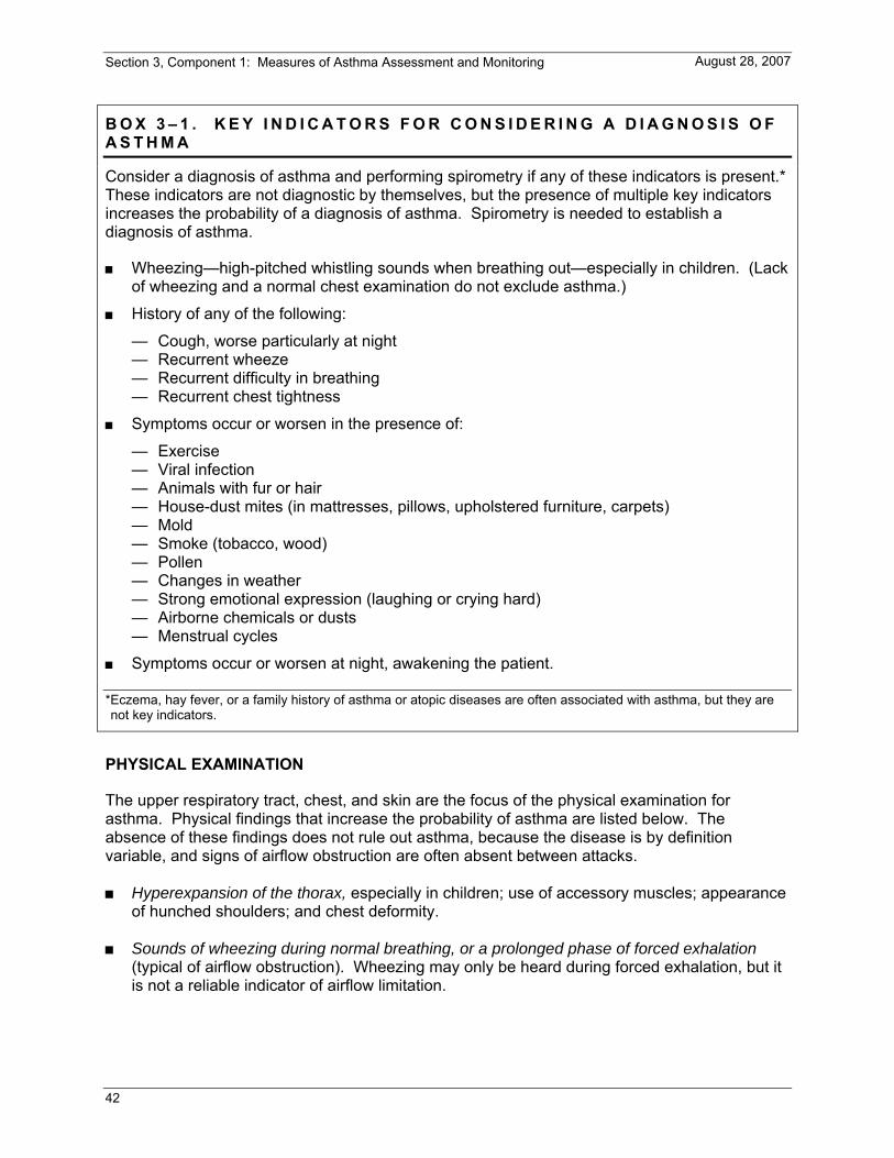
B O X 3 – 1 . K E Y I N D I C A T O R S F O R C O N S I D E R I N G A D I A G N O S I S O F A S T H M A
Consider a diagnosis of asthma and performing spirometry if any of these indicators is present.* These indicators are not diagnostic by themselves, but the presence of multiple key indicators increases the probability of a diagnosis of asthma. Spirometry is needed to establish a diagnosis of asthma.
Wheezing—high-pitched whistling sounds when breathing out—especially in children. (Lack of wheezing and a normal chest examination do not exclude asthma.)
History of any of the following:
— Cough, worse particularly at night — Recurrent wheeze — Recurrent difficulty in breathing — Recurrent chest tightness
Symptoms occur or worsen in the presence of:
— Exercise — Viral infection — Animals with fur or hair — House-dust mites (in mattresses, pillows, upholstered furniture, carpets) — Mold — Smoke (tobacco, wood) — Pollen — Changes in weather — Strong emotional expression (laughing or crying hard) — Airborne chemicals or dusts — Menstrual cycles
Symptoms occur or worsen at night, awakening the patient.
*Eczema, hay fever, or a family history of asthma or atopic diseases are often associated with asthma, but they are not key indicators.
PHYSICAL EXAMINATION
The upper respiratory tract, chest, and skin are the focus of the physical examination for asthma. Physical findings that increase the probability of asthma are listed below. The absence of these findings does not rule out asthma, because the disease is by definition variable, and signs of airflow obstruction are often absent between attacks.
Hyperexpansion of the thorax, especially in children; use of accessory muscles; appearance of hunched shoulders; and chest deformity.
Sounds of wheezing during normal breathing, or a prolonged phase of forced exhalation (typical of airflow obstruction). Wheezing may only be heard during forced exhalation, but it is not a reliable indicator of airflow limitation.
Section 3, Component 1: Measures of Asthma Assessment and Monitoring
43
August 28, 2007
Increased nasal secretion, mucosal swelling, and/or nasal polyps.
Atopic dermatitis/eczema or any other manifestation of an allergic skin condition.
PULMONARY FUNCTION TESTING (SPIROMETRY)
The Expert Panel recommends that spirometry measurements—FEV1, forced expiratory volume in 6 seconds (FEV6), FVC, FEV1/FVC—before and after the patient inhales a short-acting bronchodilator should be undertaken for patients in whom the diagnosis of asthma is being considered, including children ≥5 years of age (EPR⎯2 1997). These measurements help to determine whether there is airflow obstruction, its severity, and whether it is reversible over the short term (Bye et al. 1992; Li and O'Connell 1996). (See box 3–2 for further information.) Patients’ perception of airflow obstruction is highly variable, and spirometry sometimes reveals obstruction much more severe than would have been estimated from the history and physical examination.
B O X 3 – 2 . I M P O R T A N C E O F S P I R O M E T R Y I N A S T H M A D I A G N O S I S
Objective assessments of pulmonary function are necessary for the diagnosis of asthma because medical history and physical examination are not reliable means of excluding other diagnoses or of characterizing the status of lung impairment. Although physicians generally seem able to identify a lung abnormality as obstructive (Russell et al. 1986), they have a poor ability to assess the degree of airflow obstruction (Nair et al. 2005; Shim and Williams 1980) or to predict whether the obstruction is reversible (Russell et al. 1986). Furthermore, pulmonary function measures often do not correlate directly with symptoms. One study reports that one-third of the children who had moderate-to-severe asthma were reclassified to a more severe asthma category when pulmonary function reports of FEV1 were considered in addition to symptom frequency (Stout et al. 2006).
Conversely, a majority of children in another study who had mild-to-moderate asthma classified by symptoms had normal FEV1 (Bacharier et al. 2004). These findings emphasize the importance of using multiple measures and the value of pulmonary function testing in a comprehensive assessment of asthma.
For diagnostic purposes, spirometry is generally recommended over measurements by a peak flow meter in the clinician’s office because there is wide variability even in the published predicted peak expiratory flow (PEF) reference values. Reference values need to be specific to each brand of peak flow meter, and such normative brand-specific values currently are not available for most brands. Peak flow meters are designed as monitoring, not as diagnostic, tools in the office.
Spirometry typically measures the maximal volume of air forcibly exhaled from the point of maximal inhalation (FVC) and the volume of air exhaled during the first second of this maneuver (FEV1). Spirometry is generally valuable in children ≥5 years of age, although some children cannot conduct the maneuver adequately until after age 7. Healthy young children complete exhalation of their entire vital capacity in a few seconds, but it can take older patients much longer, especially patients who have airflow obstruction, because expiratory flow is so low at low lung volumes. In these patients, sustaining a maximal expiratory effort for the time necessary for complete exhalation may be more than 12 or 15 seconds—long enough for some patients to find the maneuver uncomfortable or associated with light headedness. This accounts for the interest in measurement of the FEV6 as a substitute for measurement of FVC in adults. In

Section 3, Component 1: Measures of Asthma Assessment and Monitoring
44
August 28, 2007
adults, FEV6 has been shown to be equivalent to FVC for identifying obstructive and restrictive patterns, using the American Thoracic Society (ATS) algorithm, and to be more reproducible and less physically demanding than FVC (Swanney et al. 2004). Airflow obstruction is indicated by a reduction in the values for both the FEV1 and the FEV1/FVC (or FEV1/ FEV6) relative to reference or predicted values. See figure 3–3a and 3–3b for an example of a spirometric curve for this test. Predicted values for FEV1/FVC are based on National Health and Nutrition Examination Survey (NHANES) data, National Center for Health Statistics, Centers for Disease Control and Prevention (CDC).
Significant reversibility is indicated by ATS standards as an increase in FEV1 of >200 mL and ≥12 percent from the baseline measure after inhalation of a short-acting bronchodilator (e.g., albuterol, 2–4 puffs of 90 mcg/puff) (ATS 1995; ATS/ERS et al. 2005; Pellegrino et al. 2005). Some studies indicate that an increase ≥10 percent of the predicted FEV1 after inhalation of a short-acting bronchodilator may be less subject to bias than measuring percent change from baseline and may have a higher likelihood of separating patients who have asthma from those who have chronic obstructive pulmonary disease (COPD) (Appleton et al. 2005; Brand et al. 1992; Dales et al. 1988; Meslier et al. 1989). Some patients who have signs and symptoms of asthma may not demonstrate reversibility until after a 2- to 3-week trial of oral corticosteroid therapy is administered to help improve their asthma control. Furthermore, the spirometry measured after a single treatment with SABA or after a short course of oral systemic corticosteroid treatment plus acute administration of a bronchodilator may not indicate the patient’s best achievable lung function; thus, followup spirometry measures are indicated as asthma control improves.
Abnormalities of lung function are categorized as restrictive and obstructive defects. A reduced ratio of FEV1/FVC or FEV1/FEV6 indicates obstruction to the flow of air from the lungs, whereas a proportionately reduced FVC (or FEV6 in adults) with a normal or increased FEV1/FVC (or FEV1/FEV6) ratio suggests a restrictive pattern. The severity of abnormality of spirometric measurements is evaluated by comparison of the patient’s results with reference values based on age, height, sex, and race (ATS 1995). Furthermore, chronic asthma may be associated with decreased lung function with a loss of response to bronchodilator. Although asthma is typically associated with an obstructive impairment that is reversible, neither this finding nor any other single test or measure is adequate to diagnose asthma. Many diseases are associated with this pattern of abnormality. The patient’s pattern of symptoms (along with other information from the patient’s medical history) and exclusion of other possible diagnoses also are needed to establish a diagnosis of asthma. In severe cases, the FVC also may be reduced due to trapping of air in the lungs.
When pulmonary function measures are obtained, measuring pulmonary function before and after bronchodilator treatment to determine reversibility is recommended. The degree of airway reversibility correlates with airway inflammation, as measured by sputum eosinophilia and FeNO (Covar et al. 2004a). In addition, those patients who have the greatest degree of reversibility in response to SABA may be at the greatest risk of developing fixed airflow obstruction and have the greatest loss of lung function (Ulrik and Backer 1999). The postbronchodilator FEV1 measure can then be used to follow lung growth patterns over time (Covar et al. 2004b).
The Expert Panel recommends that office-based physicians who care for asthma patients should have access to spirometry, which is useful in both diagnosis and periodic monitoring. Spirometry should be performed using equipment and techniques that meet standards developed by the ATS (EPR⎯2 1997). Correct technique, calibration methods, and maintenance of equipment are necessary to achieve consistently accurate test results

Section 3, Component 1: Measures of Asthma Assessment and Monitoring
45
August 28, 2007
(ATS/ERS et al. 2005). Maximal effort by the patient in performing the test is required to avoid important errors in diagnosis and management. Training courses in the performance of spirometry that are approved by the National Institute for Occupational Safety and Health are available (800–35–NIOSH).
The Expert Panel recommends that when office spirometry shows severe abnormalities, or if questions arise regarding test accuracy or interpretation, further assessment should be performed in a specialized pulmonary function laboratory (EPR⎯2 1997).
DIFFERENTIAL DIAGNOSIS OF ASTHMA
The Expert Panel recommends consideration of alternative diagnoses, as appropriate. Box 3–3 lists examples of possible alternative diagnoses for asthma that may be considered during the evaluation of medical history, physical examination, and pulmonary function. Additional studies are not routinely necessary but may be useful when considering alternative diagnoses (EPR⎯2 1997):
Additional pulmonary function studies (e.g., measurement of lung volumes and evaluation of inspiratory loops) may be indicated, especially if there are questions about possible coexisting COPD, a restrictive defect, VCD, or possible central airway obstruction. A diffusing capacity test is helpful in differentiating between asthma and emphysema in patients, such as smokers and older patients, who are at risk for both illnesses.
Bronchoprovocation with methacholine, histamine, cold air, or exercise challenge may be useful when asthma is suspected and spirometry is normal or near normal. For safety reasons, bronchoprovocation testing should be carried out by a trained individual in an appropriate facility and is not generally recommended if the FEV1 is <65 percent predicted. A positive methacholine bronchoprovocation test is diagnostic for the presence of airway hyperresponsiveness, a characteristic feature of asthma that also can be present in other conditions (e.g., allergic rhinitis, cystic fibrosis, COPD, among others). Thus, although a positive test is consistent with asthma, a negative bronchoprovocation may be more helpful to rule out asthma.
Chest x ray may be needed to exclude other diagnoses.
Allergy testing (see component 3—Control of Environmental Factors and Comorbid Conditions That Affect Asthma).
Biomarkers of inflammation. The usefulness of measurements of biomarkers of inflammation (e.g., total and differential cell count and mediator assays) in sputum, blood, urine, and exhaled air as aids to the diagnosis and assessment of asthma is currently being evaluated in clinical research trials (see “Monitoring Asthma Control With Minimally Invasive Markers and Pharmacogenetics,” in the following section on “Periodic Assessment and Monitoring of Asthma Control Essential for Asthma Management”).
Recurrent episodes of cough and wheezing are due most often to asthma in both children and adults. Underdiagnosis of asthma is a frequent problem, especially in children who wheeze when they have respiratory infections. These children are often labeled as having bronchitis, bronchiolitis, or pneumonia even though the signs and symptoms are most compatible with a diagnosis of asthma. The clinician needs, however, to be aware of other causes of airway
Section 3, Component 1: Measures of Asthma Assessment and Monitoring
46
August 28, 2007
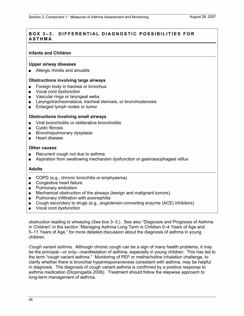
B O X 3 – 3 . D I F F E R E N T I A L D I A G N O S T I C P O S S I B I L I T I E S F O R A S T H M A
Infants and Children
Upper airway diseases Allergic rhinitis and sinusitis
Obstructions involving large airways Foreign body in trachea or bronchus Vocal cord dysfunction Vascular rings or laryngeal webs Laryngotracheomalacia, tracheal stenosis, or bronchostenosis Enlarged lymph nodes or tumor
Obstructions involving small airways Viral bronchiolitis or obliterative bronchiolitis Cystic fibrosis Bronchopulmonary dysplasia Heart disease
Other causes Recurrent cough not due to asthma Aspiration from swallowing mechanism dysfunction or gastroesophageal reflux
Adults
COPD (e.g., chronic bronchitis or emphysema) Congestive heart failure Pulmonary embolism Mechanical obstruction of the airways (benign and malignant tumors) Pulmonary infiltration with eosinophilia Cough secondary to drugs (e.g., angiotensin-converting enzyme (ACE) inhibitors) Vocal cord dysfunction
obstruction leading to wheezing (See box 3–3.). See also “Diagnosis and Prognosis of Asthma in Children” in the section “Managing Asthma Long Term in Children 0–4 Years of Age and 5–11 Years of Age,” for more detailed discussion about the diagnosis of asthma in young children.
Cough variant asthma. Although chronic cough can be a sign of many health problems, it may be the principal—or only—manifestation of asthma, especially in young children. This has led to the term “cough variant asthma.” Monitoring of PEF or methacholine inhalation challenge, to clarify whether there is bronchial hyperresponsiveness consistent with asthma, may be helpful in diagnosis. The diagnosis of cough variant asthma is confirmed by a positive response to asthma medication (Dicpinigaitis 2006). Treatment should follow the stepwise approach to long-term management of asthma.

Section 3, Component 1: Measures of Asthma Assessment and Monitoring
47
August 28, 2007
Vocal cord dysfunction often mimics asthma. VCD is characterized by episodic dyspnea and wheezing caused by intermittent paradoxical vocal cord adduction during inspiration (sometimes with abnormal adduction during expiration as well). The cause of VCD is not well understood, although some patients develop VCD in response to irritant triggers, such as fumes, cold air, and exercise. Although VCD is clearly distinct from asthma, it is often confused with asthma, leading to inappropriate medication of affected individuals with anti-asthma medications. Asthma medications typically do little, if anything, to relieve symptoms if the patient has pure VCD. VCD should be considered in the differential of difficult-to-treat, atypical asthma patients. It is important to note, however, that VCD and asthma may coexist and that VCD may complicate asthma management. Elite athletes, in particular, are prone to both exercise-induced bronchospasm (EIB) and VCD, so careful workup is warranted for athletes who present with exercise-related breathlessness (Rundell and Spiering 2003). During severe VCD episodes, respiratory distress may be severe and lead to intubation. Once the trachea is intubated, the wheezing and distress abate in VCD but not in asthma.
VCD can be difficult to diagnose. Variable flattening of the inspiratory flow loop on spirometry is strongly suggestive of the diagnosis, but abnormalities of the inspiratory loop may well be absent between episodes. The diagnosis of VCD comes from indirect or direct vocal cord visualization during an episode, during which the abnormal adduction can be documented. Therapy generally consists of speech therapy and relaxation techniques (Bucca et al. 1995; Christopher et al. 1983; Newman et al. 1995).
Several conditions that may coexist with asthma can complicate diagnosis: ABPA, OSA, and GERD (See “Component 3: Control of Environmental Factors and Comorbid Conditions That Affect Asthma.”).
Initial Assessment: Characterization of Asthma and Classification of Asthma Severity
K E Y P O I N T S : I N I T I A L A S S E S S M E N T O F A S T H M A
Once the diagnosis has been established, information obtained from the diagnostic evaluation, and additional information, if necessary, should be used to characterize the patient’s asthma in order to guide decisions for therapy (EPR⎯2 1997):
— Identify precipitating factors (e.g., exposure at home, work, daycare, or school to inhalant allergens, or irritants such as tobacco smoke, or viral respiratory infections) (Evidence A)
— Identify comorbidities that may aggravate asthma (e.g., sinusitis, rhinitis, GERD) (Evidence B)
— Classify asthma severity, using measures in both the impairment (Evidence B) and risk domains (Evidence C)
Measures of pulmonary function, using spirometry, are recommended for assessing asthma severity. Low FEV1 indicates current obstruction (impairment domain) and risk for future exacerbation (risk domain) (Evidence C). For children, FEV1/FVC appears to be a more sensitive measure of severity in the impairment domain; FEV1 is a useful measure of risk for exacerbations (Evidence C).

Section 3, Component 1: Measures of Asthma Assessment and Monitoring
48
August 28, 2007
K E Y D I F F E R E N C E S F R O M 1 9 9 7 A N D 2 0 0 2 E X P E R T P A N E L R E P O R T S
The severity classification for asthma changed the category of mild intermittent to intermittent in order to emphasize that even patients who have intermittent asthma can have severe exacerbations. A note of emphasis has also been added that acute exacerbations can be mild, moderate, or severe in any category of persistent asthma.
Severity classification is defined in terms of two domains—impairment and risk—to emphasize the need to consider separately asthma’s effects on quality of life and functional capacity on an ongoing basis (i.e., in the present) and the risks asthma presents for adverse events in the future, such as exacerbations and progressive loss of pulmonary function. These domains of asthma may respond differentially to treatment.
A new emphasis on using FEV1/FVC has been added for to classifying severity in children because it may be a more sensitive measure than FEV1.
The Expert Panel recommends that clinicians use information obtained from the diagnostic evaluation, and any additional information, if necessary, to (EPR⎯2 1997):
Identify precipitating factors Identify comorbid conditions that may aggravate asthma Assess the patient’s knowledge and skills for self-management Classify asthma severity
Once the diagnosis of asthma has been established, the next step in the initial assessment is to characterize the patient’s asthma in order to guide decisions for selecting therapy. This characterization is a basic description of the patient’s asthma phenotype.
As noted earlier, the usefulness of measurements of biomarkers of inflammation (e.g., total and differential cell count and mediator assays) in sputum, blood, urine, and exhaled air as aids to the diagnosis and assessment of asthma is currently being evaluated in clinical research trials (See “Monitoring Asthma Control With Minimally Invasive Markers and Pharmacogenetics,” in the following section on “Periodic Assessment and Monitoring of Asthma Control Essential for Asthma Management.”).
IDENTIFY PRECIPITATING FACTORS
The identification of factors that precipitate worsening of asthma—such as exposure to allergens (e.g., pets, molds, seasonal pollens), irritants (e.g., environmental tobacco smoke (ETS) and industrial pollutants (such as sulfur dioxide and ozone), or respiratory viruses (including “common cold” viruses)—can assist in educating the patient to avoid unnecessary exposures or at least to be alert to exposures that might indicate a need for increased treatment. Information obtained from the medical history (See figure 3–1.) will aid this assessment. See “Component 3: Control of Environmental Factors and Comorbid Conditions That Affect Asthma” for additional tools to assess allergies and other relevant exposures, as well as key messages for patient education on this topic.

Section 3, Component 1: Measures of Asthma Assessment and Monitoring
49
August 28, 2007
IDENTIFY COMORBID CONDITIONS THAT MAY AGGRAVATE ASTHMA
It is also important to identify whether the patient has chronic comorbid conditions that may complicate the presentation or the treatment of asthma, such as sinusitis, rhinitis, GERD, OSA, or ABPA (See “Component 3: Control of Environmental Factors and Comorbid Conditions That Affect Asthma.”). Identification of these comorbid conditions is helpful, because treating them adequately may improve overall control of asthma and lessen requirements for asthma medications.
ASSESS THE PATIENT’S KNOWLEDGE AND SKILLS FOR SELF-MANAGEMENT
Successful management of asthma requires that the patient or patient’s caregiver have a fundamental understanding of and skills for following the therapeutic recommendations, including pharmacotherapy and measures to control factors that contribute to asthma severity. Initial assessment of the patient, therefore, should include an evaluation of the patient’s self-management skills. This evaluation will guide decisions about appropriate educational training. See component 2—Education for a Partnership in Asthma Care for detailed discussion and tools for integrating assessment and education into all phases of clinical management, including the initial patient assessment.
CLASSIFY ASTHMA SEVERITY
The Expert Panel recommends that clinicians classify asthma severity by using the domains of current impairment and future risk (Evidence B—secondary analyses of clinical trials, and Evidence C—observational studies, for assessing impairment; Evidence C, for distinguishing intermittent versus persistent asthma by risk of exacerbations; Evidence D, for distinguishing different categories of persistent asthma by varying frequencies of exacerbations).
Asthma severity is the intrinsic intensity of disease. Initial assessment of patients who have confirmed asthma begins with a severity classification because the selection of type, amount, and scheduling of therapy should then correspond to the level of asthma severity. This initial assessment of asthma severity is made immediately after diagnosis, or when the patient is first encountered, generally before the patient is taking some form of long-term control medication. Assessment is made on the basis of current spirometry and the patient’s recall of symptoms over the previous 2–4 weeks, because detailed recall of symptoms decreases over time. If the assessment is made during a visit in which the patient is treated for an acute exacerbation, then asking the patient to recall symptoms in the period before the onset of the current exacerbation will suffice until a followup visit can be made.
For population-based evaluations, clinical research, or subsequent characterization of the patient’s overall severity, asthma severity can be inferred after optimal therapy is established by correlating levels of severity with the lowest level of treatment required to maintain control. For clinical management, however, the emphasis is to assess asthma severity prior to initiating therapy and, then, assess control for monitoring and adjusting therapy.
The severity classification of asthma shown in figures 3–4 a, b, and c uses the two domains of current impairment and future risk. The specific measures for classifying severity—symptoms, use of SABA for quick relief, exacerbations, and pulmonary function—that were presented in EPR—2 remain in the current report, although they have been organized into the new

Section 3, Component 1: Measures of Asthma Assessment and Monitoring
50
August 28, 2007
framework of measures of impairment and risk. As noted in the “Overview” section of this component, the distinction between impairment and risk emphasizes the need to consider separately asthma’s effects on quality of life and functional capacity on an ongoing basis (i.e., in the present) and the risks asthma presents for adverse events in the future, such as exacerbations and progressive loss of pulmonary function. Clinical trial data demonstrate that these “domains” of asthma may respond differentially to treatment. Data further suggest that, in estimating severity or control in either domain, different manifestations of asthma must be assessed, because they do not necessarily correlate with each other (Bacharier et al. 2004; Colice et al. 1999; Fuhlbrigge et al. 2002; Strunk et al. 2002). Thus, a composite of measures, with a distinction between domains of impairment and risk, will be useful in classifying severity.
Assessment of Impairment
Assessment of severity requires assessing the following components of current impairment:
Symptoms
— Nighttime awakenings — Need for SABA for quick relief of symptoms — Work/school days missed — Ability to engage in normal daily activities or in desired activities — Quality-of-life assessments
Lung function, measured by spirometry: FEV1, FVC (or FEV6), FEV1/FVC (or FEV6 in adults). Spirometry is the preferred method for measuring lung function to classify severity. Peak flow has not been found to be a reliable variable for classifying severity (Eid et al. 2000; Llewellin et al. 2002), but it may serve as a useful tool for monitoring trends in asthma control over time (See section, “Monitoring Lung Function.”).
Secondary analyses of clinical trial data and observational studies using the EPR—2 1997 or similar Global Initiative for Asthma (GINA) criteria have confirmed that the parameters for the impairment domain (symptom, activity levels, and pulmonary function) reflect increasing gradients of severity in adults (Antonicelli et al. 2004; Diette et al. 2004; EPR⎯2 1997; Schatz et al. 2003, 2005b).
Whether the ranges of pulmonary function for severity of asthma previously defined in guidelines (EPR⎯2 1997) apply well to children has been questioned in cross-sectional studies that found normal FEV1 values (many over 90 percent predicted) in a majority of the children, 5–18 years of age, regardless of their asthma severity as classified on the basis of symptoms (Bacharier et al. 2004; Paull et al. 2005; Spahn et al. 2004). Two of those studies reported that, in contrast to FEV1 measures, FEV1/FVC decreased with increasing asthma severity and thus appeared to be a more sensitive measure of severity (Bacharier et al. 2004; Paull et al. 2005). On the other hand, analysis of a large, longitudinal study of children confirmed a relationship between the severity of airflow obstruction and the risk of exacerbations (Fuhlbrigge et al. 2001). Increasing risk correlated with the FEV1 cutoffs for increasing levels of severity as defined in EPR—2 (Fuhlbrigge et al. 2006). It is emphasized that these studies also found that even children who had normal values of lung function experienced exacerbations. In addition, children who have low lung function are at greatest risk of developing fixed airflow obstruction over time (Rasmussen et al. 2002). Cumulatively, these studies underscore the importance of measuring several variables in the assessment of asthma. Making treatment decisions for children should be based on frequency and severity of past exacerbations and symptoms, with
Section 3, Component 1: Measures of Asthma Assessment and Monitoring
51
August 28, 2007
pulmonary function measures as an additional guide. FEV1 appears to be a useful measure indicating risk for exacerbations; FEV1/FVC appears to be a more sensitive measure of severity in the impairment domain. The Expert Panel has updated the pulmonary function measures for assessing asthma severity and control in children by adding suggested ranges for FEV1/FVC.
Assessment of Risk
A closely related and second dimension of severity is the concept of risk of adverse events, including exacerbations and risk of death. Assessment of the risk of future adverse events requires careful medical history, observation, and clinician judgment. Documentation of warning signs and adverse events will be necessary when a patient is felt to be at increased risk. Patients who are deemed at increased risk of adverse outcomes will need close monitoring and frequent assessment by their clinicians.
Exacerbations of asthma are acute or subacute episodes of progressively worsening shortness of breath, cough, wheezing, and chest tightness—or some combination of these symptoms. Exacerbations are characterized by decreases in expiratory airflow that can be documented and quantified by simple measurement of lung function (spirometry or PEF). Exacerbations of asthma can vary widely among individuals and within individuals, from very rare to frequent. Although the classification of severity focuses on the frequency of exacerbations, it is important to note that the severity of disease does not necessarily correlate with the intensity of exacerbations, which can vary from mild to very severe and life-threatening. Patients at any level of severity, even intermittent asthma, can have severe exacerbations. For example, a person who has intermittent asthma can have a severe exacerbation during a viral illness or when exposed to allergens to which he or she is sensitized or to noxious fumes and irritants. Accordingly, the Expert Panel has modified the designation of “mild intermittent asthma” in the previous guidelines (EPR⎯2 1997; EPR⎯Update 2002) to become “intermittent asthma” to emphasize that patients at any level of severity—including intermittent—can have severe exacerbations. The duration of exacerbations may vary from a few hours to a few days. These unpredictable variations in exacerbations can present treatment dilemmas for the clinician who strives to prevent future exacerbations and considers when to initiate chronic anti-inflammatory therapy.
The frequency of exacerbations requiring intervention with oral systemic corticosteroids has been correlated in observational studies with the designation of persistent, rather than intermittent, asthma (Fuhlbrigge et al. 2001, 2006). Determination of whether the level of severity is mild, moderate, or severe will depend on consideration of both the frequency and the intensity of the exacerbations. No data are available to correspond specific numbers with each severity category. In general, the more frequent and the more intense the exacerbations (e.g., requiring urgent, unscheduled clinical care, hospitalization, or ICU admission), the greater the degree of underlying disease severity.
Predictors that have been reported to be associated with increased risk of exacerbations (See Evidence Table 1, Predictors of Exacerbations.) or death include:
— Severe airflow obstruction, as detected by spirometry (Adams et al. 2000; Connolly et al. 1998; Fuhlbrigge et al. 2001, 2006; Kitch et al. 2004).
— Persistent severe airflow obstruction (Kitch et al. 2004).

Section 3, Component 1: Measures of Asthma Assessment and Monitoring
52
August 28, 2007
— Two or more ED visits or hospitalizations for asthma in the past year; any history of intubation or ICU admission, especially if in the past 5 years (Belessis et al. 2004; Cowie et al. 2001).
— Patients report that they feel in danger or frightened by their asthma (Janson-Bjerklie et al. 1993; Ng 2000).
— Certain demographic or patient characteristics: female, nonwhite (Diette et al. 2002), nonuse of ICS therapy, and current smoking (Eisner et al. 2001).
— Psychosocial factors: depression (Eisner et al. 2005; Goodwin et al. 2004), increased stress (Goodwin et al. 2004), socioeconomic factors (Griswold et al. 2005).
— Attitudes and beliefs about taking medications (Adams et al. 2000; Apter and Szefler 2004).
For population-based management, risk stratification is used to identify patients at increased risk of morbidity and health care resource use. Several validated psychometric instruments have been shown to predict future risk of hospitalization and ED visits (Schatz et al. 2005a).
Periodic Assessment and Monitoring of Asthma Control Essential for Asthma Management
K E Y P O I N T S : P E R I O D I C A S S E S S M E N T O F A S T H M A C O N T R O L
The goals of therapy are to achieve asthma control by (Evidence A):
— Reducing impairment:
♦ Prevent chronic and troublesome symptoms (e.g., coughing or breathlessness in the daytime, in the night, or after exertion)
♦ Require infrequent use (≤2 days a week) of inhaled SABA for quick relief of symptoms
♦ Maintain (near) “normal” pulmonary function
♦ Maintain normal activity levels (including exercise and other physical activity and attendance at work or school)
♦ Meet patients’ and families’ expectations of and satisfaction with asthma care

Section 3, Component 1: Measures of Asthma Assessment and Monitoring
53
August 28, 2007
— Reducing risk:
♦ Prevent recurrent exacerbations of asthma and minimize the need for ED visits or hospitalizations
♦ Prevent progressive loss of lung function; for children, prevent reduced lung growth
♦ Provide optimal pharmacotherapy with minimal or no adverse effects
Periodic assessments (at 1- to 6-month intervals) and ongoing monitoring of asthma control are recommended to determine if the goals of therapy are being met and if adjustments in therapy are needed (Evidence B, extrapolation from clinical trials; and Evidence C, observational studies). Measurements of the following are recommended:
— Signs and symptoms of asthma
— Pulmonary function
— Quality of life/functional status
— History of asthma exacerbations
— Pharmacotherapy (checking for adherence to therapy and potential side effects from medication)
— Patient–provider communication and patient satisfaction
Clinician assessment and patient self-assessment are the primary methods for monitoring asthma. Population-based assessment is used by health organizations, such as managed care organizations and disease management programs (EPR⎯2 1997).
The following frequencies for spirometry tests are recommended: (1) at the time of initial assessment (Evidence C), (2) after treatment is initiated and symptoms and PEF have stabilized, (3) during periods of progressive or prolonged loss of asthma control, and (4) at least every 1–2 years (Evidence D).
Use of minimally invasive markers (“biomarkers”) to monitor asthma control and guide treatment decisions for therapy is of increasing interest. Some markers, such as spirometry measures, are currently and widely used in clinical care; others, such as sputum eosinophils and FeNO, may also be useful, but they require further evaluation in both children and adults before they can be recommended as clinical tools for routine asthma management (Evidence D).
Provide to all patients a written asthma action plan based on signs and symptoms and/or PEF; written action plans are particularly recommended for patients who have moderate or severe persistent asthma, a history of severe exacerbations, or poorly controlled asthma (Evidence B).
Whether peak flow monitoring, symptom monitoring (available data show similar benefits for each), or a combination of approaches is used, self-monitoring is important to the effective self-management of asthma (Evidence A).
Section 3, Component 1: Measures of Asthma Assessment and Monitoring
54
August 28, 2007
Patients should be taught to recognize symptom patterns indicating inadequate asthma control and the need for additional therapy (Evidence A).
Consider peak flow monitoring for patients who have moderate or severe persistent asthma, patients who have a history of severe exacerbations (Evidence B), and patients who poorly perceive airflow obstruction and worsening asthma (Evidence D). Long-term daily peak flow monitoring can be helpful to (Evidence B):
— Detect early changes in asthma control that require adjustment in treatment. — Evaluate responses to changes in treatment. — Provide a quantitative measure of impairment.
K E Y D I F F E R E N C E S F R O M 1 9 9 7 A N D 2 0 0 2 E X P E R T P A N E L R E P O R T S
Periodic assessment of asthma control is emphasized.
This update (EPR—3: Full Report 2007) makes a stronger distinction than previous guidelines between classifying asthma severity and assessing asthma control. Interpretation of previous asthma guidelines raised questions about applying the severity classifications once treatment is established and also resulted in placing more emphasis on severity than on ongoing monitoring of whether therapeutic goals were met. This update (EPR—3: Full Report 2007) clarifies the issue:
— For initiating treatment, asthma severity should be classified, and the initial treatment should correspond to the appropriate severity category.
— Once treatment is established, the emphasis is on assessing asthma control to determine if the goals for therapy have been met and if adjustments in therapy (step up or step down) would be appropriate.
Assessment of asthma control includes the two domains of impairment and risk.
Peak flow monitoring: The recommendation to assess diurnal variation was deleted. New text was added regarding the patients most likely to benefit from routine peak flow monitoring. Emphasis was added that evidence suggests equal benefits to either peak flow or symptom-based monitoring; the important issue continues to be having a monitoring plan in place.
Parameters for lung function, specifically FEV1/FVC, were added as measures of asthma control for children.
Minimally invasive markers and pharmacogenetic approaches for monitoring asthma. New text was added. These approaches have gained increasing attention in clinical research, and some applications may be useful in the near future for the clinical management of asthma. The concepts are introduced here, although most require further evaluation before they can be recommended as tools for routine asthma management.
Section 3, Component 1: Measures of Asthma Assessment and Monitoring
55
August 28, 2007
GOALS OF THERAPY: ASTHMA CONTROL
The purpose of periodic assessment and ongoing monitoring is to determine whether the goals of asthma therapy are being achieved and asthma is controlled. When asthma is not controlled, it is associated with significant asthma burden (Fuhlbrigge et al. 2002), decreased quality of life (Schatz et al. 2005b), and increased health care utilization (Schatz et al. 2005a; Vollmer et al. 2002). The level of asthma control (well controlled, not well controlled, or poorly controlled) is the degree to which both dimensions of the manifestations of asthma—impairment and risk—are minimized by therapeutic intervention. The level of control at the time of followup assessment will determine clinical actions—that is, whether to maintain or adjust therapy. In previous guidelines (EPR⎯2 1997; GINA 2002), parameters for control were selected on the basis of research that used individual outcomes for evaluating the effectiveness of asthma treatments. The composite list of goals reflected the Panel’s opinions of a complete list of relevant outcomes that could define asthma control. A recent large international trial demonstrated that significant reductions in the rate of severe exacerbations and improvements in quality of life were achieved by aiming at achieving guideline-defined asthma control and by adjusting therapy to achieve it. At the end of 1 year, 30 percent of the patients achieved total control (i.e., the absence of any sign or symptom of asthma), and 60 percent had achieved well-controlled asthma (Bateman et al. 2004).
Interpretation of previous asthma guidelines, in which severity classifications before treatment corresponded to recommended steps of treatment, has raised questions about applying severity classifications once treatment is established and what elements of asthma should be used to monitor asthma during clinical followup (Graham 2006; Wolfenden et al. 2003). This update (EPR—3: Full Report 2007) clarifies the issue. For initiating treatment, asthma severity should be classified, and the initial treatment should correspond to the appropriate category of severity. Once treatment is established, the emphasis is on assessing asthma control to determine if the goals for therapy have been met and if adjustments in therapy (step up or step down) would be appropriate.
The Expert Panel recommends that asthma control be defined as follows (Evidence A):
Asthma Control
Reduce impairment
— Prevent chronic and troublesome symptoms (e.g., coughing or breathlessness in the daytime, in the night, or after exertion)
— Require infrequent use (<2 days a week) of SABA for quick relief of symptoms
— Maintain (near) “normal” pulmonary function
— Maintain normal activity levels (including exercise and other physical activity and attendance at work or school)
— Meet patients’ and families’ expectations of and satisfaction with asthma care

Section 3, Component 1: Measures of Asthma Assessment and Monitoring
56
August 28, 2007
Reduce risk
— Prevent recurrent exacerbations of asthma and minimize the need for ED visits or hospitalizations
— Prevent progressive loss of lung function; for children, prevent reduced lung growth
— Provide optimal pharmacotherapy with minimal or no adverse effects
See figures 3–5a, b, and c for classification of asthma control in three different age groups. Specific discussion of measures for assessment are in the following section. In general:
Assessment of impairment is in the form of questions, such as those presented in figure 3–6 and within figure 3–7. The focus of these questions is to assess the degree of asthma control in the present. The key elements include current pulmonary function and patient’s recall of symptoms, physical activity, quality of life, and need for SABA for quick relief of symptoms over the previous 2–4 weeks.
Assessing the risk of exacerbations is through questions regarding the use of medications, particularly oral corticosteroids, or urgent care visits. Low FEV1 is associated with increased risk for severe exacerbations (Fuhlbrigge et al. 2001).
Assessment of the risk of progressive loss function, or, for children, the risk of reduced lung growth (measured by prolonged failure to attain predicted lung function values for age) requires longitudinal assessment of lung function, preferably using spirometry.
Assessment of the risk of side effects from medication does not directly correspond to the varying levels of asthma control. For example, a patient might have well-controlled asthma with high doses of ICS and chronic oral corticosteroids but is likely to experience some adverse effects from this intense therapy. The risk of side effects can vary in intensity from none to very troublesome and worrisome; see component 4—Medications for discussion of potential adverse effects associated with different asthma medications. Although not directly correlated to control, the risk or evidence of side effects should be included in the overall assessment of the risk domain of asthma control.
Future work on assessment of asthma control tools will define the relative value of including specific biological markers and test how well the tool predicts the risk of exacerbations.
MEASURES FOR PERIODIC ASSESSMENT AND MONITORING OF ASTHMA CONTROL
The Expert Panel recommends that ongoing monitoring of asthma control be performed to determine whether all the goals of therapy are met—that is, reducing both impairment and risk (Evidence B); see figures 3–5 a, b, and c for assessing asthma control for different age groups.
The Expert Panel recommends that the frequency of visits to a clinician for review of asthma control is a matter of clinical judgment; in general, patients who have intermittent or mild persistent asthma that has been under control for at least 3 months should be seen by a clinician about every 6 months, and patients who have uncontrolled and/or severe persistent asthma and those who need additional supervision to help them follow their treatment plan need to be seen more often (EPR⎯2 1997).

Section 3, Component 1: Measures of Asthma Assessment and Monitoring
57
August 28, 2007
The assessment measures for control monitor six areas described in this section and are recommended based on the opinion of the Expert Panel and review of the scientific literature. A seventh area, monitoring asthma control with minimally invasive markers, is of increasing interest, but many of these markers require further evaluation before they can be recommended widely for routine asthma care.
Monitoring signs and symptoms of asthma
Monitoring pulmonary function
— Spirometry — Peak flow monitoring
Monitoring quality of life
Monitoring history of asthma exacerbations
Monitoring pharmacotherapy for adherence and for potential side effects
Monitoring patient–provider communication and patient satisfaction
Monitoring asthma control with minimally invasive markers and pharmacogenetics (requires further evaluation)
Monitoring Signs and Symptoms of Asthma
The Expert Panel recommends that every patient who has asthma should be taught to recognize symptom patterns that indicate inadequate asthma control (Evidence A) (See also “Component 2: Education for a Partnership in Asthma Care.”). Either symptom and/or PEF monitoring should be used as a means to determine the need for intervention, including additional medication, in the context of a written asthma action plan.
The Expert Panel recommends that symptoms and clinical signs of asthma should be assessed at each health care visit through physical examination and appropriate questions (EPR⎯2 1997). This is important for optimal asthma care.
The Expert Panel recommends that the detailed symptoms history should be based on a short (2–4 weeks) recall period (EPR⎯2 1997). Patients’ detailed recall of symptoms decreases over time; therefore, the clinician may choose to assess over a 2-week, 3-week, or 4-week recall period. Symptom assessment for periods longer than 4 weeks should reflect more global symptom assessment, such as inquiring whether the patient’s asthma has been better or worse since the last visit and inquiring whether the patient has encountered any particular difficulties during specific seasons or events. Figure 3–7 provides an example of a set of questions that can be used to characterize both global (long-term recall) and recent (short-term recall) asthma symptoms.
Section 3, Component 1: Measures of Asthma Assessment and Monitoring
58
August 28, 2007
The Expert Panel recommends that assessment of the patient’s symptom history should include at least four key symptom expressions (Evidence B, extrapolation from clinical trials; and Evidence C, from observational studies):
Daytime asthma symptoms (including wheezing, cough, chest tightness, or shortness of breath)
Nocturnal awakening as a result of asthma symptoms
Frequency of use of SABA for relief of symptoms
Inability or difficulty performing normal activities (including exercise) because of asthma symptoms
Monitoring Pulmonary Function
The Expert Panel recommends that, in addition to assessing symptoms, it is also important to assess pulmonary function periodically (Evidence B, extrapolation from clinical trials; and Evidence C, from observational studies). The main methods are spirometry and peak flow monitoring.
Low FEV1 is associated with increased risk of severe asthma exacerbations (Fuhlbrigge et al. 2001). Regular monitoring of pulmonary function is particularly important for asthma patients who do not perceive their symptoms until airflow obstruction is severe. There is no readily available method of detecting the “poor perceivers.” The literature reports that patients who had a near-fatal asthma exacerbation, as well as older patients, are more likely to have poor perception of airflow obstruction (Connolly et al. 1992; Kikuchi et al. 1994).
Spirometry
The Expert Panel recommends the following frequencies for spirometry measurements: (1) at the time of initial assessment (Evidence C); (2) after treatment is initiated and symptoms and PEF have stabilized, to document attainment of (near) “normal” airway function; (3) during a period of progressive or prolonged loss of asthma control; and (4) at least every 1–2 years to assess the maintenance of airway function (Evidence B, extrapolation from clinical trials). Spirometry may be indicated more often than every 1–2 years, depending on the clinical severity and response to management (Evidence D). These spirometry measures should be followed over the patient’s lifetime to detect potential for decline and rate of decline of pulmonary function over time (Evidence C).
As noted previously, adjusting therapy according to the level of asthma control improves the patient’s quality of life and reduces morbidity due to asthma (Bateman et al. 2004). Measures of control in this and related studies, as well as in numerous clinical trials that examine drug efficacy, include measures of lung function obtained by spirometry. Lung function declines in adults as they grow older, and adults who have asthma have greater declines, on average, than adults who do not have asthma and do not smoke. For children, lung function increases as they grow older, until maximal lung function is achieved, which occurs for most individuals by 20 years of age. Children who have asthma may have reductions in lung growth compared to children who do not have asthma. The postbronchodilator FEV1 measure can be used to follow lung growth patterns over time (Covar et al. 2004a). Observations of reduced lung growth may reflect a progressive worsening of asthma control that should be treated accordingly.
Section 3, Component 1: Measures of Asthma Assessment and Monitoring
59
August 28, 2007
Spirometry with measurement of the FEV1 is also useful:
As a periodic (e.g., yearly) check on the accuracy of the peak flow meter (Miles et al. 1995) for patients who are monitoring PEF.
When more precision is desired in measuring lung function (e.g., when evaluating response to bronchodilator or nonspecific airway responsiveness or when assessing response to a “step down” in pharmacotherapy).
When PEF results are unreliable (e.g., in some very young or elderly patients, when neuromuscular or orthopedic problems are present, or technical artifact is suspected (see below)) and the physician needs the quality checks that are available only with spirometry (Hankinson and Wagner 1993).
Peak Flow Monitoring
The Expert Panel recommends the following:
If peak flow monitoring is performed, the written asthma action plan should use the patient’s personal best peak flow as the reference value (EPR⎯Update 2002).
Consider long-term daily peak flow monitoring for:
— Patients who have moderate or severe persistent asthma (Evidence B). — Patients who have a history of severe exacerbations (Evidence B). — Patients who poorly perceive airflow obstruction and worsening asthma
(Evidence D). — Patients who prefer this monitoring method (Evidence D).
Long-term daily peak flow monitoring can be helpful to (EPR⎯Update 2002):
— Detect early changes in disease states that require treatment. — Evaluate responses to changes in therapy. — Afford a quantitative measure of impairment.
Peak flow monitoring during exacerbations will help determine the severity of the exacerbations and guide therapeutic decisions in the home, school, clinicians’ office, or ED (See “Component 2: Education for a Partnership in Asthma Care” and section 5, “Managing Exacerbations of Asthma.”).
Consider home peak flow monitoring during exacerbations of asthma for:
— Patients who have a history of severe exacerbations (Evidence B). — Patients who have moderate or severe persistent asthma (Evidence B). — Patients who have difficulty perceiving signs of worsening asthma (Evidence D).
PEF measurements, using either handheld mechanical or electronic metered devices, provide a means to obtain simple, quantitative, and reproducible assessments of the existence and severity of airflow obstruction. It must be stressed that peak flow meters function best as tools for ongoing monitoring, not diagnosis. Because the measurement of PEF is dependent on effort and technique, patients need instructions, demonstrations, and frequent reviews of
Section 3, Component 1: Measures of Asthma Assessment and Monitoring
60
August 28, 2007
technique. See “Component 2: Education for a Partnership in Asthma Care” for detailed instructions on using peak flow meters. The accuracy of peak flow monitoring devices may decrease over time (Irvin et al. 1997); therefore, measurements that are at odds with the clinical status of the patient may be related to technical and not physiologic factors, and consideration should be given to reviewing technique with the patient or replacing the device the patient is currently using. The patient’s measured personal best peak flow is the most appropriate reference value for the patient’s action plan.
In clinical trials, peak flow values have been used as major outcome measures to monitor both asthma control and treatment responses, short (Lazarus et al. 2001) and long term (Boushey et al. 2005). In the context of both impairment and risk domains for asthma severity reviewed previously, it should be noted that peak flow values may not correlate with other asthma outcome measures such as treatment failure (Leone et al. 2001) or asthma exacerbations (Lazarus et al. 2001). Although peak flow monitoring to guide chronic asthma management has been reported to be valuable in studies more reflective of clinical practice, the results are not consistent enough for this tool to be recommended uniformly for all asthma patients (Jain et al. 1998) (See Evidence Table 2, Usefulness of Peak Flow Measurement, and EPR—Update 2002.). Thus, the relative usefulness of peak flow measurements as monitoring tools can be individualized, based on the patient’s age (decreased utility in preschool children and the elderly), socioeconomic status (minority and poor children show greatest benefit) (Yoos et al. 2002), asthma pattern (of questionable utility to monitor individuals who have histories of rapid onset of severe airflow obstruction), asthma severity (Llewellin et al. 2002), ability to perceive signs and symptoms of early worsening of asthma (Jain et al. 1998), and the clinician’s and patient’s opinions as to their contribution in achieving and maintaining acceptable asthma control.
Peak Flow Versus Symptom-Based Monitoring Action Plan
A systematic review of the evidence in 2002 concluded that, although studies available at that time were limited, studies did not clearly show that a peak flow monitoring-based action plan was better than a symptom monitoring-based plan in improving outcomes but that it did show similar benefits.
Evidence generated since the 2002 review does not change these recommendations.
The Expert Panel recommends the following:
Either peak flow monitoring or symptom monitoring, if taught and followed correctly, may be equally effective (Evidence B).
Whether peak flow monitoring, symptom monitoring, or a combination of approaches is used, self-monitoring is important to the effective self-management of asthma (Evidence A). The nature and intensity of self-monitoring should be individualized, based on such factors as asthma severity, the patient’s ability to perceive airflow obstruction, availability of peak flow meters, and patient preferences. Patient preferences for objective measures or certain patient circumstances, such as inability either to perceive or to report signs and symptoms of worsening asthma, warrant the use of peak flow monitoring and justify the associated time, energy, and costs to the clinician and patient (Evidence D).
Section 3, Component 1: Measures of Asthma Assessment and Monitoring
61
August 28, 2007
Provide to all patients a written asthma action plan that includes daily treatment and recognizing and handing worsening asthma, including self-adjustment of medications in response to acute symptoms or changes in PEF measures. Written action plans are particularly recommended for patients who have moderate or severe persistent asthma, a history of severe exacerbations, or poorly controlled asthma (Evidence B). Either peak flow or symptom self-monitoring appears to increase patients’ awareness of the disease status and control, thereby helping patients “tune in” to their disease; and action plans enhance clinician–patient communication. Thus, the nature of the plan, whether it is based on symptoms or based on peak flow, is not the important issue; rather, it is having a plan in place versus not having one at all. For additional discussion of written asthma action plans, see component 2—Education for Partnership in Asthma Care and section 4, “Managing Asthma Long Term in Children, School Issues.”
Monitoring Quality of Life
The Expert Panel recommends that several key areas of quality of life and related loss of physical function should be assessed periodically for each person who has asthma (Evidence C). These include:
Any work or school missed because of asthma
Any reduction in usual activities (either home/work/school or recreation/exercise)
Any disturbances in sleep due to asthma
Any change in caregivers’ activities due to a child’s asthma (for caregivers of children who have asthma)
See figure 3–7 for sample questions that characterize quality-of-life concerns for persons who have asthma.
The goals of asthma treatment include improving quality of life for people who have asthma in addition to controlling symptoms, reducing the risk of exacerbations, and preventing asthma-related death. It is important, therefore, to examine how the disease expression and control are affecting the patient’s quality of life. Several dimensions of quality of life may be important to track; these include physical function, role function, and mental health function. Clinical asthma status parameters correlate only moderately with quality-of-life measures. Correlations between symptoms and quality of life are often in the low-to-moderate range, while correlations with pulmonary function measures are quite weak. These observations suggest that perceptions and experiences of patients must be assessed directly and not imputed from measures of clinical status. Quality of life appears to be a distinct component of asthma health status, along with nighttime symptoms, daytime symptoms, and SABA use (Juniper et al. 2004).
In general, the impact of asthma is greater on the physical functioning component of life quality than on mental functioning (Adams et al. 2006; Graham et al. 2000; Stahl et al. 2003). However, when loss of physical functioning in valued life activities occurs, a higher correlation with quality of life is found among adults who have asthma. Valued life activities are those that individuals find most meaningful or pleasurable, and loss of these has been found to have a significant association with an increase in clinical asthma severity, patients’ perception of asthma severity, and decrease in general physical function (Katz et al. 2004). Similarly, among adolescents who have asthma, quality of life was found to correlate with shortness of breath during exercise (Hallstrand et al. 2003). In contrast, in younger children (mean age of

Section 3, Component 1: Measures of Asthma Assessment and Monitoring
62
August 28, 2007
9.3 ± 2.2 years), quality of life was more associated with the level of anxiety (Annett et al. 2001). Significant reduction in quality of life is also apparent when people who have asthma also have comorbid chronic conditions, such as diabetes, arthritis, heart disease, stroke, cancer, and osteoporosis (Adams et al. 2006).
The predictors of quality of life among people who have asthma may be related to levels of asthma severity. Lung function, however, was not found to be an independent predictor of quality of life at any level of severity, whereas shortness of breath was found to predict quality of life at all levels of asthma severity (Moy et al. 2001; Wijnhoven et al. 2001). Asthma symptom frequency has been found to be the most significant determinant of the subjective experience of asthma and perception of quality of life (Schatz et al. 2005a). Another important reason to monitor health-related quality of life is that it predicts health care utilization among patients who have asthma (Eisner et al. 2002; Magid et al. 2004) and for this reason may be a useful method of identifying patients who are at risk of exacerbation. Patients’ reports of impaired quality of life to their primary care providers (PCPs) also were found to result in increased interventions, especially patient education and counseling, as well as medication changes (Jacobs et al. 2001).
Quality of life, perceptions of asthma control, and depression are psychosocial factors worth assessing over time, because they may affect directly the ability to engage in self-management of asthma and affect indirectly asthma morbidity and mortality outcomes. Both asthma-specific and generic quality-of-life measures are associated with patients’ perceived control of asthma (Katz et al. 2002). The coping resources and specific coping style used by patients who have respiratory disease have been associated with quality of life. Among patients who have asthma, a more emotional or avoidant coping style, low self-efficacy, and low mastery feelings were found to be independently associated with poor quality of life (Hesselink et al. 2004).
Many instruments have been developed and tested to assess quality of life among persons who have asthma in all age groups. Both asthma-specific and generic quality-of-life instruments have been tested and validated (See box 3–4.). Specific measures are more useful for assessing an individual’s response to treatment and are more sensitive than generic measures in detecting the impact of changes in asthma severity or control (Graham et al. 2000). Generic measures are more useful in assessing the broad impact of asthma on the quality of life and functioning in a population of people (Graham et al. 2000; Noonan et al. 1995) and for comparing populations across diagnoses of chronic illness (Graham et al. 2000; Mancuso et al. 2001).
B O X 3 – 4 . I N S T R U M E N T S F O R A S S E S S I N G A S T H M A - S P E C I F I C A N D G E N E R I C Q U A L I T Y O F L I F E
Asthma-Specific Quality of Life Mini Asthma Quality of Life Questionnaire (Juniper et al. 1999a) Asthma Quality of Life Questionnaire (Katz et al. 1999; Marks et al. 1993) ITG Asthma Short Form (Bayliss et al. 2000) Asthma Quality of Life for Children (Juniper et al. 1996)
Generic Quality of Life SF-36 (Bousquet et al. 1994) SF-12 (Ware et al. 1996)

Section 3, Component 1: Measures of Asthma Assessment and Monitoring
63
August 28, 2007
Most of these instruments, however, are more suited for use in research studies than in clinical settings. Certain concerns preclude the Expert Panel’s recommendation of the general adoption of these instruments at this time for routine encounters. These concerns include lack of experience with the use of the instruments in clinical practice and the time involved in administering the surveys. A few questionnaires have been shortened (Juniper et al. 1996) or tested by alternate methods of administration, such as telephone surveys (Pinnock et al. 2005).
Still, the importance of this concept to people who have asthma warrants that clinicians assess and monitor the effect of asthma on quality of life. See figure 3–7 for sample questions that may be used in the clinical setting for characterizing quality-of-life concerns for persons who have asthma.
Monitoring History of Asthma Exacerbations
The Expert Panel recommends that, during periodic assessments, clinicians should question the patient and evaluate any records of patient self-monitoring (figure 3–7) to detect exacerbations, both those that are self-treated and those treated by other health care providers (Evidence C). Exacerbations of asthma are episodes of marked increases in symptoms and reductions in lung function that interfere with the ability to perform usual activities unless quick relief therapy, such as SABA and additional corticosteroid treatment, is used. (See section 5 on “Managing Exacerbations of Asthma,” for the classification of severity of exacerbations.) The most common cause of severe exacerbations is infection with a respiratory virus, especially rhinovirus, but exacerbations may be brought on by exposures to allergens or irritants, air pollutants, certain medications, and, possibly, emotional stress. Exacerbations also can be triggered by withdrawal of ICS or other long-term-control therapy. (See “Component 3: Control of Environmental Factors and Comorbid Conditions That Affect Asthma” for a review of literature on causes of exacerbations.)
It is important to evaluate the frequency, rate of onset, severity, and causes of exacerbations. A history of previous exacerbations, especially in the past year, is the strongest predictor of future severe exacerbations leading to ED visits and hospitalizations (Adams et al. 2000; Eisner et al. 2001; Ford et al. 2001; Lieu et al. 1998). The patient should be asked about precipitating exposures and other factors. Specific inquiry into unscheduled visits to health care providers, telephone calls for assistance, and use of urgent or emergency care facilities is helpful. Severity of the exacerbation can be estimated by the increased need for oral corticosteroids. Finally, any hospitalizations should be documented, including the facility, duration of stay, and any use of critical care or intubation. To facilitate continuity of care, the clinician then can request summaries of all care received.
Monitoring Pharmacotherapy for Adherence and Potential Side Effects
The Expert Panel recommends monitoring the following factors at each visit: patient’s adherence to the regimen, inhaler technique, and side effects of medications (Evidence C). See sample questions in figure 3–7 for assessing the patient’s adherence to, concerns about, or adverse experiences with the drug regimen. See component 2—Education for a Partnership in Asthma Care for further discussion of patient’s adherence to treatment.
Monitoring Patient–Provider Communication and Patient Satisfaction
The Expert Panel recommends that health care providers should routinely assess the effectiveness of patient–clinician communication (Evidence D). (See figure 3–7 for sample
Section 3, Component 1: Measures of Asthma Assessment and Monitoring
64
August 28, 2007
questions.) Open and unrestricted communication among the clinician, the patient, and the patient’s family is essential to ensure successful self-management by the patient who has asthma. A patient’s negative attitude toward medication and/or reluctance toward self-management are risk factors for severe exacerbations (Adams et al. 2000). Every effort should be made to encourage open discussion of concerns and expectation of therapy. See “Component 2: Education for a Partnership in Asthma Care” for specific strategies to enhance communication and patient adherence to the treatment plan.
The Expert Panel recommends that two aspects of patient satisfaction should be monitored: satisfaction with asthma control and satisfaction with the quality of care (Evidence D). Patients’ satisfaction with their asthma care and resolution of fears and concerns are important goals and will increase adherence to the treatment plan (Haynes et al. 1979; Meichenbaum and Turk 1987). See figures 3–2, 3–7, and 3–8 for examples of questions to use in monitoring patient satisfaction.
Monitoring Asthma Control With Minimally Invasive Markers and Pharmacogenetics
The Expert Panel recommends some minimally invasive markers for monitoring asthma control—such as spirometry and airway hyperresponsiveness—that are appropriately used, currently and widely, in asthma care (Evidence B). Other markers, such as sputum eosinophils and FeNO, are increasingly used in clinical research and will require further evaluation in adults and children before they can be recommended as a clinical tool for routine asthma management (Evidence D).
The interest in minimally invasive markers of asthma control arises from concerns over the possible dissociation between the severity of symptoms and impairments in function in the present, and the severity of the risk of exacerbations or progressive loss of pulmonary function in the future. For example, in a patient who reported daily symptoms, twice weekly nocturnal awakenings from asthma, shortness of breath on climbing stairs, and two exacerbations requiring ED treatment in the previous 12 months when first seen, does the resolution of all symptoms while taking treatment with a low dose of an ICS necessarily mean that his/her risk of exacerbations in the future is now acceptably low? A similar question might be asked of a patient treated with a high dose of an ICS and a LABA. If symptoms are completely controlled, can treatment be tapered without jeopardizing the patient’s protection against future exacerbations? Must high-dose therapy for asthma be continued in a patient whose symptoms and function are well controlled but whose spirometry reveals a severely reduced but stable airflow obstruction (e.g., FEV1 = 55 percent predicted)? Thus, although direct questioning is the best approach for assessing impairment, measurements of “biomarkers” are being examined as a way of assessing risk and thereby guiding adjustments in treatment.
The goal is to find a marker for asthma akin to hemoglobin A1C for diabetes (Its elevation is an index of the control of diabetes, and its reduction by therapy is known to reduce the risks of cardiovascular and renal complications.). To be practical, the marker should be measurable with minimal discomfort and risk to the patient and at minimal cost.
Spirometry: Perhaps the oldest marker of asthma impairment and risk is maximal expiratory flow, most commonly measured as FEV1 and expressed as a percentage of predicted. Two large, retrospective cohort studies have shown that a reduction in FEV1 at an annual visit is associated with increases in the risk of an attack of wheezing and shortness of breath over the next 12 or 36 months for pediatric and adult cohorts, respectively, and that the risk is greatest for those who have values consistent with “severe asthma,” as described by the guidelines

Section 3, Component 1: Measures of Asthma Assessment and Monitoring
65
August 28, 2007
(<60 percent predicted); the risk is next greatest for those who have an FEV1 qualifying as “moderate asthma” (60–79 percent predicted); and the risk is least for those who have an FEV1 for “mild asthma” (80–100 percent predicted) (Fuhlbrigge et al. 2001; Fuhlbrigge et al. 2006; Kitch et al. 2004). The validity is less well established of using a reduction in FEV1 as a marker of increased risk of progressive loss of pulmonary function in patients.
Airway responsiveness is measured by delivering serially increasing doses of a provocative agent, like methacholine, and calculating the “provocative dose” causing a 20 percent fall in FEV1 (“PC20”). Making this measurement is time consuming, expensive, and so far has been disappointing in predicting exacerbations in patients weaned from ICS treatment (Deykin et al. 2005). More promising, but still under investigation, is measurement of the PD15 to mannitol (Leuppi et al. 2005), possibly because it provokes bronchoconstriction indirectly, through the activation of mast cells in the bronchial mucosa. A system for delivering progressively increasing doses from simple inhaler devices has been developed (Leuppi et al. 2002), but at the time of this writing, the system has been approved for use only in Australia.
Sputum eosinophils: Two approaches to measuring the intensity of eosinophilic inflammation deserve mention. One is to analyze the cells and mediators in the sputum induced by inhalation of hypertonic saline aerosol (Djukanovic et al. 2002). The other is to measure the concentration of gases or volatile substances in exhaled air.
Analysis of induced sputum has attracted much attention, and analysis of the number or proportion of eosinophils in the sample holds up well in distinguishing patients who have or do not have asthma in repeatability, in association with other markers of asthma severity, and in predicting responsiveness to starting or withdrawing ICS treatment (Deykin et al. 2005). Its principal drawbacks are the difficulties in standardizing the methods for obtaining, preparing, and analyzing the samples, even across specialized centers, and the demands on the time of highly trained technical staff for obtaining and processing the samples. Still, a controlled prospective study has shown that adjusting ICS treatment to control sputum eosinophilia—as opposed to controlling symptoms, SABA use, nocturnal awakenings, and pulmonary function—significantly reduced both the rate of exacerbations and the cumulative dose of ICS (Green et al. 2002).
Fractional exhaled nitric oxide: Increases in FeNO are thought to reflect the intensity of eosinophilic inflammation of the bronchial mucosa. Like sputum eosinophil counts, measurement of FeNO distinguishes patients who do or do not have asthma, is repeatable, is associated with other markers of asthma severity, and, in some but not all studies, predicts responsiveness to starting or withdrawing ICS or oral corticosteroid treatment (Kharitonov et al. 1997; Pijnenburg et al. 2005; Taylor 2006). A device for measuring FeNO has been approved by the U.S. Food and Drug Administration (FDA); and a prospective, controlled study has shown that when ICS treatment was adjusted to control FeNO, as opposed to controlling the standard indices of asthma, the cumulative dose of ICS was reduced, with no worsening of the frequency of asthma exacerbations (Smith et al. 2005).
Other methods include measurement of compounds, like hydrogen ion (pH), isoprostanes, leukotriene metabolites, and products of nitrosylation in EBC (Hunt 2002). The condensate is collected by passing exhaled air through a cold tube for 10–20 minutes. Several studies have shown differences in the concentrations of various compounds in the EBC of healthy persons and those who have asthma, but work remains to be done to establish the range of normal values, repeatability, association with other markers of asthma severity, and responsiveness to treatment.

Section 3, Component 1: Measures of Asthma Assessment and Monitoring
66
August 28, 2007
A recent study in children suggests that low pulmonary function and high indicators of markers of allergic airway inflammation—such as FeNO, blood eosinophil count, and IgE—predict greater response to ICS than to LTRAs in children (Szefler et al. 2005). Several studies indicate that monitoring biomarkers—such as measures of hyperresponsiveness, sputum eosinophils, and FeNO—can be used to guide treatment decisions (Green et al. 2002; Smith et al. 2005; Sont et al. 1999). Each of these studies has shown a reduction in asthma exacerbations with the biomarker-based treatment approach, as compared to treatment based on symptoms and pulmonary function, although the trend toward decreased exacerbations did not reach statistical significance in one of the studies (Smith et al. 2005). In addition, FeNO and sputum eosinophilis may be used in diagnosing asthma, as their sensitivity and specificity approach that of methacholine challenges, and both have sensitivities greater than SABA reversibility (Dupont et al. 2003; Smith et al. 2004).
Once these tools are refined for application to the clinical setting, they could be useful in guiding treatment selection to achieve and monitor asthma control quickly. It is important that tools for using biomarkers to diagnose or monitor asthma be tested in both children and adults, because the presentation of the disease may differ between age groups.
Pharmacogenetics in Managing Asthma
Pharmacogenetics is the study of the genetic causes of between-person variation in drug treatment response. To date, three genes have been identified that influence response to specific asthma medications: LTRA (Alox 5) (Drazen 1999; Lima et al. 2006), SABA (B2AR) (Israel et al. 2000, 2004; Silverman et al. 2003; Taylor et al. 2000), and ICS (CRHR1) (Tantisira et al. 2004). It is not clear that the functional variants responsible for these associations have been identified. The ADRB2 gene has been studied the most. Multiple studies have shown that individuals homozygous for Arg/Arg at position 16 of the protein have about a 3 percent reduction in peak flow when compared to Gly/Gly homozygotes. Because individuals having Arg/Arg homozygotes account for only 16 percent of the Caucasian population in the United States, this is a small amount of variability in the clinical phenotype in a small percentage of the population and thus is of questionable clinical significance. Studies of the influence of the homozygous Arg-16 genetic variant on response to LABA are inconclusive. Some studies show reduced lung function and increased symptoms (Wechsler et al. 2006); others show no adverse effects (Bleecker et al. 2006; Taylor et al. 2000) (see component 4—Medications). None of these genotypes, in isolation, explains a sufficient amount of variation in the drug-response phenotype to warrant clinical testing at this time. It is likely, however, that prediction of response to asthma treatment will be a clinical reality in the near future.
METHODS FOR PERIODIC ASSESSMENT AND MONITORING OF ASTHMA CONTROL
Each of the key measures used in the periodic assessment of asthma (i.e., signs and symptoms, pulmonary function, quality of life, history of exacerbations, pharmacotherapy, and patient–provider communication and patient satisfaction) can be obtained by several methods. The principal methods include the clinician’s assessment and the patient’s (and/or parent’s or caregiver’s) self-assessment. In addition, population-based assessment of asthma care is being developed in the managed care field.
Section 3, Component 1: Measures of Asthma Assessment and Monitoring
67
August 28, 2007
Clinician Assessment
The Expert Panel recommends that patients who have intermittent or mild or moderate persistent asthma (i.e., requiring steps 1, 2, 3, or 4 treatment) that has been under control for at least 3 months should be seen by a clinician about every 6 months. Patients who have uncontrolled and/or severe persistent asthma (i.e., requiring steps 5 or 6 treatment) and those who need additional supervision to help them follow their treatment plan should be seen more often (EPR⎯2 1997).
The frequency of visits to a clinician for review of asthma control is a matter of clinical judgment. Clinical assessment of asthma should be obtained through medical history and physical examination with appropriate pulmonary function testing. Optimal followup assessment of medical history may be achieved best via a consistent set of questions (figure 3–7).
Patient Self-Assessment
The Expert Panel recommends that clinicians should encourage patients to use self-assessment tools to determine from the perspective of the patient and/or the patient’s family whether the asthma is well controlled (EPR⎯2 1997). The two general methods are (1) a daily diary and (2) a periodic self-assessment form to be filled out by the patient and/or family member, usually at the time of the followup visits to the clinician. Patients are less likely to see completion of diaries and forms as a burden if they receive feedback from the clinician that allows them to see value in self-monitoring.
The daily diary should include the key factors to be monitored at home: symptoms and/or peak flow, medication use, and restricted activity (See “Component 2: Education for a Partnership in Asthma Care.”). Monitoring with a daily diary will be most useful to patients whose asthma is not yet under control and who are trying new treatments. It is also useful for those who need help in identifying environmental or occupational exposures that make their asthma worse.
The self-assessment questionnaires that can be completed at office visits are intended to capture the patient’s and family’s impression of asthma control, self-management skills, and overall satisfaction with care. Several multidimensional instruments have been developed to assess control. Four of those that have been validated in more than one study for their psychometric quality are listed in figure 3–8. Two that have given permission are reproduced in that figure. Each of these four validated tools includes the impairment domain by measuring the dimension of symptoms, activity limitations, and need for quick relief medication, but not all include the physiological dimension of lung function. Only one includes a biological marker. Most of the questionnaires do not assess the risk domain of asthma control. Figure 3–9 is a sample self-assessment tool that incorporates both impairment and risk domains; however, this instrument has not had standardized assessment for validity and reliability.
Population-Based Assessment
Asthma care is of increasing interest in various health care settings. Important regulatory organizations for the health care industry (e.g., the National Committee on Quality Assurance) have included the care of persons who have asthma as a key indicator of the quality of managed care. In this context, periodic population-based assessment of asthma care has begun to emerge as an issue for patients and their clinical care providers. This type of

Section 3, Component 1: Measures of Asthma Assessment and Monitoring
68
August 28, 2007
assessment often uses population experience, such as hospitalization or ED visit rates, to examine care within different clinical settings and among different providers. Complex, standardized population surveys (including lengthy health-status instruments) are being tested experimentally in the managed care setting.
Referral to an Asthma Specialist for Consultation or Comanagement
The Expert Panel recommends referral for consultation or care to a specialist in asthma care (usually, a fellowship-trained allergist or pulmonologist; occasionally, other physicians who have expertise in asthma management, developed through additional training and experience) when (Evidence D):
Patient has had a life-threatening asthma exacerbation.
Patient is not meeting the goals of asthma therapy after 3–6 months of treatment. An earlier referral or consultation is appropriate if the physician concludes that the patient is unresponsive to therapy.
Signs and symptoms are atypical, or there are problems in differential diagnosis.
Other conditions complicate asthma or its diagnosis (e.g., sinusitis, nasal polyps, aspergillosis, severe rhinitis, VCD, GERD, COPD).
Additional diagnostic testing is indicated (e.g., allergy skin testing, rhinoscopy, complete pulmonary function studies, provocative challenge, bronchoscopy).
Patient requires additional education and guidance on complications of therapy, problems with adherence, or allergen avoidance.
Patient is being considered for immunotherapy.
Patient requires step 4 care or higher (step 3 for children 0–4 years of age). Consider referral if patient requires step 3 care (step 2 for children 0–4 years of age).
Patient has required more than two bursts of oral corticosteroids in 1 year or has an exacerbation requiring hospitalization.
Patient requires confirmation of a history that suggests that an occupational or environmental inhalant or ingested substance is provoking or contributing to asthma. Depending on the complexities of diagnosis, treatment, or the intervention required in the work environment, it may be appropriate in some cases for the specialist to manage the patient over a period of time or to comanage with the PCP.
In addition, patients who have significant psychiatric, psychosocial, or family problems that interfere with their asthma therapy may need referral to an appropriate mental health professional for counseling or treatment. These problems have been shown to interfere with a patient’s ability to adhere to treatment (Strunk et al. 1985, 1987).
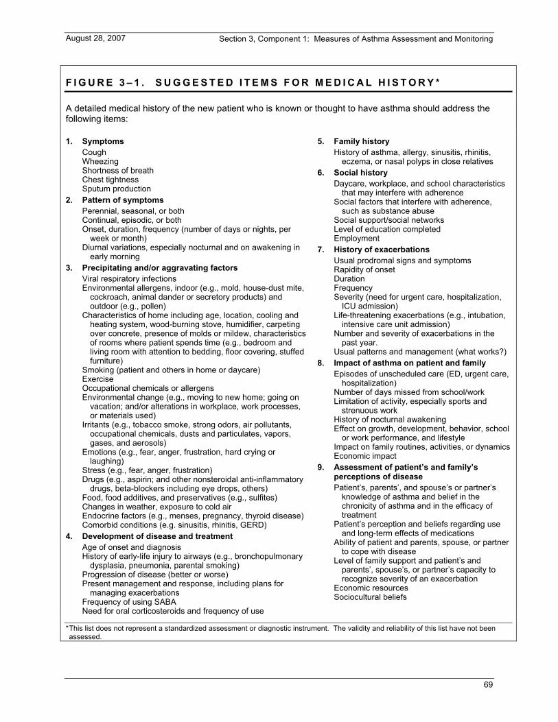
Section 3, Component 1: Measures of Asthma Assessment and Monitoring
69
August 28, 2007
F I G U R E 3 – 1 . S U G G E S T E D I T E M S F O R M E D I C A L H I S T O R Y *
A detailed medical history of the new patient who is known or thought to have asthma should address the following items:
1. Symptoms Cough Wheezing Shortness of breath Chest tightness Sputum production
2. Pattern of symptoms Perennial, seasonal, or both Continual, episodic, or both Onset, duration, frequency (number of days or nights, per
week or month) Diurnal variations, especially nocturnal and on awakening in
early morning 3. Precipitating and/or aggravating factors
Viral respiratory infections Environmental allergens, indoor (e.g., mold, house-dust mite,
cockroach, animal dander or secretory products) and outdoor (e.g., pollen)
Characteristics of home including age, location, cooling and heating system, wood-burning stove, humidifier, carpeting over concrete, presence of molds or mildew, characteristics of rooms where patient spends time (e.g., bedroom and living room with attention to bedding, floor covering, stuffed furniture)
Smoking (patient and others in home or daycare) Exercise Occupational chemicals or allergens Environmental change (e.g., moving to new home; going on
vacation; and/or alterations in workplace, work processes, or materials used)
Irritants (e.g., tobacco smoke, strong odors, air pollutants, occupational chemicals, dusts and particulates, vapors, gases, and aerosols)
Emotions (e.g., fear, anger, frustration, hard crying or laughing)
Stress (e.g., fear, anger, frustration) Drugs (e.g., aspirin; and other nonsteroidal anti-inflammatory
drugs, beta-blockers including eye drops, others) Food, food additives, and preservatives (e.g., sulfites) Changes in weather, exposure to cold air Endocrine factors (e.g., menses, pregnancy, thyroid disease) Comorbid conditions (e.g. sinusitis, rhinitis, GERD)
4. Development of disease and treatment Age of onset and diagnosis History of early-life injury to airways (e.g., bronchopulmonary
dysplasia, pneumonia, parental smoking) Progression of disease (better or worse) Present management and response, including plans for
managing exacerbations Frequency of using SABA Need for oral corticosteroids and frequency of use
5. Family history History of asthma, allergy, sinusitis, rhinitis,
eczema, or nasal polyps in close relatives 6. Social history
Daycare, workplace, and school characteristics that may interfere with adherence
Social factors that interfere with adherence, such as substance abuse
Social support/social networks Level of education completed Employment
7. History of exacerbations Usual prodromal signs and symptoms Rapidity of onset Duration Frequency Severity (need for urgent care, hospitalization,
ICU admission) Life-threatening exacerbations (e.g., intubation,
intensive care unit admission) Number and severity of exacerbations in the
past year. Usual patterns and management (what works?)
8. Impact of asthma on patient and family Episodes of unscheduled care (ED, urgent care,
hospitalization) Number of days missed from school/work Limitation of activity, especially sports and
strenuous work History of nocturnal awakening Effect on growth, development, behavior, school
or work performance, and lifestyle Impact on family routines, activities, or dynamics Economic impact
9. Assessment of patient’s and family’s perceptions of disease Patient’s, parents’, and spouse’s or partner’s
knowledge of asthma and belief in the chronicity of asthma and in the efficacy of treatment
Patient’s perception and beliefs regarding use and long-term effects of medications
Ability of patient and parents, spouse, or partner to cope with disease
Level of family support and patient’s and parents’, spouse’s, or partner’s capacity to recognize severity of an exacerbation
Economic resources Sociocultural beliefs
* This list does not represent a standardized assessment or diagnostic instrument. The validity and reliability of this list have not been assessed.
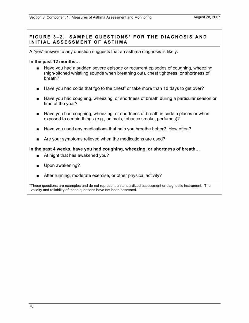
Section 3, Component 1: Measures of Asthma Assessment and Monitoring
70
August 28, 2007
F I G U R E 3 – 2 . S A M P L E Q U E S T I O N S * F O R T H E D I A G N O S I S A N D I N I T I A L A S S E S S M E N T O F A S T H M A
A “yes” answer to any question suggests that an asthma diagnosis is likely.
In the past 12 months… Have you had a sudden severe episode or recurrent episodes of coughing, wheezing
(high-pitched whistling sounds when breathing out), chest tightness, or shortness of breath?
Have you had colds that “go to the chest” or take more than 10 days to get over?
Have you had coughing, wheezing, or shortness of breath during a particular season or time of the year?
Have you had coughing, wheezing, or shortness of breath in certain places or when exposed to certain things (e.g., animals, tobacco smoke, perfumes)?
Have you used any medications that help you breathe better? How often?
Are your symptoms relieved when the medications are used?
In the past 4 weeks, have you had coughing, wheezing, or shortness of breath… At night that has awakened you?
Upon awakening?
After running, moderate exercise, or other physical activity?
* These questions are examples and do not represent a standardized assessment or diagnostic instrument. The validity and reliability of these questions have not been assessed.

Section 3, Component 1: Measures of Asthma Assessment and Monitoring
71
August 28, 2007
F I G U R E 3 – 3 a . S A M P L E S P I R O M E T R Y V O L U M E T I M E A N D F L O W V O L U M E C U R V E S
Key: FEV1, forced expiratory volume in 1 second
F I G U R E 3 – 3 b . R E P O R T O F S P I R O M E T R Y F I N D I N G S P R E - A N D P O S T B R O N C H O D I L A T O R
Prebronchodilator Postbronchodilator
Study: bronch Age: 59
ID: Height: 175 cm
Test date: 8/7/06 Sex: M
Time: 9:38 a.m. System: 7 20 17
Study: bronch Age: 59
ID: Height:175 cm
Test date: 8/7/06 Sex: M
Time: 9:58 a.m. System: 7 20 17
Trial FVC FEV1 FEV1/ FVC (%)
Trial FVC FEV1
FEV1/ FVC (%)
1 4.34 2.68 61.8% 1 4.73 2.94 62.2%
2 4.44 2.62 58.9% 2 4.76 3.07 64.5%
3 4.55 2.71 59.6% 3 4.78 3.04 63.5%
Best Values 4.56 2.71 59.4% Best Values 4.78 3.07 64.3% Predicted Values*
4.23 3.40 80.5% Reference Values
4.56 2.71
Percent Predicted
107.8% 79.7% 73.8% Difference (L) 0.22 0.36
Difference (%) 4.8% 13.4% Interpretations: FEV1 and FEV1/FVC are below normal range. The reduced rate at which air is exhaled indicates obstruction to airflow. *Predicted values from Knudson et al. (1983)
Interpretations: Significant increases in FEV1, with bronchodilator (≥12% increase after bronchodilator indicates a significant change).
Key: FEV1, forced expiratory volume in 1 second; FVC, forced vital capacity
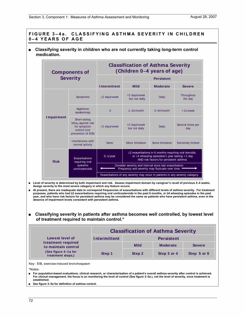
Section 3, Component 1: Measures of Asthma Assessment and Monitoring
72
August 28, 2007
F I G U R E 3 – 4 a . C L A S S I F Y I N G A S T H M A S E V E R I T Y I N C H I L D R E N 0 – 4 Y E A R S O F A G E
Classifying severity in children who are not currently taking long-term control medication.
Consider severity and interval since last exacerbation.Frequency and severity may fluctuate over time.
Extremely limitedSome limitationMinor limitationNoneInterference with normal activity
Several times per dayDaily>2 days/week
but not daily≤2 days/week
Short-actingbeta2-agonist use
for symptom control (not
prevention of EIB)
Exacerbations of any severity may occur in patients in any severity category
Exacerbations requiring oral
systemiccorticosteroids
Risk
Impairment
>1x/week3−4x/month1−2x/month0Nighttimeawakenings
Classification of Asthma Severity(Children 0−4 years of age)
Persistent
Components ofSeverity
≥2 exacerbations in 6 months requiring oral steroids,or ≥4 wheezing episodes/1 year lasting >1 day
AND risk factors for persistent asthma0−1/year
Throughoutthe dayDaily>2 days/week
but not daily≤2 days/weekSymptoms
SevereModerateMildIntermittent
Level of severity is determined by both impairment and risk. Assess impairment domain by caregiver’s recall of previous 2–4 weeks.
Assign severity to the most severe category in which any feature occurs. At present, there are inadequate data to correspond frequencies of exacerbations with different levels of asthma severity. For treatment
purposes, patients who had ≥2 exacerbations requiring oral corticosteroids in the past 6 months, or ≥4 wheezing episodes in the past year, and who have risk factors for persistent asthma may be considered the same as patients who have persistent asthma, even in the absence of impairment levels consistent with persistent asthma.
Classifying severity in patients after asthma becomes well controlled, by lowest level of treatment required to maintain control.*
SevereModerateMild
Classification of Asthma SeverityPersistentIntermittentLowest level of
treatment requiredto maintain control(See figure 4−1a for
treatment steps.) Step 5 or 6Step 2 Step 3 or 4Step 1
Key: EIB, exercise-induced bronchospasm
*Notes: For population-based evaluations, clinical research, or characterization of a patient’s overall asthma severity after control is achieved.
For clinical management, the focus is on monitoring the level of control (See figure 3–5a.), not the level of severity, once treatment is established.
See figure 3–5a for definition of asthma control.

Section 3, Component 1: Measures of Asthma Assessment and Monitoring
73
August 28, 2007
F I G U R E 3 – 4 b . C L A S S I F Y I N G A S T H M A S E V E R I T Y I N C H I L D R E N 5 – 1 1 Y E A R S O F A G E
Classifying severity in children who are not currently taking long-term control medication.
• Normal FEV1between exacerbations
Extremely limitedSome limitationMinor limitationNoneInterference with
normal activity
Several times per dayDaily>2 days/week
but not daily≤2 days/week
Short-actingbeta2-agonist use
for symptomcontrol (not
prevention of EIB)
• FEV1 <60% predicted
• FEV1 = 60−80% predicted
• FEV1 = >80% predicted
• FEV1 >80% predicted
≥2 in 1 year (see note)
Relative annual risk of exacerbations may be related to FEV1
Classification of Asthma Severity(Children 5−11 years of age)
Consider severity and interval since last exacerbation. Frequency and severity may fluctuate over time for patients in any severity category.
Impairment
Risk
Persistent
Components of Severity
0−1/year (see note)Exacerbations requiring oral systemic corticosteroids
• FEV1/FVC <75%
• FEV1/FVC =75−80%
• FEV1/FVC >80%
• FEV1/FVC >85%
Lung function
Often 7x/week
>1x/week butnot nightly3−4x/month≤2x/monthNighttime
awakenings
Throughoutthe day
Daily>2 days/week but not daily
≤2 days/weekSymptoms
SevereModerateMildIntermittent
Level of severity is determined by both impairment and risk. Assess impairment domain by patient’s/caregiver’s recall of the previous 2–4 weeks and spirometry. Assign severity to the most severe category in which any feature occurs.
At present, there are inadequate data to correspond frequencies of exacerbations with different levels of asthma severity. In general, more frequent and intense exacerbations (e.g., requiring urgent, unscheduled care, hospitalization, or ICU admission) indicate greater underlying disease severity. For treatment purposes, patients who had ≥2 exacerbations requiring oral systemic corticosteroids in the past year may be considered the same as patients who have persistent asthma, even in the absence of impairment levels consistent with persistent asthma.
Classifying severity in patients after asthma becomes well controlled, by lowest level of treatment required to maintain control.*
SevereModerateMild
Classification of Asthma SeverityPersistentIntermittentLowest level of
treatment requiredto maintain control
(See figure 4−1bfor treatment steps.) Step 5 or 6Step 2 Step 3 or 4Step 1
Key: EIB, exercise-induced bronchospasm; FEV1, forced expiratory volume in second; FVC, forced vital capacity; ICU, intensive care unit
*Notes: For population-based evaluations, clinical research, or characterization of a patient’s overall asthma severity after control is achieved.
For clinical management, the focus is on monitoring the level of control (See figure 3–5b.), not the level of severity, once treatment is established.
See figure 3–5b for definition of asthma control.

Section 3, Component 1: Measures of Asthma Assessment and Monitoring
74
August 28, 2007
F I G U R E 3 – 4 c . C L A S S I F Y I N G A S T H M A S E V E R I T Y I N Y O U T H S ≥ 1 2 Y E A R S O F A G E A N D A D U L T S
Classifying severity for patients who are not currently taking long-term control medications.
• Normal FEV1between exacerbations
Extremely limitedSome limitationMinor limitationNoneInterference with normal activity
Several timesper day
Daily>2 days/weekbut not >1x/day
≤2 days/weekShort-actingbeta2-agonist use
for symptom control (not prevention
of EIB)
≥2/year (see note)0−1/year(see note)
• FEV1 <60% predicted
• FEV1 >60% but <80% predicted
• FEV1 ≥80% predicted
• FEV1 >80% predicted
• FEV1/FVCreduced >5%
• FEV1/FVC reduced 5%
• FEV1/FVC normal
• FEV1/FVC normal
Risk
Relative annual risk of exacerbations may be related to FEV1
Classification of Asthma Severity(Youths ≥12 years of age and adults)
Consider severity and interval since last exacerbation. Frequency andseverity may fluctuate over time for patients in any severity category.
Impairment
Normal FEV1/FVC:8−19 yr 85%
20 −39 yr 80%40 −59 yr 75%60 −80 yr 70%
Persistent
Components of Severity
Exacerbationsrequiring oral
systemic corticosteroids
Lung function
Often 7x/week>1x/week butnot nightly
3−4x/month≤2x/monthNighttime awakenings
Throughout the day
Daily>2 days/week but not daily
≤2 days/weekSymptoms
SevereModerateMildIntermittent
Level of severity is determined by assessment of both impairment and risk. Assess impairment domain by patient’s/caregiver’s recall of
previous 2–4 weeks and spirometry. Assign severity to the most severe category in which any feature occurs. At present, there are inadequate data to correspond frequencies of exacerbations with different levels of asthma severity. In general,
more frequent and intense exacerbations (e.g., requiring urgent, unscheduled care, hospitalization, or ICU admission) indicate greater underlying disease severity. For treatment purposes, patients who had ≥2 exacerbations requiring oral systemic corticosteroids in the past year may be considered the same as patients who have persistent asthma, even in the absence of impairment levels consistent with persistent asthma.
Classifying severity in patients after asthma becomes well controlled, by lowest level of treatment required to maintain control.*
SevereModerateMild
Classification of Asthma SeverityPersistentIntermittentLowest level of
treatment requiredto maintain control
(See figure 4−5for treatment steps.) Step 5 or 6Step 2 Step 3 or 4Step 1
Key: EIB, exercise-induced bronchospasm; FEV1, forced expiratory volume in 1 second; FVC, forced vital capacity; ICU, intensive care unit
*Notes: For population-based evaluations, clinical research, or characterization of a patient’s overall asthma severity after control is achieved.
For clinical management, the focus is on monitoring the level of control (See figure 3–5c.), not the level of severity, once treatment is established.
See figure 3–5c for definition of asthma control.
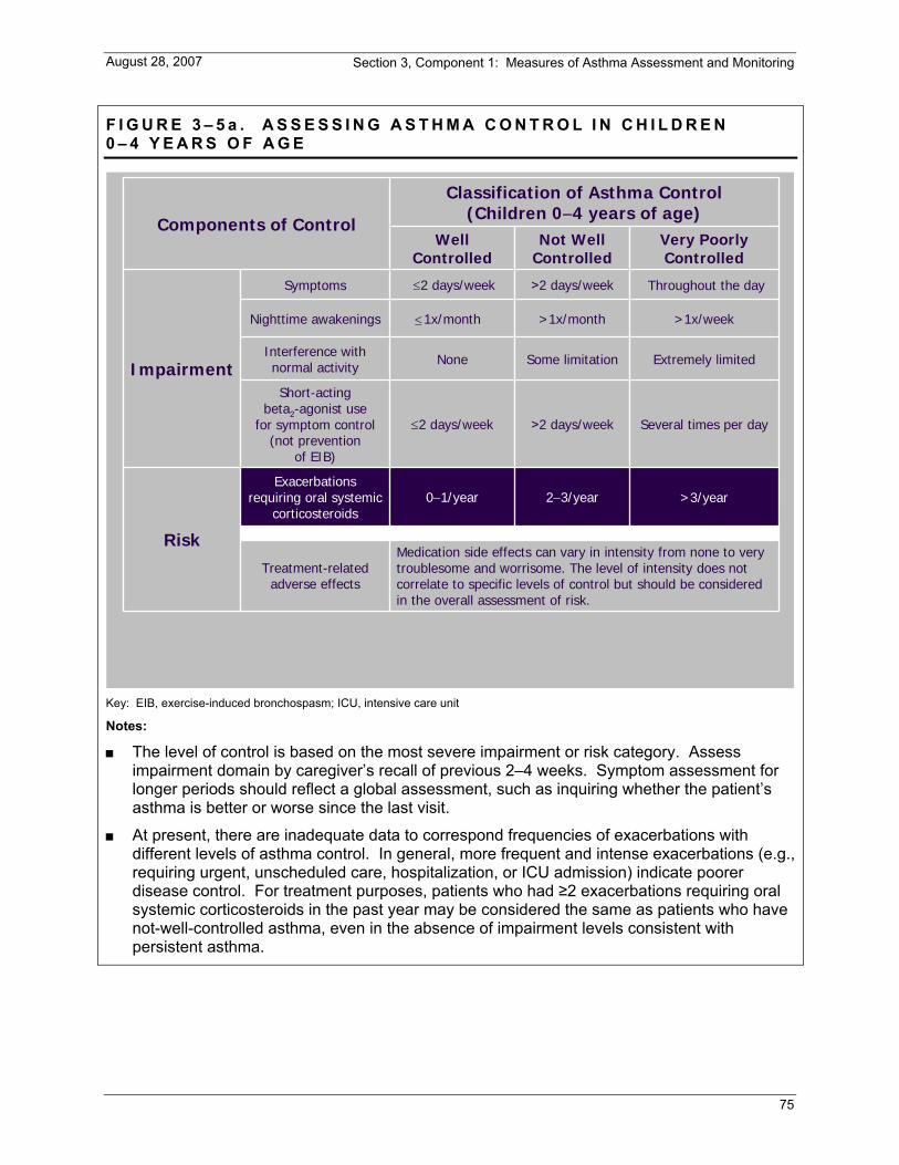
Section 3, Component 1: Measures of Asthma Assessment and Monitoring
75
August 28, 2007
F I G U R E 3 – 5 a . A S S E S S I N G A S T H M A C O N T R O L I N C H I L D R E N 0 – 4 Y E A R S O F A G E
>3/year2−3/year0−1/yearExacerbations
requiring oral systemic corticosteroids
Risk
Several times per day>2 days/week≤2 days/week
Short-actingbeta2-agonist use
for symptom control (not prevention
of EIB)
Extremely limitedSome limitationNoneInterference with normal activity
Medication side effects can vary in intensity from none to very troublesome and worrisome. The level of intensity does not correlate to specific levels of control but should be consideredin the overall assessment of risk.
Classification of Asthma Control(Children 0−4 years of age)
Impairment
Components of Control
Treatment-related adverse effects
>1x/week>1x/month1x/monthNighttime awakenings
Throughout the day>2 days/week≤2 days/weekSymptoms
Very Poorly Controlled
Not Well Controlled
Well Controlled
Key: EIB, exercise-induced bronchospasm; ICU, intensive care unit
Notes:
The level of control is based on the most severe impairment or risk category. Assess impairment domain by caregiver’s recall of previous 2–4 weeks. Symptom assessment for longer periods should reflect a global assessment, such as inquiring whether the patient’s asthma is better or worse since the last visit.
At present, there are inadequate data to correspond frequencies of exacerbations with different levels of asthma control. In general, more frequent and intense exacerbations (e.g., requiring urgent, unscheduled care, hospitalization, or ICU admission) indicate poorer disease control. For treatment purposes, patients who had ≥2 exacerbations requiring oral systemic corticosteroids in the past year may be considered the same as patients who have not-well-controlled asthma, even in the absence of impairment levels consistent with persistent asthma.
≤
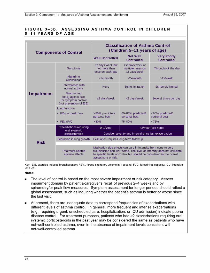
Section 3, Component 1: Measures of Asthma Assessment and Monitoring
76
August 28, 2007
F I G U R E 3 – 5 b . A S S E S S I N G A S T H M A C O N T R O L I N C H I L D R E N 5 – 1 1 Y E A R S O F A G E
Impairment
≥2/year (see note)0−1/yearExacerbations requiring oral systemic corticosteroids
Lung function
<60% predicted/personal best
60−80% predicted/personal best
>80% predicted/personal best
FEV1 or peak flow
Evaluation requires long-term followup.
Medication side effects can vary in intensity from none to very troublesome and worrisome. The level of intensity does not correlate to specific levels of control but should be considered in the overall assessment of risk.
Treatment-related adverse effects
Consider severity and interval since last exacerbation
Risk
Several times per day>2 days/week≤2 days/week
Short-actingbeta2-agonist use
for symptom control(not prevention of EIB)
Extremely limitedSome limitationNoneInterference withnormal activity
Classification of Asthma Control(Children 5−11 years of age)Components of Control
Reduction in lung growth
<75%75−80%>80%FEV1/FVC
≥2x/week≥2x/month≤1x/monthNighttimeawakenings
Throughout the day>2 days/week or multiple times on≤2 days/week
≤2 days/week but not more than
once on each daySymptoms
Very Poorly Controlled
Not Well ControlledWell Controlled
Key: EIB, exercise-induced bronchospasm; FEV1, forced expiratory volume in 1 second; FVC, forced vital capacity; ICU, intensive care unit
Notes:
The level of control is based on the most severe impairment or risk category. Assess impairment domain by patient’s/caregiver’s recall of previous 2–4 weeks and by spirometry/or peak flow measures. Symptom assessment for longer periods should reflect a global assessment, such as inquiring whether the patient’s asthma is better or worse since the last visit.
At present, there are inadequate data to correspond frequencies of exacerbations with different levels of asthma control. In general, more frequent and intense exacerbations (e.g., requiring urgent, unscheduled care, hospitalization, or ICU admission) indicate poorer disease control. For treatment purposes, patients who had ≥2 exacerbations requiring oral systemic corticosteroids in the past year may be considered the same as patients who have not-well-controlled asthma, even in the absence of impairment levels consistent with not-well-controlled asthma.

Section 3, Component 1: Measures of Asthma Assessment and Monitoring
77
August 28, 2007
F I G U R E 3 – 5 c . A S S E S S I N G A S T H M A C O N T R O L I N Y O U T H S ≥ 1 2 Y E A R S O F A G E A N D A D U L T S
Consider severity and interval since last exacerbation
Evaluation requires long-term followup care
Medication side effects can vary in intensity from none to very troublesome and worrisome. The level of intensity does not correlate to specific levels of control but should be considered in the overall assessment of risk.
Treatment-related adverse effects
Progressive loss of lung functionRisk
Validated Questionnaires
≥2/year (see note)
Throughout the day>2 days/week≤2 days/weekSymptoms
Impairment
3–4N/A≤15
1–2≥1.516−19
0≤0.75*≥20
ATAQACQACT
<60% predicted/personal best
60−80% predicted/personal best
>80% predicted/personal best
FEV1 or peak flow
Several times per day>2 days/week≤2 days/weekShort-acting beta2-agonist use for symptom control (not prevention of EIB)
0−1/yearExacerbations
Classification of Asthma Control(Youths ≥12 years of age and adults)
Components of Control
Extremely limitedSome limitationNoneInterference with normal activity
≥4x/week1−3x/week≤2x/monthNighttime awakening
Very PoorlyControlled
NotWell-ControlledWell-Controlled
*ACQ values of 0.76–1.4 are indeterminate regarding well-controlled asthma.
Key: EIB, exercise-induced bronchospasm; FEV1, forced expiratory volume in 1 second. See figure 3–8 for full name and source of ATAQ, ACQ, ACT.
Notes:
The level of control is based on the most severe impairment or risk category. Assess impairment domain by patient’s recall of previous 2–4 weeks and by spirometry/or peak flow measures. Symptom assessment for longer periods should reflect a global assessment, such as inquiring whether the patient’s asthma is better or worse since the last visit.
At present, there are inadequate data to correspond frequencies of exacerbations with different levels of asthma control. In general, more frequent and intense exacerbations (e.g., requiring urgent, unscheduled care, hospitalization, or ICU admission) indicate poorer disease control. For treatment purposes, patients who had ≥2 exacerbations requiring oral systemic corticosteroids in the past year may be considered the same as patients who have not-well-controlled asthma, even in the absence of impairment levels consistent with not-well-controlled asthma.

Section 3, Component 1: Measures of Asthma Assessment and Monitoring
78
August 28, 2007
F I G U R E 3 – 6 . S A M P L E Q U E S T I O N S F O R A S S E S S I N G A N D M O N I T O R I N G A S T H M A C O N T R O L
Monitoring Asthma Control
Ask the patient: Has your asthma awakened you at night or early morning?
Have you needed more quick-relief bronchodilator medication (inhaled short-acting beta2-agonist) than usual?
Have you needed any urgent medical care for your asthma, such as unscheduled visits to your doctor, an urgent care clinic, or the emergency department?
Are you participating in your usual and desired activities?
If you are measuring your peak flow, has it been below your personal best?
Actions to consider: Assess whether the medications are being taken as prescribed.
Assess whether the medications are being inhaled with correct technique.
Assess lung function with spirometry and compare to previous measurement.
Adjust medications, as needed; either step up if control is inadequate or step down if control is maximized, to achieve the best control with the lowest dose of medication.
Source: Adapted and reprinted from “Global Initiative for Asthma: Pocket Guide for Asthma Management and Prevention.” NIH Publication No. 96-3659B. Bethesda, MD: Department of Health and Human Services, National Institutes of Health, National Heart, Lung, and Blood Institute. 1995
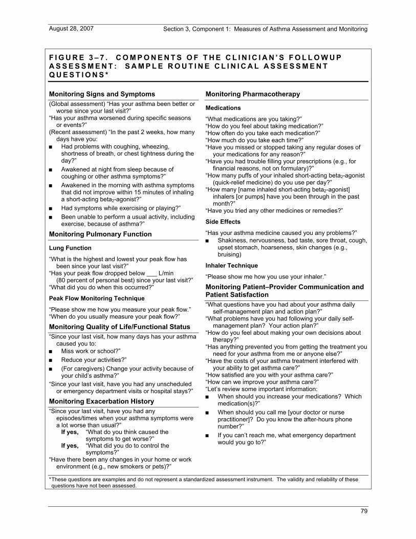
Section 3, Component 1: Measures of Asthma Assessment and Monitoring
79
August 28, 2007
F I G U R E 3 – 7 . C O M P O N E N T S O F T H E C L I N I C I A N ’ S F O L L O W U P A S S E S S M E N T : S A M P L E R O U T I N E C L I N I C A L A S S E S S M E N T Q U E S T I O N S *
Monitoring Signs and Symptoms (Global assessment) “Has your asthma been better or
worse since your last visit?” “Has your asthma worsened during specific seasons
or events?” (Recent assessment) “In the past 2 weeks, how many
days have you: Had problems with coughing, wheezing,
shortness of breath, or chest tightness during the day?”
Awakened at night from sleep because of coughing or other asthma symptoms?”
Awakened in the morning with asthma symptoms that did not improve within 15 minutes of inhaling a short-acting beta2-agonist?”
Had symptoms while exercising or playing?” Been unable to perform a usual activity, including
exercise, because of asthma?”
Monitoring Pulmonary Function
Lung Function
“What is the highest and lowest your peak flow has been since your last visit?”
“Has your peak flow dropped below ___ L/min (80 percent of personal best) since your last visit?”
“What did you do when this occurred?”
Peak Flow Monitoring Technique
“Please show me how you measure your peak flow.” “When do you usually measure your peak flow?”
Monitoring Quality of Life/Functional Status “Since your last visit, how many days has your asthma
caused you to: Miss work or school?” Reduce your activities?” (For caregivers) Change your activity because of
your child’s asthma?” “Since your last visit, have you had any unscheduled
or emergency department visits or hospital stays?”
Monitoring Exacerbation History “Since your last visit, have you had any
episodes/times when your asthma symptoms were a lot worse than usual?”
If yes, “What do you think caused the symptoms to get worse?”
If yes, “What did you do to control the symptoms?”
“Have there been any changes in your home or work environment (e.g., new smokers or pets)?”
Monitoring Pharmacotherapy
Medications
“What medications are you taking?” “How do you feel about taking medication?” “How often do you take each medication?” “How much do you take each time?” “Have you missed or stopped taking any regular doses of
your medications for any reason?” “Have you had trouble filling your prescriptions (e.g., for
financial reasons, not on formulary)?” “How many puffs of your inhaled short-acting beta2-agonist
(quick-relief medicine) do you use per day?” “How many [name inhaled short-acting beta2-agonist]
inhalers [or pumps] have you been through in the past month?”
“Have you tried any other medicines or remedies?”
Side Effects
“Has your asthma medicine caused you any problems?” Shakiness, nervousness, bad taste, sore throat, cough,
upset stomach, hoarseness, skin changes (e.g., bruising)
Inhaler Technique
“Please show me how you use your inhaler.”
Monitoring Patient–Provider Communication and Patient Satisfaction “What questions have you had about your asthma daily
self-management plan and action plan?” “What problems have you had following your daily self-
management plan? Your action plan?” “How do you feel about making your own decisions about
therapy?” “Has anything prevented you from getting the treatment you
need for your asthma from me or anyone else?” “Have the costs of your asthma treatment interfered with
your ability to get asthma care?” “How satisfied are you with your asthma care?” “How can we improve your asthma care?” “Let’s review some important information: When should you increase your medications? Which
medication(s)?” When should you call me [your doctor or nurse
practitioner]? Do you know the after-hours phone number?”
If you can’t reach me, what emergency department would you go to?”
* These questions are examples and do not represent a standardized assessment instrument. The validity and reliability of these questions have not been assessed.
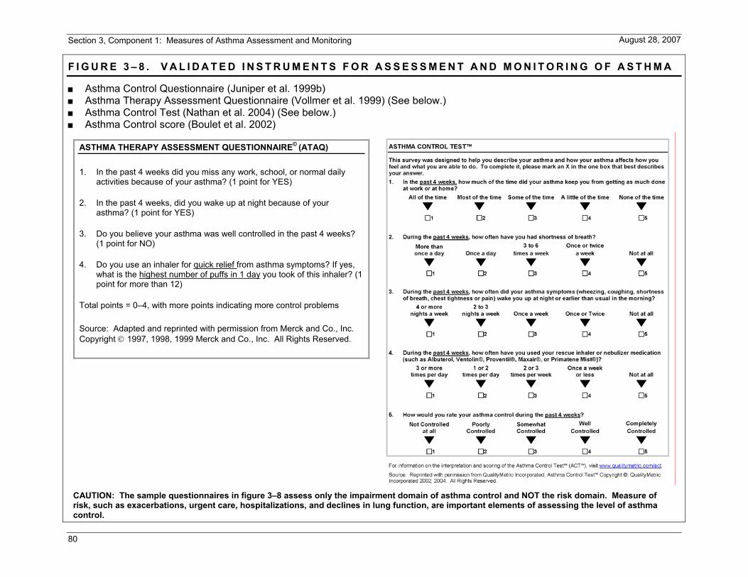
Section 3, Component 1: Measures of Asthma Assessment and Monitoring
80
August 28, 2007
F I G U R E 3 – 8 . V A L I D A T E D I N S T R U M E N T S F O R A S S E S S M E N T A N D M O N I T O R I N G O F A S T H M A
Asthma Control Questionnaire (Juniper et al. 1999b) Asthma Therapy Assessment Questionnaire (Vollmer et al. 1999) (See below.) Asthma Control Test (Nathan et al. 2004) (See below.) Asthma Control score (Boulet et al. 2002)
ASTHMA THERAPY ASSESSMENT QUESTIONNAIRE© (ATAQ)
1. In the past 4 weeks did you miss any work, school, or normal daily activities because of your asthma? (1 point for YES)
2. In the past 4 weeks, did you wake up at night because of your asthma? (1 point for YES)
3. Do you believe your asthma was well controlled in the past 4 weeks? (1 point for NO)
4. Do you use an inhaler for quick relief from asthma symptoms? If yes, what is the highest number of puffs in 1 day you took of this inhaler? (1 point for more than 12)
Total points = 0–4, with more points indicating more control problems Source: Adapted and reprinted with permission from Merck and Co., Inc. Copyright © 1997, 1998, 1999 Merck and Co., Inc. All Rights Reserved.
CAUTION: The sample questionnaires in figure 3–8 assess only the impairment domain of asthma control and NOT the risk domain. Measure of risk, such as exacerbations, urgent care, hospitalizations, and declines in lung function, are important elements of assessing the level of asthma control.

Section 3, Component 1: Measures of Asthma Assessment and Monitoring
81
August 28, 2007
F I G U R E 3 – 9 . S A M P L E * P A T I E N T S E L F - A S S E S S M E N T S H E E T F O R F O L L O W U P V I S I T S
Name: Date:
Your Asthma Control
How many days in the past week have you had chest tightness, cough, shortness of breath, or wheezing (whistling in your chest)?
_____ 0 _____ 1 _____ 2 _____ 3 _____ 4 _____ 5 _____ 6 _____ 7
How many nights in the past week have you had chest tightness, cough, shortness of breath, or wheezing (whistling in your chest)?
_____ 0 _____ 1 _____ 2 _____ 3 _____ 4 _____ 5 _____ 6 _____ 7
Do you perform peak flow readings at home?
______ yes ______ no
If yes, did you bring your peak flow chart? ______ yes ______ no
How many days in the past week has asthma restricted your physical activity?
_____ 0 _____ 1 _____ 2 _____ 3 _____ 4 _____ 5 _____ 6 _____ 7
Have you had any asthma attacks since your last visit?
______ yes ______ no
Have you had any unscheduled visits to a doctor, including to the emergency department, since your last visit?
______ yes ______ no
____very well controlled ____somewhat controlled ____not well controlled
How well controlled is your asthma, in your opinion?
Average number of puffs per day
Taking your medicine
What problems have you had taking your medicine or following your asthma action plan?
Please ask the doctor or nurse to review how you take your medicine.
Your questions What questions or concerns would you like to discuss with the doctor?
How satisfied are you with your asthma care?
____very satisfied ____somewhat satisfied ____not satisfied
* These questions are examples and do not represent a standardized assessment instrument. Other examples of asthma control questions: Asthma Control Questionnaire (Juniper); Asthma Therapy Assessment Questionnaire (Volmer); Asthma Control Test (Nathan); Asthma Control Score (Boulet)
Section 3, Component 1: Measures of Asthma Assessment and Monitoring
82
August 28, 2007
REFERENCES
Adams RJ, Smith BJ, Ruffin RE. Factors associated with hospital admissions and repeat emergency department visits for adults with asthma. Thorax 2000;55(7):566–73.
Adams RJ, Wilson DH, Taylor AW, Daly A, Tursan d'Espaignet E, Dal GE, Ruffin RE. Coexistent chronic conditions and asthma quality of life: a population-based study. Chest 2006;129(2):285–91.
American Thoracic Society. Standardization of spirometry, 1994 update. Am J Respir Crit Care Med 1995;152(3):1107–36.
American Thoracic Society and European Respiratory Society Task Force, Pellegrino R, Viegi G, Brusasco V, Crapo RO, Burgos F, Casaburi R, Coates A, van der Grinten C, Gustafsson P, et al. Standardization of lung function testing. Eur Respir J 2005;26:948–68.
Annett RD, Bender BG, Lapidus J, Duhamel TR, Lincoln A. Predicting children's quality of life in an asthma clinical trial: what do children's reports tell us? J Pediatr 2001;139(6):854–61.
Antonicelli L, Bucca C, Neri M, De Benedetto F, Sabbatani P, Bonifazi F, Eichler HG, Zhang Q, Yin DD. Asthma severity and medical resource utilisation. Eur Respir J 2004;23(5):723–9.
Appleton SL, Adams RJ, Wilson DH, Taylor AW, Ruffin RE. Spirometric criteria for asthma: adding further evidence to the debate. J Allergy Clin Immunol 2005;116(5):976–82.
Apter AJ, Szefler SJ. Advances in adult and pediatric asthma. J Allergy Clin Immunol 2004;113(3):407–14.
Ayres JG, Jyothish D, Ninan T. Brittle asthma. Paediatr Respir Rev 2004;5(1):40–4. Review.
Bacharier LB, Strunk RC, Mauger D, White D, Lemanske RF Jr, Sorkness CA. Classifying asthma severity in children: mismatch between symptoms, medication use, and lung function. Am J Respir Crit Care Med 2004;170(4):426–32.
Barnes PJ, Woolcock AJ. Difficult asthma. Eur Respir J 1998;12(5):1209–18.
Bateman ED, Boushey HA, Bousquet J, Busse WW, Clark TJ, Pauwels RA, Pedersen SE; GOAL Investigators Group. Can guideline-defined asthma control be achieved? The Gaining Optimal Asthma ControL study. Am J Respir Crit Care Med 2004;170(8):836–44.
Bayliss MS, Espindle DM, Buchner D, Blaiss MS, Ware JE. A new tool for monitoring asthma outcomes: the ITG Asthma Short Form. Qual Life Res 2000;9(4):451–66.
Belessis Y, Dixon S, Thomsen A, Duffy B, Rawlinson W, Henry R, Morton J. Risk factors for an intensive care unit admission in children with asthma. Pediatr Pulmonol 2004;37(3):201–9.

Section 3, Component 1: Measures of Asthma Assessment and Monitoring
83
August 28, 2007
Bijl-Hofland ID, Cloosterman SG, Van Schayck CP, Elshout FJ, Akkermans RP, Folgering HT. Perception of respiratory sensation assessed by means of histamine challenge and threshold loading tests. Chest 2000;117(4):954–9.
Bleecker ER, Yancey SW, Baitinger LA, Edwards LD, Klotsman M, Anderson WH, Dorinsky PM. Salmeterol response is not affected by beta2-adrenergic receptor genotype in subjects with persistent asthma. J Allergy Clin Immunol 2006;118(4):809–16.
Boulet LP, Boulet V, Milot J. How should we quantify asthma control? A proposal. Chest 2002;122(6):2217–23.
Boushey HA, Sorkness CA, King TS, Sullivan SD, Fahy JV, Lazarus SC, Chinchilli VM, Craig TJ, DiMango EA, Deykin A, et al.; National Heart, Lung, and Blood Institute's Asthma Clinical Research Network. Daily versus as-needed corticosteroids for mild persistent asthma. N Engl J Med 2005;352(15):1519–28.
Bousquet J, Knani J, Dhivert H, Richard A, Chicoye A, Ware JE Jr, Michel FB. Quality of life in asthma. I. Internal consistency and validity of the SF-36 questionnaire. Am J Respir Crit Care Med 1994;149(2 Pt 1):371–5.
Bousquet J, Wenzel S, Holgate S, Lumry W, Freeman P, Fox H. Predicting response to omalizumab, an anti-IgE antibody, in patients with allergic asthma. Chest 2004;125(4):1378–86.
Brand PL, Quanjer PH, Postma DS, Kerstjens HA, Koeter GH, Dekhuijzen PN, Sluiter HJ. Interpretation of bronchodilator response in patients with obstructive airways disease. The Dutch Chronic Non-Specific Lung Disease (CNSLD) Study Group. Thorax 1992;47(6):429–36.
Bucca C, Rolla G, Brussino L, De Rose V, Bugiani M. Are asthma-like symptoms due to bronchial or extrathoracic airway dysfunction? Lancet 1995;346(8978):791–5.
Bye MR, Kerstein D, Barsh E. The importance of spirometry in the assessment of childhood asthma. Am J Dis Child 1992;146(8):977–8.
Christopher KL, Wood RP, Eckert RC, Blager FB, Raney RA, Souhrada JF. Vocal-cord dysfunction presenting as asthma. N Engl J Med 1983;308(26):1566–70.
Colice GL, Burgt JV, Song J, Stampone P, Thompson PJ. Categorizing asthma severity. Am J Respir Crit Care Med 1999;160(6):1962–7.
Connolly CK, Mamun M, Alcock SM, Prescott RJ. The Darlington and Northallerton Prospective Asthma Study: best function predicts mortality during the first 10 years. Respir Med 1998;92(11):1274–80.
Connolly MJ, Crowley JJ, Charan NB, Nielson CP, Vestal RE. Reduced subjective awareness of bronchoconstriction provoked by methacholine in elderly asthmatic and normal subjects as measured on a simple awareness scale. Thorax 1992;47(6):410–3.

Section 3, Component 1: Measures of Asthma Assessment and Monitoring
84
August 28, 2007
Covar RA, Spahn JD, Martin RJ, Silkoff PE, Sundstrom DA, Murphy J, Szefler SJ. Safety and application of induced sputum analysis in childhood asthma. J Allergy Clin Immunol 2004a;114(3):575–82.
Covar RA, Spahn JD, Murphy JR, Szefler SJ; Childhood Asthma Management Program Research Group. Progression of asthma measured by lung function in the childhood asthma management program. Am J Respir Crit Care Med 2004b;170(3):234–41. Epub March 2004.
Cowie RL, Underwood MF, Revitt SG, Field SK. Predicting emergency department utilization in adults with asthma: a cohort study. J Asthma 2001;38(2):179–84.
Dales RE, Spitzer WO, Tousignant P, Schechter M, Suissa S. Clinical interpretation of airway response to a bronchodilator. Epidemiologic considerations. Am Rev Respir Dis 1988;138(2):317–20.
Deykin A, Lazarus SC, Fahy JV, Wechsler ME, Boushey HA, Chinchilli VM, Craig TJ, DiMango E, Kraft M, Leone F, et al.; Asthma Clinical Research Network, National Heart, Lung, and Blood Institute/NIH. Sputum eosinophil counts predict asthma control after discontinuation of inhaled corticosteroids. J Allergy Clin Immunol 2005;115(4):720–7.
Dicpinigaitis PV. Chronic cough due to asthma: ACCP evidence-based clinical practice guidelines. Chest 2006;129(1 Suppl):75S–79S.
Diette GB, Krishnan JA, Dominici F, Haponik E, Skinner EA, Steinwachs D, Wu AW. Asthma in older patients: factors associated with hospitalization. Arch Intern Med 2002;162(10):1123–32.
Diette GB, Krishnan JA, Wolfenden LL, Skinner EA, Steinwachs DM, Wu AW. Relationship of physician estimate of underlying asthma severity to asthma outcomes. Ann Allergy Asthma Immunol 2004;93(6):546–52.
Djukanovic R, Sterk PJ, Fahy JV, Hargreave FE. Standardised methodology of sputum induction and processing. Eur Respir J Suppl 2002;37:1s–2s.
Drazen JM. Asthma therapy with agents preventing leukotriene synthesis or action. Proc Assoc Am Physicians 1999;111(6):547–59.
Dupont LJ, Demedts MG, Verleden GM. Prospective evaluation of the validity of exhaled nitric oxide for the diagnosis of asthma. Chest 2003;123(3):751–6.
Eid N, Yandell B, Howell L, Eddy M, Sheikh S. Can peak expiratory flow predict airflow obstruction in children with asthma? Pediatrics 2000;105(2):354–8.
Eisner MD, Ackerson LM, Chi F, Kalkbrenner A, Buchner D, Mendoza G, Lieu T. Health-related quality of life and future health care utilization for asthma. Ann Allergy Asthma Immunol 2002;89(1):46–55.
Eisner MD, Katz PP, Lactao G, Iribarren C. Impact of depressive symptoms on adult asthma outcomes. Ann Allergy Asthma Immunol 2005;94(5):566–74.
Section 3, Component 1: Measures of Asthma Assessment and Monitoring
85
August 28, 2007
Eisner MD, Katz PP, Yelin EH, Shiboski SC, Blanc PD. Risk factors for hospitalization among adults with asthma: the influence of sociodemographic factors and asthma severity. Respir Res 2001;2(1):53–60. Epub December 2000.
EPR⎯2. Expert Panel Report 2: Guidelines for the Diagnosis and Management of Asthma (EPR⎯2 1997). NIH Publication No. 97-4051. Bethesda, MD, U.S. Department of Health and Human Services; National Institutes of Health; National Heart, Lung, and Blood Institute; National Asthma Education and Prevention Program, 1997.
EPR⎯Update 2002. Expert Panel Report: Guidelines for the Diagnosis and Management of Asthma. Update on Selected Topics 2002 (EPR⎯Update 2002). NIH Publication No. 02-5074. Bethesda, MD, U.S. Department of Health and Human Services; National Institutes of Health; National Heart, Lung, and Blood Institute; National Asthma Education and Prevention Program, June 2003.
Ford JG, Meyer IH, Sternfels P, Findley SE, McLean DE, Fagan JK, Richardson L. Patterns and predictors of asthma-related emergency department use in Harlem. Chest 2001;120(4):1129–35.
Fuhlbrigge AL, Adams RJ, Guilbert TW, Grant E, Lozano P, Janson SL, Martinez F, Weiss KB, Weiss ST. The burden of asthma in the United States: level and distribution are dependent on interpretation of the National Asthma Education and Prevention Program guidelines. Am J Respir Crit Care Med 2002;166(8):1044–9.
Fuhlbrigge AL, Kitch BT, Paltiel AD, Kuntz KM, Neumann PJ, Dockery DW, Weiss ST. FEV(1) is associated with risk of asthma attacks in a pediatric population. J Allergy Clin Immunol 2001;107(1):61–7.
Fuhlbrigge AL, Weiss ST, Kuntz KM, Paltiel AD. Forced expiratory volume in 1 second percentage improves the classification of severity among children with asthma. Pediatrics 2006;118(2):e347–e355. Epub July 2006.
Global Initiative for Asthma Management and Prevention (GINA). NHLBI/WHO Workshop Report. NIH Publication No. 02-3659. Bethesda, MD: Department of Health and Human Services; National Institutes of Health; National Heart, Lung, and Blood Institute, 2002.
Goodwin RD, Fergusson DM, Horwood LJ. Asthma and depressive and anxiety disorders among young persons in the community. Psychol Med 2004;34(8):1465–74.
Graham DM, Blaiss MS, Bayliss MS, Espindle DM, Ware JE Jr. Impact of changes in asthma severity on health-related quality of life in pediatric and adult asthma patients: results from the asthma outcomes monitoring system. Allergy Asthma Proc 2000;21(3):151–8.
Graham LM. Classifying asthma. Chest 2006;130(1 Suppl):13S–20S. Review.
Green RH, Brightling CE, McKenna S, Hargadon B, Parker D, Bradding P, Wardlaw AJ, Pavord ID. Asthma exacerbations and sputum eosinophil counts: a randomised controlled trial. Lancet 2002;360(9347):1715–21.
Section 3, Component 1: Measures of Asthma Assessment and Monitoring
86
August 28, 2007
Griswold SK, Nordstrom CR, Clark S, Gaeta TJ, Price ML, Camargo CA Jr. Asthma exacerbations in North American adults: who are the "frequent fliers" in the emergency department? Chest 2005;127(5):1579–86.
Hallstrand TS, Curtis JR, Aitken ML, Sullivan SD. Quality of life in adolescents with mild asthma. Pediatr Pulmonol 2003;36(6):536–43.
Hankinson JL, Wagner GR. Medical screening using periodic spirometry for detection of chronic lung disease. Occup Med 1993;8(2):353–61.
Haynes RB, Taylor DW, Sackett DL, eds. Compliance in Health Care. Baltimore: Johns Hopkins University Press, 1979.
Heaney LG, Conway E, Kelly C, Johnston BT, English C, Stevenson M, Gamble J. Predictors of therapy resistant asthma: outcome of a systematic evaluation protocol. Thorax 2003;58(7):561–6.
Hesselink AE, Penninx BW, Schlosser MA, Wijnhoven HA, van der Windt DA, Kriegsman DM, van Eijk JT. The role of coping resources and coping style in quality of life of patients with asthma or COPD. Qual Life Res 2004;13(2):509–18.
Hunt J. Exhaled breath condensate: an evolving tool for noninvasive evaluation of lung disease. J Allergy Clin Immunol 2002;110(1):28–34.
Irvin CG, Martin RJ, Chinchilli VM, Kunselman SJ, Cherniack RM. Quality control of peak flow meters for multicenter clinical trials. The Asthma Clinical Research Network (ACRN). Am J Respir Crit Care Med 1997;156(2 Pt 1):396–402.
Israel E, Chinchilli VM, Ford JG, Boushey HA, Cherniack R, Craig TJ, Deykin A, Fagan JK, Fahy JV, Fish J, et al.; National Heart, Lung, and Blood Institute's Asthma Clinical Research Network. Use of regularly scheduled albuterol treatment in asthma: genotype-stratified, randomised, placebo-controlled cross-over trial. Lancet 2004;364(9444):1505–12.
Israel E, Drazen JM, Liggett SB, Boushey HA, Cherniack RM, Chinchilli VM, Cooper DM, Fahy JV, Fish JE, Ford JG, et al. The effect of polymorphisms of the beta(2)-adrenergic receptor on the response to regular use of albuterol in asthma. Am J Respir Crit Care Med 2000;162(1):75–80.
Jacobs JE, van de Lisdonk EH, Smeele I, Van Weel C, Grol RP. Management of patients with asthma and COPD: monitoring quality of life and the relationship to subsequent GP interventions. Fam Pract 2001;18(6):574–80.
Jain P, Kavuru MS, Emerman CL, Ahmad M. Utility of peak expiratory flow monitoring. Chest 1998;114(3):861–76.
Janson-Bjerklie S, Ferketich S, Benner P. Predicting the outcomes of living with asthma. Res Nurs Health 1993;16(4):241–50.
Juniper EF, Guyatt GH, Cox FM, Ferrie PJ, King DR. Development and validation of the Mini Asthma Quality of Life Questionnaire. Eur Respir J 1999a;14(1):32–8.
Section 3, Component 1: Measures of Asthma Assessment and Monitoring
87
August 28, 2007
Juniper EF, Guyatt GH, Feeny DH, Ferrie PJ, Griffith LE, Townsend M. Measuring quality of life in children with asthma. Qual Life Res 1996;5(1):35–46.
Juniper EF, O'Byrne PM, Guyatt GH, Ferrie PJ, King DR. Development and validation of a questionnaire to measure asthma control. Eur Respir J 1999b;14(4):902–7.
Juniper EF, Wisniewski ME, Cox FM, Emmett AH, Nielsen KE, O'Byrne PM. Relationship between quality of life and clinical status in asthma: a factor analysis. Eur Respir J 2004;23(2):287–91.
Katz PP, Eisner MD, Henke J, Shiboski S, Yelin EH, Blanc PD. The Marks Asthma Quality of Life Questionnaire: further validation and examination of responsiveness to change. J Clin Epidemiol 1999;52(7):667–75.
Katz PP, Yelin EH, Eisner MD, Blanc PD. Perceived control of asthma and quality of life among adults with asthma. Ann Allergy Asthma Immunol 2002;89(3):251–8.
Katz PP, Yelin EH, Eisner MD, Earnest G, Blanc PD. Performance of valued life activities reflected asthma-specific quality of life more than general physical function. J Clin Epidemiol 2004;57(3):259–67.
Kharitonov S, Alving K, Barnes PJ. Exhaled and nasal nitric oxide measurements: recommendations. The European Respiratory Society Task Force. Eur Respir J 1997;10(7):1683–93.
Kikuchi Y, Okabe S, Tamura G, Hida W, Homma M, Shirato K, Takishima T. Chemosensitivity and perception of dyspnea in patients with a history of near-fatal asthma. N Engl J Med 1994;330(19):1329–34.
Kitch BT, Paltiel AD, Kuntz KM, Dockery DW, Schouten JP, Weiss ST, Fuhlbrigge AL. A single measure of FEV1 is associated with risk of asthma attacks in long-term follow-up. Chest 2004;126(6):1875–82.
Knudson RJ, Lebowitz MD, Holberg CJ, Burrows B. Changes in the normal maximal expiratory flow-volume curve with growth and aging. Am Rev Respir Dis 1983;127(6):725–34.
Lazarus SC, Boushey HA, Fahy JV, Chinchilli VM, Lemanske RF Jr, Sorkness CA, Kraft M, Fish JE, Peters SP, Craig T, et al.; Asthma Clinical Research Network for the National Heart, Lung, and Blood Institute. Long-acting beta2-agonist monotherapy vs. continued therapy with inhaled corticosteroids in patients with persistent asthma: a randomized controlled trial. JAMA 2001;285(20):2583–93.
Leone FT, Mauger EA, Peters SP, Chinchilli VM, Fish JE, Boushey HA, Cherniack RM, Drazen JM, Fahy JV, Ford J, et al. The utility of peak flow, symptom scores, and beta-agonist use as outcome measures in asthma clinical research. Chest 2001;119(4):1027–33.
Leuppi JD, Brannan JD, Anderson SD. Bronchial provocation tests: the rationale for using inhaled mannitol as a test for airway hyperresponsiveness. Swiss Med Wkly 2002;132(13−14):151–8.
Section 3, Component 1: Measures of Asthma Assessment and Monitoring
88
August 28, 2007
Leuppi JD, Tandjung R, Anderson SD, Stolz D, Brutsche MH, Bingisser R, Perruchoud AP, Surber C, Knoblauch A, Andersson M, et al. Prediction of treatment-response to inhaled corticosteroids by mannitol-challenge test in COPD. A proof of concept. Pulm Pharmacol Ther 2005;18(2):83–8.
Li JT, O'Connell EJ. Clinical evaluation of asthma. Ann Allergy Asthma Immunol 1996;76(1):1–13; quiz 13−5. Review.
Lieu TA, Quesenberry CP, Sorel ME, Mendoza GR, Leong AB. Computer-based models to identify high-risk children with asthma. Am J Respir Crit Care Med 1998;157(4 Pt 1):1173–80.
Lima JJ, Zhang S, Grant A, Shao L, Tantisira KG, Allayee H, Wang J, Sylvester J, Holbrook J, Wise R, et al. Influence of leukotriene pathway polymorphisms on response to montelukast in asthma. Am J Respir Crit Care Med 2006;173(4):379–85. Epub November 2005.
Liu AH, Zeiger R, Sorkness C, Mahr T, Ostrom N, Burgess S, Rosenzweig JC, Manjunath R. Development and cross-sectional validation of the Childhood Asthma Control Test. J Allergy Clin Immunol 2007;119(4):817–25. Epub March 2007.
Llewellin P, Sawyer G, Lewis S, Cheng S, Weatherall M, Fitzharris P, Beasley R. The relationship between FEV1 and PEF in the assessment of the severity of airways obstruction. Respirology 2002;7(4):333–7.
Magid DJ, Houry D, Ellis J, Lyons E, Rumsfeld JS. Health-related quality of life predicts emergency department utilization for patients with asthma. Ann Emerg Med 2004;43(5):551–7.
Mancuso CA, Peterson MG, Charlson ME. Comparing discriminative validity between a disease-specific and a general health scale in patients with moderate asthma. J Clin Epidemiol 2001;54(3):263–74.
Marks GB, Dunn SM, Woolcock AJ. An evaluation of an asthma quality of life questionnaire as a measure of change in adults with asthma. J Clin Epidemiol 1993;46(10):1103–11.
Meichenbaum D, Turk DC. Facilitating Treatment Adherence: A Practitioner's Guidebook. New York: Plenum Press, 1987.
Meslier N, Racineux JL, Six P, Lockhart A. Diagnostic value of reversibility of chronic airway obstruction to separate asthma from chronic bronchitis: a statistical approach. Eur Respir J 1989;2(6):497–505.
Miles JF, Bright P, Ayres JG, Cayton RM, Miller MR. The performance of Mini Wright peak flow meters after prolonged use. Respir Med 1995;89(9):603–5.
Moy ML, Israel E, Weiss ST, Juniper EF, Dube L, Drazen JM; NHBLI Asthma Clinical Research Network. Clinical predictors of health-related quality of life depend on asthma severity. Am J Respir Crit Care Med 2001;163(4):924–9.
Nair SJ, Daigle KL, DeCuir P, Lapin CD, Schramm CM. The influence of pulmonary function testing on the management of asthma in children. J Pediatr 2005;147(6):797–801.

Section 3, Component 1: Measures of Asthma Assessment and Monitoring
89
August 28, 2007
Nathan RA, Sorkness CA, Kosinski M, Schatz M, Li JT, Marcus P, Murray JJ, Pendergraft TB. Development of the Asthma Control Test: a survey for assessing asthma control. J Allergy Clin Immunol 2004;113(1):59–65.
Newman KB, Mason UG III, Schmaling KB. Clinical features of vocal cord dysfunction. Am J Respir Crit Care Med 1995;152(4 Pt 1):1382–6.
Ng TP. Validity of symptom and clinical measures of asthma severity for primary outpatient assessment of adult asthma. Br J Gen Pract 2000;50(450):7–12.
Noonan M, Chervinsky P, Busse WW, Weisberg SC, Pinnas J, de Boisblanc BP, Boltansky H, Pearlman D, Repsher L, Kellerman D. Fluticasone propionate reduces oral prednisone use while it improves asthma control and quality of life. Am J Respir Crit Care Med 1995;152(5 Pt 1):1467–73.
Paull K, Covar R, Jain N, Gelfand EW, Spahn JD. Do NHLBI lung function criteria apply to children? A cross-sectional evaluation of childhood asthma at National Jewish Medical and Research Center, 1999−2002. Pediatr Pulmonol 2005;39(4):311–7.
Pellegrino R, Viegi G, Brusasco V, Crapo RO, Burgos F, Casaburi R, Coates A, van der Grinten CP, Gustafsson P, Hankinson J, et al. Interpretative strategies for lung function tests. Eur Respir J 2005;26(5):948–68.
Pijnenburg MW, Hofhuis W, Hop WC, de Jongste JC. Exhaled nitric oxide predicts asthma relapse in children with clinical asthma remission. Thorax 2005;60(3):215–8.
Pinnock H, Juniper EF, Sheikh A. Concordance between supervised and postal administration of the Mini Asthma Quality of Life Questionnaire (MiniAQLQ) and Asthma Control Questionnaire (ACQ) was very high. J Clin Epidemiol 2005;58(8):809–14.
Rasmussen F, Taylor DR, Flannery EM, Cowan JO, Greene JM, Herbison GP, Sears MR. Risk factors for airway remodeling in asthma manifested by a low postbronchodilator FEV1/vital capacity ratio: a longitudinal population study from childhood to adulthood. Am J Respir Crit Care Med 2002;165(11):1480–8.
Rundell KW, Spiering BA. Inspiratory stridor in elite athletes. Chest 2003;123(2):468–74.
Russell NJ, Crichton NJ, Emerson PA, Morgan AD. Quantitative assessment of the value of spirometry. Thorax 1986;41(5):360–3.
Schatz M, Dombrowski MP, Wise R, Thom EA, Landon M, Mabie W, Newman RB, Hauth JC, Lindheimer M, Caritis SN, et al. Asthma morbidity during pregnancy can be predicted by severity classification. J Allergy Clin Immunol 2003;112(2):283–8.
Schatz M, Mosen D, Apter AJ, Zeiger RS, Vollmer WM, Stibolt TB, Leong A, Johnson MS, Mendoza G, Cook EF. Relationship of validated psychometric tools to subsequent medical utilization for asthma. J Allergy Clin Immunol 2005a;115(3):564–70.
Section 3, Component 1: Measures of Asthma Assessment and Monitoring
90
August 28, 2007
Schatz M, Mosen D, Apter AJ, Zeiger RS, Vollmer WM, Stibolt TB, Leong A, Johnson MS, Mendoza G, Cook EF. Relationships among quality of life, severity, and control measures in asthma: an evaluation using factor analysis. J Allergy Clin Immunol 2005b;115(5):1049–55.
Shim CS, Williams MH Jr. Evaluation of the severity of asthma: patients versus physicians. Am J Med 1980;68(1):11–3.
Silverman EK, Kwiatkowski DJ, Sylvia JS, Lazarus R, Drazen JM, Lange C, Laird NM, Weiss ST. Family-based association analysis of beta2-adrenergic receptor polymorphisms in the Childhood Asthma Management Program. J Allergy Clin Immunol 2003;112(5):870–6.
Smith AD, Cowan JO, Brassett KP, Herbison GP, Taylor DR. Use of exhaled nitric oxide measurements to guide treatment in chronic asthma. N Engl J Med 2005;352(21):2163–73. Epub May 2005.
Smith AD, Cowan JO, Filsell S, McLachlan C, Monti-Sheehan G, Jackson P, Taylor DR. Diagnosing asthma: comparisons between exhaled nitric oxide measurements and conventional tests. Am J Respir Crit Care Med 2004;169(4):473–78. Epub November 2003.
Sont JK, Willems LN, Bel EH, van Krieken JH, Vandenbroucke JP, Sterk PJ. Clinical control and histopathologic outcome of asthma when using airway hyperresponsiveness as an additional guide to long-term treatment. The AMPUL Study Group. Am J Respir Crit Care Med 1999;159(4 Pt 1):1043–51.
Spahn JD, Cherniack R, Paull K, Gelfand EW. Is forced expiratory volume in one second the best measure of severity in childhood asthma? Am J Respir Crit Care Med 2004;169(7):784–6. Epub January 2004.
Stahl E, Postma DS, Juniper EF, Svensson K, Mear I, Lofdahl CG. Health-related quality of life in asthma studies. Can we combine data from different countries? Pulm Pharmacol Ther 2003;16(1):53–9.
Stempel DA, Fuhlbrigge AL. Defining the responder in asthma therapy. J Allergy Clin Immunol 2005;115(3):466–9.
Stout JW, Visness CM, Enright P, Lamm C, Shapiro G, Gan VN, Adams GK III, Mitchell HE. Classification of asthma severity in children: the contribution of pulmonary function testing. Arch Pediatr Adolesc Med 2006;160(8):844–50.
Strunk RC. Asthma deaths in childhood: identification of patients at risk and intervention. J Allergy Clin Immunol 1987;80(3 Pt 2):472–7.
Strunk RC, Mrazek DA, Fuhrmann GS, LaBrecque JF. Physiologic and psychological characteristics associated with deaths due to asthma in childhood. A case-controlled study. JAMA 1985;254(9):1193–8.
Section 3, Component 1: Measures of Asthma Assessment and Monitoring
91
August 28, 2007
Strunk RC, Sternberg AL, Bacharier LB, Szefler SJ. Nocturnal awakening caused by asthma in children with mild-to-moderate asthma in the childhood asthma management program. J Allergy Clin Immunol 2002;110(3):395–403.
Swanney MP, Beckert LE, Frampton CM, Wallace LA, Jensen RL, Crapo RO. Validity of the American Thoracic Society and other spirometric algorithms using FVC and forced expiratory volume at 6 s for predicting a reduced total lung capacity. Chest 2004;126(6):1861–6.
Szefler SJ, Phillips BR, Martinez FD, Chinchilli VM, Lemanske RF, Strunk RC, Zeiger RS, Larsen G, Spahn JD, Bacharier LB, et al. Characterization of within-subject responses to fluticasone and montelukast in childhood asthma. J Allergy Clin Immunol 2005;115(2):233–42.
Tantisira KG, Hwang ES, Raby BA, Silverman ES, Lake SL, Richter BG, Peng SL, Drazen JM, Glimcher LH, Weiss ST. TBX21: a functional variant predicts improvement in asthma with the use of inhaled corticosteroids. Proc Natl Acad Sci U S A 2004;101(52):18099–104.
Taylor DR. Nitric oxide as a clinical guide for asthma management. J Allergy Clin Immunol 2006;117(2):259–62.
Taylor DR, Drazen JM, Herbison GP, Yandava CN, Hancox RJ, Town GI. Asthma exacerbations during long term beta agonist use: influence of beta(2) adrenoceptor polymorphism. Thorax 2000;55(9):762–7.
Ulrik CS, Backer V. Nonreversible airflow obstruction in life-long nonsmokers with moderate to severe asthma. Eur Respir J 1999;14(4):892–6.
Vollmer WM, Markson LE, O'Connor E, Frazier EA, Berger M, Buist AS. Association of asthma control with health care utilization: a prospective evaluation. Am J Respir Crit Care Med 2002;165(2):195–9.
Vollmer WM, Markson LE, O'Connor E, Sanocki LL, Fitterman L, Berger M, Buist AS. Association of asthma control with health care utilization and quality of life. Am J Respir Crit Care Med 1999;160(5 Pt 1):1647–52.
Ware J Jr, Kosinski M, Keller SD. A 12-Item Short-Form Health Survey: construction of scales and preliminary tests of reliability and validity. Med Care 1996;34(3):220–33.
Wechsler ME, Lehman E, Lazarus SC, Lemanske RF Jr, Boushey HA, Deykin A, Fahy JV, Sorkness CA, Chinchilli VM, Craig TJ, et al.; National Heart, Lung, and Blood Institute's Asthma Clinical Research Network. Beta-adrenergic receptor polymorphisms and response to salmeterol. Am J Respir Crit Care Med 2006;173(5):519–26. Epub December 2005.
Wijnhoven HA, Kriegsman DM, Hesselink AE, Penninx BW, de Haan M. Determinants of different dimensions of disease severity in asthma and COPD: pulmonary function and health-related quality of life. Chest 2001;119(4):1034–42.
Wolfenden LL, Diette GB, Krishnan JA, Skinner EA, Steinwachs DM, Wu AW. Lower physician estimate of underlying asthma severity leads to undertreatment. Arch Intern Med 2003;163(2):231–6.

Section 3, Component 1: Measures of Asthma Assessment and Monitoring
92
August 28, 2007
Yoos HL, Kitzman H, McMullen A, Henderson C, Sidora K. Symptom monitoring in childhood asthma: a randomized clinical trial comparing peak expiratory flow rate with symptom monitoring. Ann Allergy Asthma Immunol 2002;88(3):283–91.
Zhang J, Yu C, Holgate ST, Reiss TF. Variability and lack of predictive ability of asthma end-points in clinical trials. Eur Respir J 2002;20(5):1102–9.