section a - basics of occlusion - brian palmer, dds
TRANSCRIPT

OcclusionOcclusionThe KEY to dentistry.
The KEY to total health.The KEY to this website.
A1

Basics of Occlusion
A2
Simplistic definition of occlusion: The way teeth meet and function.

A3
The BEST textbook on dentistry. Every dentist
should read.
Peter E. Dawson. Evaluation, Diagnosis, and Treatment of
Occlusal Problems, 2nd ed.. Mosby.
A4
I am standing beside, in my opinion, one of the
best dentists in the world, Dr. Peter Dawson.

Centric Relation (CR)Refers to the RELATIONSHIP of the
MANDIBLE TO THE SKULL as it rotates around the ‘hinge-axis” before any
translatory movement of the condyles from their “upper-most and mid-most position”.
It is irrespective of tooth position or vertical dimension.
A5Peter E. Dawson. Evaluation, Diagnosis, and Treatment
of Occlusal Problems, 2nd ed.. Mosby.

Transcranial radiograph of TMJ.
Condyles in socket. Condyles advanced.
Right TMJ
Left TMJ
White arrows: Articular tubercle.Green arrows: Head of condyle.
Red arrows: Glenoid fossa.A6

Condyle: The rounded articular surface at the end of the mandible (lower jaw).
Glenoid fossa: A deep concavity in the temporal bone a the root of the zygomatic arch that receives the condyle of the mandible.
Tubercle: A slight elevation from the surface of the bone giving attachment to a muscle or ligament.
A7

Mandible &TMJ
Working side.Condyle pivots.
Balancing side.Condyle has downward path.
A8

Working side: (Mandible moving toward the cheek)
Balancing side: (Mandible moving toward the tongue)
Working side condyle pivots within the socket and is better supported.
Balancing side condyle has a downward orbiting path. It is traveling a greater distance in ‘space’
and is more prone to injury or damage.
A9

Centric Occlusion (CO)Refers to the RELATIONSHIP of the
MANDIBLE TO THE MAXILLA when the teeth are in maximum occlusal
contact, irrespective of the position or alignment of the condyle-disk
assemblies.
A10Peter E. Dawson. Evaluation, Diagnosis, and Treatment
of Occlusal Problems, 2nd ed.. Mosby.

Five requirementsof a stable occlusion.
A11
Peter E. Dawson. Evaluation, Diagnosis, and Treatment of Occlusal Problems, 2nd ed.. Mosby. Chapter 27, p470-476.

1. Stable stops on all the teethwhen the condyles are in Centric Relation (CR).
A12

Centric Stops
Note ‘U’ shape of dental arches.
Point contacts are on lingual cusp tips of maxillary posterior teeth, buccal cusp tips of mandibular posterior teeth, central pits or marginal ridges on posterior teeth, incisals of lower anteriors and linguals of upper anteriors.
A13

Teeth are designed to absorb heavy forces in the direction of
the long axis of the tooth.
Most teeth are not designed to absorb damaging lateral forces.
A14

Ideal bite
• Should have point contacts of the maxillary posterior lingual cusp tips and the mandibular posterior buccal cusp tips to the central fossa or marginal ridges of opposing posterior teeth.
• Forces exerted on the posterior teeth should be directed through the long axis of the teeth.
• ‘Normal’ buccal positioning of the maxillary buccal cusps should be ‘outside’ or buccal to the mandibular teeth.
A15

A16
Note ‘U’ shape of dental arches.
To naturally accommodate all the teeth in an ideal bite relationship,
the arches need to have a ‘U’ shape (as apposed to a ‘V’
shape).
2. Anterior Guidance in harmony with the border movements of the Envelope of Function.
A17

Envelope of function
Note angulation of the maxillary anterior tooth
(red arrow).Most normal chewing stays within the red area, but the lower teeth have the range of the black line.
Lower teeth are ‘guided’ by a gentle slanted slope of the
upper lingual surfaces.
A18

Too upright. Should have position of
dotted tooth.
Mal-aligned cuspid.
A too upright tooth interferes with the
“Envelope of Function”.A19

3. Disclusion (separation) of all the posterior (back) teeth in protrusive (forward) movements by the MOST ANTERIOR (front) teeth (Anterior Guidance).
A20
Ideally this should be the 6 front teeth, but in some cases of an open bite for example, the
most anterior tooth could be a bicuspid.

In this case, the bicuspid is the most anterior tooth.
Anterior open bite.
A21

A22Correct excursive marks (green).
Note that there are no green excursive marks
made during lateral movements on any of
the posterior teeth.

4. Disclusion of all the posterior teethon the non-working or balancing side (side where the lower teeth are moving toward the tongue).
A23

5. Disclusion of all the posterior teethon the working side during excursions (side where the lower teeth are moving toward the cheek).
A24

A25
Excursive interferences
These markings are indicators of occlusal trauma.

A light lateral force can loosen a post, just as it can loosen a tooth. It
could also make a tooth sensitive.
A26

Exception for rule 5.
• Teeth may be in GROUP FUNCTIONif they are in precise harmony with anterior and condylar guidance.
A27

A28
Group function.
Balanced working stroke.

Other exception:• The patient provides a substitute
• i.e.- If a tongue thrust holds the teeth apart.
A29

An anterior tongue thrust.A30

Other exception:• The patient eliminates the need:
–i.e. – People with a Class III occlusion usually have a “chop-chop” bite and the mandible is already forward.
A31

Class III malocclusion (Lateral view)A32

Key Points:
• Teeth could loosen.• Teeth could wear excessively.• Teeth could move out of alignment.• Teeth could get sore.• Teeth could get cervical notching – abfractions.• Open contact could develop.• TMJ could break down.• Bone loss could occur.• Tori could develop.
All five requirements must be fulfilled or one or more signs of instability will be seen in time.
A33

Williamson EH.,Lundquist DO., Anterior guidance: its effect on electromyographic activity of the temporal and masseter muscles. J. Prosthetic Dentistry.49(6):816-23, 1983 June.
Summary of article by Williamson and Lundquist:“The elimination of posterior contacts by an
appropriate anterior guidance reduces the elevating activity of the temporal muscles.”
A34

Curve of Spee:Allows for the normal functional
protrusive movement of the mandible.
Curve of Wilson:Allows for those exquisite movements which are used in chewing functions..
A35Peter E. Dawson. Evaluation, Diagnosis, and
Treatment of Occlusal Problems, 2nd ed.. Mosby.

Curve of Spee.
Anteroposterior curvature of the occlusal surfaces.
4” radius
Peter E. Dawson. Evaluation, Diagnosis, and Treatment of Occlusal Problems, 2nd ed.. Mosby. P85.
A36

Curve of SpeeThe curve of Spee begins at the tip of the lower
cuspid and touches the buccal cusp tips of all the mandibular posterior teeth and continues to the
anterior border of the ramus. (p85)
An ideal curve of Spee is aligned so that a continuation of this arc would extend through the
condyles.
The curvature of this arc would relate, on average, to part of a circle with a 4-inch radius.
Peter E. Dawson. Evaluation, Diagnosis, and Treatment of Occlusal Problems, 2nd ed.. Mosby. P85-91.
A37

Curve of Wilson.
Buccal - Lingual Curvature.
For mastication.
A38

Curve of Wilson.
The curve of Wilson is the mediolateral curve that contacts the buccal and lingual cusp tips of each side of the arch. It results from the inward inclination of the lower posterior teeth, making the lingual cusps lower than the buccal cups on the mandibular arch; the buccal cusps are higher than the lingual cusps on the maxillary arch because of the outward inclination of the upper posterior teeth.
Peter E. Dawson. Evaluation, Diagnosis, and Treatment of Occlusal Problems, 2nd ed.. Mosby. P88.
A39
Curve of Wilson. Impact on chewing.
The lingual inclination of the lower posterior teeth positions the lingual cusps lower than the buccal cusps. This design permits easy access to the occlusal table. As the tongue lays the food on the occlusal surfaces, it is stopped from going past the chewing position by the taller buccal cusps.
Peter E. Dawson. Evaluation, Diagnosis, and Treatment of Occlusal Problems, 2nd ed.. Mosby. P89.
A40

One of the functions of our tongue:
Dump food into our mouth.
A41

Functions of the tongue.
Dumps food down our throat after having ‘mashed’ the
food against our palate during mastication.
Dumps food laterally onto our teeth during mastication.
Note importance of curve of Wilson here.
Also note man squirting digestive enzymes onto the food.A42

A 13 year-old who is severely tongue-tied. He has stomach pains, irregular stools and trouble saying some words.
A43

He has trouble directing food for proper chewing and swallowing.Would have difficulty mashing food against his palate.
A44

Tight frenum limits elevation of tongue in this 13-year-old boy.
Tight frenum makes ‘mashing of food’
against palate difficult, plus impacts ability to
enunciate.A45

26-year-old with classic ankyloglossia. She has digestive problems as well.A46

Restricted lateral movement. Age 26. A47

Ineffective dumping of food onto teeth due to restricted lateral movement of tongue.A48

Restricted lateral movement. Age 26. A49

Ineffective dumping of food onto teeth due to restricted lateral movement of tongue.A50
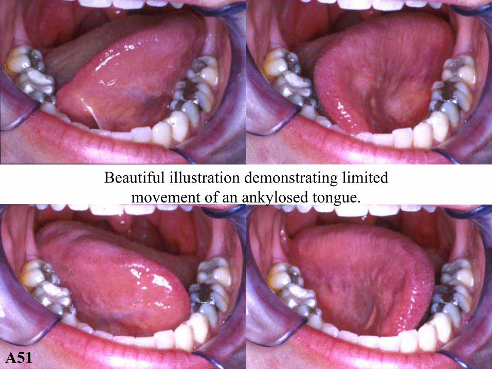
Beautiful illustration demonstrating limited movement of an ankylosed tongue.
A51

Curve of Wilson
Curve of Spee
A52Reverse Curve of Wilson

A53 A prehistoric skull with a reverse curve of Wilson.
Looks like the jaw had been broken.

A54 Another skull with a reverse curve of Wilson.
See next 3 slides.

A55 Close up of same skull - right side. See next 2 slides.

A56 Close up of same skull - left side. See next slide.

A57 Maxillary arch of same skull.

The ‘Neutral Zone’
A58

Tongue, teeth and cheeks are at rest in a “neutral”position. There are no abnormal forces within the mouth. This allows for the proper alignment of the teeth and dental
arches.
This also allows for normal facial development.
A59

The jaw is like a nutcracker.• Strongest muscle forces are exerted
close to the hinge.• The force diminishes as the distance
from the fulcrum increases.• Strong anterior stops protect the
posterior teeth.
A60

Jaw as a nutcracker.
A61

A62 Square jaw demonstrating strong masseters.

A63 Square jaw demonstrating strong masseters.

A64 Strong muscle attachment stimulated bone development.

Important Dental Concept
Cuspid Rise / Anterior Guidance
A65

A66 Nicely aligned teeth. (Class I occlusion)
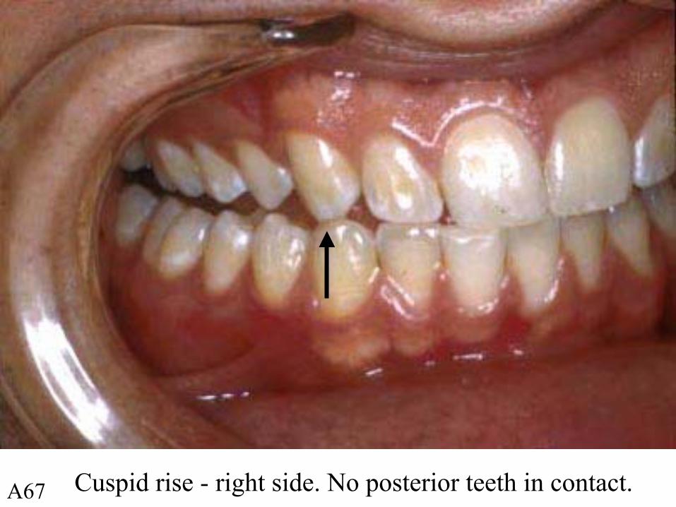
A67 Cuspid rise - right side. No posterior teeth in contact.

A68 During crossover, guidance is from anterior teeth.

A69During crossover, none of the posterior teeth on other side are contacting either.

A70Cuspid rise going in other direction. No posterior contacts.

A71Crossover going in other direction. No posterior contacts.

Traumatic Occlusion
Malocclusion
A72

A73 Anterior view during closure.

A74
Guidance coming from posterior tooth during excursion.

A75 Problem tooth causing interference and discomfort.

Maxillary lingual cusp tip below the Curve of Wilson. Interferes during excursions.
Curve of Wilson
Curve of Wilson
A76

“X” Factors that effect breakdown.• Emotional stress• Physical stress• Ability to cope• Heredity• Age• Diet• Growth and Development• Illness and disease• Physical environment• Habits• Factors still unknown.
A77
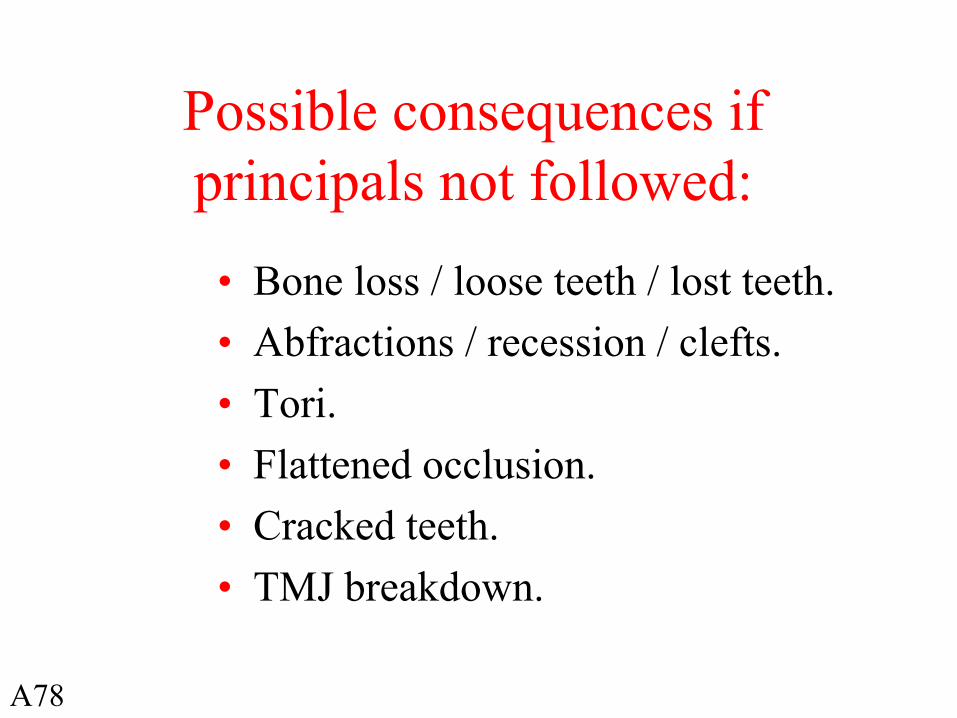
Possible consequences if principals not followed:
• Bone loss / loose teeth / lost teeth. • Abfractions / recession / clefts.• Tori.• Flattened occlusion.• Cracked teeth.• TMJ breakdown.
A78

Occlusion Terms
• Overjet / Overbite.• Class I occlusion: Adult and Pedo.• Class II malocclusion, Div 1 & 2.• Class III malocclusion.• Cross-bite.
A79

Overjet is a horizontal measurement.
Overbite is a vertical measurement.
A80

Class I occlusion.
Best occlusion.
Breastfed individual have the best chance of having this ideal occlusion.
A81

Adult Class I (Lateral view)
Arrows are where they should be.
Has ideal overjet and overbite.
A82

Adult Class I (Anterior view)
Mid-line is in the middle of the mouth / smile.
A83

Adult Class IBest occlusion.
A84

Child’s Class I (Lateral view)A85

Child’s Class I (Anterior view)A86

Child’s Class I
A87

Class II malocclusion.
Retrognathic - ‘pushed back jaw’- malocclusion.
Potentially deadly if it contributes to, or is a causative factor in, the development obstructive sleep apnea (OSA). It is the
most common malocclusion in individuals with OSA.
Major contributors to this malocclusion are: bottle feeding, pacifier use, excessive noxious habits and early obstruction / airway resistance of the airway.
A88

Class II - retrognathic malocclusionA89
Arrows should be aligned opposite to each other.

Previous models positioned to a Class I occlusionA90
Models hand articulated so arrows better approximate where they
should be in a Class I relationship.

Class II, Division I (Lateral view)
Red arrows should line up opposite each other.
Overjet
A91

Class II, Division I (Anterior view)A92

Class II, Division 1
Upper anterior teeth flare out.
A93

Class II, Division 2 (Lateral view)
Arrows should line up opposite each other.
A94

Class II, Division 2 (Anterior view)
Note slant of upper 4 anterior teeth.A95
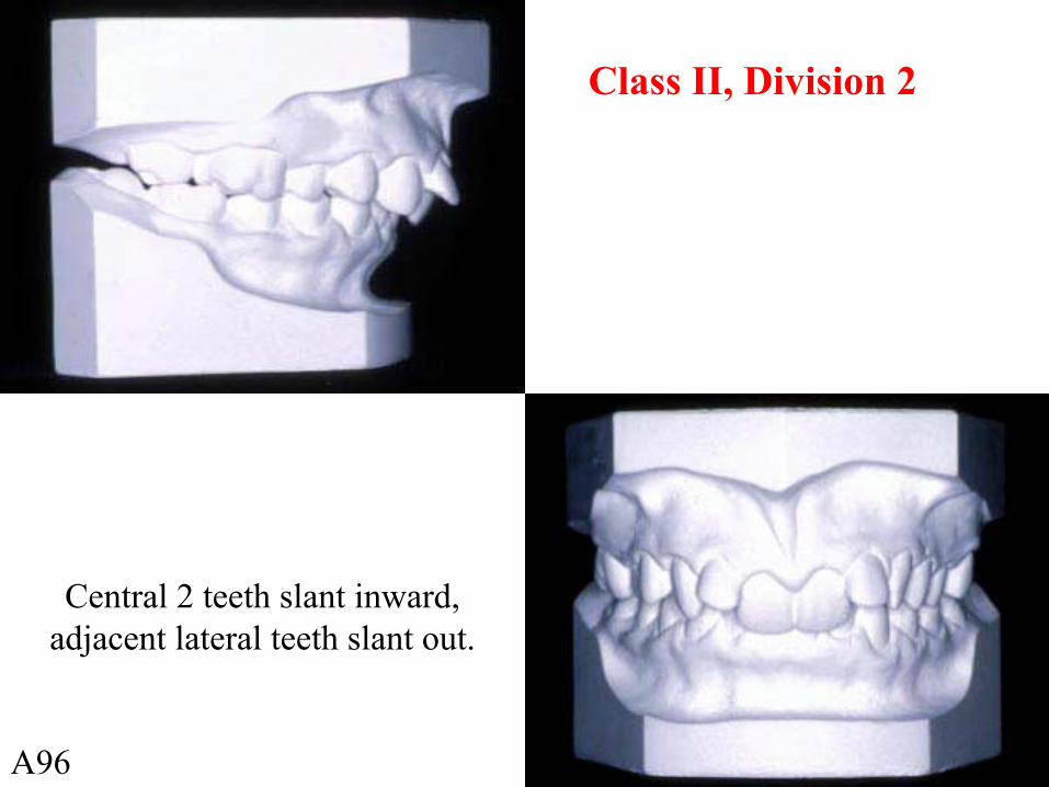
Class II, Division 2
Central 2 teeth slant inward, adjacent lateral teeth slant out.
A96

Note significant overbite on this Class II malocclusion.
A97

Class III malocclusion.
The ‘bulldog look’.
A98

Class III (Lateral view)
Arrows should line up.
A99

Class III (Anterior view)A100

Class III malocclusion.
Sometimes called an ‘under-bite’.
A101

Infant with Class III malocclusion.A102

Cross-bite
A103

In ‘B’, upper teeth are ‘inside’ lower teeth - due to a narrow arch. A104

Models demonstrating an example of a high palate and narrow upper arch.
A105
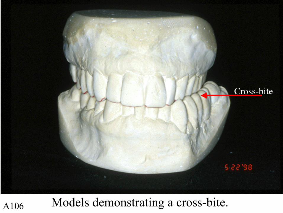
Models demonstrating a cross-bite.
Cross-bite
A106

Close-up of a cross-bite malocclusion.A107

Cross-bite has contributed to development of abfractions / recession and small tori developing on buccal #31.
A108

AAPD Vision Statement (1996)
• “89% of youth, ages 12 - 17 years, have some occlusal disharmony.”
• “16% of youth have a severe handicapping malocclusion that requires mandatory treatment.”
Pediatr Dent, (J Amer Acad Pediatr Dent), Spec Issue:Reference Manual 1995-96,17(6).
A109

Pacifier use (1997)
• 85% of children in her study used pacifiers by age one month. Children weaned from breastfeeding early use a pacifier more often than those who are breastfed longer.
Victora CG, Behague DP, et al. Pacifier use and short breastfeeding duration: cause, consequence, or coincidence? Pediatr. 1997 Mar;99(3):445-53.
A110

Craniofacial Development
• Largest increment occurs within the first 4 years of life.
• Is 90% complete by 12 years of age
Shepard J, et al. Evaluation of the upper airway in patients with OSA, Sleep 1991;14(4):361-71.
A111

End of section A
Brian Palmer, D.D.S. Leawood, Kansas December 2004.