selective attention on representations in working memory: cognitive … · · 2018-04-02working...
TRANSCRIPT
Submitted 23 February 2018Accepted 18 March 2018Published 2 April 2018
Corresponding authorYixuan Ku, [email protected]
Academic editorTifei Yuan
Additional Information andDeclarations can be found onpage 9
DOI 10.7717/peerj.4585
Copyright2018 Ku
Distributed underCreative Commons CC-BY 4.0
OPEN ACCESS
Selective attention on representations inworking memory: cognitive and neuralmechanismsYixuan KuFaculty of Education, East China Normal Unviersity, Shanghai, ChinaThe Key Lab of Brain Functional Genomics, MOE & STCSM, Shanghai Changning-ECNUMental HealthCenter, School of Psychology and Cognitive Science, East China Normal University, Shanghai, ChinaNYU-ECNU Institute of Brain and Cognitive Science, NYU Shanghai and Collaborative Innovation Centerfor Brain Science, Shanghai, China
ABSTRACTSelective attention and working memory are inter-dependent core cognitive functions.It is critical to allocate attention on selected targets during the capacity-limited workingmemory processes to fulfill the goal-directed behavior. The trends of research on bothtopics are increasing exponentially in recent years, and it is considered that selectiveattention and working memory share similar underlying neural mechanisms. Differenttypes of attention orientation in working memory are introduced by distinctive cues,and the means using retrospective cues are strengthened currently as it is manipulatingthe representation in memory, instead of the perceptual representation. The cognitiveand neural mechanisms of the retro-cue effects are further reviewed, as well as thepotential molecular mechanism. The frontal-parietal network that is involved in bothattention and working memory is also the neural candidate for attention orientationduring working memory. Neural oscillations in the gamma and alpha/beta oscillationsmay respectively be employed for the feedforward and feedback information transferbetween the sensory cortices and the association cortices. Dopamine and serotoninsystemsmight interactwith each other subserving the communication betweenmemoryand attention. In conclusion, representations which attention shifts towards arestrengthened, while representations which attention moves away from are degraded.Studies on attention orientation during working memory indicates the flexibility ofthe processes of working memory, and the beneficial way that overcome the limitedcapacity of working memory.
Subjects Psychiatry and PsychologyKeywords Attention orientation, Working memory, Object-based attention, Feature-basedattention, Retrospective cue, Selective attention
INTRODUCTIONWorkingmemory (WM) is a fundamental cognitive system thatmaintains andmanipulatesinformation from the outside world in a short period for goal-directed actions (Baddeley,2012). WM is critical to support everyday behaviors including language comprehension,learning and reasoning (Baddeley, 2003). In spite of its core position in cognition, WMhas severely limited capacity (Luck & Vogel, 2013). From the magic number seven (Miller,1956) to the magic number four (Cowan, 2001), the limit in WM reflects the bottleneck of
How to cite this article Ku (2018), Selective attention on representations in working memory: cognitive and neural mechanisms. PeerJ6:e4585; DOI 10.7717/peerj.4585

information processing in cognition. Researchers are fascinated about the mechanisms ofWM capacity as the capacity is highly correlated with general intelligence (IQ) (Redick et al.,2011). Given the restricted resource of WM, it is essential to rely on selective attention, thegoal-directed focus on certain aspects of the environment, while ignoring other irrelevantaspects. Empirical studies suggests that individual differences inWMcapacity are correlatedwith the ability to control attention (Kane et al., 2001), and those who have lower WMcapacity are not able to filter out distractors during WM maintenance (Vogel, McCollough& Machizawa, 2005). Therefore, effectively orienting attention during WM is importantfor goal-directed processes and behaviors.
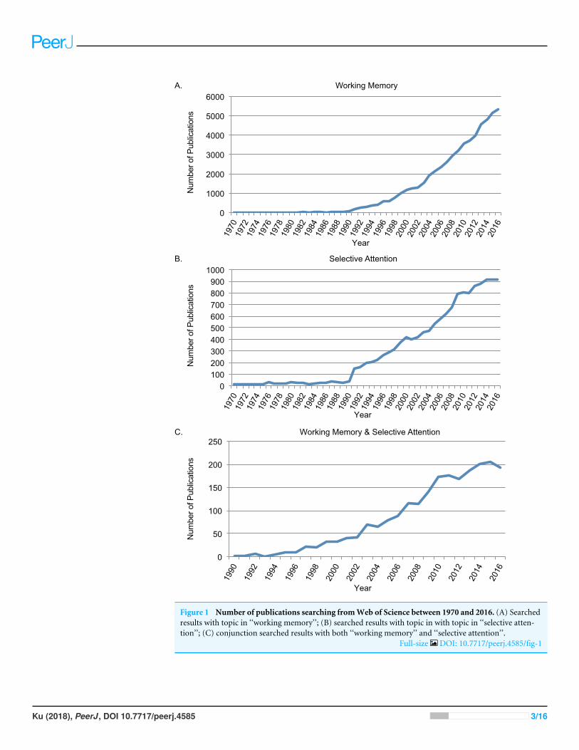
SURVEY METHODOLOGYSince 1970s, research on WM and selective attention has increased exponentially(Baddeley, 2003; Baddeley, 2012; Carrasco, 2011). Searching through Web of Science(http://www.webofknowledge.com/) was used to identify the number of publicationson both topics. As the two topics are both proper nouns, double quotation marks wereused for precisely matched results. First, searching with topic of ‘‘working memory’’from Web of Science between 1970 and 2016, there were 56,256 papers in total (See DataS1). Second, searching with topic of ‘‘selective attention’’ from Web of Science between1970 and 2016, there were 14,214 papers in total (See Data S1). Third, conjunctionsearching with topic of ‘‘working memory’’ AND topic of ‘‘selective attention’’ from Webof Science between 1970 and 2016, there were 2199 papers in total (See Data S1). Searchingin PubMed (http://www.ncbi.nlm.nih.gov) lead to similar results for the trends. First,searching with ‘‘working memory’’ in title or abstract (narrowed down from ‘‘topic’’) fromPubMed between 1970 and 2016, there were 22,930 papers in total. Second, searching with‘‘selective attention’’ in title or abstract from PubMed between 1970 and 2016, there were4,699 papers in total. Third, conjunction searching of the above two in title or abstractfrom PubMed between 1970 and 2016, there were 434 papers in total.
Research on working memory and selective attentionThe rapid increasing number of studies on WM and selective attention indicated thatboth of the concepts were within the focus of research interests on cognition. Figure 1depicted searching results from Web of Science between 1970 and 2016 using topics of‘‘working memory’’ (Fig. 1A), ‘‘selective attention’’ (Fig. 1B). The trend on both topicsincreased significantly after 1990s, when functional magnetic resonance imaging (fMRI)was developed and widely applied in research (Ogawa et al., 1990). The starting year in theconjunction search with both ‘‘working memory’’ and ‘‘selective attention’’ between 1970and 2016 (Fig. 1C)was 1990 aswell. It was increasingly acknowledged that selective attentionand WM were inter-dependent cognitive functions (Awh, Vogel & Oh, 2006; Ku, 2015).
Based on the shared neural correlates between spatial WM and spatial selective attention(LaBar et al., 1999), Awh & Jonides (2001) proposed that the mechanisms of the twoprocesses were overlapped. Some WM models even assumed that WM was merelyrepresentations from long-term memory that were under the focus of attention (Cowan,1998).However, long-termmemory plus attentionmodelmay not explain allWMprocesses
Ku (2018), PeerJ, DOI 10.7717/peerj.4585 2/16
0
50
100
150
200
250
1990
19
92
1994
19
96
1998
20
00
2002
20
04
2006
20
08
2010
20
12
2014
20
16
0 100 200 300 400 500 600 700 800 900
1000
1970
19
72
1974
19
76
1978
19
80
1982
19
84
1986
19
88
1990
19
92
1994
19
96
1998
20
00
2002
20
04
2006
20
08
2010
20
12
2014
20
16
Selective Attention
A.
B.
Working Memory
0
1000
2000
3000
4000
5000
6000
1970
19
72
1974
19
76
1978
19
80
1982
19
84
1986
19
88
1990
19
92
1994
19
96
1998
20
00
2002
20
04
2006
20
08
2010
20
12
2014
20
16
C. Working Memory & Selective Attention
Year
Year
Year
Num
ber o
f Pub
licat
ions
Num
ber o
f Pub
licat
ions
Num
ber o
f Pub
licat
ions
Figure 1 Number of publications searching fromWeb of Science between 1970 and 2016. (A) Searchedresults with topic in ‘‘working memory’’; (B) searched results with topic in with topic in ‘‘selective atten-tion’’; (C) conjunction searched results with both ‘‘working memory’’ and ‘‘selective attention’’.
Full-size DOI: 10.7717/peerj.4585/fig-1
Ku (2018), PeerJ, DOI 10.7717/peerj.4585 3/16
as the representation in WM needs to integrates new incoming sensory information, andis much more flexible than that in long-term memory.
WM processes can influence selective attention, when high WM load introduces moredistraction and less inhibition in selective attention (De Fockert et al., 2001). Meanwhile,attentional selection may also be guided by the template in WM (Downing, 2000; Soto etal., 2008). In contrary, selective attention can bias WM processing at multiple stages, fromsensory encoding till memory retrieval, even during the stage prior to sensory stimuli,i.e., the expectation period (Gazzaley & Nobre, 2012). While traditionally the majority ofstudies focus on the influence of selective attention on perception (Carrasco, 2011), recentstudies use retrospective cues to manipulate representations maintained in WM (Souza &Oberauer, 2016).
Orienting attention during working memory maintenance byretrospective cuesStandard WM paradigm includes a stimulus (sample) that needs to remember, a shortperiod (delay) when sample disappears and memorandums are maintained, the secondstimulus (probe) for participants to judge whether it match the first one. Cues about theprobed feature or spatial information could be presented at different stages: cues presentedprior to the sample (sensory) stimuli are named pre-cues; cues presented when the samplestimuli are existing are named sensory cues; cues presented briefly after the sample stimulidisappeared are named iconic cues; cues presented after a couple of hundred milliseconds(usually 500 ms after the sample onset) are named retro-cues; cues presented after the probestimuli are named post-cues. Different cues influence WM processes at distinctive stages asstated above.
Traditional work assumed that the performance of WM could only be affected duringvery short intervals after the offset of the sample stimuli, when the representations werethought as in an ‘iconic’ format, which had vast capacity (Phillips, 1974). After therepresentations were consolidated into WM, they became stable but then had limitedcapacity. Some theories further distinguished WM states as fragile vs. stable, based on thetemporal progresses after sensory encoding (Sligte, Scholte & Lamme, 2008). The effects oficonic cues were very similar to those sensory cues during the sensory encoding period.Their effects were also similar to those pre-cues presented before the sample stimuli, whichwere originally introduced by Posner (1980). Seminar neuroimaging study by Kastner andcolleagues in 1999 revealed that spatial attention induced by pre-cues changed the activityin human visual cortices during the expectation period (Kastner et al., 1999). The changedbaseline neural activity would then biased the processing of incoming sensory information(Nobre & Van Ede, 2018). The pre-cues, sensory cues and iconic cues were thought toinfluence the perceptual representation, which might be different from the retro-cues thattook impact on the representation in WM.
The retro-cue effects were first discovered independently by two groups (Landman,Spekreijse & Lamme, 2003; Griffin & Nobre, 2003), and they tended to be similar but a bitsmaller than the pre-cue effects as the predictive cues before the sample stimuli influencedthe perceptual processing that seemed to be more efficient than the memory processing
Ku (2018), PeerJ, DOI 10.7717/peerj.4585 4/16
(Griffin & Nobre, 2003). However, the processes on perception andmemory representationwere similar as reflected in ERP waveforms (Griffin & Nobre, 2003).
Retrospective cues could vary in different dimensions. First, there were spatial vs.feature/object cues (Li et al., 2015), which had similar effects. Second, there were also validvs. invalid cues, when valid cues lead to better performance and invalid cues lead to worseperformance (Gunseli et al., 2015). Third, the reliability of the cues could vary from 50%to 100%, while reliability increased the retro-cue effects (Shimi et al., 2013; Gunseli et al.,2015). Fourth, there were cues in different sensory domain, including visual (Landman,Spekreijse & Lamme, 2003; Griffin & Nobre, 2003), auditory (Backer & Alain, 2012) andtactile (Katus & Eimer, 2015), even crossmodal cues (Katus, Grubert & Eimer, 2016). Fifth,the retro-cue could be presented either centrally or peripherally, when the effects werecomparable (Matsukura et al., 2014). Last, the time interval between the retro-cue and theprobe could vary, and it was suggested that at least 300 ms were needed for the processesof retro-cue to take effects (Souza et al., 2014).
The distinctive effects induced by different retro-cues gave evidence for the flexibilityof WM, indicating that more information could be extracted after the cue. Traditionallyit was assumed that WM had fixed capacity limitations. It again suggested that long-termmemory plus attention could not explain the processes of WM.
Interference during working memory maintenanceAttention could be allocated towards the representations in WM and make additiveenhancement to the task performance. On the other side, attention could also bedirected away from WM representations by interference during the delay period,leading to worse task performance. There are two types of external interference,distraction (goal-irrelevant information that should be ignored) and interruption(information requiring attention as a secondary task). Both of them deteriorate WMperformance, but to different extents and utilize distinct neural mechanisms (Clapp,Rubens & Gazzaley, 2010; Clapp et al., 2011; Clapp & Gazzaley, 2012). The filtering ofdistraction is thought to be dependent on top-down suppression signals from theprefrontal cortex (PFC) (Knight et al., 1999; Chadick, Zanto & Gazzaley, 2014), while aninterruption requires a reallocation of cognitive resources, as well as processes involvedin reactivating the disrupted representation afterwards, which is reliant on medialtemporal lobe structures and the PFC (Sakai, Rowe & Passingham, 2002). Functionalconnectivity between stimulus-selective visual cortex and the prefrontal cortex, measuredvia functional magnetic resonance imaging (fMRI), has indicated that distractiondoes not change frontal-posterior functional connectivity during the delay, whereasinterruptions result in a functional disconnection of the network that is reinstantiatedafter the interruption and prior to WM recall (Clapp, Rubens & Gazzaley, 2010).
Neural representation in the sensory cortices was thought to be fragile to interferenceand neural representation in the associative cortices (such as the prefrontal cortex, PFCand the posterior parietal cortex, PPC) was thought to be more stable (Bettencourt & Xu,2015). The former and the latter were proposed to represent the quality and quantity ofWM, respectively (Ku, Bodner & Zhou, 2015).
Ku (2018), PeerJ, DOI 10.7717/peerj.4585 5/16
Cognitive mechanism of attention orientation during workingmemorySouza and Oberauer tested six hypotheses about the cognitive mechanisms underlyingthe retro-cue effect: (i) Protection from decay; (ii) Prioritizing for probe comparison; (iii)Enhancing the cued representations; (iv) Removing non-cued representations; (v) Affectingdecision making processes; (vi) Protection from perceptual interference. The evidencediscussed in their review provides support for the last four of these hypotheses (Souza &Oberauer, 2016).
It should be noted that there could be other cases of cognitive mechanisms underlyingthe retro-cue effect. Recently it was proposed that retro-cues first reoriented attention andthen reconfigured theWM representation in the service of upcoming task demands (Myers,Stokes & Nobre, 2017). Besides, as attention was suggested to implement serially towards aspatial/temporal object in nature (Jia et al., 2017), the retro-cue might take effect throughstabilizing the processes of attentional shift.
Since behavioral analysis could only show summarized experimental effects altogether,neural process along the entire temporal axis is critical to reveal the detailed dynamics of themechanism. Future studies are still needed to make progresses on such neural mechanisms.
Changing concepts of the neural mechanism underlying workingmemoryThe concept of neural mechanism underlying WM has changed for a couple of times.Originally as the sustained delay activity in the prefrontal cortex was discovered to representthe mnemonic information (Fuster & Alexander, 1971), it was proposed that the prefrontalcortex was critical to maintain the representations of WM (Goldman-Rakic, 1995), thebehavioral goals, as well as the means to achieve these goals (Miller & Cohen, 2001).
Afterwards, human neuroimaging studies further suggested the posterior parietal cortexas additional neural niche. Both fMRI and electroencephalography (EEG) revealed that theposterior neural activity changed with WM load and reached a plateau, which was similarto those behavioral findings (Todd & Marois, 2004; Vogel & Machizawa, 2004; Xu & Chun,2006). However, both the behavioral and the neural plateau was recently challenged (Vanden Berg & Ma, 2014; Bays, 2018).
The recent ten years witnessed the growing evidence that the posterior sensoryregions were where the precise WM information is primarily stored (Ku, Bodner &Zhou, 2015; Christophel et al., 2017), and the role of the PFC was more emphasized inproviding top-down control (D’Esposito et al., 1995; Smith & Jonides, 1999; Gazzaley &Nobre, 2012). Although traditional neuroimaging and neurophysiological studies indicatedthat the early sensory areas lacked of persistent delay activity (Bisley et al., 2004; Offen,Schluppeck & Heeger, 2009), the primary somatosensory cortex in rhesus monkey did showsustained and informative firing during the delay period of a tactile unimodal WM task(Zhou & Fuster, 1996).
Multivariate pattern analysis (MVPA) applied in neuroimaging data (Haynes & Rees,2006; Davis & Poldrack, 2013) revealed that content-specific representations could be
Ku (2018), PeerJ, DOI 10.7717/peerj.4585 6/16
decoded during WM delay period from the primary visual areas (Serences et al., 2009;Harrison & Tong, 2009), as well as the primary auditory cortex (Kumar et al., 2016).Furthermore, visual motion patterns could not only be decoded from visual areas butalso from the primary somatosensory cortex when the task is a pure visual WM task(Christophel & Haynes, 2014), suggesting the cross-modal modulation in the primarysomatosensory cortex (Ku et al., 2007). The causal role of the primary sensory cortexin both unimodal and crossmodal WM were verified by recent transcranial magneticstimulation (TMS) studies (Ku et al., 2015a; Ku et al., 2015b; Zhao et al., in press).
However, neurophysiological findings that the persistent modulation of activity inthe primary visual cortex (Super, Spekreijse & Lamme, 2001) was argued to be feedbackinformation from the associative cortices such as the PPC (Xu, 2017). The debating wouldbe carried on unless there is solid evidence combing spatio-temporal neural recording andcausal methods to manipulate the activity in the PFC or PPC, such as TMS or transcranialcurrent stimulation (tCS).
Neural mechanism of attention orientation during working memoryWhile filtering out distracting information is performed through directing attention awayfrom distractors (Vogel, McCollough & Machizawa, 2005), attention orientation towardsthe target may implement similar mechanisms that the PFC controls accesses to WM(McNab & Klingberg, 2008; Reinhart et al., 2012). Different features of WM may involvedistinctive frontal areas, since the frontal lobe could be divided into subdivisions based onthe abstraction of processed goals (Badre, 2008).
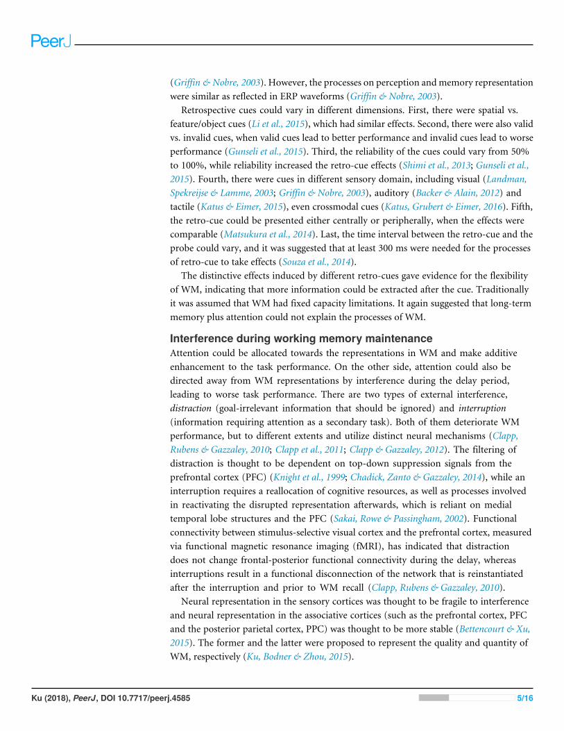
The neural dynamics of attention orientation during WM was revealed by EEG andMagnetoencephalography (MEG) studies. Most studies were within the visual domain andrevealed both event related potentials (ERPs) and neural oscillations relating to the retro-cue effects (Kuo, Stokes & Nobre, 2012;Myers et al., 2015). MEG studies also confirmed theneural oscillations for spatial attention orientation during WM maintenance (Worden etal., 2000). Backer, Binns & Alain (2015) used an auditory delayed matching-to-sample taskand a visual retro-cue directing attention to either the spatial information or the semanticcategory of the auditory target. Similar ERPs and neural oscillations of EEG comparedwith those in the visual domain were able to explain the behavioral benefits and dissociatethe feature-specific vs. object-specific processes of attention orientation during WM. Theattention orientation during tactile WM task also showed similar neural dynamics (Katus& Eimer, 2015). Such findings of retro-cue effects in visual, auditory and tactile domainssupport the theory of amodal attention orientation during WM (Shinn-Cunningham,2008). The illustration of the process is depicted in Fig. 2. However, it is still unknownwhether the object-based or feature-based attention orientation are the same in differentsensory domains.
Moreover, neuroimaging studies in the visual domain suggested that the fronto-parietal network exerted top-down control (Corbetta & Shulman, 2002), which might alsoinfluenced attention orientation during WM as the retro-cues activated similar brainnetworks (Lepsien et al., 2005).
Ku (2018), PeerJ, DOI 10.7717/peerj.4585 7/16

Cued Visual Features
Cued Auditoy Features
Cued Tactile Features
Cued Object
Un-cued Object
Featur
e-bas
ed A
ttenti
on
Object-
base
d Atte
ntion
Un-cued Visual Features
Un-cued Auditoy Features
Un-cued Tactile Features
ntion
Spatia
l Atte
ntion
Figure 2 Attention orientation during working memory (WM). The lower plane is the feature-based at-tention space. Round patches with different colors indicate features represented in WM (red, visual; blue,auditory; yellow, tactile). The cued features are in dark color and the un-cued features are in light color.The middle plane is the object-based attention space. Grey round patches depict objects maintained inWM. Orienting attention to a cued object (darker grey) strengthen the representation of this object com-pared with other un-cued objects (lighter grey) in WM. It may strengthen some features connected to thisobject (thicker dashed lines), while other features remain (thinner dashed lines). The connection can bebi-directional, i.e. when attention is oriented to one feature; the object representation connected to thisfeature will be strengthened, but may not affect other feature representations from the same object. Theupper plane is the spatial attention space. Dashed circles indicate attention allocation in the spatial map.The thicker circle indicates the prioritized focus of attention. The thinner circle indicates the divided focusof attention.
Full-size DOI: 10.7717/peerj.4585/fig-2
It should be noted that the fate of uncued item in WM was still debating. Some studiessuggested that the uncued representations were removed out of the memory buffer ordegraded at the cost of enhancing the cued item (Matsukura, Luck & Vecera, 2007; Kuo,Stokes & Nobre, 2012), while others indicate that they remained unaffected (Rerko &Oberauer, 2013). Future studies using MVPA with EEG/MEG may help resolve thesearguments, by looking into the dynamic changes of the representation in WM.
Meanwhile, the development of the theory in attention orientation during WM willfacilitate our understanding of the direction of information transfer between the sensoryareas and the associative cortices during WM, which can be either feedforward or feedback(Corbetta & Shulman, 2002; Xu, 2017). Recent neurophysiological findings have revealedthat the feedforward processing may rely on the gamma oscillation (40–90 Hz) and thefeedback processing may react through alpha/beta (8–30 Hz) (Van Kerkoerle et al., 2014;
Ku (2018), PeerJ, DOI 10.7717/peerj.4585 8/16
Bastos et al., 2015; Van Kerkoerle, Self & Roelfsema, 2017). Combining neural modulationmethod (TMS or tCS) with neurophysiological recordings will help to validate thesehypotheses.
Besides these neuronal mechanisms, rare molecular mechanisms were discovered forattention orientation during WM. Studies with animals have shown that dopamine D1receptor in the prefrontal cortex is key in regulating WM, but not attention orientationin saccadic searching (Sawaguchi & Goldman-Rakic, 1991). Positron emission tomography(PET) study further reveal that within the prefrontal cortex dopamine D2 release is alsomore prominent in WM than sustained attention task (Aalto, 2005). On the other side,Psilocybin, a serotonin (5-HT) receptor agonist, affects attentional tracking task butnot WM task (Carter et al., 2005). Therefore, dopamine may influence WM more thanattention, and serotoninmay take stronger effect on the other way. It would be interesting tosee how these two neural transmitters interact with each other during attention orientationin WM, which needs elegant experimental design with animals.
CONCLUSIONSSelective attention and working memory interact with each other and share similar neuralmechanisms. Using retrospective cues during WM is an efficient way to overcome thelimited capacity of WM. The cued representations are strengthened, while the fate of theun-cued representations are still in debate, either degraded or unchanged. Future studiesimplementing both neural modulation methods (TMS and tCS) and neurophysiologicalrecording (EEG/MEG) are critical to consolidate the existing hypotheses and to help resolvethe controversial theories in this expanding field.
ADDITIONAL INFORMATION AND DECLARATIONS
FundingThis work was sponsored by the Peak Discipline Construction Project of Education atEast China Normal University, Fundamental Research Funds for the Central Universities(No. 2017ECNU-YYJ050), Shanghai Pujiang Talents Plan Project (No. 16PJC022), andthe Major Program of Science and Technology Commission Shanghai Municipal (No.17JC1404100). The funders had no role in study design, data collection and analysis,decision to publish, or preparation of the manuscript.
Grant DisclosuresThe following grant information was disclosed by the author:East China Normal University.Fundamental Research Funds for the Central Universities: 2017ECNU-YYJ050.Shanghai Pujiang Talents Plan Project: 16PJC022.Major Programof Science andTechnologyCommission ShanghaiMunicipal: 17JC1404100.
Competing InterestsThe author declares there are no competing interests.
Ku (2018), PeerJ, DOI 10.7717/peerj.4585 9/16
Author Contributions• Yixuan Ku conceived and designed the experiments, performed the experiments,analyzed the data, contributed reagents/materials/analysis tools, prepared figures and/ortables, authored or reviewed drafts of the paper, approved the final draft.
Data AvailabilityThe following information was supplied regarding data availability:
The raw data are provided in a Data S1.
Supplemental InformationSupplemental information for this article can be found online at http://dx.doi.org/10.7717/peerj.4585#supplemental-information.
REFERENCESAalto S. 2005. Frontal and temporal dopamine release during working memory and
attention tasks in healthy humans: a positron emission tomography study usingthe high-affinity dopamine D2 receptor ligand [11C]FLB 457. The Journal ofNeuroscience 25:2471–2477 DOI 10.1523/JNEUROSCI.2097-04.2005.
Awh E, Jonides J. 2001. Overlapping mechanisms of attention and spatial working mem-ory. Trends in Cognitive Sciences 5:119–126 DOI 10.1016/S1364-6613(00)01593-X.
Awh E, Vogel EK, Oh SH. 2006. Interactions between attention and working memory.Neuroscience 139:201–208 DOI 10.1016/j.neuroscience.2005.08.023.
Backer KC, Alain C. 2012. Orienting attention to sound object representations attenuateschange deafness. Journal of Experimental Psychology. Human Perception and Perfor-mance 38:1554–1566 DOI 10.1037/a0027858.
Backer KC, BinnsMA, Alain C. 2015. Neural dynamics underlying attentional orientingto auditory representations in short-term memory. The Journal of Neuroscience35:1307–1318 DOI 10.1523/JNEUROSCI.1487-14.2015.
Baddeley A. 2003.Working memory: looking back and looking forward. Nature ReviewsNeuroscience 4:829–839 DOI 10.1038/nrn1201.
Baddeley A. 2012.Working memory: theories, models, and controversies. Annual Reviewof Psychology 63:1–29 DOI 10.1146/annurev-psych-120710-100422.
Badre D. 2008. Cognitive control, hierarchy, and the rostro-caudal organization of thefrontal lobes. Trends in Cognitive Sciences 12:193–200 DOI 10.1016/j.tics.2008.02.004.
Bastos AM, Vezoli J, Bosman CA, Schoffelen J-M, Oostenveld R, Dowdall JR,DeWeerd P, Kennedy H, Fries P. 2015. Visual areas exert feedforward andfeedback influences through distinct frequency channels. Neuron 85:390–401DOI 10.1016/j.neuron.2014.12.018.
Bays PM. 2018. Reassessing the evidence for capacity limits in neural signals related toworking memory. Cerebral Cortex 28:1432–1438 DOI 10.1093/cercor/bhx351.
Bettencourt KC, Xu Y. 2015. Decoding the content of visual short-term memoryunder distraction in occipital and parietal areas. Nature Neuroscience 19:150–157DOI 10.1038/nn.4174.
Ku (2018), PeerJ, DOI 10.7717/peerj.4585 10/16
Bisley JW, Zaksas D, Droll JA, Pasternak T. 2004. Activity of neurons in cortical areaMT during a memory for motion task. Journal of Neurophysiology 91:286–300DOI 10.1152/jn.00870.2003.
CarrascoM. 2011. Visual attention: the past 25 years. Vision Research 51:1484–1525DOI 10.1016/j.visres.2011.04.012.
Carter OL, Burr DC, Pettigrew JD,Wallis GM, Hasler F, Vollenweider FX. 2005. Usingpsilocybin to investigate the relationship between attention, working memory, andthe serotonin 1A and 2A receptors. Journal of Cognitive Neuroscience 17:1497–1508DOI 10.1162/089892905774597191.
Chadick JZ, Zanto TP, Gazzaley A. 2014. Structural and functional differences in medialprefrontal cortex underlie distractibility and suppression deficits in ageing. NatureCommunications 5:Article 4223 DOI 10.1038/ncomms5223.
Christophel TB, Haynes J-D. 2014. Decoding complex flow-field patterns in visualworking memory. NeuroImage 91:43–51 DOI 10.1016/j.neuroimage.2014.01.025.
Christophel TB, Klink PC, Spitzer B, Roelfsema PR, Haynes J-D. 2017. The dis-tributed nature of working memory. Trends in Cognitive Sciences 21:112–124DOI 10.1016/j.tics.2016.12.007.
ClappWC, Gazzaley A. 2012. Distinct mechanisms for the impact of distraction andinterruption on working memory in aging. Neurobiology of Aging 33:134–148DOI 10.1016/j.neurobiolaging.2010.01.012.
ClappWC, RubensMT, Gazzaley A. 2010.Mechanisms of working memory disruptionby external interference. Cerebral Cortex 20:859–872 DOI 10.1093/cercor/bhp150.
ClappWC, RubensMT, Sabharwal J, Gazzaley A. 2011. Deficit in switching betweenfunctional brain networks underlies the impact of multitasking on working memoryin older adults. Proceedings of the National Academy of Sciences of the United States ofAmerica 108:7212–7217 DOI 10.1073/pnas.1015297108.
Corbetta M, Shulman GL. 2002. Control of goal-directed and stimulus-driven attentionin the brain. Nature Reviews Neuroscience 3:201–215 DOI 10.1038/nrn755.
Cowan N. 1998. Attention and memory: an integrated framework. New York: OxfordUniversity Press.
Cowan N. 2001. The magical number 4 in short-term memory: a reconsidera-tion of mental storage capacity. Behavioral and Brain Sciences 24:87–114DOI 10.1017/S0140525X01003922.
Davis T, Poldrack RA. 2013.Measuring neural representations with fMRI: prac-tices and pitfalls. Annals of the New York Academy of Sciences 1296:108–134DOI 10.1111/nyas.12156.
De Fockert J, Rees G, Frith C, Lavie N. 2001. The role of working memory in visualselective attention. Science 291:1803–1806 DOI 10.1126/science.1056496.
D’Esposito M, Detre JA, Alsop DC, Shin RK, Atlas S, GrossmanM. 1995. The neuralbasis of the central executive system of working memory. Nature 378:279–281DOI 10.1038/378279a0.
Ku (2018), PeerJ, DOI 10.7717/peerj.4585 11/16
Downing PE. 2000. Interactions between visual working memory and selective attention.Psychological Science 11:467–473 DOI 10.1111/1467-9280.00290.
Fuster JM, Alexander GE. 1971. Neuron activity related to short-term memory. Science173:652–654 DOI 10.1126/science.173.3997.652.
Gazzaley A, Nobre AC. 2012. Top-down modulation: bridging selective attention andworking memory. Trends in Cognitive Sciences 16:128–134DOI 10.1016/j.tics.2011.11.014.
Goldman-Rakic PS. 1995. Cellular basis of working memory. Neuron 14:477–485DOI 10.1016/0896-6273(95)90304-6.
Griffin IC, Nobre AC. 2003. Orienting attention to locations in internal representations.Journal of Cognitive Neuroscience 15:1176–1194 DOI 10.1162/089892903322598139.
Gunseli E, VanMoorselaar D, Meeter M, Olivers CNL. 2015. The reliability of retro-cuesdetermines the fate of noncued visual working memory representations. PsychonomicBulletin & Review 22:1334–1341.
Harrison SA, Tong F. 2009. Decoding reveals the contents of visual working memory inearly visual areas. Nature 458:632–635 DOI 10.1038/nature07832.
Haynes J-D, Rees G. 2006. Decoding mental states from brain activity in humans. NatureReviews Neuroscience 7:523–534 DOI 10.1038/nrn1931.
Jia J, Liu L, Fang F, Luo H. 2017. Sequential sampling of visual objects during sustainedattention. PLOS Biology 15:e2001903 DOI 10.1371/journal.pbio.2001903.
KaneMJ, Bleckley MK, Conway ARA, Engle RW. 2001. A controlled-attention view ofworking-memory capacity. Journal of Experimental Psychology. General 130:169–183DOI 10.1037/0096-3445.130.2.169.
Kastner S, PinskMA, DeWeerd P, Desimone R, Ungerleider LG. 1999. Increasedactivity in human visual cortex during directed attention in the absence of visualstimulation. Neuron 22:751–761 DOI 10.1016/S0896-6273(00)80734-5.
Katus T, EimerM. 2015. Lateralized delay period activity marks the focus of spatialattention in working memory: evidence from Somatosensory event-Rrlated brainpotentials. The Journal of Neuroscience 35:6689–6695DOI 10.1523/JNEUROSCI.5046-14.2015.
Katus T, Grubert A, EimerM. 2016. Intermodal attention shifts in multimodal workingmemory. Journal of Cognitive Neuroscience 83:1–9 DOI 10.1162/jocn_a_01072.
Knight RT, StainesWR, Swick D, Chao LL. 1999. Prefrontal cortex regulates inhibitionand excitation in distributed neural networks. Acta Psychologica 101:159–178DOI 10.1016/S0001-6918(99)00004-9.
Ku Y. 2015. Feature-based and object-based attention orientation during short-termmemory maintenance. Journal of Neurophysiology 114:3036–3038DOI 10.1152/jn.00342.2015.
Ku Y, Bodner M, Zhou Y-D. 2015. Prefrontal cortex and sensory cortices duringworking memory: quantity and quality. Neuroscience Bulletin 31:175–182DOI 10.1007/s12264-014-1503-7.
Ku (2018), PeerJ, DOI 10.7717/peerj.4585 12/16
Ku Y, Ohara S, Wang L, Lenz FA, Hsiao SS, Bodner M, Hong B, Zhou Y-D. 2007.Prefrontal cortex and somatosensory cortex in tactile crossmodal association:an independent component analysis of ERP recordings. PLOS ONE 2:e771DOI 10.1371/journal.pone.0000771.
Ku Y, Zhao D, Bodner M, Zhou Y-D. 2015a. Cooperative processing in primarysomatosensory cortex and posterior parietal cortex during tactile working memory.The European Journal of Neuroscience 42:1905–1911 DOI 10.1111/ejn.12950.
Ku Y, Zhao D, Hao N, Hu Y, Bodner M, Zhou Y-D. 2015b. Sequential roles of primarySomatosensory cortex and posterior parietal cortex in tactile-visual cross-modalworking memory: a single-pulse transcranial magnetic stimulation (spTMS) study.Brain Stimulation 8:88–91 DOI 10.1016/j.brs.2014.08.009.
Kumar S, Joseph S, Gander PE, Barascud N, Halpern AR, Griffiths TD. 2016. A brainsystem for auditory working memory. The Journal of Neuroscience 36:4492–4505DOI 10.1523/JNEUROSCI.4341-14.2016.
Kuo B-C, Stokes MG, Nobre AC. 2012. Attention modulates maintenance of represen-tations in visual short-term memory. Journal of Cognitive Neuroscience 24:51–60DOI 10.1162/jocn_a_00087.
LaBar KS, Gitelman DR, Parrish TB, MesulamM. 1999. Neuroanatomic overlap ofworking memory and spatial attention networks: a functional MRI comparisonwithin subjects. NeuroImage 10:695–704 DOI 10.1006/nimg.1999.0503.
Landman R, Spekreijse H, Lamme VAF. 2003. Large capacity storage of integratedobjects before change blindness. Vision Research 43:149–164DOI 10.1016/S0042-6989(02)00402-9.
Lepsien J, Griffin IC, Devlin JT, Nobre AC. 2005. Directing spatial attention in mentalrepresentations: interactions between attentional orienting and working-memoryload. NeuroImage 26:733–743 DOI 10.1016/j.neuroimage.2005.02.026.
Li X, Cheng X, Li J, Pan Y, Hu Y, Ku Y. 2015. Examination of mechanisms underlyingenhanced memory performance in action video game players: a pilot study. Frontiersin Psychology 6:Article 843 DOI 10.3389/fpsyg.2015.00843.
Luck SJ, Vogel EK. 2013. Visual working memory capacity: from psychophysics andneurobiology to individual differences. Trends in Cognitive Sciences 17:391–400DOI 10.1016/j.tics.2013.06.006.
MatsukuraM, Cosman JD, Roper ZJJ, Vatterott DB, Vecera SP. 2014. Location-specific effects of attention during visual short-term memory maintenance. Journalof Experimental Psychology. Human Perception and Performance 40:1103–1116DOI 10.1037/a0035685.
MatsukuraM, Luck SJ, Vecera SP. 2007. Attention effects during visual short-termmemory maintenance: protection or prioritization? Perception & Psychophysics69:1422–1434 DOI 10.3758/BF03192957.
McNab F, Klingberg T. 2008. Prefrontal cortex and basal ganglia control access toworking memory. Nature Neuroscience 11:103–107 DOI 10.1038/nn2024.
Ku (2018), PeerJ, DOI 10.7717/peerj.4585 13/16
Miller GA. 1956. The magical number seven, plus or minus two: some limitson our capacity for processing information. Psychological Review 63:81–97DOI 10.1037/h0043158.
Miller EK, Cohen JD. 2001. An integrative theory of prefrontal cortex function. AnnualReview of Neuroscience 24:167–202 DOI 10.1146/annurev.neuro.24.1.167.
Myers NE, Stokes MG, Nobre AC. 2017. Prioritizing information during working mem-ory: beyond sustained internal attention. Trends in Cognitive Sciences 21:449–461DOI 10.1016/j.tics.2017.03.010.
Myers NE,Walther L, Wallis G, Stokes MG, Nobre AC. 2015. Temporal dynamics ofattention during encoding versus maintenance of working memory: complementaryviews from event-related potentials and alpha-band oscillations. Journal of CognitiveNeuroscience 27:492–508 DOI 10.1162/jocn_a_00727.
Nobre AC, Van Ede F. 2018. Anticipated moments: temporal structure in attention.Nature Reviews Neuroscience 19(1):34–48 DOI 10.1038/nrn.2017.141.
Offen S, Schluppeck D, Heeger DJ. 2009. The role of early visual cortex in vi-sual short-term memory and visual attention. Vision Research 49:1352–1362DOI 10.1016/j.visres.2007.12.022.
Ogawa S, Lee TM, Kay AR, Tank DW. 1990. Brain magnetic resonance imaging withcontrast dependent on blood oxygenation. Proceedings of the National Academy ofSciences of the United States of America 87:9868–9872 DOI 10.1073/pnas.87.24.9868.
PhillipsWA. 1974. On the distinction between sensory storage and short-term visualmemory. Perception & Psychophysics 16:283–290 DOI 10.3758/BF03203943.
Posner MI. 1980. Orienting of attention. The Quarterly Journal of Experimental Psychol-ogy 32:3–25 DOI 10.1080/00335558008248231.
Redick TS, Calvo A, Gay CE, Engle RW. 2011.Working memory capacity and go/no-go task performance: selective effects of updating, maintenance, and inhibition.Journal of Experimental Psychology: Learning, Memory, and Cognition 37:308–324DOI 10.1037/a0022216.
Reinhart RMG, Heitz RP, Purcell BA,Weigand PK, Schall JD,Woodman GF. 2012.Homologous mechanisms of visuospatial working memory maintenance in macaqueand human: properties and sources. The Journal of Neuroscience 32:7711–7722DOI 10.1523/JNEUROSCI.0215-12.2012.
Rerko L, Oberauer K. 2013. Focused, unfocused, and defocused information in workingmemory. Journal of Experimental Psychology: Learning, Memory, and Cognition39:1075–1096 DOI 10.1037/a0031172.
Sakai K, Rowe JB, Passingham RE. 2002. Active maintenance in prefrontal area 46 cre-ates distractor-resistant memory. Nature Neuroscience 5:479–484 DOI 10.1038/nn846.
Sawaguchi T, Goldman-Rakic PS. 1991. D1 dopamine receptors in prefrontal cortex: in-volvement in working memory. Science 251:947–950 DOI 10.1126/science.1825731.
Serences JT, Ester EF, Vogel EK, Awh E. 2009. Stimulus-specific delay activity in humanprimary visual cortex. Psychological Science 20:207–214DOI 10.1111/j.1467-9280.2009.02276.x.
Ku (2018), PeerJ, DOI 10.7717/peerj.4585 14/16

Shimi A, Nobre AC, Astle D, Scerif G. 2013. Orienting attention within visual short-term memory: development and mechanisms. Child Development 85:578–592DOI 10.1111/cdev.12150.
Shinn-Cunningham BG. 2008. Object-based auditory and visual attention. Trends inCognitive Sciences 12:182–186 DOI 10.1016/j.tics.2008.02.003.
Sligte IG, Scholte HS, Lamme VAF. 2008. Are there multiple visual short-term memorystores? PLOS ONE 3:e1699 DOI 10.1371/journal.pone.0001699.
Smith EE, Jonides J. 1999. Storage and executive processes in the frontal lobes. Science283:1657–1661 DOI 10.1126/science.283.5408.1657.
Soto D, Hodsoll J, Rotshtein P, Humphreys GW. 2008. Automatic guidance ofattention from working memory. Trends in Cognitive Sciences 12:342–348DOI 10.1016/j.tics.2008.05.007.
Souza AS, Oberauer K. 2016. In search of the focus of attention in working memory:13 years of the retro-cue effect. Attention, Perception & Psychophysics 78:1839–1860DOI 10.3758/s13414-016-1108-5.
Souza AS, Rerko L, Lin H-Y, Oberauer K. 2014. Focused attention improves workingmemory: implications for flexible-resource and discrete-capacity models. Attention,Perception & Psychophysics 76:2080–2102 DOI 10.3758/s13414-014-0687-2.
Super H, Spekreijse H, Lamme VA. 2001. A neural correlate of working memory in themonkey primary visual cortex. Science 293:120–124 DOI 10.1126/science.1060496.
Todd JJ, Marois R. 2004. Capacity limit of visual short-term memory in human posteriorparietal cortex. Nature 428:751–754 DOI 10.1038/nature02466.
Van den Berg R, MaWJ. 2014. ‘‘Plateau-’’ related summary statistics are uninformativefor comparing working memory models. Attention, Perception & Psychophysics76:2117–2135 DOI 10.3758/s13414-013-0618-7.
Van Kerkoerle T, Self MW, Dagnino B, Gariel-Mathis M-A, Poort J, Van der TogtC, Roelfsema PR. 2014. Alpha and gamma oscillations characterize feedbackand feedforward processing in monkey visual cortex. Proceedings of the Na-tional Academy of Sciences of the United States of America 111:14332–14341DOI 10.1073/pnas.1402773111.
Van Kerkoerle T, Self MW, Roelfsema PR. 2017. Layer-specificity in the effects ofattention and working memory on activity in primary visual cortex. Nature Com-munications 8:Article 13804 DOI 10.1038/ncomms13804.
Vogel EK, MachizawaMG. 2004. Neural activity predicts individual differences in visualworking memory capacity. Nature 428:748–751 DOI 10.1038/nature02447.
Vogel EK, McCollough AW,MachizawaMG. 2005. Neural measures reveal individ-ual differences in controlling access to working memory. Nature 438:500–503DOI 10.1038/nature04171.
WordenMS, Foxe JJ, Wang N, Simpson GV. 2000. Anticipatory biasing of visuospatialattention indexed by retinotopically specific alpha-band electroencephalographyincreases over occipital cortex. The Journal of Neuroscience 20(6):RC63.
Ku (2018), PeerJ, DOI 10.7717/peerj.4585 15/16
Xu Y. 2017. Reevaluating the sensory account of visual working memory storage. Trendsin Cognitive Sciences 21:794–815 DOI 10.1016/j.tics.2017.06.013.
Xu Y, ChunMM. 2006. Dissociable neural mechanisms supporting visual short-termmemory for objects. Nature 440:91–95 DOI 10.1038/nature04262.
Zhao D, Zhou Y-D, Bodner M, Ku Y. 2017. The causal role of the prefrontal cortexand Somatosensory cortex in tactile working memory. Cerebral Cortex In PressDOI 10.1093/cercor/bhx213.
Zhou Y-D, Fuster JM. 1996.Mnemonic neuronal activity in somatosensory cortex.Proceedings of the National Academy of Sciences of the United States of America93:10533–10537 DOI 10.1073/pnas.93.19.10533.
Ku (2018), PeerJ, DOI 10.7717/peerj.4585 16/16